Filters
▼Clonality
▼Type
▼Reactivity
▼Gene Name
▼Isotype
▼Host
▼Application
▼Clone
▼Polyclonal Antibodies
At AAA Biotech also known as AAA Bio or AAABio, we provide a broad range of purified polyclonal antibodies (pAbs) that are able to all be browsed online through our website. Due to their high specificity and strong binding affinity, these antibodies are ideal for wide swathes of research and experimental applications.
Our polyclonal antibodies can easily support your work, whether you use them for Western Blotting, Immunocytochemistry (with or without Immunofluorescence used in conjunction), Immunohistochemistry, Immunoprecipitation, and ELISA tests. We highly encourage you to browse our range of pAbs and choose the one that best suits your experimental model.
Viewing 9350-9400 of 96805 product results
PTGIS, Polyclonal Antibody (Cat# AAA235695)
TAT, Polyclonal Antibody (Cat# AAA235696)
 IF (Immunofluorescence)
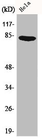
(Immunofluorescent analysis of Hela cells using AAA235697 at dilution of 1:200 and Alexa Fluor 488-congugated AffiniPure Goat Anti-Rabbit IgG(H+L))
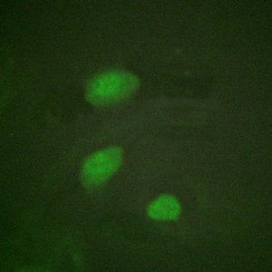
IF (Immunofluorescence)
(Immunofluorescent analysis of Hela cells using AAA235697 at dilution of 1:200 and Alexa Fluor 488-congugated AffiniPure Goat Anti-Rabbit IgG(H+L))
HIRA, Polyclonal Antibody (Cat# AAA235697)
BAI1, Polyclonal Antibody (Cat# AAA235700)
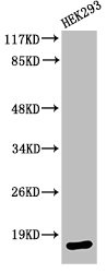
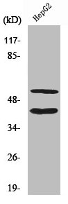
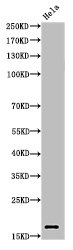
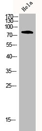
 WB (Western Blot)
(Western Blot analysis of HepG2 cells using Cleaved-Caspase-3 p17 (D175) Polyclonal Antibody)
WB (Western Blot)
(Western Blot analysis of HepG2 cells using Cleaved-Caspase-3 p17 (D175) Polyclonal Antibody)
CASP3, Polyclonal Antibody (Cat# AAA235703)
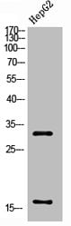
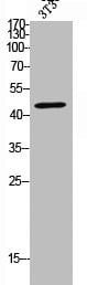 WB (Western Blot)
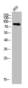
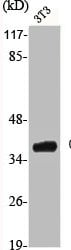
(Western Blot analysis of NIH-3T3 cells using Cleaved-MMP-10 (F99) Polyclonal Antibody)
WB (Western Blot)
(Western Blot analysis of NIH-3T3 cells using Cleaved-MMP-10 (F99) Polyclonal Antibody)
MMP10, Polyclonal Antibody (Cat# AAA235712)
 WB (Western Blot)
(Western Blot analysis of customer's (cat sample) using Cleaved-Spectrin α II (D1185) Polyclonal Antibody)
WB (Western Blot)
(Western Blot analysis of customer's (cat sample) using Cleaved-Spectrin α II (D1185) Polyclonal Antibody)
SPTAN1, Polyclonal Antibody (Cat# AAA235717)
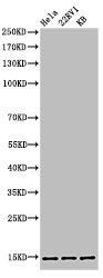 WB (Western Blot)
(Western Blot analysis of HELA 22RV1 KB cells using Acetyl-Histone H3 (K27) Polyclonal Antibody)
WB (Western Blot)
(Western Blot analysis of HELA 22RV1 KB cells using Acetyl-Histone H3 (K27) Polyclonal Antibody)
Histone H3, Polyclonal Antibody (Cat# AAA235723)
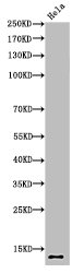
 WB (Western Blot)
(Western Blot analysis of HELA cells using Acetyl-Histone H4 (K5) Polyclonal Antibody)
WB (Western Blot)
(Western Blot analysis of HELA cells using Acetyl-Histone H4 (K5) Polyclonal Antibody)
Histone H4, Polyclonal Antibody (Cat# AAA235724)
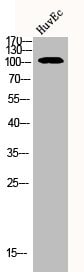
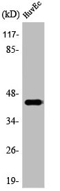
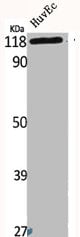
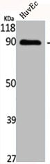
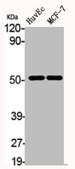 WB (Western Blot)
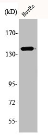
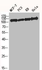
(Western Blot analysis of HuvEc MCF7 cells using AChRalpha10 Polyclonal Antibody)
WB (Western Blot)
(Western Blot analysis of HuvEc MCF7 cells using AChRalpha10 Polyclonal Antibody)
CHRNA10, Polyclonal Antibody (Cat# AAA235892)
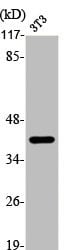
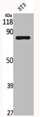
 WB (Western Blot)
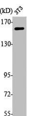
(Western Blot analysis of NIH-3T3 cells using Actinin-alpha3 Polyclonal Antibody)
WB (Western Blot)
(Western Blot analysis of NIH-3T3 cells using Actinin-alpha3 Polyclonal Antibody)
ACTN3, Polyclonal Antibody (Cat# AAA235901)
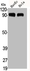 WB (Western Blot)
(Western Blot analysis of HuvEc HELA cells using ADAMTS-1 Polyclonal Antibody)
WB (Western Blot)
(Western Blot analysis of HuvEc HELA cells using ADAMTS-1 Polyclonal Antibody)
ADAMTS1, Polyclonal Antibody (Cat# AAA235902)
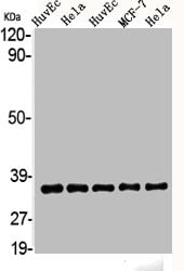 WB (Western Blot)
(Western Blot analysis of HELA HuvEc MCF7 cells using AKR1A1 Polyclonal Antibody)
WB (Western Blot)
(Western Blot analysis of HELA HuvEc MCF7 cells using AKR1A1 Polyclonal Antibody)
AKR1A1, Polyclonal Antibody (Cat# AAA235911)
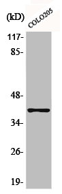
 WB (Western Blot)
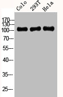
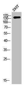
(Western Blot analysis of COLO 293T HELA cells using Amyloid-beta Polyclonal Antibody)
WB (Western Blot)
(Western Blot analysis of COLO 293T HELA cells using Amyloid-beta Polyclonal Antibody)
APP, Polyclonal Antibody (Cat# AAA235917)
 WB (Western Blot)
(Western Blot analysis of NIH-3T3 cells using Annexin I Polyclonal Antibody)
WB (Western Blot)
(Western Blot analysis of NIH-3T3 cells using Annexin I Polyclonal Antibody)
ANXA1, Polyclonal Antibody (Cat# AAA235918)
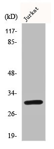
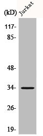
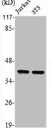 WB (Western Blot)
(Western Blot analysis of Jurkat NIH-3T3 cells using Annexin II Polyclonal Antibody)
WB (Western Blot)
(Western Blot analysis of Jurkat NIH-3T3 cells using Annexin II Polyclonal Antibody)
ANXA2, Polyclonal Antibody (Cat# AAA235919)
 WB (Western Blot)
(Western Blot analysis of NIH-3T3 cells using AP-1/Jun D Polyclonal Antibody)
WB (Western Blot)
(Western Blot analysis of NIH-3T3 cells using AP-1/Jun D Polyclonal Antibody)
JUN/JUND, Polyclonal Antibody (Cat# AAA235922)
 WB (Western Blot)
(Western Blot analysis of NIH-3T3 cells using API5 Polyclonal Antibody)
WB (Western Blot)
(Western Blot analysis of NIH-3T3 cells using API5 Polyclonal Antibody)
API5, Polyclonal Antibody (Cat# AAA235925)
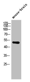
 WB (Western Blot)
(Western Blot analysis of MOUSE-BRAIN cells using ARMCX1 Polyclonal Antibody)
WB (Western Blot)
(Western Blot analysis of MOUSE-BRAIN cells using ARMCX1 Polyclonal Antibody)
ARMCX1, Polyclonal Antibody (Cat# AAA235931)
 WB (Western Blot)
(Western Blot analysis of NIH-3T3 cells using ASK 1 Polyclonal Antibody)
WB (Western Blot)
(Western Blot analysis of NIH-3T3 cells using ASK 1 Polyclonal Antibody)
MAP3K5, Polyclonal Antibody (Cat# AAA235935)
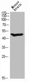
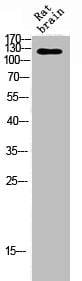 WB (Western Blot)
(Western Blot analysis of RAT-BRAIN cells using Ataxin-2 Polyclonal Antibody)
WB (Western Blot)
(Western Blot analysis of RAT-BRAIN cells using Ataxin-2 Polyclonal Antibody)
ATXN2, Polyclonal Antibody (Cat# AAA235936)
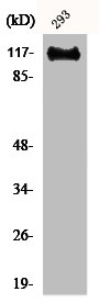
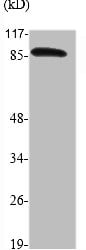 WB (Western Blot)
(Western Blot analysis of various cells using ATRIP Polyclonal Antibody)
WB (Western Blot)
(Western Blot analysis of various cells using ATRIP Polyclonal Antibody)
ATRIP, Polyclonal Antibody (Cat# AAA235940)

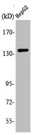
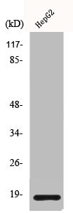
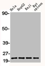 WB (Western Blot)
(Western Blot analysis of HELA HepG2 RAJI RAT-SPLEEN cells using Bax Polyclonal Antibody)
WB (Western Blot)
(Western Blot analysis of HELA HepG2 RAJI RAT-SPLEEN cells using Bax Polyclonal Antibody)
BAX, Polyclonal Antibody (Cat# AAA235946)
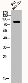
 WB (Western Blot)
(Western Blot analysis of RAT-MUSCLE cells using Bcl-x Polyclonal Antibody)
WB (Western Blot)
(Western Blot analysis of RAT-MUSCLE cells using Bcl-x Polyclonal Antibody)
BCL2L1, Polyclonal Antibody (Cat# AAA235952)
 WB (Western Blot)
(Western Blot analysis of HELA cells using Bcl-x Polyclonal Antibody)
WB (Western Blot)
(Western Blot analysis of HELA cells using Bcl-x Polyclonal Antibody)
BCL2L1, Polyclonal Antibody (Cat# AAA235953)
 WB (Western Blot)
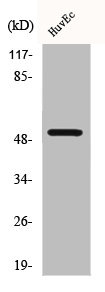
(Western Blot analysis of HuvEc cells using Bek Polyclonal Antibody)
WB (Western Blot)
(Western Blot analysis of HuvEc cells using Bek Polyclonal Antibody)
FGFR2, Polyclonal Antibody (Cat# AAA235954)
 WB (Western Blot)
(Western Blot analysis of MCF7 PC-3 KB HELA cells using BM28 Polyclonal Antibody)
WB (Western Blot)
(Western Blot analysis of MCF7 PC-3 KB HELA cells using BM28 Polyclonal Antibody)
MCM2, Polyclonal Antibody (Cat# AAA235956)
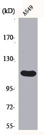
 WB (Western Blot)
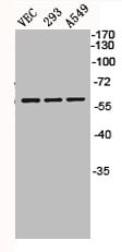
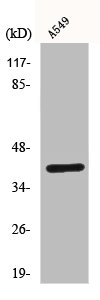
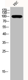
(Western Blot analysis of VEC 293 A549 cells using BMP-5 Polyclonal Antibody)
WB (Western Blot)
(Western Blot analysis of VEC 293 A549 cells using BMP-5 Polyclonal Antibody)
BMP5, Polyclonal Antibody (Cat# AAA235959)
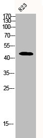
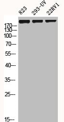 WB (Western Blot)
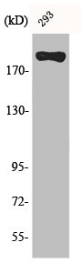
(Western Blot analysis of 823 293-UV 22RV1 cells using BRCA1 Polyclonal Antibody)
WB (Western Blot)
(Western Blot analysis of 823 293-UV 22RV1 cells using BRCA1 Polyclonal Antibody)
BRCA1, Polyclonal Antibody (Cat# AAA235964)
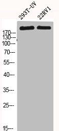 WB (Western Blot)
(Western Blot analysis of 293T-UV 22RV1 cells using BRCA1 Polyclonal Antibody)
WB (Western Blot)
(Western Blot analysis of 293T-UV 22RV1 cells using BRCA1 Polyclonal Antibody)
BRCA1, Polyclonal Antibody (Cat# AAA235965)
 WB (Western Blot)
(Western Blot analysis of various cells using BRS-3 Polyclonal Antibody)
WB (Western Blot)
(Western Blot analysis of various cells using BRS-3 Polyclonal Antibody)
BRS3, Polyclonal Antibody (Cat# AAA235968)
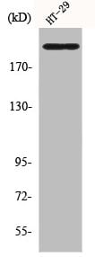
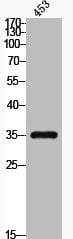 WB (Western Blot)
(Western Blot analysis of 453 cells using C/EBP epsilon Polyclonal Antibody)
WB (Western Blot)
(Western Blot analysis of 453 cells using C/EBP epsilon Polyclonal Antibody)
CEBPE, Polyclonal Antibody (Cat# AAA235973)
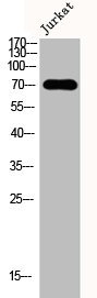
 WB (Western Blot)
(Western Blot analysis of Jurkat cells using C1INH Polyclonal Antibody)
WB (Western Blot)
(Western Blot analysis of Jurkat cells using C1INH Polyclonal Antibody)
SERPING1, Polyclonal Antibody (Cat# AAA235974)
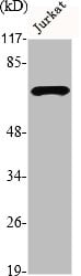
 WB (Western Blot)
(Western Blot analysis of Jurkat cells using Abl1 Polyclonal Antibody)
WB (Western Blot)
(Western Blot analysis of Jurkat cells using Abl1 Polyclonal Antibody)
ABL1, Polyclonal Antibody (Cat# AAA235977)
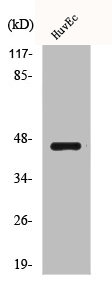
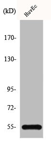
 WB (Western Blot)
(Western Blot analysis of HuvEc cells using Cadherin-16 Polyclonal Antibody)
WB (Western Blot)
(Western Blot analysis of HuvEc cells using Cadherin-16 Polyclonal Antibody)
CDH16, Polyclonal Antibody (Cat# AAA235978)
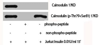 WB (Western Blot)
(Western Blot analysis of Jukat+Insulin cells using Calmodulin Polyclonal Antibody)
WB (Western Blot)
(Western Blot analysis of Jukat+Insulin cells using Calmodulin Polyclonal Antibody)
CALM1, Polyclonal Antibody (Cat# AAA235979)
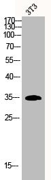
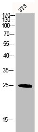
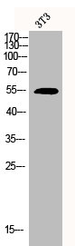
 WB (Western Blot)
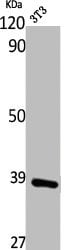
(Western Blot analysis of NIH-3T3 cells using Casein Kinase Ialpha Polyclonal Antibody)
WB (Western Blot)
(Western Blot analysis of NIH-3T3 cells using Casein Kinase Ialpha Polyclonal Antibody)
CSNK1A1, Polyclonal Antibody (Cat# AAA235982)
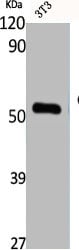
 WB (Western Blot)
(Western Blot analysis of NIH-3T3 cells using Catenin-gamma Polyclonal Antibody)
WB (Western Blot)
(Western Blot analysis of NIH-3T3 cells using Catenin-gamma Polyclonal Antibody)
JUP, Polyclonal Antibody (Cat# AAA235992)
 WB (Western Blot)
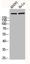
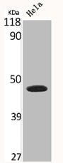
(Western Blot analysis of AD293 HELA cells using CBP Polyclonal Antibody)
WB (Western Blot)
(Western Blot analysis of AD293 HELA cells using CBP Polyclonal Antibody)
CREBBP, Polyclonal Antibody (Cat# AAA235994)
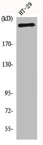
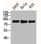 WB (Western Blot)
(Western Blot analysis of 293T Hela 823 cells using CD44 Polyclonal Antibody)
WB (Western Blot)
(Western Blot analysis of 293T Hela 823 cells using CD44 Polyclonal Antibody)
CD44, Polyclonal Antibody (Cat# AAA235999)
 WB (Western Blot)
(Western Blot analysis of L929 cells using CD9 Polyclonal Antibody)
WB (Western Blot)
(Western Blot analysis of L929 cells using CD9 Polyclonal Antibody)
CD9, Polyclonal Antibody (Cat# AAA236000)
 WB (Western Blot)
(Western Blot analysis of NIH-3T3 cells using CD95 Polyclonal Antibody)
WB (Western Blot)
(Western Blot analysis of NIH-3T3 cells using CD95 Polyclonal Antibody)
FAS, Polyclonal Antibody (Cat# AAA236001)
 WB (Western Blot)
(Western Blot analysis of NIH-3T3 cells using Cdc20 Polyclonal Antibody)
WB (Western Blot)
(Western Blot analysis of NIH-3T3 cells using Cdc20 Polyclonal Antibody)
CDC20, Polyclonal Antibody (Cat# AAA236002)
 WB (Western Blot)
(Western Blot analysis of NIH-3T3 cells using Cdc23 Polyclonal Antibody)
WB (Western Blot)
(Western Blot analysis of NIH-3T3 cells using Cdc23 Polyclonal Antibody)
CDC23, Polyclonal Antibody (Cat# AAA236003)
 WB (Western Blot)
(Western Blot analysis of K562 Jurkat cells using CDYL2 Polyclonal Antibody)
WB (Western Blot)
(Western Blot analysis of K562 Jurkat cells using CDYL2 Polyclonal Antibody)
CDYL2, Polyclonal Antibody (Cat# AAA236010)
 WB (Western Blot)
(Western Blot analysis of NIH-3T3 cells using CLC-4 Polyclonal Antibody)
WB (Western Blot)
(Western Blot analysis of NIH-3T3 cells using CLC-4 Polyclonal Antibody)
CLCN4, Polyclonal Antibody (Cat# AAA236022)
 WB (Western Blot)
(Western Blot analysis of NIH-3T3 cells using CNT2 Polyclonal Antibody)
WB (Western Blot)
(Western Blot analysis of NIH-3T3 cells using CNT2 Polyclonal Antibody)
SLC28A2, Polyclonal Antibody (Cat# AAA236033)
 WB (Western Blot)
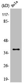
(Western Blot analysis of HELA cells using Connexin 43 Polyclonal Antibody)
WB (Western Blot)
(Western Blot analysis of HELA cells using Connexin 43 Polyclonal Antibody)
GJA1, Polyclonal Antibody (Cat# AAA236037)
 WB (Western Blot)
(Western Blot analysis of Jurkat cells using DMPK Polyclonal Antibody)
WB (Western Blot)
(Western Blot analysis of Jurkat cells using DMPK Polyclonal Antibody)
DMPK, Polyclonal Antibody (Cat# AAA236096)
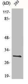
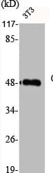
 WB (Western Blot)
(Western Blot analysis of 293 A549 cells using DNA Ligase IV Polyclonal Antibody)
WB (Western Blot)
(Western Blot analysis of 293 A549 cells using DNA Ligase IV Polyclonal Antibody)
LIG4, Polyclonal Antibody (Cat# AAA236098)
What are Polyclonal Antibodies?
Polyclonal antibodies are antibodies that come from multiple B cell clones of a host animal. The typical hosts used for the majority of polyclonal antibody production are rabbits, goats, sheep, and donkeys. These polyclonal antibodies, once having identified their target, will bind to different epitopes located at different regions or sequences on the same protein/antigen. As a result, they are ideal at locating and binding to the target, even if the target is in very low concentrations (due to many different antibodies being able to bind to the same target molecule, which allows for significant amplification of a downstream signal).
Polyclonal antibodies are typically produced by injecting an antigen into a host animal, which causes the animal’s immune system to attack the foreign antigen by mass generating antibodies against it. After a period of time, serum is collected from the animal and purified using physicochemical fractionation, class-specific affinity purification, and/or antigen-affinity purification.
Key Uses of Polyclonal Antibodies
- Western Blotting: This method is used to find specific proteins in biological samples after separating them by size.
- Immunohistochemistry: IHC helps visualize the location of proteins in tissue sections using various staining techniques.
- ELISA: (Enzyme-Linked Immunosorbent Assay) is typically used to identify specific protein quantities in a sample. ELISAs can be either “Quantitative” or “Qualitative”.
- Flow Cytometry: technique that identifies and measures the specific protein on the surface or inside the cells in a fluid suspension.
- Immunoprecipitation: IP isolates and studies a specific protein from a complex mixture using antibodies.
Why Buy Polyclonal Antibodies from AAA Biotech?
1. Ideal for Various Applications
Our antibodies are generally going to be validated for use in multiple types of assays, including ELISA, Western Blotting, Immunohistochemistry, Immunoprecipitation, amongst others. They are ideal for a wide range of research applications.
2. Rigorous Quality Control
All of the antibodies in our catalog undergo strict quality testing to ensure specificity, sensitivity, and consistent performance. We are confident in the ability of our antibodies to provide you with accurate results.
3. Wide Assortment of Antibodies
Antibodies in are catalog can be found for both common and exotic species, and these antibodies are also available in both conjugated and recombinant forms to suit many diverse experimental needs.
4. Highly Purified
Our antibodies are available in purified forms with over 85% purity, as confirmed by SDS-PAGE. They are also available with tags such as His, Flag, GST, or MBP. We cater to customers worldwide.
FAQ
1. How are polyclonal antibodies produced?
Traditionally, polyclonal antibodies are produced by injecting an antigen into a host animal (such as a rabbit or goat), which then triggers an immune response from the host animal. The animal’s B cells produce antibodies that will recognize different parts of the injected antigen. These antibodies are then collected from the animal’s blood and purified for use.
2. How do polyclonal antibodies differ from monoclonal antibodies?
Polyclonal antibodies are a mix of antibodies that bind to different locations (epitopes) of the same antigen, while monoclonal antibodies are identical and bind to just one specific epitope. This makes polyclonal antibodies more versatile and better at detecting proteins that may be present in low quantities or in altered/modified forms.
3. How should I store polyclonal antibodies?
Polyclonal antibodies should be stored at 4°C for short-term use (up to a few weeks) and at -20°C or -80°C for long-term storage. Avoid repeated freeze-thaw cycles by dividing them into small aliquots. Always check the datasheet for specific storage instructions.