Filters
▼Clonality
▼Type
▼Reactivity
▼Gene Name
▼Isotype
▼Host
▼Application
▼Clone
▼Polyclonal Antibodies
At AAA Biotech also known as AAA Bio or AAABio, we provide a broad range of purified polyclonal antibodies (pAbs) that are able to all be browsed online through our website. Due to their high specificity and strong binding affinity, these antibodies are ideal for wide swathes of research and experimental applications.
Our polyclonal antibodies can easily support your work, whether you use them for Western Blotting, Immunocytochemistry (with or without Immunofluorescence used in conjunction), Immunohistochemistry, Immunoprecipitation, and ELISA tests. We highly encourage you to browse our range of pAbs and choose the one that best suits your experimental model.
Viewing 9500-9550 of 96805 product results
 IHC (Immunohistochemisry)
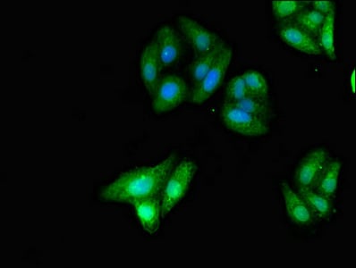
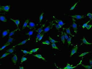
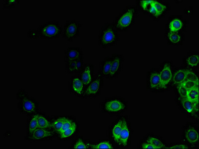
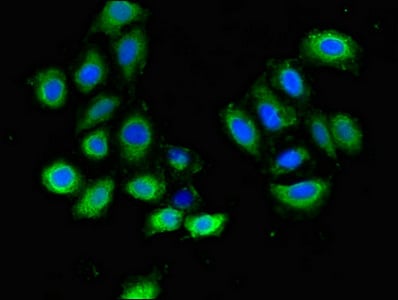
(Immunofluorescent analysis of Hela cells using AAA233216 at a dilution of 1:100 and Alexa Fluor 488-congugated AffiniPure Goat Anti-Rabbit IgG(H+L))
IHC (Immunohistochemisry)
(Immunofluorescent analysis of Hela cells using AAA233216 at a dilution of 1:100 and Alexa Fluor 488-congugated AffiniPure Goat Anti-Rabbit IgG(H+L))
GNAT1, Polyclonal Antibody (Cat# AAA233216)
 IHC (Immunohistochemisry)
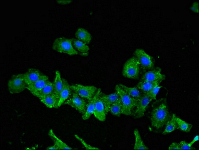
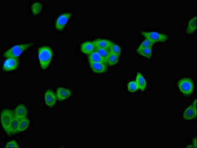
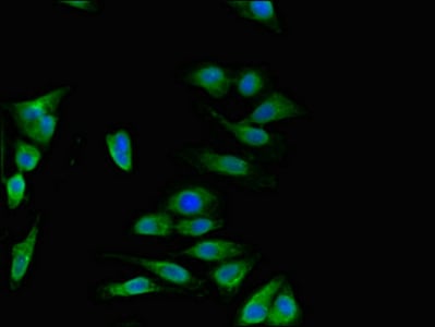
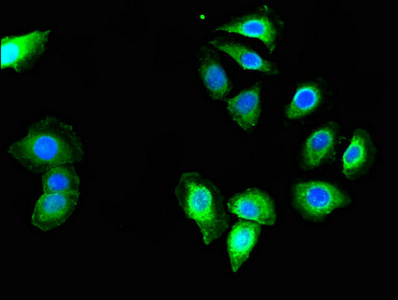
(Immunofluorescent analysis of HepG2 cells using AAA233222 at a dilution of 1:100 and Alexa Fluor 488-congugated AffiniPure Goat Anti-Rabbit IgG(H+L))
IHC (Immunohistochemisry)
(Immunofluorescent analysis of HepG2 cells using AAA233222 at a dilution of 1:100 and Alexa Fluor 488-congugated AffiniPure Goat Anti-Rabbit IgG(H+L))
GZMK, Polyclonal Antibody (Cat# AAA233222)
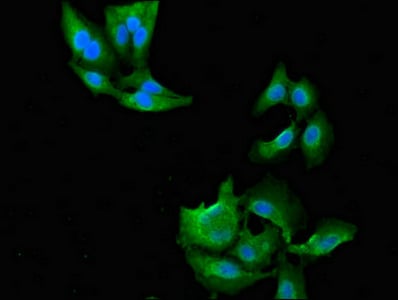
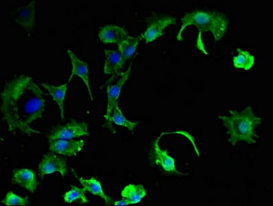
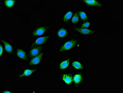
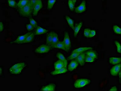
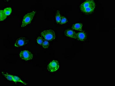
 IF (Immunofluorescence)
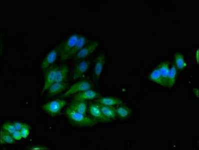
(Immunofluorescent analysis of HepG2 cells using AAA233227 at a dilution of 1:100 and Alexa Fluor 488-congugated AffiniPure Goat Anti-Rabbit IgG(H+L))
IF (Immunofluorescence)
(Immunofluorescent analysis of HepG2 cells using AAA233227 at a dilution of 1:100 and Alexa Fluor 488-congugated AffiniPure Goat Anti-Rabbit IgG(H+L))
IDH1, Polyclonal Antibody (Cat# AAA233227)
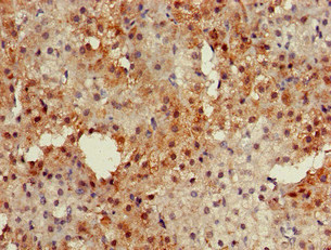
 IHC (Immunohistochemisry)
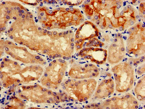
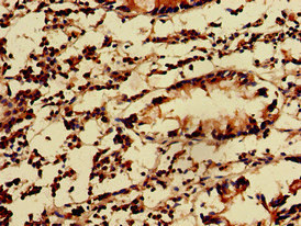
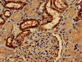
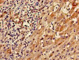
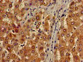
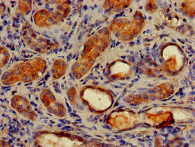
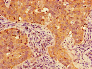
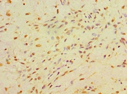
(Immunohistochemistry analysis of human kidney tissue using AAA233229 at dilution of 1:100)
IHC (Immunohistochemisry)
(Immunohistochemistry analysis of human kidney tissue using AAA233229 at dilution of 1:100)
IGHD, Polyclonal Antibody (Cat# AAA233229)

 IHC (Immunohistochemisry)
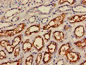
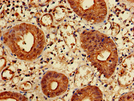
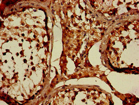
(Immunohistochemistry of paraffin-embedded human kidney tissue using AAA233234 at dilution of 1:100)
IHC (Immunohistochemisry)
(Immunohistochemistry of paraffin-embedded human kidney tissue using AAA233234 at dilution of 1:100)
IREB2, Polyclonal Antibody (Cat# AAA233234)
 IHC (Immunohistochemisry)
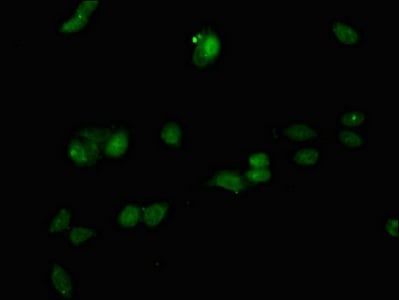
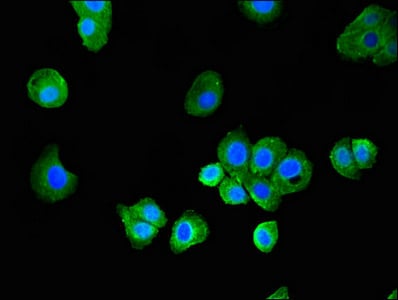
(Immunofluorescent analysis of Hela cells using AAA233235 at a dilution of 1:100 and Alexa Fluor 488-congugated AffiniPure Goat Anti-Rabbit IgG(H+L))
IHC (Immunohistochemisry)
(Immunofluorescent analysis of Hela cells using AAA233235 at a dilution of 1:100 and Alexa Fluor 488-congugated AffiniPure Goat Anti-Rabbit IgG(H+L))
IRS2, Polyclonal Antibody (Cat# AAA233235)
 IF (Immunofluorescence)
(Immunofluorescent analysis of HepG2 cells using AAA233237 at a dilution of 1:100 and Alexa Fluor 488-congugated AffiniPure Goat Anti-Rabbit IgG(H+L))
IF (Immunofluorescence)
(Immunofluorescent analysis of HepG2 cells using AAA233237 at a dilution of 1:100 and Alexa Fluor 488-congugated AffiniPure Goat Anti-Rabbit IgG(H+L))
KIDINS220, Polyclonal Antibody (Cat# AAA233237)
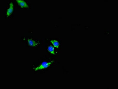
 IF (Immunofluorescence)
(Immunofluorescent analysis of PC3 cells using AAA233240 at a dilution of 1:100 and Alexa Fluor 488-congugated AffiniPure Goat Anti-Rabbit IgG(H+L))
IF (Immunofluorescence)
(Immunofluorescent analysis of PC3 cells using AAA233240 at a dilution of 1:100 and Alexa Fluor 488-congugated AffiniPure Goat Anti-Rabbit IgG(H+L))
MARK3, Polyclonal Antibody (Cat# AAA233240)
 IF (Immunofluorescence)
(Immunofluorescent analysis of Hela cells using AAA233248 at a dilution of 1:100 and Alexa Fluor 488-congugated AffiniPure Goat Anti-Rabbit IgG(H+L))
IF (Immunofluorescence)
(Immunofluorescent analysis of Hela cells using AAA233248 at a dilution of 1:100 and Alexa Fluor 488-congugated AffiniPure Goat Anti-Rabbit IgG(H+L))
NARF, Polyclonal Antibody (Cat# AAA233248)
 IHC (Immunohistochemisry)
(Immunofluorescent analysis of Hela cells using AAA233249 at a dilution of 1:100 and Alexa Fluor 488-congugated AffiniPure Goat Anti-Rabbit IgG(H+L))
IHC (Immunohistochemisry)
(Immunofluorescent analysis of Hela cells using AAA233249 at a dilution of 1:100 and Alexa Fluor 488-congugated AffiniPure Goat Anti-Rabbit IgG(H+L))
NAT1, Polyclonal Antibody (Cat# AAA233249)
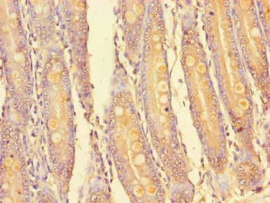
 IHC (Immunohistochemisry)
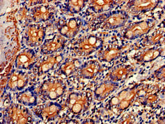
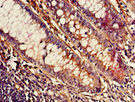
(Immunohistochemistry of paraffin-embedded human small intestine tissue using AAA233253 at dilution of 1:100)
IHC (Immunohistochemisry)
(Immunohistochemistry of paraffin-embedded human small intestine tissue using AAA233253 at dilution of 1:100)
NR0B1, Polyclonal Antibody (Cat# AAA233253)
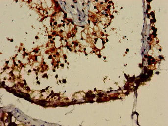
 IHC (Immunohistochemisry)
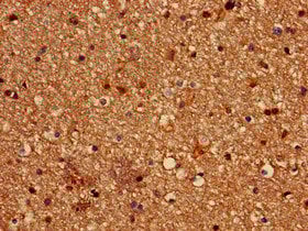
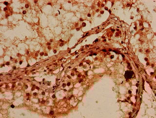
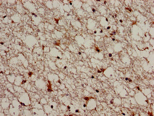
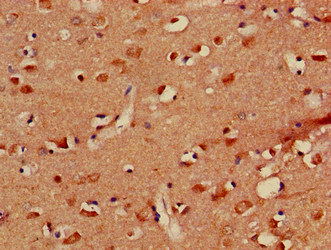
(Immunohistochemistry of paraffin-embedded human brain tissue using AAA233257 at dilution of 1:100)
IHC (Immunohistochemisry)
(Immunohistochemistry of paraffin-embedded human brain tissue using AAA233257 at dilution of 1:100)
PALM, Polyclonal Antibody (Cat# AAA233257)
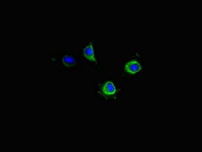 IHC (Immunohistochemisry)
(Immunofluorescent analysis of Hela cells using AAA233062 at a dilution of 1:100 and Alexa Fluor 488-congugated AffiniPure Goat Anti-Rabbit IgG(H+L))
IHC (Immunohistochemisry)
(Immunofluorescent analysis of Hela cells using AAA233062 at a dilution of 1:100 and Alexa Fluor 488-congugated AffiniPure Goat Anti-Rabbit IgG(H+L))
PIM2, Polyclonal Antibody (Cat# AAA233062)
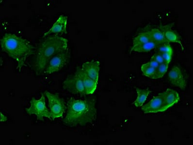
 IF (Immunofluorescence)
(Immunofluorescent analysis of HepG2 cells using AAA233067 at a dilution of 1:100 and Alexa Fluor 488-congugated AffiniPure Goat Anti-Rabbit IgG(H+L))
IF (Immunofluorescence)
(Immunofluorescent analysis of HepG2 cells using AAA233067 at a dilution of 1:100 and Alexa Fluor 488-congugated AffiniPure Goat Anti-Rabbit IgG(H+L))
SEMA6A, Polyclonal Antibody (Cat# AAA233067)
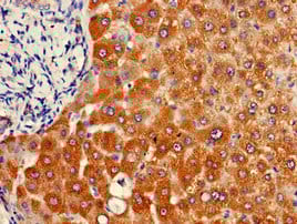
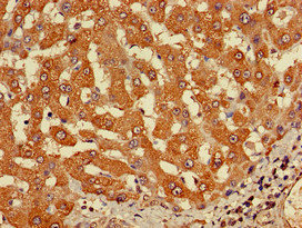
 IHC (Immunohistochemisry)
(Immunofluorescent analysis of HepG2 cells using AAA233080 at a dilution of 1:100 and Alexa Fluor 488-congugated AffiniPure Goat Anti-Rabbit IgG(H+L))
IHC (Immunohistochemisry)
(Immunofluorescent analysis of HepG2 cells using AAA233080 at a dilution of 1:100 and Alexa Fluor 488-congugated AffiniPure Goat Anti-Rabbit IgG(H+L))
SLCO1B3, Polyclonal Antibody (Cat# AAA233080)
 IHC (Immunohistochemisry)
(Immunofluorescent analysis of HepG2 cells using AAA233081 at a dilution of 1:100 and Alexa Fluor 488-congugated AffiniPure Goat Anti-Rabbit IgG(H+L))
IHC (Immunohistochemisry)
(Immunofluorescent analysis of HepG2 cells using AAA233081 at a dilution of 1:100 and Alexa Fluor 488-congugated AffiniPure Goat Anti-Rabbit IgG(H+L))
SNX15, Polyclonal Antibody (Cat# AAA233081)
 IHC (Immunohistochemisry)
(Immunofluorescent analysis of MCF-7 cells using AAA233091 at a dilution of 1:100 and Alexa Fluor 488-congugated AffiniPure Goat Anti-Rabbit IgG(H+L))
IHC (Immunohistochemisry)
(Immunofluorescent analysis of MCF-7 cells using AAA233091 at a dilution of 1:100 and Alexa Fluor 488-congugated AffiniPure Goat Anti-Rabbit IgG(H+L))
PKIB, Polyclonal Antibody (Cat# AAA233091)
 IF (Immunofluorescence)
(Immunofluorescent analysis of HepG2 cells using AAA233092 at a dilution of 1:100 and Alexa Fluor 488-congugated AffiniPure Goat Anti-Rabbit IgG(H+L))
IF (Immunofluorescence)
(Immunofluorescent analysis of HepG2 cells using AAA233092 at a dilution of 1:100 and Alexa Fluor 488-congugated AffiniPure Goat Anti-Rabbit IgG(H+L))
RACGAP1, Polyclonal Antibody (Cat# AAA233092)
 IHC (Immunohistochemisry)
(Immunofluorescent analysis of Hela cells using AAA233099 at a dilution of 1:100 and Alexa Fluor 488-congugated AffiniPure Goat Anti-Rabbit IgG(H+L))
IHC (Immunohistochemisry)
(Immunofluorescent analysis of Hela cells using AAA233099 at a dilution of 1:100 and Alexa Fluor 488-congugated AffiniPure Goat Anti-Rabbit IgG(H+L))
PRMT7, Polyclonal Antibody (Cat# AAA233099)
 IF (Immunofluorescence)
(Immunofluorescent analysis of HepG2 cells using AAA233100 at a dilution of 1:100 and Alexa Fluor 488-congugated AffiniPure Goat Anti-Rabbit IgG(H+L))
IF (Immunofluorescence)
(Immunofluorescent analysis of HepG2 cells using AAA233100 at a dilution of 1:100 and Alexa Fluor 488-congugated AffiniPure Goat Anti-Rabbit IgG(H+L))
ALKBH4, Polyclonal Antibody (Cat# AAA233100)
 IF (Immunofluorescence)
(Immunofluorescent analysis of HepG2 cells using AAA233103 at a dilution of 1:100 and Alexa Fluor 488-congugated AffiniPure Goat Anti-Rabbit IgG(H+L))
IF (Immunofluorescence)
(Immunofluorescent analysis of HepG2 cells using AAA233103 at a dilution of 1:100 and Alexa Fluor 488-congugated AffiniPure Goat Anti-Rabbit IgG(H+L))
GULP1, Polyclonal Antibody (Cat# AAA233103)
 IHC (Immunohistochemisry)
(Immunofluorescent analysis of HepG2 cells using AAA233104 at a dilution of 1:100 and Alexa Fluor 488-congugated AffiniPure Goat Anti-Rabbit IgG(H+L))
IHC (Immunohistochemisry)
(Immunofluorescent analysis of HepG2 cells using AAA233104 at a dilution of 1:100 and Alexa Fluor 488-congugated AffiniPure Goat Anti-Rabbit IgG(H+L))
MAP1LC3C, Polyclonal Antibody (Cat# AAA233104)
 IHC (Immunohistochemisry)
(Immunofluorescent analysis of A549 cells using AAA233106 at a dilution of 1:100 and Alexa Fluor 488-congugated AffiniPure Goat Anti-Rabbit IgG(H+L))
IHC (Immunohistochemisry)
(Immunofluorescent analysis of A549 cells using AAA233106 at a dilution of 1:100 and Alexa Fluor 488-congugated AffiniPure Goat Anti-Rabbit IgG(H+L))
WNK1, Polyclonal Antibody (Cat# AAA233106)
 IF (Immunofluorescence)
(Immunofluorescent analysis of HepG2 cells using AAA233107 at a dilution of 1:100 and Alexa Fluor 488-congugated AffiniPure Goat Anti-Rabbit IgG(H+L))
IF (Immunofluorescence)
(Immunofluorescent analysis of HepG2 cells using AAA233107 at a dilution of 1:100 and Alexa Fluor 488-congugated AffiniPure Goat Anti-Rabbit IgG(H+L))
ABCB9, Polyclonal Antibody (Cat# AAA233107)
 IHC (Immunohistochemisry)
(Immunofluorescent analysis of Hela cells using AAA233108 at a dilution of 1:100 and Alexa Fluor 488-congugated AffiniPure Goat Anti-Rabbit IgG(H+L))
IHC (Immunohistochemisry)
(Immunofluorescent analysis of Hela cells using AAA233108 at a dilution of 1:100 and Alexa Fluor 488-congugated AffiniPure Goat Anti-Rabbit IgG(H+L))
TIGAR, Polyclonal Antibody (Cat# AAA233108)
 IF (Immunofluorescence)
(Immunofluorescent analysis of Hela cells using AAA233109 at a dilution of 1:100 and Alexa Fluor 488-congugated AffiniPure Goat Anti-Rabbit IgG(H+L))
IF (Immunofluorescence)
(Immunofluorescent analysis of Hela cells using AAA233109 at a dilution of 1:100 and Alexa Fluor 488-congugated AffiniPure Goat Anti-Rabbit IgG(H+L))
DISC1, Polyclonal Antibody (Cat# AAA233109)
MCM8, Polyclonal Antibody (Cat# AAA233113)
 IF (Immunofluorescence)
(Immunofluorescent analysis of HepG2 cells using AAA233114 at a dilution of 1:100 and Alexa Fluor 488-congugated AffiniPure Goat Anti-Rabbit IgG(H+L))
IF (Immunofluorescence)
(Immunofluorescent analysis of HepG2 cells using AAA233114 at a dilution of 1:100 and Alexa Fluor 488-congugated AffiniPure Goat Anti-Rabbit IgG(H+L))
GGA1, Polyclonal Antibody (Cat# AAA233114)
 IF (Immunofluorescence)
(Immunofluorescent analysis of A549 cells using AAA233115 at a dilution of 1:100 and Alexa Fluor 488-congugated AffiniPure Goat Anti-Rabbit IgG(H+L))
IF (Immunofluorescence)
(Immunofluorescent analysis of A549 cells using AAA233115 at a dilution of 1:100 and Alexa Fluor 488-congugated AffiniPure Goat Anti-Rabbit IgG(H+L))
MED23, Polyclonal Antibody (Cat# AAA233115)
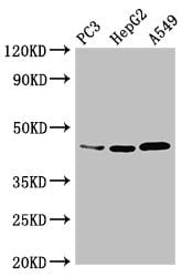 WB (Western Blot)
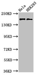
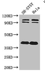
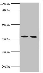
(Western BlotPositive WB detected in: PC3 whole cell lysate,HepG2 whole cell lysate,A549 whole cell lysateAll lanes: DNMT3L antibody at 3.4ug/mlSecondaryGoat polyclonal to rabbit IgG at 1/50000 dilutionPredicted band size: 44 KDaObserved band size: 44 KDa)
WB (Western Blot)
(Western BlotPositive WB detected in: PC3 whole cell lysate,HepG2 whole cell lysate,A549 whole cell lysateAll lanes: DNMT3L antibody at 3.4ug/mlSecondaryGoat polyclonal to rabbit IgG at 1/50000 dilutionPredicted band size: 44 KDaObserved band size: 44 KDa)
DNMT3L, Polyclonal Antibody (Cat# AAA233117)
 IF (Immunofluorescence)
(Immunofluorescent analysis of HepG2 cells using AAA233118 at a dilution of 1:100 and Alexa Fluor 488-congugated AffiniPure Goat Anti-Rabbit IgG(H+L))
IF (Immunofluorescence)
(Immunofluorescent analysis of HepG2 cells using AAA233118 at a dilution of 1:100 and Alexa Fluor 488-congugated AffiniPure Goat Anti-Rabbit IgG(H+L))
FBXO9, Polyclonal Antibody (Cat# AAA233118)
 IHC (Immunohistochemisry)
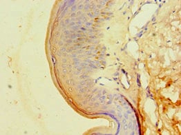
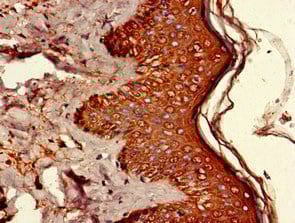
(Immunohistochemistry of paraffin-embedded human skin tissue using AAA233119 at dilution 1:100)
IHC (Immunohistochemisry)
(Immunohistochemistry of paraffin-embedded human skin tissue using AAA233119 at dilution 1:100)
CA14, Polyclonal Antibody (Cat# AAA233119)
 IF (Immunofluorescence)
(Immunofluorescent analysis of MCF-7 cells using AAA233121 at a dilution of 1:100 and Alexa Fluor 488-congugated AffiniPure Goat Anti-Rabbit IgG(H+L))
IF (Immunofluorescence)
(Immunofluorescent analysis of MCF-7 cells using AAA233121 at a dilution of 1:100 and Alexa Fluor 488-congugated AffiniPure Goat Anti-Rabbit IgG(H+L))
TRIM33, Polyclonal Antibody (Cat# AAA233121)
PDZRN3, Polyclonal Antibody (Cat# AAA233123)
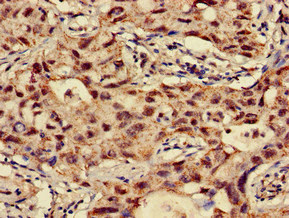
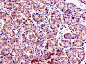
 IHC (Immunohiostchemistry)
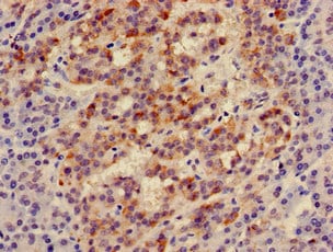
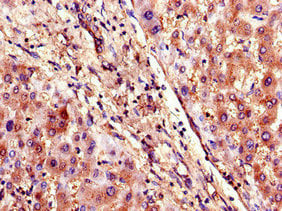
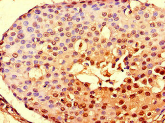
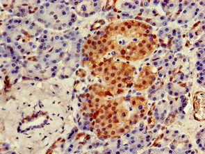
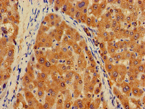
(Immunohistochemistry analysis of human pancreatic cancer using AAA233124 at dilution of 1:100)
IHC (Immunohiostchemistry)
(Immunohistochemistry analysis of human pancreatic cancer using AAA233124 at dilution of 1:100)
ICK, Polyclonal Antibody (Cat# AAA233124)
 WB (Western Blot)
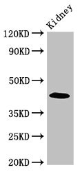
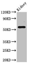
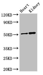
(Western BlotPositive WB detected in: Mouse kidney tissueAll lanes: FBXO3 antibody at 4ug/mlSecondaryGoat polyclonal to rabbit IgG at 1/50000 dilutionPredicted band size: 55,48,47 KDaObserved band size: 55 KDa)
WB (Western Blot)
(Western BlotPositive WB detected in: Mouse kidney tissueAll lanes: FBXO3 antibody at 4ug/mlSecondaryGoat polyclonal to rabbit IgG at 1/50000 dilutionPredicted band size: 55,48,47 KDaObserved band size: 55 KDa)
FBXO3, Polyclonal Antibody (Cat# AAA233129)
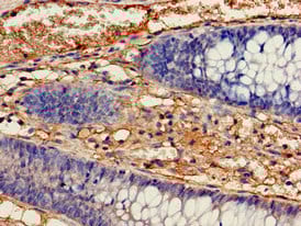
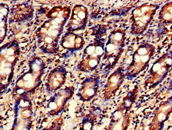
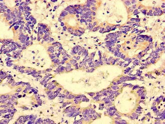
 IHC (Immunohiostchemistry)
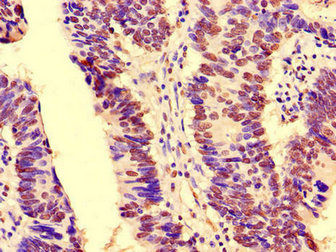
(Immunohistochemistry of paraffin-embedded human colon cancer using AAA233131 at dilution of 1:100)
IHC (Immunohiostchemistry)
(Immunohistochemistry of paraffin-embedded human colon cancer using AAA233131 at dilution of 1:100)
NOTCH3, Polyclonal Antibody (Cat# AAA233131)
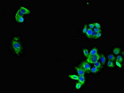
 IF (Immunofluorescence)
(Immunofluorescent analysis of HepG2 cells using AAA233134 at a dilution of 1:100 and Alexa Fluor 488-congugated AffiniPure Goat Anti-Rabbit IgG(H+L))
IF (Immunofluorescence)
(Immunofluorescent analysis of HepG2 cells using AAA233134 at a dilution of 1:100 and Alexa Fluor 488-congugated AffiniPure Goat Anti-Rabbit IgG(H+L))
UBQLN1, Polyclonal Antibody (Cat# AAA233134)

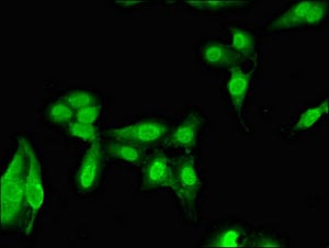
 IF (Immunofluorescence)
(Immunofluorescent analysis of HepG2 cells using AAA233136 at a dilution of 1:100 and Alexa Fluor 488-congugated AffiniPure Goat Anti-Rabbit IgG(H+L))
IF (Immunofluorescence)
(Immunofluorescent analysis of HepG2 cells using AAA233136 at a dilution of 1:100 and Alexa Fluor 488-congugated AffiniPure Goat Anti-Rabbit IgG(H+L))
SIGLEC7, Polyclonal Antibody (Cat# AAA233136)
porB, Polyclonal Antibody (Cat# AAA233340)
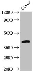
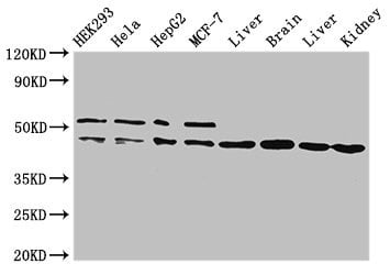
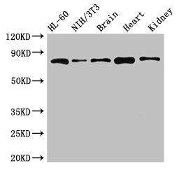
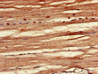
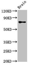
 WB (Western Blot)
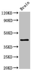
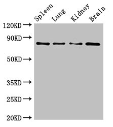
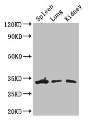
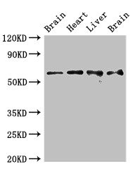
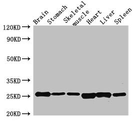
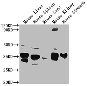
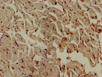
(Western BlotPositive WB detected in: Mouse Liver tissue, Mouse Spleen tissue, Mouse Lung tissue, Mouse Kidney tissue, Mouse Stomach tissueAll lanes: Fgf15 antibody at 1:2000SecondaryGoat polyclonal to rabbit IgG at 1/50000 dilutionPredicted band size: 26 kDaObserved band size: 26 kDa)
WB (Western Blot)
(Western BlotPositive WB detected in: Mouse Liver tissue, Mouse Spleen tissue, Mouse Lung tissue, Mouse Kidney tissue, Mouse Stomach tissueAll lanes: Fgf15 antibody at 1:2000SecondaryGoat polyclonal to rabbit IgG at 1/50000 dilutionPredicted band size: 26 kDaObserved band size: 26 kDa)
Fgf15, Polyclonal Antibody (Cat# AAA233341)
MYLK, Polyclonal Antibody (Cat# AAA233357)
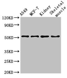
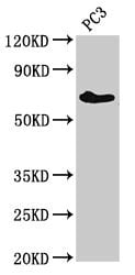
 WB (Western Blot)
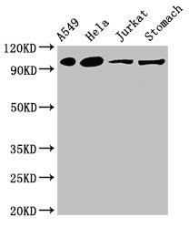
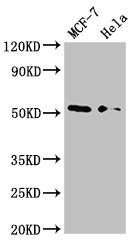
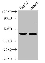
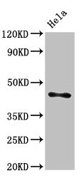
(Western BlotPositive WB detected in: Hela whole cell lysateAll lanes: ASAH1 antibody at 3ug/mlSecondaryGoat polyclonal to rabbit IgG at 1/50000 dilutionPredicted band size: 45,47 KDaObserved band size: 45 KDa)
WB (Western Blot)
(Western BlotPositive WB detected in: Hela whole cell lysateAll lanes: ASAH1 antibody at 3ug/mlSecondaryGoat polyclonal to rabbit IgG at 1/50000 dilutionPredicted band size: 45,47 KDaObserved band size: 45 KDa)
ASAH1, Polyclonal Antibody (Cat# AAA233358)
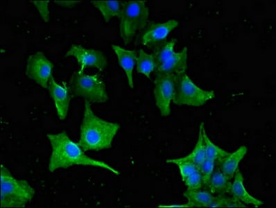
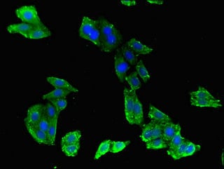
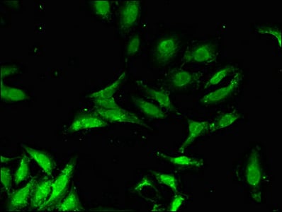
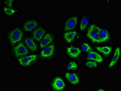
 IF (Immunofluorescence)
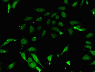
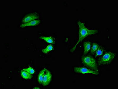
(Immunofluorescent analysis of A549 cells using AAA233360 at a dilution of 1:100 and Alexa Fluor 488-congugated AffiniPure Goat Anti-Rabbit IgG(H+L))
IF (Immunofluorescence)
(Immunofluorescent analysis of A549 cells using AAA233360 at a dilution of 1:100 and Alexa Fluor 488-congugated AffiniPure Goat Anti-Rabbit IgG(H+L))
ZMYND11, Polyclonal Antibody (Cat# AAA233360)
 IHC (Immunohistochemisry)
(Immunofluorescent analysis of A549 cells using AAA233370 at a dilution of 1:100 and Alexa Fluor 488-congugated AffiniPure Goat Anti-Rabbit IgG(H+L))
IHC (Immunohistochemisry)
(Immunofluorescent analysis of A549 cells using AAA233370 at a dilution of 1:100 and Alexa Fluor 488-congugated AffiniPure Goat Anti-Rabbit IgG(H+L))
PLXND1, Polyclonal Antibody (Cat# AAA233370)
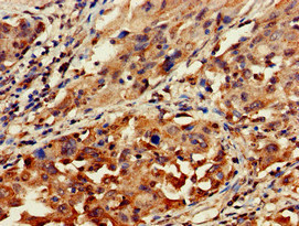
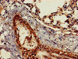
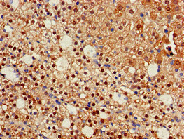
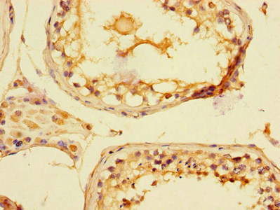
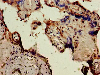
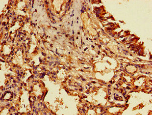
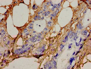
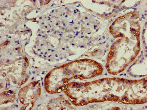
 IHC (Immunohiostchemistry)
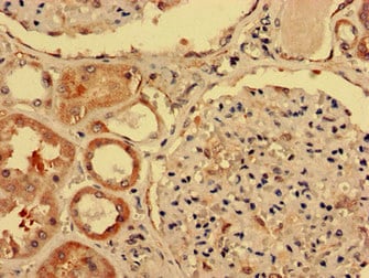
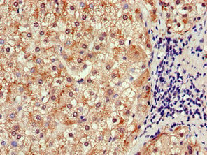
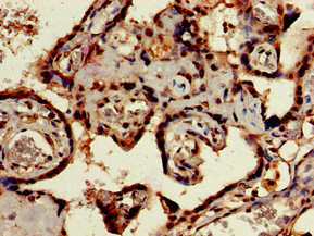
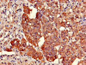
(Immunohistochemistry of paraffin-embedded human adrenal gland tissue using AAA233372 at dilution of 1:100)
IHC (Immunohiostchemistry)
(Immunohistochemistry of paraffin-embedded human adrenal gland tissue using AAA233372 at dilution of 1:100)
EARS2, Polyclonal Antibody (Cat# AAA233372)
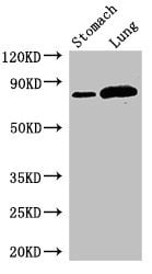 WB (Western Blot)
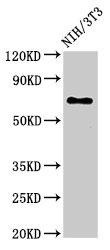
(Western BlotPositive WB detected in: Mouse stomach tissue,Rat lung tissueAll lanes: SLC3A1 antibody at 2.8ug/mlSecondaryGoat polyclonal to rabbit IgG at 1/50000 dilutionPredicted band size: 79,47,46,36,58,67,64 KDaObserved band size: 79 KDa)
WB (Western Blot)
(Western BlotPositive WB detected in: Mouse stomach tissue,Rat lung tissueAll lanes: SLC3A1 antibody at 2.8ug/mlSecondaryGoat polyclonal to rabbit IgG at 1/50000 dilutionPredicted band size: 79,47,46,36,58,67,64 KDaObserved band size: 79 KDa)
SLC3A1, Polyclonal Antibody (Cat# AAA233375)
 IHC (Immunohistochemisry)
(Immunofluorescent analysis of MCF-7 cells using AAA233378 at a dilution of 1:100 and Alexa Fluor 488-congugated AffiniPure Goat Anti-Rabbit IgG(H+L))
IHC (Immunohistochemisry)
(Immunofluorescent analysis of MCF-7 cells using AAA233378 at a dilution of 1:100 and Alexa Fluor 488-congugated AffiniPure Goat Anti-Rabbit IgG(H+L))
CYP20A1, Polyclonal Antibody (Cat# AAA233378)
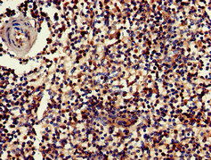
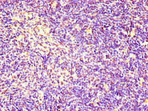
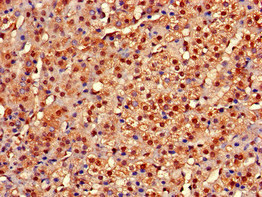
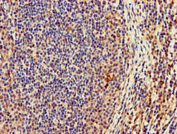 IHC (Immunohiostchemistry)
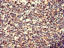
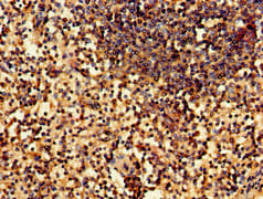
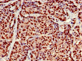
(Immunohistochemistry of paraffin-embedded human lymph node tissue using AAA233381 at dilution of 1:100)
IHC (Immunohiostchemistry)
(Immunohistochemistry of paraffin-embedded human lymph node tissue using AAA233381 at dilution of 1:100)
CASC1, Polyclonal Antibody (Cat# AAA233381)
esxA, Polyclonal Antibody (Cat# AAA233383)
What are Polyclonal Antibodies?
Polyclonal antibodies are antibodies that come from multiple B cell clones of a host animal. The typical hosts used for the majority of polyclonal antibody production are rabbits, goats, sheep, and donkeys. These polyclonal antibodies, once having identified their target, will bind to different epitopes located at different regions or sequences on the same protein/antigen. As a result, they are ideal at locating and binding to the target, even if the target is in very low concentrations (due to many different antibodies being able to bind to the same target molecule, which allows for significant amplification of a downstream signal).
Polyclonal antibodies are typically produced by injecting an antigen into a host animal, which causes the animal’s immune system to attack the foreign antigen by mass generating antibodies against it. After a period of time, serum is collected from the animal and purified using physicochemical fractionation, class-specific affinity purification, and/or antigen-affinity purification.
Key Uses of Polyclonal Antibodies
- Western Blotting: This method is used to find specific proteins in biological samples after separating them by size.
- Immunohistochemistry: IHC helps visualize the location of proteins in tissue sections using various staining techniques.
- ELISA: (Enzyme-Linked Immunosorbent Assay) is typically used to identify specific protein quantities in a sample. ELISAs can be either “Quantitative” or “Qualitative”.
- Flow Cytometry: technique that identifies and measures the specific protein on the surface or inside the cells in a fluid suspension.
- Immunoprecipitation: IP isolates and studies a specific protein from a complex mixture using antibodies.
Why Buy Polyclonal Antibodies from AAA Biotech?
1. Ideal for Various Applications
Our antibodies are generally going to be validated for use in multiple types of assays, including ELISA, Western Blotting, Immunohistochemistry, Immunoprecipitation, amongst others. They are ideal for a wide range of research applications.
2. Rigorous Quality Control
All of the antibodies in our catalog undergo strict quality testing to ensure specificity, sensitivity, and consistent performance. We are confident in the ability of our antibodies to provide you with accurate results.
3. Wide Assortment of Antibodies
Antibodies in are catalog can be found for both common and exotic species, and these antibodies are also available in both conjugated and recombinant forms to suit many diverse experimental needs.
4. Highly Purified
Our antibodies are available in purified forms with over 85% purity, as confirmed by SDS-PAGE. They are also available with tags such as His, Flag, GST, or MBP. We cater to customers worldwide.
FAQ
1. How are polyclonal antibodies produced?
Traditionally, polyclonal antibodies are produced by injecting an antigen into a host animal (such as a rabbit or goat), which then triggers an immune response from the host animal. The animal’s B cells produce antibodies that will recognize different parts of the injected antigen. These antibodies are then collected from the animal’s blood and purified for use.
2. How do polyclonal antibodies differ from monoclonal antibodies?
Polyclonal antibodies are a mix of antibodies that bind to different locations (epitopes) of the same antigen, while monoclonal antibodies are identical and bind to just one specific epitope. This makes polyclonal antibodies more versatile and better at detecting proteins that may be present in low quantities or in altered/modified forms.
3. How should I store polyclonal antibodies?
Polyclonal antibodies should be stored at 4°C for short-term use (up to a few weeks) and at -20°C or -80°C for long-term storage. Avoid repeated freeze-thaw cycles by dividing them into small aliquots. Always check the datasheet for specific storage instructions.