Filters
▼Clonality
▼Type
▼Reactivity
▼Gene Name
▼Isotype
▼Host
▼Application
▼Clone
▼Polyclonal Antibodies
At AAA Biotech also known as AAA Bio or AAABio, we provide a broad range of purified polyclonal antibodies (pAbs) that are able to all be browsed online through our website. Due to their high specificity and strong binding affinity, these antibodies are ideal for wide swathes of research and experimental applications.
Our polyclonal antibodies can easily support your work, whether you use them for Western Blotting, Immunocytochemistry (with or without Immunofluorescence used in conjunction), Immunohistochemistry, Immunoprecipitation, and ELISA tests. We highly encourage you to browse our range of pAbs and choose the one that best suits your experimental model.
Viewing 9550-9600 of 96805 product results

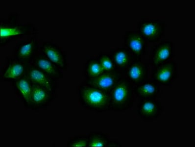
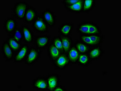
 IF (Immunofluorescence)
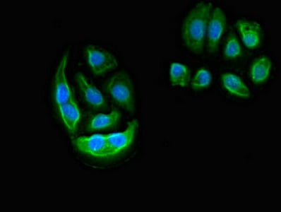
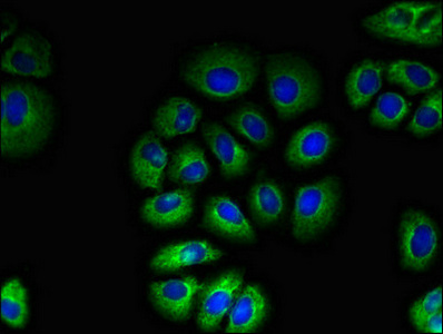
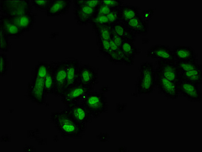
(Immunofluorescent analysis of A549 cells using AAA233384 at a dilution of 1:100 and Alexa Fluor 488-congugated AffiniPure Goat Anti-Rabbit IgG(H+L))
IF (Immunofluorescence)
(Immunofluorescent analysis of A549 cells using AAA233384 at a dilution of 1:100 and Alexa Fluor 488-congugated AffiniPure Goat Anti-Rabbit IgG(H+L))
RAI1, Polyclonal Antibody (Cat# AAA233384)
 IF (Immunofluorescence)
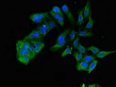
(Immunofluorescent analysis of A549 cells using AAA233385 at a dilution of 1:100 and Alexa Fluor 488-congugated AffiniPure Goat Anti-Rabbit IgG(H+L))
IF (Immunofluorescence)
(Immunofluorescent analysis of A549 cells using AAA233385 at a dilution of 1:100 and Alexa Fluor 488-congugated AffiniPure Goat Anti-Rabbit IgG(H+L))
VASH2, Polyclonal Antibody (Cat# AAA233385)
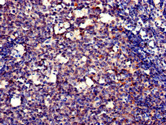
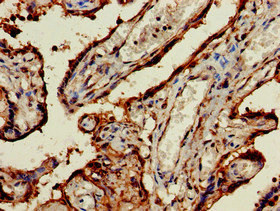
 IHC (Immunohistochemisry)
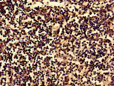
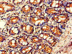
(Immunohistochemistry of paraffin-embedded human lung tissue using AAA233389 at dilution of 1:100)
IHC (Immunohistochemisry)
(Immunohistochemistry of paraffin-embedded human lung tissue using AAA233389 at dilution of 1:100)
DENND1C, Polyclonal Antibody (Cat# AAA233389)
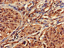
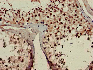
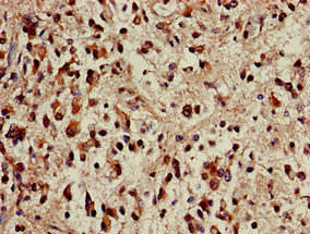
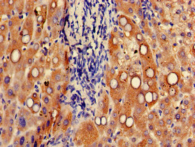
 IHC (Immunohiostchemistry)
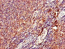
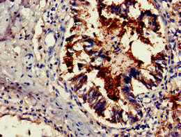
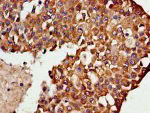
(Immunohistochemistry of paraffin-embedded human glioma cancer using AAA233392 at dilution of 1:100)
IHC (Immunohiostchemistry)
(Immunohistochemistry of paraffin-embedded human glioma cancer using AAA233392 at dilution of 1:100)
FUNDC1, Polyclonal Antibody (Cat# AAA233392)
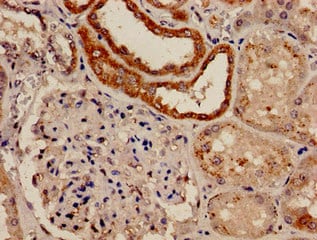
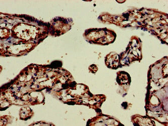
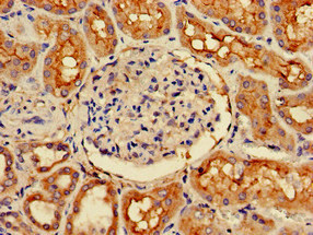
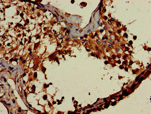
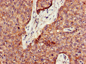
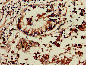
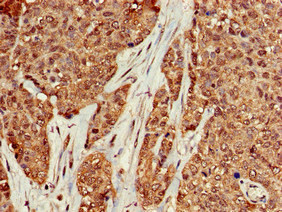
 IHC (Immunohiostchemistry)
(Immunohistochemistry of paraffin-embedded human kidney tissue using AAA233393 at dilution of 1:100)
IHC (Immunohiostchemistry)
(Immunohistochemistry of paraffin-embedded human kidney tissue using AAA233393 at dilution of 1:100)
PHOSPHO1, Polyclonal Antibody (Cat# AAA233393)
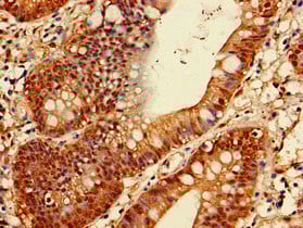
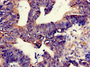
 IHC (Immunohiostchemistry)
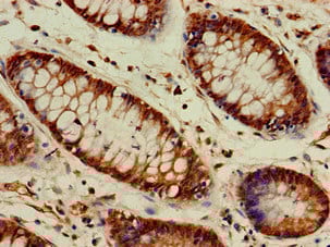
(Immunohistochemistry of paraffin-embedded human colon cancer using AAA233394 at dilution of 1:100)
IHC (Immunohiostchemistry)
(Immunohistochemistry of paraffin-embedded human colon cancer using AAA233394 at dilution of 1:100)
ARAP3, Polyclonal Antibody (Cat# AAA233394)
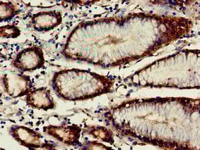
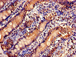
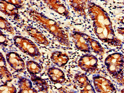
 ICC (Immunocytochemistry)
(Immunocytochemistry analysis of human small intestine tissue using AAA233395 at dilution of 1:100)
ICC (Immunocytochemistry)
(Immunocytochemistry analysis of human small intestine tissue using AAA233395 at dilution of 1:100)
FBXW7, Polyclonal Antibody (Cat# AAA233395)
 ICC (Immunocytochemistry)
(Immunocytochemistry analysis of human glioma cancer using AAA233396 at dilution of 1:100)
ICC (Immunocytochemistry)
(Immunocytochemistry analysis of human glioma cancer using AAA233396 at dilution of 1:100)
TBC1D31, Polyclonal Antibody (Cat# AAA233396)
 IHC (Immunohistochemisry)
(Immunofluorescent analysis of PC3 cells using AAA233397 at a dilution of 1:100 and Alexa Fluor 488-congugated AffiniPure Goat Anti-Rabbit IgG(H+L))
IHC (Immunohistochemisry)
(Immunofluorescent analysis of PC3 cells using AAA233397 at a dilution of 1:100 and Alexa Fluor 488-congugated AffiniPure Goat Anti-Rabbit IgG(H+L))
PLIN4, Polyclonal Antibody (Cat# AAA233397)
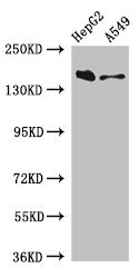
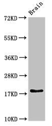
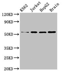
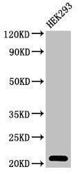 WB (Western Blot)
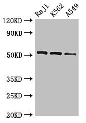
(Western BlotPositive WB detected in: HEK293 whole cell lysateAll lanes: TSR2 antibody at 2ug/mlSecondaryGoat polyclonal to rabbit IgG at 1/50000 dilutionPredicted band size: 21 KDaObserved band size: 21 KDa)
WB (Western Blot)
(Western BlotPositive WB detected in: HEK293 whole cell lysateAll lanes: TSR2 antibody at 2ug/mlSecondaryGoat polyclonal to rabbit IgG at 1/50000 dilutionPredicted band size: 21 KDaObserved band size: 21 KDa)
TSR2, Polyclonal Antibody (Cat# AAA233398)
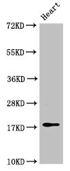
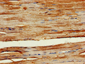
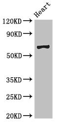
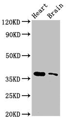
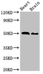
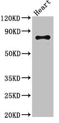 WB (Western Blot)
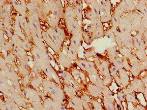
(Western BlotPositive WB detected in: Mouse heart tissueAll lanes: ELMO2 antibody at 3.4ug/mlSecondaryGoat polyclonal to rabbit IgG at 1/50000 dilutionPredicted band size: 83,73 KDaObserved band size: 83 KDa)
WB (Western Blot)
(Western BlotPositive WB detected in: Mouse heart tissueAll lanes: ELMO2 antibody at 3.4ug/mlSecondaryGoat polyclonal to rabbit IgG at 1/50000 dilutionPredicted band size: 83,73 KDaObserved band size: 83 KDa)
ELMO2, Polyclonal Antibody (Cat# AAA233401)
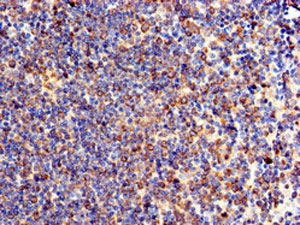
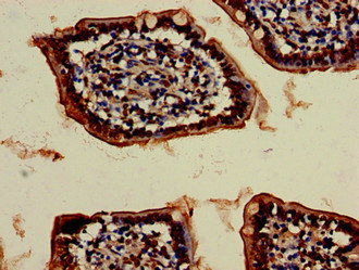
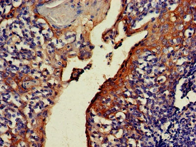 IHC (Immunohiostchemistry)
(IHC image of AAA233404 diluted at 1:500 and staining in paraffin-embedded human tonsil tissue performed on a Leica BondTM system. After dewaxing and hydration, antigen retrieval was mediated by high pressure in a citrate buffer (pH 6.0). Section was blocked with 10% normal goat serum 30min at RT. Then primary antibody (1% BSA) was incubated at 4 degree C overnight. The primary is detected by a biotinylated secondary antibody and visualized using an HRP conjugated SP system.)
IHC (Immunohiostchemistry)
(IHC image of AAA233404 diluted at 1:500 and staining in paraffin-embedded human tonsil tissue performed on a Leica BondTM system. After dewaxing and hydration, antigen retrieval was mediated by high pressure in a citrate buffer (pH 6.0). Section was blocked with 10% normal goat serum 30min at RT. Then primary antibody (1% BSA) was incubated at 4 degree C overnight. The primary is detected by a biotinylated secondary antibody and visualized using an HRP conjugated SP system.)
NOA1, Polyclonal Antibody (Cat# AAA233404)
 IF (Immunofluorescence)
(Immunofluorescent analysis of A549 cells using AAA233409 at a dilution of 1:100 and Alexa Fluor 488-congugated AffiniPure Goat Anti-Rabbit IgG(H+L))
IF (Immunofluorescence)
(Immunofluorescent analysis of A549 cells using AAA233409 at a dilution of 1:100 and Alexa Fluor 488-congugated AffiniPure Goat Anti-Rabbit IgG(H+L))
TAS1R2, Polyclonal Antibody (Cat# AAA233409)
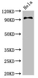
 IHC (Immunohistochemisry)
(Immunofluorescent analysis of Hela cells using AAA233410 at a dilution of 1:100 and Alexa Fluor 488-congugated AffiniPure Goat Anti-Rabbit IgG(H+L))
IHC (Immunohistochemisry)
(Immunofluorescent analysis of Hela cells using AAA233410 at a dilution of 1:100 and Alexa Fluor 488-congugated AffiniPure Goat Anti-Rabbit IgG(H+L))
BLNK, Polyclonal Antibody (Cat# AAA233410)
 IHC (Immunohiostchemistry)
(Immunohistochemistry of paraffin-embedded human breast cancer using AAA233411 at dilution of 1:100)
IHC (Immunohiostchemistry)
(Immunohistochemistry of paraffin-embedded human breast cancer using AAA233411 at dilution of 1:100)
PIGK, Polyclonal Antibody (Cat# AAA233411)
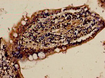
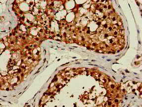
 IHC (Immunohiostchemistry)
(Immunohistochemistry of paraffin-embedded human adrenal gland tissue using AAA233412 at dilution of 1:100)
IHC (Immunohiostchemistry)
(Immunohistochemistry of paraffin-embedded human adrenal gland tissue using AAA233412 at dilution of 1:100)
USP6NL, Polyclonal Antibody (Cat# AAA233412)
 IHC (Immunohistochemisry)
(Immunofluorescent analysis of A549 cells using AAA233413 at a dilution of 1:100 and Alexa Fluor 488-congugated AffiniPure Goat Anti-Rabbit IgG(H+L))
IHC (Immunohistochemisry)
(Immunofluorescent analysis of A549 cells using AAA233413 at a dilution of 1:100 and Alexa Fluor 488-congugated AffiniPure Goat Anti-Rabbit IgG(H+L))
LHFPL5, Polyclonal Antibody (Cat# AAA233413)
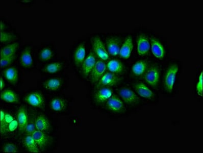
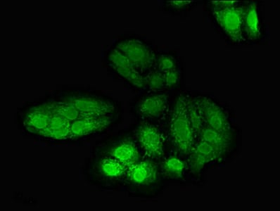
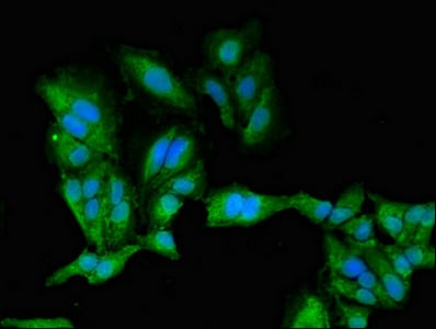
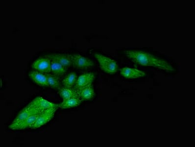
 IF (Immunofluorescence)
(Immunofluorescent analysis of MCF-7 cells using AAA233415 at a dilution of 1:100 and Alexa Fluor 488-congugated AffiniPure Goat Anti-Rabbit IgG(H+L))
IF (Immunofluorescence)
(Immunofluorescent analysis of MCF-7 cells using AAA233415 at a dilution of 1:100 and Alexa Fluor 488-congugated AffiniPure Goat Anti-Rabbit IgG(H+L))
STOML3, Polyclonal Antibody (Cat# AAA233415)
 IHC (Immunohistochemisry)
(Immunofluorescent analysis of A549 cells using AAA233416 at a dilution of 1:100 and Alexa Fluor 488-congugated AffiniPure Goat Anti-Rabbit IgG(H+L))
IHC (Immunohistochemisry)
(Immunofluorescent analysis of A549 cells using AAA233416 at a dilution of 1:100 and Alexa Fluor 488-congugated AffiniPure Goat Anti-Rabbit IgG(H+L))
CDK5RAP2, Polyclonal Antibody (Cat# AAA233416)
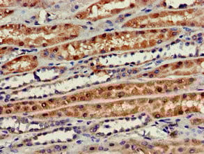
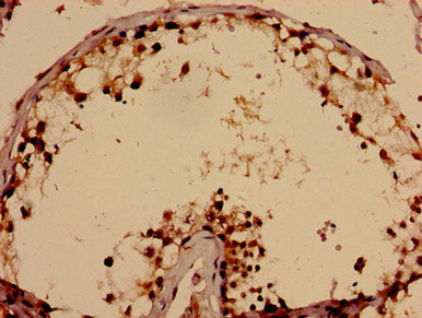
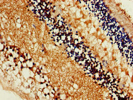
 IHC (Immunohistochemisry)
(Immunohistochemistry of paraffin-embedded human testis tissue using AAA233418 at dilution of 1:100)
IHC (Immunohistochemisry)
(Immunohistochemistry of paraffin-embedded human testis tissue using AAA233418 at dilution of 1:100)
DOT1L, Polyclonal Antibody (Cat# AAA233418)
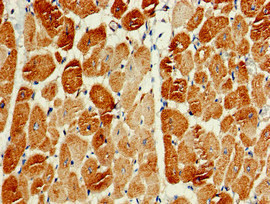
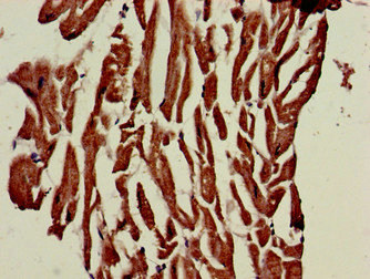
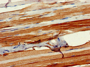
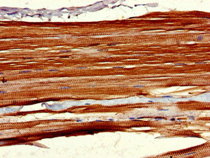
 IHC (Immunohistochemisry)
(Immunohistochemistry of paraffin-embedded human skeletal muscle tissue using AAA233422 at dilution of 1:100)
IHC (Immunohistochemisry)
(Immunohistochemistry of paraffin-embedded human skeletal muscle tissue using AAA233422 at dilution of 1:100)
PIK3C3, Polyclonal Antibody (Cat# AAA233422)
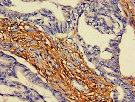
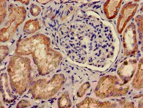
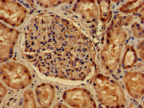 IHC (Immunohiostchemistry)
(Immunohistochemistry of paraffin-embedded human kidney tissue using AAA233425 at dilution of 1:100)
IHC (Immunohiostchemistry)
(Immunohistochemistry of paraffin-embedded human kidney tissue using AAA233425 at dilution of 1:100)
PDLIM2, Polyclonal Antibody (Cat# AAA233425)
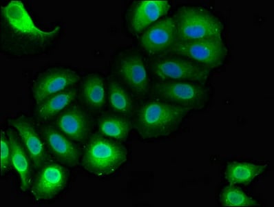
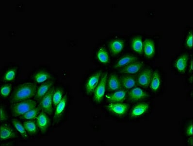
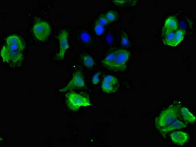
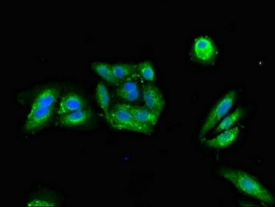
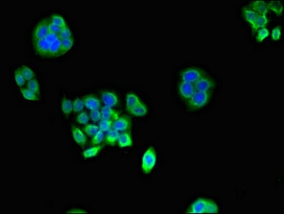
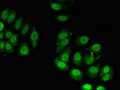
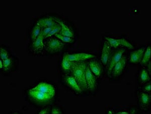
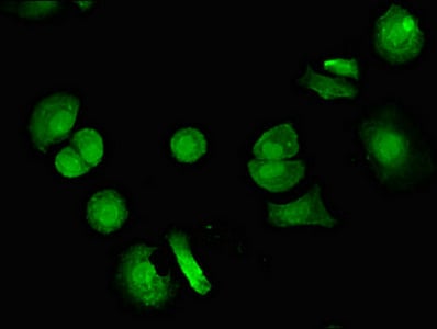
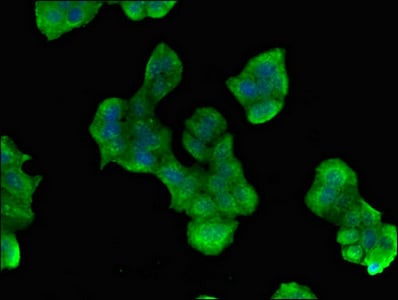 IF (Immunofluorescence)
(Immunofluorescent analysis of PC3 cells using AAA233260 at a dilution of 1:100 and Alexa Fluor 488-congugated AffiniPure Goat Anti-Rabbit IgG(H+L))
IF (Immunofluorescence)
(Immunofluorescent analysis of PC3 cells using AAA233260 at a dilution of 1:100 and Alexa Fluor 488-congugated AffiniPure Goat Anti-Rabbit IgG(H+L))
PES1, Polyclonal Antibody (Cat# AAA233260)
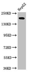
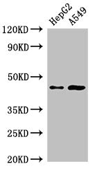
 IHC (Immunohistochemisry)
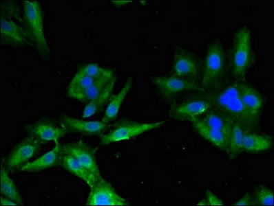
(Immunofluorescent analysis of HepG2 cells using AAA233266 at a dilution of 1:100 and Alexa Fluor 488-congugated AffiniPure Goat Anti-Rabbit IgG(H+L))
IHC (Immunohistochemisry)
(Immunofluorescent analysis of HepG2 cells using AAA233266 at a dilution of 1:100 and Alexa Fluor 488-congugated AffiniPure Goat Anti-Rabbit IgG(H+L))
POLH, Polyclonal Antibody (Cat# AAA233266)
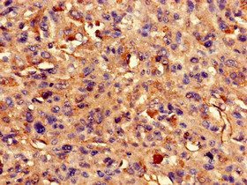
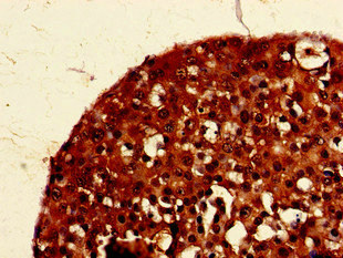
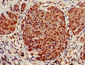
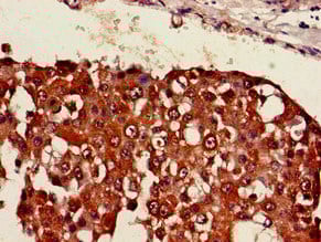
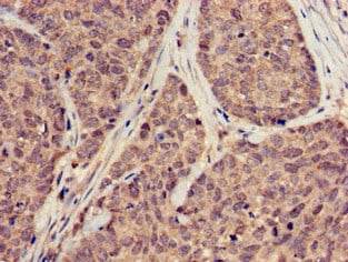
 IHC (Immunohiostchemistry)
(Immunohistochemistry of paraffin-embedded human ovarian cancer using AAA233271 at dilution of 1:100)
IHC (Immunohiostchemistry)
(Immunohistochemistry of paraffin-embedded human ovarian cancer using AAA233271 at dilution of 1:100)
PPT1, Polyclonal Antibody (Cat# AAA233271)
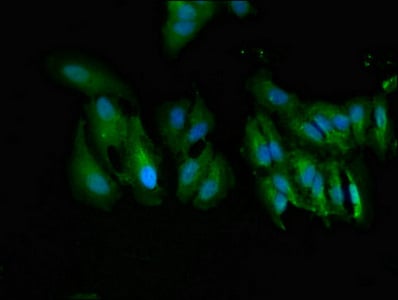
 IF (Immunofluorescence)
(Immunofluorescent analysis of Hela cells using AAA233272 at a dilution of 1:100 and Alexa Fluor 488-congugated AffiniPure Goat Anti-Rabbit IgG(H+L))
IF (Immunofluorescence)
(Immunofluorescent analysis of Hela cells using AAA233272 at a dilution of 1:100 and Alexa Fluor 488-congugated AffiniPure Goat Anti-Rabbit IgG(H+L))
PRKACB, Polyclonal Antibody (Cat# AAA233272)

 IF (Immunofluorescence)
(Immunofluorescent analysis of HepG2 cells using AAA233273 at a dilution of 1:100 and Alexa Fluor 488-congugated AffiniPure Goat Anti-Rabbit IgG(H+L))
IF (Immunofluorescence)
(Immunofluorescent analysis of HepG2 cells using AAA233273 at a dilution of 1:100 and Alexa Fluor 488-congugated AffiniPure Goat Anti-Rabbit IgG(H+L))
PRKAR2A, Polyclonal Antibody (Cat# AAA233273)
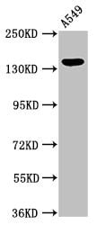
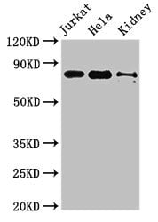
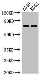
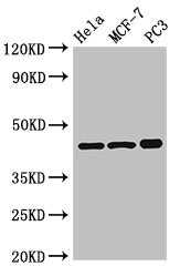
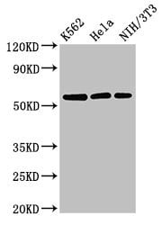
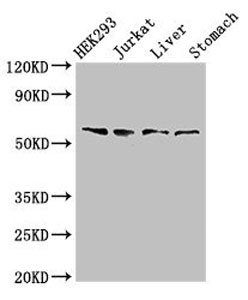
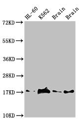
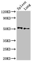 WB (Western Blot)
(Western BlotPositive WB detected in: Rat spleen tissue,Rat lung tissueAll lanes: PSEN2 antibody at 3ug/mlSecondaryGoat polyclonal to rabbit IgG at 1/50000 dilutionPredicted band size: 51,47 KDaObserved band size: 51 KDa)
WB (Western Blot)
(Western BlotPositive WB detected in: Rat spleen tissue,Rat lung tissueAll lanes: PSEN2 antibody at 3ug/mlSecondaryGoat polyclonal to rabbit IgG at 1/50000 dilutionPredicted band size: 51,47 KDaObserved band size: 51 KDa)
PSEN2, Polyclonal Antibody (Cat# AAA233276)

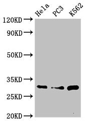
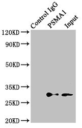 IP (Immunoprecipitation)
(Immunoprecipitating PSMA1 in K562 whole cell lysateLane 1: Rabbit control IgG (1ug)instead of AAA233277 in K562 whole cell lysate. For western blotting,a HRP-conjugated light chain specific antibody was used as the secondary antibody (1/50000)Lane 2: AAA233277 (8ug)+ K562 whole cell lysate (500ug)Lane 3: K562 whole cell lysate (10ug))
IP (Immunoprecipitation)
(Immunoprecipitating PSMA1 in K562 whole cell lysateLane 1: Rabbit control IgG (1ug)instead of AAA233277 in K562 whole cell lysate. For western blotting,a HRP-conjugated light chain specific antibody was used as the secondary antibody (1/50000)Lane 2: AAA233277 (8ug)+ K562 whole cell lysate (500ug)Lane 3: K562 whole cell lysate (10ug))
PSMA1, Polyclonal Antibody (Cat# AAA233277)
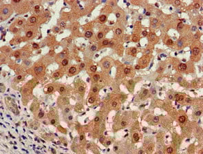
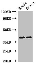
 IHC (Immunohistochemisry)
(Immunohistochemistry of paraffin-embedded human brain tissue using AAA233278 at dilution of 1:100)
IHC (Immunohistochemisry)
(Immunohistochemistry of paraffin-embedded human brain tissue using AAA233278 at dilution of 1:100)
PSMA5, Polyclonal Antibody (Cat# AAA233278)
 IHC (Immunohistochemisry)
(Immunofluorescent analysis of A549 cells using AAA233285 at a dilution of 1:100 and Alexa Fluor 488-congugated AffiniPure Goat Anti-Rabbit IgG(H+L))
IHC (Immunohistochemisry)
(Immunofluorescent analysis of A549 cells using AAA233285 at a dilution of 1:100 and Alexa Fluor 488-congugated AffiniPure Goat Anti-Rabbit IgG(H+L))
RPL23A, Polyclonal Antibody (Cat# AAA233285)
 IF (Immunofluorescence)
(Immunofluorescent analysis of HepG2 cells using AAA233286 at a dilution of 1:100 and Alexa Fluor 488-congugated AffiniPure Goat Anti-Rabbit IgG(H+L))
IF (Immunofluorescence)
(Immunofluorescent analysis of HepG2 cells using AAA233286 at a dilution of 1:100 and Alexa Fluor 488-congugated AffiniPure Goat Anti-Rabbit IgG(H+L))
RPS4Y1, Polyclonal Antibody (Cat# AAA233286)
 IHC (Immunohiostchemistry)
(Immunohistochemistry of paraffin-embedded human breast cancer using AAA233290 at dilution of 1:100)
IHC (Immunohiostchemistry)
(Immunohistochemistry of paraffin-embedded human breast cancer using AAA233290 at dilution of 1:100)
SESN1, Polyclonal Antibody (Cat# AAA233290)
 IHC (Immunohistochemisry)
(Immunofluorescent analysis of A549 cells using AAA233291 at a dilution of 1:100 and Alexa Fluor 488-congugated AffiniPure Goat Anti-Rabbit IgG(H+L))
IHC (Immunohistochemisry)
(Immunofluorescent analysis of A549 cells using AAA233291 at a dilution of 1:100 and Alexa Fluor 488-congugated AffiniPure Goat Anti-Rabbit IgG(H+L))
SLC16A8, Polyclonal Antibody (Cat# AAA233291)

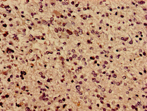
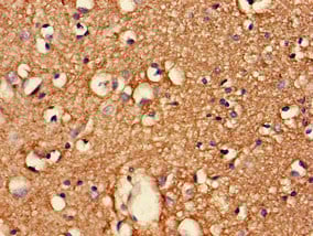
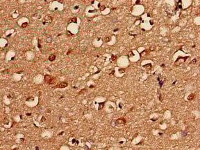
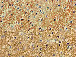
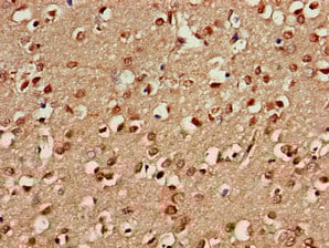
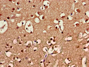 IHC (Immunohistochemisry)
(Immunohistochemistry of paraffin-embedded human brain tissue using AAA233295 at dilution of 1:100)
IHC (Immunohistochemisry)
(Immunohistochemistry of paraffin-embedded human brain tissue using AAA233295 at dilution of 1:100)
SRR, Polyclonal Antibody (Cat# AAA233295)
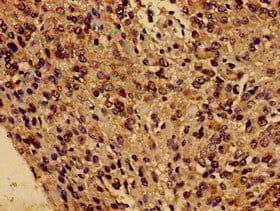
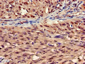
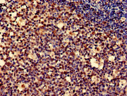
 IHC (Immunohiostchemistry)
(Immunohistochemistry of paraffin-embedded human glioma cancer using AAA233298 at dilution of 1:100)
IHC (Immunohiostchemistry)
(Immunohistochemistry of paraffin-embedded human glioma cancer using AAA233298 at dilution of 1:100)
TERF1, Polyclonal Antibody (Cat# AAA233298)
 IHC (Immunohistochemisry)
(Immunofluorescent analysis of Hela cells using AAA233303 at a dilution of 1:100 and Alexa Fluor 488-congugated AffiniPure Goat Anti-Rabbit IgG(H+L))
IHC (Immunohistochemisry)
(Immunofluorescent analysis of Hela cells using AAA233303 at a dilution of 1:100 and Alexa Fluor 488-congugated AffiniPure Goat Anti-Rabbit IgG(H+L))
TNIK, Polyclonal Antibody (Cat# AAA233303)
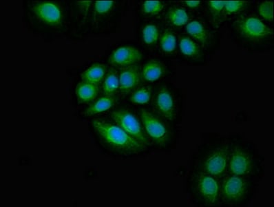
 IF (Immunofluorescence)
(Immunofluorescent analysis of HepG2 cells using AAA233307 at a dilution of 1:100 and Alexa Fluor 488-congugated AffiniPure Goat Anti-Rabbit IgG(H+L))
IF (Immunofluorescence)
(Immunofluorescent analysis of HepG2 cells using AAA233307 at a dilution of 1:100 and Alexa Fluor 488-congugated AffiniPure Goat Anti-Rabbit IgG(H+L))
TXK, Polyclonal Antibody (Cat# AAA233307)
 IF (Immunofluorescence)
(Immunofluorescent analysis of A549 cells using AAA233308 at a dilution of 1:100 and Alexa Fluor 488-congugated AffiniPure Goat Anti-Rabbit IgG(H+L))
IF (Immunofluorescence)
(Immunofluorescent analysis of A549 cells using AAA233308 at a dilution of 1:100 and Alexa Fluor 488-congugated AffiniPure Goat Anti-Rabbit IgG(H+L))
TYK2, Polyclonal Antibody (Cat# AAA233308)
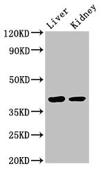
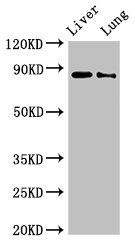
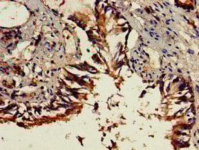
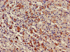
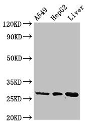
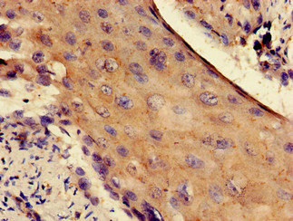
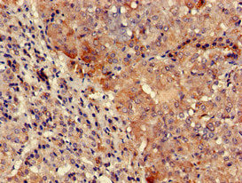
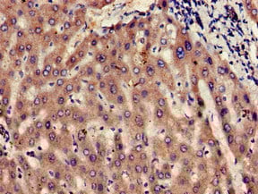 IHC (Immunohiostchemistry)
(Immunohistochemistry of paraffin-embedded human liver tissue using AAA233309 at dilution of 1:100)
IHC (Immunohiostchemistry)
(Immunohistochemistry of paraffin-embedded human liver tissue using AAA233309 at dilution of 1:100)
UBE2V2, Polyclonal Antibody (Cat# AAA233309)
 IHC (Immunohistochemisry)
(Immunofluorescent analysis of Hela cells using AAA233310 at a dilution of 1:100 and Alexa Fluor 488-congugated AffiniPure Goat Anti-Rabbit IgG(H+L))
IHC (Immunohistochemisry)
(Immunofluorescent analysis of Hela cells using AAA233310 at a dilution of 1:100 and Alexa Fluor 488-congugated AffiniPure Goat Anti-Rabbit IgG(H+L))
UGT1A9, Polyclonal Antibody (Cat# AAA233310)
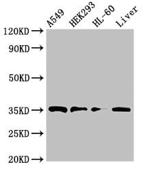
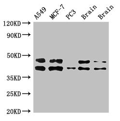
 WB (Western Blot)
(Western BlotPositive WB detected in: Mouse heart tissue,Mouse brain tissueAll lanes: VPS4B antibody at 2.5ug/mlSecondaryGoat polyclonal to rabbit IgG at 1/50000 dilutionPredicted band size: 50 KDaObserved band size: 50 KDa)
WB (Western Blot)
(Western BlotPositive WB detected in: Mouse heart tissue,Mouse brain tissueAll lanes: VPS4B antibody at 2.5ug/mlSecondaryGoat polyclonal to rabbit IgG at 1/50000 dilutionPredicted band size: 50 KDaObserved band size: 50 KDa)
VPS4B, Polyclonal Antibody (Cat# AAA233312)
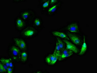
 IF (Immunofluorescence)
(Immunofluorescent analysis of Hela cells using AAA233315 at a dilution of 1:100 and Alexa Fluor 488-congugated AffiniPure Goat Anti-Rabbit IgG(H+L))
IF (Immunofluorescence)
(Immunofluorescent analysis of Hela cells using AAA233315 at a dilution of 1:100 and Alexa Fluor 488-congugated AffiniPure Goat Anti-Rabbit IgG(H+L))
SUMO3, Polyclonal Antibody (Cat# AAA233315)
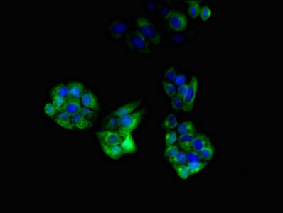
 IF (Immunofluorescence)
(Immunofluorescent analysis of HepG2 cells using AAA233316 at a dilution of 1:100 and Alexa Fluor 488-congugated AffiniPure Goat Anti-Rabbit IgG(H+L))
IF (Immunofluorescence)
(Immunofluorescent analysis of HepG2 cells using AAA233316 at a dilution of 1:100 and Alexa Fluor 488-congugated AffiniPure Goat Anti-Rabbit IgG(H+L))
IL23A, Polyclonal Antibody (Cat# AAA233316)
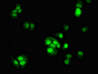
 IHC (Immunohistochemisry)
(Immunofluorescent analysis of HepG2 cells using AAA233319 at a dilution of 1:100 and Alexa Fluor 488-congugated AffiniPure Goat Anti-Rabbit IgG(H+L))
IHC (Immunohistochemisry)
(Immunofluorescent analysis of HepG2 cells using AAA233319 at a dilution of 1:100 and Alexa Fluor 488-congugated AffiniPure Goat Anti-Rabbit IgG(H+L))
KRAS, Polyclonal Antibody (Cat# AAA233319)
N, Polyclonal Antibody (Cat# AAA233325)
LMP2, Polyclonal Antibody (Cat# AAA233327)
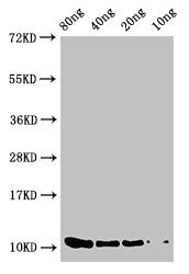 WB (Western Blot)
(Western BlotPositive WB detected in Recombinant proteinAll lanes: prgJ antibody at 2ug/mlSecondaryGoat polyclonal to rabbit IgG at 1/50000 dilutionpredicted band size: 11 kDaobserved band size: 11 kDa)
WB (Western Blot)
(Western BlotPositive WB detected in Recombinant proteinAll lanes: prgJ antibody at 2ug/mlSecondaryGoat polyclonal to rabbit IgG at 1/50000 dilutionpredicted band size: 11 kDaobserved band size: 11 kDa)
prgJ, Polyclonal Antibody (Cat# AAA233330)
C3, Polyclonal Antibody (Cat# AAA233334)
mscL, Polyclonal Antibody (Cat# AAA233338)
What are Polyclonal Antibodies?
Polyclonal antibodies are antibodies that come from multiple B cell clones of a host animal. The typical hosts used for the majority of polyclonal antibody production are rabbits, goats, sheep, and donkeys. These polyclonal antibodies, once having identified their target, will bind to different epitopes located at different regions or sequences on the same protein/antigen. As a result, they are ideal at locating and binding to the target, even if the target is in very low concentrations (due to many different antibodies being able to bind to the same target molecule, which allows for significant amplification of a downstream signal).
Polyclonal antibodies are typically produced by injecting an antigen into a host animal, which causes the animal’s immune system to attack the foreign antigen by mass generating antibodies against it. After a period of time, serum is collected from the animal and purified using physicochemical fractionation, class-specific affinity purification, and/or antigen-affinity purification.
Key Uses of Polyclonal Antibodies
- Western Blotting: This method is used to find specific proteins in biological samples after separating them by size.
- Immunohistochemistry: IHC helps visualize the location of proteins in tissue sections using various staining techniques.
- ELISA: (Enzyme-Linked Immunosorbent Assay) is typically used to identify specific protein quantities in a sample. ELISAs can be either “Quantitative” or “Qualitative”.
- Flow Cytometry: technique that identifies and measures the specific protein on the surface or inside the cells in a fluid suspension.
- Immunoprecipitation: IP isolates and studies a specific protein from a complex mixture using antibodies.
Why Buy Polyclonal Antibodies from AAA Biotech?
1. Ideal for Various Applications
Our antibodies are generally going to be validated for use in multiple types of assays, including ELISA, Western Blotting, Immunohistochemistry, Immunoprecipitation, amongst others. They are ideal for a wide range of research applications.
2. Rigorous Quality Control
All of the antibodies in our catalog undergo strict quality testing to ensure specificity, sensitivity, and consistent performance. We are confident in the ability of our antibodies to provide you with accurate results.
3. Wide Assortment of Antibodies
Antibodies in are catalog can be found for both common and exotic species, and these antibodies are also available in both conjugated and recombinant forms to suit many diverse experimental needs.
4. Highly Purified
Our antibodies are available in purified forms with over 85% purity, as confirmed by SDS-PAGE. They are also available with tags such as His, Flag, GST, or MBP. We cater to customers worldwide.
FAQ
1. How are polyclonal antibodies produced?
Traditionally, polyclonal antibodies are produced by injecting an antigen into a host animal (such as a rabbit or goat), which then triggers an immune response from the host animal. The animal’s B cells produce antibodies that will recognize different parts of the injected antigen. These antibodies are then collected from the animal’s blood and purified for use.
2. How do polyclonal antibodies differ from monoclonal antibodies?
Polyclonal antibodies are a mix of antibodies that bind to different locations (epitopes) of the same antigen, while monoclonal antibodies are identical and bind to just one specific epitope. This makes polyclonal antibodies more versatile and better at detecting proteins that may be present in low quantities or in altered/modified forms.
3. How should I store polyclonal antibodies?
Polyclonal antibodies should be stored at 4°C for short-term use (up to a few weeks) and at -20°C or -80°C for long-term storage. Avoid repeated freeze-thaw cycles by dividing them into small aliquots. Always check the datasheet for specific storage instructions.