Filters
▼Clonality
▼Type
▼Reactivity
▼Gene Name
▼Isotype
▼Host
▼Application
▼Clone
▼Polyclonal Antibodies
At AAA Biotech also known as AAA Bio or AAABio, we provide a broad range of purified polyclonal antibodies (pAbs) that are able to all be browsed online through our website. Due to their high specificity and strong binding affinity, these antibodies are ideal for wide swathes of research and experimental applications.
Our polyclonal antibodies can easily support your work, whether you use them for Western Blotting, Immunocytochemistry (with or without Immunofluorescence used in conjunction), Immunohistochemistry, Immunoprecipitation, and ELISA tests. We highly encourage you to browse our range of pAbs and choose the one that best suits your experimental model.
Viewing 9600-9650 of 96805 product results
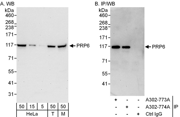 WB (Western Blot)
(Detection of human and mouse PRP6 by western blot (h&m) and immunoprecipitation (h). Samples: Whole cell lysate from HeLa (5, 15 and 50 ug for WB; 1 mg for IP, 20% of IP loaded), HEK293T (T; 50 ug), and mouse NIH 3T3 (M; 50 ug) cells. Antibodies: Affinity purified rabbit anti-PRP6 antibody AAA212011 used for WB at 0.04 ug/ml (A) and 0.4 ug/ml (B) and used for IP at 3 ug/mg lysate. PRP6 was also immunoprecipitated by rabbit anti-PRP6 antibody which recognizes an upstream epitope. Detection: Chemiluminescence with exposure times of 30 seconds (A and B).)
WB (Western Blot)
(Detection of human and mouse PRP6 by western blot (h&m) and immunoprecipitation (h). Samples: Whole cell lysate from HeLa (5, 15 and 50 ug for WB; 1 mg for IP, 20% of IP loaded), HEK293T (T; 50 ug), and mouse NIH 3T3 (M; 50 ug) cells. Antibodies: Affinity purified rabbit anti-PRP6 antibody AAA212011 used for WB at 0.04 ug/ml (A) and 0.4 ug/ml (B) and used for IP at 3 ug/mg lysate. PRP6 was also immunoprecipitated by rabbit anti-PRP6 antibody which recognizes an upstream epitope. Detection: Chemiluminescence with exposure times of 30 seconds (A and B).)
PRP6, Polyclonal Antibody (Cat# AAA212011)
 WB (Western Blot)
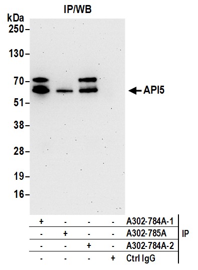
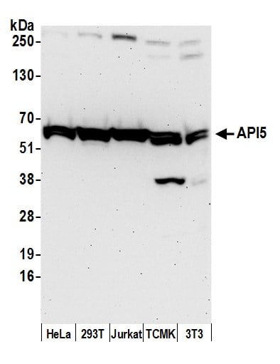
(Detection of human and mouse API5 by western blot. Samples:Whole cell lysate (50 ug) from HeLa, HEK293T, Jurkat, mouse TCMK-1, and mouse NIH 3T3 cells prepared using NETN lysis buffer. Antibody: Affinity purified rabbit anti-API5 antibody AAA212017 (lot AAA212017-2) used for WB at 0.1 ug/ml. Detection:Chemiluminescence with an exposure time of 3 minutes.)
WB (Western Blot)
(Detection of human and mouse API5 by western blot. Samples:Whole cell lysate (50 ug) from HeLa, HEK293T, Jurkat, mouse TCMK-1, and mouse NIH 3T3 cells prepared using NETN lysis buffer. Antibody: Affinity purified rabbit anti-API5 antibody AAA212017 (lot AAA212017-2) used for WB at 0.1 ug/ml. Detection:Chemiluminescence with an exposure time of 3 minutes.)
API5, Polyclonal Antibody (Cat# AAA212017)
 WB (Western Blot)
(Detection of human and mouse FKBP4/FKBP52 by western blot. Samples: Whole cell lysate from HeLa (5, 15 and 50 ug), HEK293T (T; 50 ug), and mouse NIH 3T3 (M; 50 ug) cells. Antibody: Affinity purified rabbit anti-FKBP4/FKBP52 antibody AAA211467 used for WB at 0.04 ug/ml. Detection: Chemiluminescence with an exposure time of 1 second.)
WB (Western Blot)
(Detection of human and mouse FKBP4/FKBP52 by western blot. Samples: Whole cell lysate from HeLa (5, 15 and 50 ug), HEK293T (T; 50 ug), and mouse NIH 3T3 (M; 50 ug) cells. Antibody: Affinity purified rabbit anti-FKBP4/FKBP52 antibody AAA211467 used for WB at 0.04 ug/ml. Detection: Chemiluminescence with an exposure time of 1 second.)
FKBP4/FKBP52, Polyclonal Antibody (Cat# AAA211467)
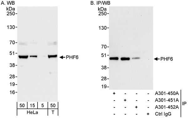 WB (Western Blot)
(Detection of human PHF6 by western blot and immunoprecipitation. Samples: Whole cell lysate from HeLa (5, 15 and 50 ug for WB; 1 mg for IP, 20% of IP loaded) and HEK293T (T; 50 ug) cells. Antibodies: Affinity purified rabbit anti-PHF6 antibody AAA211479 used for WB at 0.04 ug/ml (A) and 1 ug/ml (B) and used for IP at 3 ug/mg lysate. PHF6 was also immunoprecipitated by rabbit anti-PHF6 antibodies and which recognize upstream epitopes. Detection: Chemiluminescence with exposure times of 3 seconds (A) and 10 seconds (B).)
WB (Western Blot)
(Detection of human PHF6 by western blot and immunoprecipitation. Samples: Whole cell lysate from HeLa (5, 15 and 50 ug for WB; 1 mg for IP, 20% of IP loaded) and HEK293T (T; 50 ug) cells. Antibodies: Affinity purified rabbit anti-PHF6 antibody AAA211479 used for WB at 0.04 ug/ml (A) and 1 ug/ml (B) and used for IP at 3 ug/mg lysate. PHF6 was also immunoprecipitated by rabbit anti-PHF6 antibodies and which recognize upstream epitopes. Detection: Chemiluminescence with exposure times of 3 seconds (A) and 10 seconds (B).)
PHF6, Polyclonal Antibody (Cat# AAA211479)
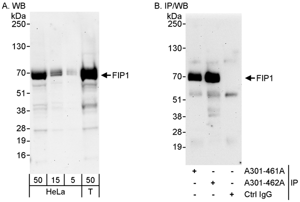 WB (Western Blot)
(Detection of human FIP1 by western blot and immunoprecipitation. Samples: Whole cell lysate from HeLa (5, 15 and 50 ug for WB; 1 mg for IP, 20% of IP loaded) and HEK293T (T; 50 ug) cells. Antibodies: Affinity purified rabbit anti-FIP1 antibody AAA211485 used for WB at 0.1 ug/ml (A) and 1 ug/ml (B) and used for IP at 3 ug/mg lysate. FIP1 was also immunoprecipitated by rabbit anti-FIP1 antibody which recognizes an upstream epitope. Detection: Chemiluminescence with exposure times of 3 minutes (A and B).)
WB (Western Blot)
(Detection of human FIP1 by western blot and immunoprecipitation. Samples: Whole cell lysate from HeLa (5, 15 and 50 ug for WB; 1 mg for IP, 20% of IP loaded) and HEK293T (T; 50 ug) cells. Antibodies: Affinity purified rabbit anti-FIP1 antibody AAA211485 used for WB at 0.1 ug/ml (A) and 1 ug/ml (B) and used for IP at 3 ug/mg lysate. FIP1 was also immunoprecipitated by rabbit anti-FIP1 antibody which recognizes an upstream epitope. Detection: Chemiluminescence with exposure times of 3 minutes (A and B).)
FIP1, Polyclonal Antibody (Cat# AAA211485)
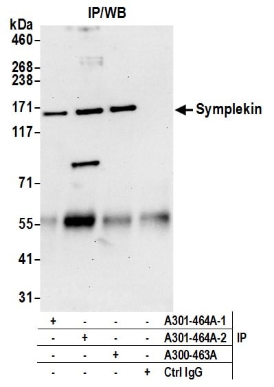
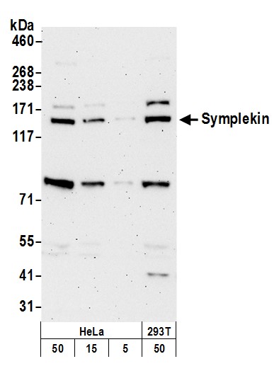 WB (Western Blot)
(Detection of human and mouse Symplekin by western blot. Samples: Whole cell lysate from HeLa, (5, 15 and 50 ug), and HEK293T (50ug) cells prepared using NETN lysis buffer. Antibody: Affinity purified rabbit anti-Symplekin antibody AAA211487 (lot AAA211487-2) used for WB at 0.02 ug/ml. Detection: Chemiluminescence with an exposure time of 3 minutes.)
WB (Western Blot)
(Detection of human and mouse Symplekin by western blot. Samples: Whole cell lysate from HeLa, (5, 15 and 50 ug), and HEK293T (50ug) cells prepared using NETN lysis buffer. Antibody: Affinity purified rabbit anti-Symplekin antibody AAA211487 (lot AAA211487-2) used for WB at 0.02 ug/ml. Detection: Chemiluminescence with an exposure time of 3 minutes.)
Symplekin, Polyclonal Antibody (Cat# AAA211487)
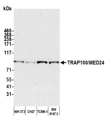
 WB (Western Blot)
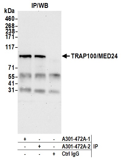
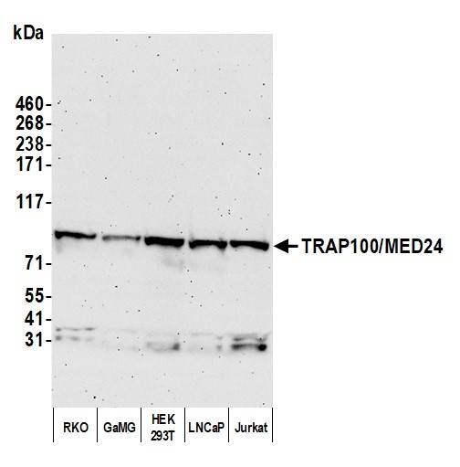
(Detection of human TRAP100/MED24 by western blot. Samples: Whole cell lysate (50 ug) from RKO, GaMG, HEK293T, LNCaP, and Jurkat cells prepared using NETN lysis buffer. Antibody: Affinity purified rabbit anti-TRAP100/MED24 antibody (AAA211493 lot 2) used for WB at 1 ug/ml. Detection: Chemiluminescence with an exposure time of 3 minutes.)
WB (Western Blot)
(Detection of human TRAP100/MED24 by western blot. Samples: Whole cell lysate (50 ug) from RKO, GaMG, HEK293T, LNCaP, and Jurkat cells prepared using NETN lysis buffer. Antibody: Affinity purified rabbit anti-TRAP100/MED24 antibody (AAA211493 lot 2) used for WB at 1 ug/ml. Detection: Chemiluminescence with an exposure time of 3 minutes.)
TRAP100/MED24, Polyclonal Antibody (Cat# AAA211493)
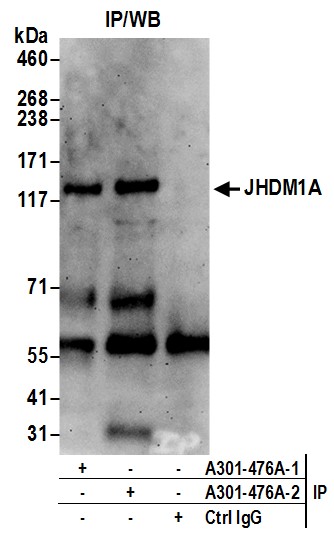
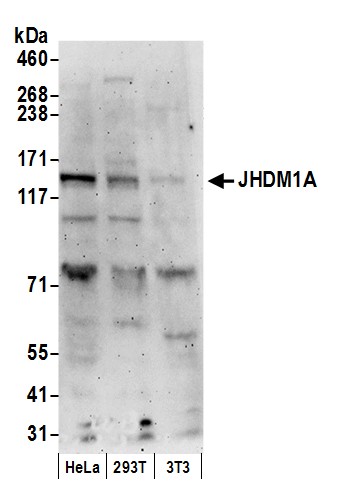 WB (Western Blot)
(Detection of human and mouse JHDM1A by western blot. Samples: Whole cell lysate (50 ug) from HeLa, HEK293T, and mouse NIH 3T3 cells prepared using NETN lysis buffer. Antibodies: Affinity purified rabbit anti-JHDM1A antibody AAA211495 (lot AAA211495-2) used for WB at 0.2 ug/ml. Detection: Chemiluminescence with an exposure time of 3 minutes.)
WB (Western Blot)
(Detection of human and mouse JHDM1A by western blot. Samples: Whole cell lysate (50 ug) from HeLa, HEK293T, and mouse NIH 3T3 cells prepared using NETN lysis buffer. Antibodies: Affinity purified rabbit anti-JHDM1A antibody AAA211495 (lot AAA211495-2) used for WB at 0.2 ug/ml. Detection: Chemiluminescence with an exposure time of 3 minutes.)
JHDM1A, Polyclonal Antibody (Cat# AAA211495)
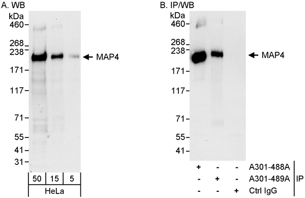 WB (Western Blot)
(Detection of human MAP4 by western blot and immunoprecipitation. Samples: Whole cell lysate (5, 15 and 50 ug for WB; 1 mg for IP, 20% of IP loaded) from HeLa cells. Antibodies: Affinity purified rabbit anti-MAP4 antibody AAA211501 used for WB at 0.04 ug/ml (A) and 0.01 ug/ml (B) and used for IP at 3 ug/mg lysate. MAP4 was also immunoprecipitated by rabbit anti-MAP4 antibody which recognizes an upstream epitope. Detection: Chemiluminescence with exposure times of 30 second (A and B).)
WB (Western Blot)
(Detection of human MAP4 by western blot and immunoprecipitation. Samples: Whole cell lysate (5, 15 and 50 ug for WB; 1 mg for IP, 20% of IP loaded) from HeLa cells. Antibodies: Affinity purified rabbit anti-MAP4 antibody AAA211501 used for WB at 0.04 ug/ml (A) and 0.01 ug/ml (B) and used for IP at 3 ug/mg lysate. MAP4 was also immunoprecipitated by rabbit anti-MAP4 antibody which recognizes an upstream epitope. Detection: Chemiluminescence with exposure times of 30 second (A and B).)
MAP4, Polyclonal Antibody (Cat# AAA211501)
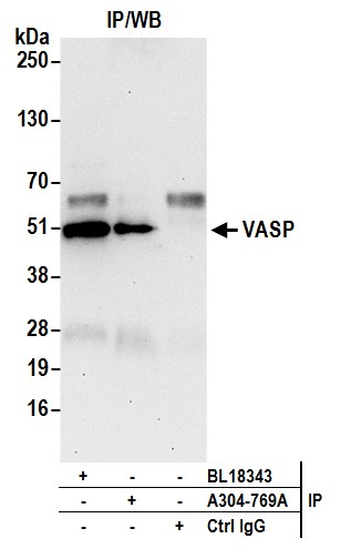
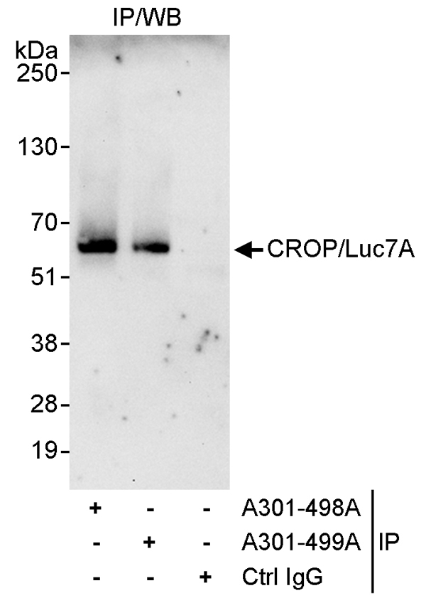 IP (Immunoprecipitation)
(Detection of human CROP/Luc7A by western blot of immunoprecipitates. Samples: Whole cell lysate (1 mg for IP, 20% of IP loaded) from HeLa cells. Antibodies: Affinity purified rabbit anti-CROP/Luc7A antibody AAA211504 used for IP at 3 ug/mg lysate. CROP/Luc7A was also immunoprecipitated by rabbit anti-CROP/Luc7A antibody which recognizes a downstream epitope. For blotting immunoprecipitated CROP/Luc7A, AAA211504 was used at 1 ug/ml. Detection: Chemiluminescence with an exposure time of 30 seconds.)
IP (Immunoprecipitation)
(Detection of human CROP/Luc7A by western blot of immunoprecipitates. Samples: Whole cell lysate (1 mg for IP, 20% of IP loaded) from HeLa cells. Antibodies: Affinity purified rabbit anti-CROP/Luc7A antibody AAA211504 used for IP at 3 ug/mg lysate. CROP/Luc7A was also immunoprecipitated by rabbit anti-CROP/Luc7A antibody which recognizes a downstream epitope. For blotting immunoprecipitated CROP/Luc7A, AAA211504 was used at 1 ug/ml. Detection: Chemiluminescence with an exposure time of 30 seconds.)
CROP/Luc7A, Polyclonal Antibody (Cat# AAA211504)
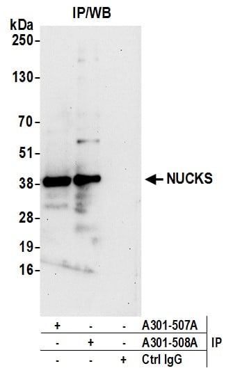 IP (Immunoprecipitation)
(Detection of human NUCKS by western blot of immunoprecipitates. Samples: Whole cell lysate (1.0 mg per IP reaction; 20% of IP loaded) from HeLa cells prepared using NETN lysis buffer. Antibodies: Affinity purified rabbit anti-NUCKS antibody AAA211508 (lot AAA211508-2) used for IP at 3 ug per reaction. NUCKS was also immunoprecipitated by rabbit anti-NUCKS antibody For blotting immunoprecipitated NUCKS, AAA211508 was used at 1 ug/ml. Detection: Chemiluminescence with an exposure time of 30 seconds.)
IP (Immunoprecipitation)
(Detection of human NUCKS by western blot of immunoprecipitates. Samples: Whole cell lysate (1.0 mg per IP reaction; 20% of IP loaded) from HeLa cells prepared using NETN lysis buffer. Antibodies: Affinity purified rabbit anti-NUCKS antibody AAA211508 (lot AAA211508-2) used for IP at 3 ug per reaction. NUCKS was also immunoprecipitated by rabbit anti-NUCKS antibody For blotting immunoprecipitated NUCKS, AAA211508 was used at 1 ug/ml. Detection: Chemiluminescence with an exposure time of 30 seconds.)
NUCKS, Polyclonal Antibody (Cat# AAA211508)
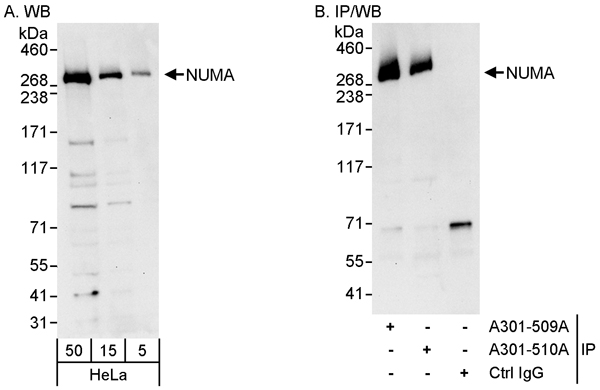 WB (Western Blot)
(Detection of human NUMA by western blot and immunoprecipitation. Samples: Whole cell lysate (5, 15 and 50 ug for WB; 1 mg for IP, 20% of IP loaded) from HeLa cells. Antibodies: Affinity purified rabbit anti-NUMA antibody AAA211510 used for WB at 0.04 ug/ml (A) and 0.1 ug/ml (B) and used for IP at 3 ug/mg lysate. NUMA was also immunoprecipitated by rabbit anti-NUMA antibody which recognizes an upstream epitope. Detection: Chemiluminescence with exposure times of 30 seconds (A and B).)
WB (Western Blot)
(Detection of human NUMA by western blot and immunoprecipitation. Samples: Whole cell lysate (5, 15 and 50 ug for WB; 1 mg for IP, 20% of IP loaded) from HeLa cells. Antibodies: Affinity purified rabbit anti-NUMA antibody AAA211510 used for WB at 0.04 ug/ml (A) and 0.1 ug/ml (B) and used for IP at 3 ug/mg lysate. NUMA was also immunoprecipitated by rabbit anti-NUMA antibody which recognizes an upstream epitope. Detection: Chemiluminescence with exposure times of 30 seconds (A and B).)
NUMA, Polyclonal Antibody (Cat# AAA211510)
 WB (Western Blot)
(Detection of human SART3/TIP110 by western blot and immunoprecipitation. Samples: Whole cell lysate (5, 15 and 50 ug for WB; 1 mg for IP, 20% of IP loaded) from HeLa cells. Antibodies: Affinity purified rabbit anti-SART3/TIP110 antibody AAA211514 used for WB at 0.02 ug/ml (A) and 0.04 ug/ml (B) and used for IP at 3 ug/mg lysate. SART3/TIP110 was also immunoprecipitated by rabbit anti-SART3/TIP110 antibody which recognizes a downstream epitope. Detection: Chemiluminescence with exposure times of 3 seconds (A and B).)
WB (Western Blot)
(Detection of human SART3/TIP110 by western blot and immunoprecipitation. Samples: Whole cell lysate (5, 15 and 50 ug for WB; 1 mg for IP, 20% of IP loaded) from HeLa cells. Antibodies: Affinity purified rabbit anti-SART3/TIP110 antibody AAA211514 used for WB at 0.02 ug/ml (A) and 0.04 ug/ml (B) and used for IP at 3 ug/mg lysate. SART3/TIP110 was also immunoprecipitated by rabbit anti-SART3/TIP110 antibody which recognizes a downstream epitope. Detection: Chemiluminescence with exposure times of 3 seconds (A and B).)
SART3/TIP110, Polyclonal Antibody (Cat# AAA211514)
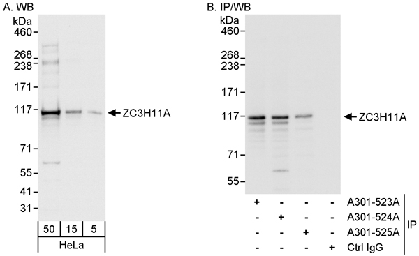 WB (Western Blot)
(Detection of human ZC3H11A by western blot and immunoprecipitation. Samples: Whole cell lysate (5, 15 and 50 ug for WB; 1 mg for IP, 20% of IP loaded) from HeLa cells. Antibodies: Affinity purified rabbit anti-ZC3H11A antibody AAA211515 used for WB at 0.04 ug/ml (A) and 0.1 ug/ml (B) and used for IP at 3 ug/mg lysate. ZC3H11A was also immunoprecipitated by rabbit anti-ZC3H11A antibodies and which recognize other epitopes. Detection: Chemiluminescence with exposure times of 3 seconds (A and B).)
WB (Western Blot)
(Detection of human ZC3H11A by western blot and immunoprecipitation. Samples: Whole cell lysate (5, 15 and 50 ug for WB; 1 mg for IP, 20% of IP loaded) from HeLa cells. Antibodies: Affinity purified rabbit anti-ZC3H11A antibody AAA211515 used for WB at 0.04 ug/ml (A) and 0.1 ug/ml (B) and used for IP at 3 ug/mg lysate. ZC3H11A was also immunoprecipitated by rabbit anti-ZC3H11A antibodies and which recognize other epitopes. Detection: Chemiluminescence with exposure times of 3 seconds (A and B).)
ZC3H11A, Polyclonal Antibody (Cat# AAA211515)
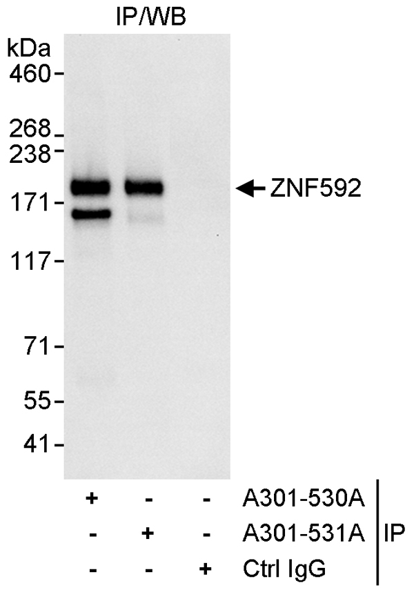 IP (Immunoprecipitation)
(Detection of human ZNF592 by western blot of immunoprecipitates. Samples: Whole cell lysate (1 mg for IP, 20% of IP loaded) from HeLa cells. Antibodies: Affinity purified rabbit anti-ZNF592 antibody AAA211519 used for IP at 3 ug/mg lysate. ZNF592 was also immunoprecipitated by rabbit anti-ZNF592 antibody which recognizes an upstream epitope. For blotting immunoprecipitated ZNF592, was used at 1 ug/ml. Detection: Chemiluminescence with an exposure time of 3 seconds.)
IP (Immunoprecipitation)
(Detection of human ZNF592 by western blot of immunoprecipitates. Samples: Whole cell lysate (1 mg for IP, 20% of IP loaded) from HeLa cells. Antibodies: Affinity purified rabbit anti-ZNF592 antibody AAA211519 used for IP at 3 ug/mg lysate. ZNF592 was also immunoprecipitated by rabbit anti-ZNF592 antibody which recognizes an upstream epitope. For blotting immunoprecipitated ZNF592, was used at 1 ug/ml. Detection: Chemiluminescence with an exposure time of 3 seconds.)
ZNF592, Polyclonal Antibody (Cat# AAA211519)
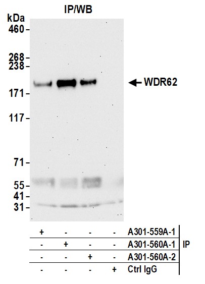
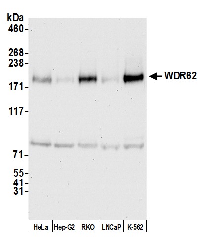 WB (Western Blot)
(Detection of human WDR62 by western blot. Samples: Whole cell lysate (50 ug) from HeLa, Hep-G2, RKO, LNCaP, and K-562 cells prepared using NETN lysis buffer. Antibody: Affinity purified rabbit anti-WDR62 antibody (AAA211529 lot 2) used for WB at 0.4 ug/ml. Detection: Chemiluminescence with an exposure time of 30 seconds.)
WB (Western Blot)
(Detection of human WDR62 by western blot. Samples: Whole cell lysate (50 ug) from HeLa, Hep-G2, RKO, LNCaP, and K-562 cells prepared using NETN lysis buffer. Antibody: Affinity purified rabbit anti-WDR62 antibody (AAA211529 lot 2) used for WB at 0.4 ug/ml. Detection: Chemiluminescence with an exposure time of 30 seconds.)
WDR62, Polyclonal Antibody (Cat# AAA211529)
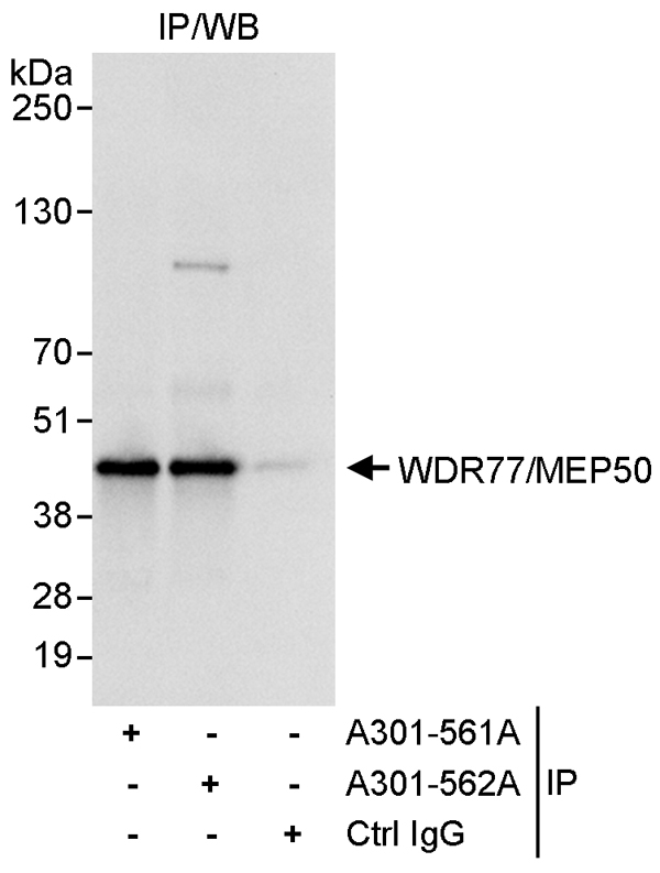 IP (Immunoprecipitation)
(Detection of human WDR77/MEP50 by western blot of immunoprecipitates. Samples: Whole cell lysate (1 mg for IP, 20% of IP loaded) from HeLa cells. Antibodies: Affinity purified rabbit anti-WDR77/MEP50 antibody AAA211530 used for IP at 3 ug/mg lysate. WDR77/MEP50 was also immunoprecipitated by rabbit anti-WDR77/MEP50 antibody which recognizes a downstream epitope. For blotting immunoprecipitated WDR77/MEP50, was used at 1 ug/ml. Detection: Chemiluminescence with an exposure time of 1 second.)
IP (Immunoprecipitation)
(Detection of human WDR77/MEP50 by western blot of immunoprecipitates. Samples: Whole cell lysate (1 mg for IP, 20% of IP loaded) from HeLa cells. Antibodies: Affinity purified rabbit anti-WDR77/MEP50 antibody AAA211530 used for IP at 3 ug/mg lysate. WDR77/MEP50 was also immunoprecipitated by rabbit anti-WDR77/MEP50 antibody which recognizes a downstream epitope. For blotting immunoprecipitated WDR77/MEP50, was used at 1 ug/ml. Detection: Chemiluminescence with an exposure time of 1 second.)
WDR77/MEP50, Polyclonal Antibody (Cat# AAA211530)
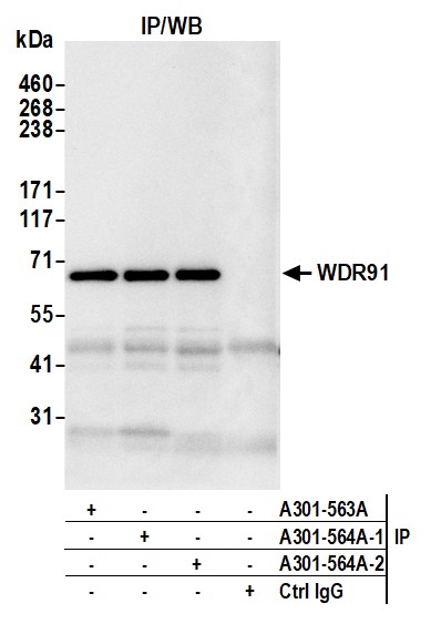
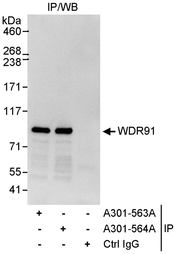 IP (Immunoprecipitation)
(Detection of human WDR91 by western blot of immunoprecipitates. Samples: Whole cell lysate (1 mg for IP, 20% of IP loaded) from HeLa cells. Antibodies: Affinity purified rabbit anti-WDR91 antibody AAA211532 used for IP at 3 ug/mg lysate. WDR91 was also immunoprecipitated by rabbit anti-WDR91 antibody which recognizes a downstream epitope. For blotting immunoprecipitated WDR91, was used at 1 ug/ml. Detection: Chemiluminescence with an exposure time of 10 seconds.)
IP (Immunoprecipitation)
(Detection of human WDR91 by western blot of immunoprecipitates. Samples: Whole cell lysate (1 mg for IP, 20% of IP loaded) from HeLa cells. Antibodies: Affinity purified rabbit anti-WDR91 antibody AAA211532 used for IP at 3 ug/mg lysate. WDR91 was also immunoprecipitated by rabbit anti-WDR91 antibody which recognizes a downstream epitope. For blotting immunoprecipitated WDR91, was used at 1 ug/ml. Detection: Chemiluminescence with an exposure time of 10 seconds.)
WDR91, Polyclonal Antibody (Cat# AAA211532)
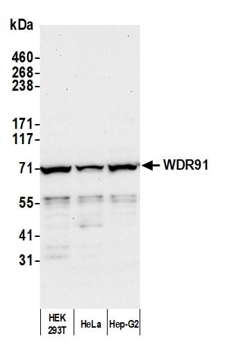 WB (Western Blot)
(Detection of human WDR91 by western blot. Samples: Whole cell lysate (50 ug) from HEK293T, HeLa, and Hep-G2 cells prepared using NETN lysis buffer. Antibody: Affinity purified rabbit anti-WDR91 antibody (AAA211533 lot 2) used for WB at 0.1 ug/ml. Detection: Chemiluminescence with an exposure time of 3 minutes.)
WB (Western Blot)
(Detection of human WDR91 by western blot. Samples: Whole cell lysate (50 ug) from HEK293T, HeLa, and Hep-G2 cells prepared using NETN lysis buffer. Antibody: Affinity purified rabbit anti-WDR91 antibody (AAA211533 lot 2) used for WB at 0.1 ug/ml. Detection: Chemiluminescence with an exposure time of 3 minutes.)
WDR91, Polyclonal Antibody (Cat# AAA211533)
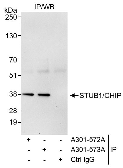
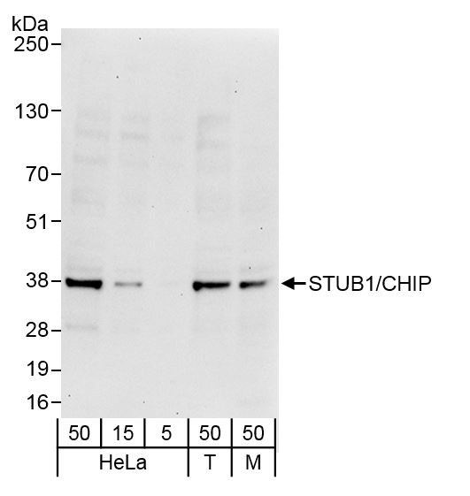 WB (Western Blot)
(Detection of human and mouse STUB1/CHIP by western blot. Samples: Whole cell lysate from HeLa (5, 15 and 50 ug), HEK293T (T; 50 ug) and mouse NIH 3T3 (M; 50 ug) cells. Antibodies: Affinity purified rabbit anti-STUB1/CHIP antibody AAA211539 (lot AAA211539-1) used for WB at 0.04 ug/ml. Detection: Chemiluminescence with exposure time of 30 seconds.)
WB (Western Blot)
(Detection of human and mouse STUB1/CHIP by western blot. Samples: Whole cell lysate from HeLa (5, 15 and 50 ug), HEK293T (T; 50 ug) and mouse NIH 3T3 (M; 50 ug) cells. Antibodies: Affinity purified rabbit anti-STUB1/CHIP antibody AAA211539 (lot AAA211539-1) used for WB at 0.04 ug/ml. Detection: Chemiluminescence with exposure time of 30 seconds.)
STUB1/CHIP, Polyclonal Antibody (Cat# AAA211539)
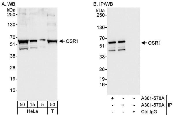
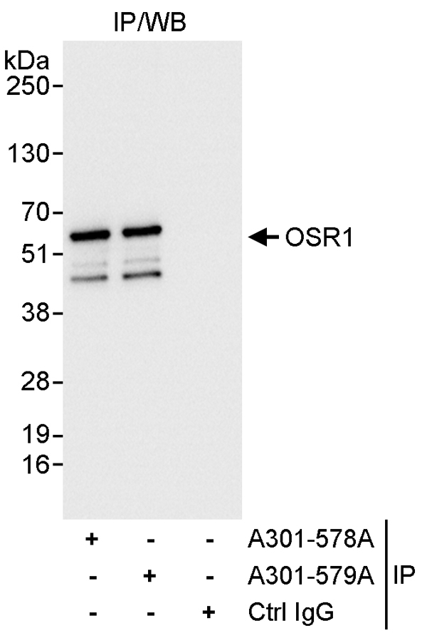 IP (Immunoprecipitation)
(Detection of human OSR1 by western blot of immunoprecipitates. Samples: Whole cell lysate (1 mg for IP, 20% of IP loaded) from HeLa cells. Antibodies: Affinity purified rabbit anti-OSR1 antibody AAA211540 used for IP at 3 ug/mg lysate. OSR1 was also immunoprecipitated by rabbit anti-OSR1 antibody which recognizes a downstream epitope. For blotting immunoprecipitated OSR1, was used at 0.1 ug/ml. Detection: Chemiluminescence with an exposure time of 1 second.)
IP (Immunoprecipitation)
(Detection of human OSR1 by western blot of immunoprecipitates. Samples: Whole cell lysate (1 mg for IP, 20% of IP loaded) from HeLa cells. Antibodies: Affinity purified rabbit anti-OSR1 antibody AAA211540 used for IP at 3 ug/mg lysate. OSR1 was also immunoprecipitated by rabbit anti-OSR1 antibody which recognizes a downstream epitope. For blotting immunoprecipitated OSR1, was used at 0.1 ug/ml. Detection: Chemiluminescence with an exposure time of 1 second.)
OSR1, Polyclonal Antibody (Cat# AAA211540)
 WB (Western Blot)
(Detection of human OSR1 by western blot and immunoprecipitation. Samples: Whole cell lysate from HeLa (5, 15 and 50 ug for WB; 1 mg for IP, 20% of IP loaded) and HEK293T (T; 50 ug) cells. Antibodies: Affinity purified rabbit anti-OSR1 antibody AAA211541 used for WB at 0.04 ug/ml (A) and 0.1 ug/ml (B) and used for IP at 3 ug/mg lysate. OSR1 was also immunoprecipitated by rabbit anti-OSR1 antibody which recognizes an upstream epitope. Detection: Chemiluminescence with exposure times of 1 second (A and B).)
WB (Western Blot)
(Detection of human OSR1 by western blot and immunoprecipitation. Samples: Whole cell lysate from HeLa (5, 15 and 50 ug for WB; 1 mg for IP, 20% of IP loaded) and HEK293T (T; 50 ug) cells. Antibodies: Affinity purified rabbit anti-OSR1 antibody AAA211541 used for WB at 0.04 ug/ml (A) and 0.1 ug/ml (B) and used for IP at 3 ug/mg lysate. OSR1 was also immunoprecipitated by rabbit anti-OSR1 antibody which recognizes an upstream epitope. Detection: Chemiluminescence with exposure times of 1 second (A and B).)
OSR1, Polyclonal Antibody (Cat# AAA211541)
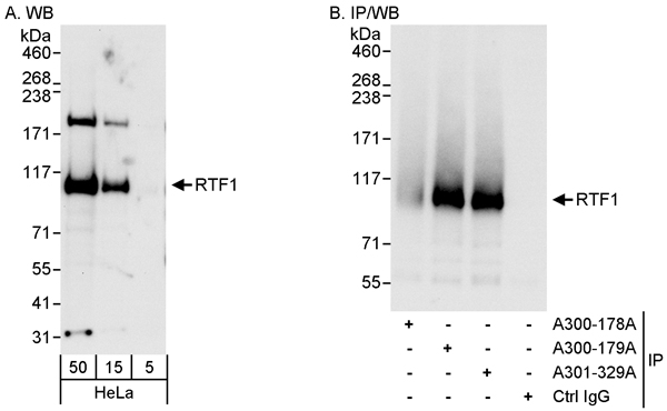 WB (Western Blot)
(Detection of human RTF1 by western blot and immunoprecipitation. Samples: Whole cell lysate (5, 15 and 50 ug for WB; 1 mg for IP, 20% of IP loaded) from HeLa cells. Antibodies: Affinity purified rabbit anti-RTF1 antibody AAA211423 used for WB at 0.04 ug/ml (A) and 1 ug/ml (B) and used for IP at 3 ug/mg lysate. RTF1 was also immunoprecipitated by rabbit anti-RTF1 antibody which recognizes an upstream epitope. Detection: Chemiluminescence with exposure times of 10 seconds (A) and 3 seconds (B).)
WB (Western Blot)
(Detection of human RTF1 by western blot and immunoprecipitation. Samples: Whole cell lysate (5, 15 and 50 ug for WB; 1 mg for IP, 20% of IP loaded) from HeLa cells. Antibodies: Affinity purified rabbit anti-RTF1 antibody AAA211423 used for WB at 0.04 ug/ml (A) and 1 ug/ml (B) and used for IP at 3 ug/mg lysate. RTF1 was also immunoprecipitated by rabbit anti-RTF1 antibody which recognizes an upstream epitope. Detection: Chemiluminescence with exposure times of 10 seconds (A) and 3 seconds (B).)
Rtf1, Polyclonal Antibody (Cat# AAA211423)
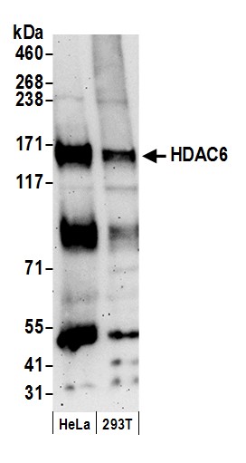
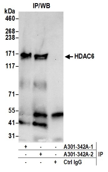
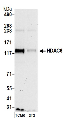 WB (Western Blot)
(Detection of mouse HDAC6 by western blot. Samples: Whole cell lysate (50 ug) from TCMK-1 and NIH 3T3 cells prepared using NETN lysis buffer. Antibody: Affinity purified rabbit anti-HDAC6 antibody AAA211432 (lot AAA211432-2) used for WB at 0.1 ug/ml. Detection: Chemiluminescence with an exposure time of 3 minutes.)
WB (Western Blot)
(Detection of mouse HDAC6 by western blot. Samples: Whole cell lysate (50 ug) from TCMK-1 and NIH 3T3 cells prepared using NETN lysis buffer. Antibody: Affinity purified rabbit anti-HDAC6 antibody AAA211432 (lot AAA211432-2) used for WB at 0.1 ug/ml. Detection: Chemiluminescence with an exposure time of 3 minutes.)
HDAC6, Polyclonal Antibody (Cat# AAA211432)
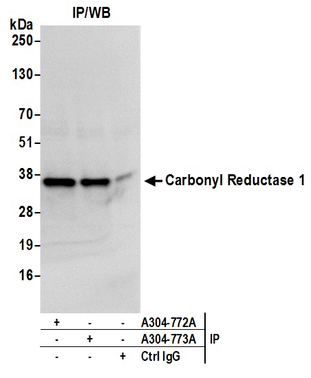
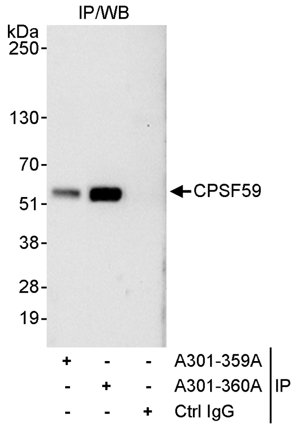 IP (Immunoprecipitation)
(Detection of human CPSF59 by western blot of immunoprecipitates. Samples: Whole cell lysate (1 mg for IP, 20% of IP loaded) from HeLa cells. Antibodies: Affinity purified rabbit anti-CPSF59 antibody AAA211439 used for IP at 3 ug/mg lysate. CPSF59 was also immunoprecipitated by rabbit anti-CPSF59 antibody which recognizes an upstream epitope. For blotting immunoprecipitated CPSF59, was used at 1 ug/ml. Detection: Chemiluminescence with an exposure time of 10 seconds.)
IP (Immunoprecipitation)
(Detection of human CPSF59 by western blot of immunoprecipitates. Samples: Whole cell lysate (1 mg for IP, 20% of IP loaded) from HeLa cells. Antibodies: Affinity purified rabbit anti-CPSF59 antibody AAA211439 used for IP at 3 ug/mg lysate. CPSF59 was also immunoprecipitated by rabbit anti-CPSF59 antibody which recognizes an upstream epitope. For blotting immunoprecipitated CPSF59, was used at 1 ug/ml. Detection: Chemiluminescence with an exposure time of 10 seconds.)
CPSF59, Polyclonal Antibody (Cat# AAA211439)
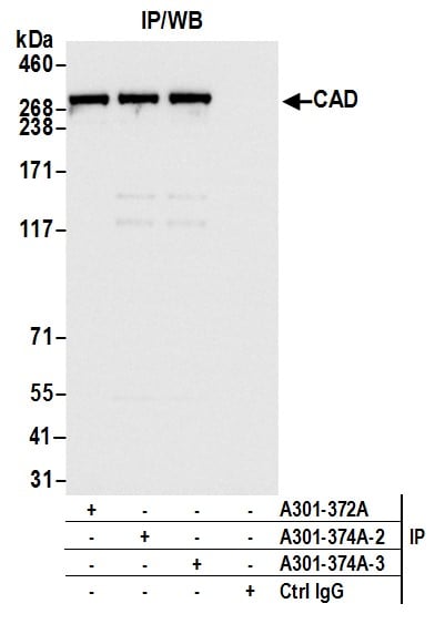
 WB (Western Blot)
(Detection of human and mouse CAD by western blot. Samples: Whole cell lysate (50 ug) from HEK293T, K-562, Jurkat, TCMK-1, and NIH 3T3 cells prepared using NETN lysis buffer. Antibody: Affinity purified rabbit anti-CAD antibody AAA211444 (lot AAA211444-3) used for WB at 0.04 ug/ml. Detection: Chemiluminescence with an exposure time of 10 seconds.)
WB (Western Blot)
(Detection of human and mouse CAD by western blot. Samples: Whole cell lysate (50 ug) from HEK293T, K-562, Jurkat, TCMK-1, and NIH 3T3 cells prepared using NETN lysis buffer. Antibody: Affinity purified rabbit anti-CAD antibody AAA211444 (lot AAA211444-3) used for WB at 0.04 ug/ml. Detection: Chemiluminescence with an exposure time of 10 seconds.)
CAD, Polyclonal Antibody (Cat# AAA211444)
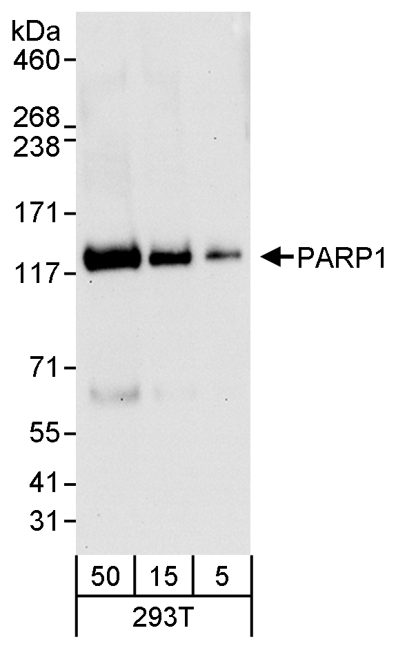 WB (Western Blot)
(Detection of human PARP1 by western blot. Samples: Whole cell lysate (5, 15 and 50 ug) from HEK293T cells. Antibody: Affinity purified rabbit anti-PARP1 antibody AAA211445 used for WB at 0.04 ug/ml. Detection: Chemiluminescence with an exposure time of 30 seconds.)
WB (Western Blot)
(Detection of human PARP1 by western blot. Samples: Whole cell lysate (5, 15 and 50 ug) from HEK293T cells. Antibody: Affinity purified rabbit anti-PARP1 antibody AAA211445 used for WB at 0.04 ug/ml. Detection: Chemiluminescence with an exposure time of 30 seconds.)
PARP1, Polyclonal Antibody (Cat# AAA211445)
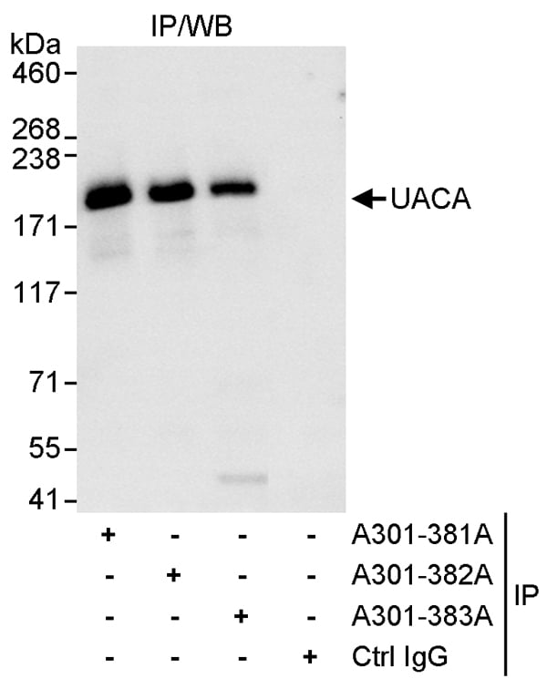 IP (Immunoprecipitation)
(Detection of human UACA by western blot of immunoprecipitates. Samples: Whole cell lysate (1 mg for IP, 20% of IP loaded) from HeLa cells. Antibodies: Affinity purified rabbit anti-UACA antibody AAA211450 used for IP at 3 ug/mg lysate. UACA was also immunoprecipitated by rabbit anti-UACA antibodies and which recognize other epitopes. For blotting immunoprecipitated UACA, was used at 1 ug/ml. Detection: Chemiluminescence with an exposure time of 3 seconds.)
IP (Immunoprecipitation)
(Detection of human UACA by western blot of immunoprecipitates. Samples: Whole cell lysate (1 mg for IP, 20% of IP loaded) from HeLa cells. Antibodies: Affinity purified rabbit anti-UACA antibody AAA211450 used for IP at 3 ug/mg lysate. UACA was also immunoprecipitated by rabbit anti-UACA antibodies and which recognize other epitopes. For blotting immunoprecipitated UACA, was used at 1 ug/ml. Detection: Chemiluminescence with an exposure time of 3 seconds.)
UACA, Polyclonal Antibody (Cat# AAA211450)
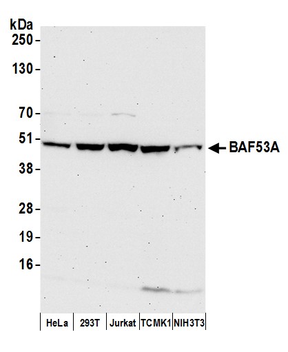 WB (Western Blot)
(Detection of human and mouse BAF53A by western blot. Samples: Whole cell lysate (50 ug) from HeLa, HEK293T, Jurkat, mouse TCMK-1, and mouse NIH 3T3 cells prepared using NETN lysis buffer. Antibody: Affinity purified rabbit anti-BAF53A antibody AAA211457 (lot AAA211457-3) used for WB at 0.04 ug/ml. Detection: Chemiluminescence with an exposure time of 75 seconds.)
WB (Western Blot)
(Detection of human and mouse BAF53A by western blot. Samples: Whole cell lysate (50 ug) from HeLa, HEK293T, Jurkat, mouse TCMK-1, and mouse NIH 3T3 cells prepared using NETN lysis buffer. Antibody: Affinity purified rabbit anti-BAF53A antibody AAA211457 (lot AAA211457-3) used for WB at 0.04 ug/ml. Detection: Chemiluminescence with an exposure time of 75 seconds.)
BAF53A, Polyclonal Antibody (Cat# AAA211457)
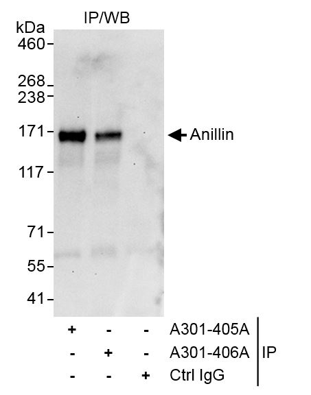
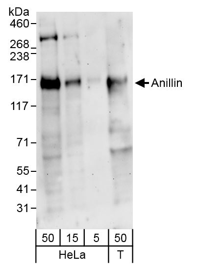 WB (Western Blot)
(Detection of human Anillin by western blot. Samples: Whole cell lysate from HeLa (5, 15 and 50 ug) and HEK293T (T; 50 ug) cells. Antibodies: Affinity purified rabbit anti-Anillin antibody AAA211460 (lot AAA211460-1) used for WB at 0.1 ug/ml. Detection: Chemiluminescence with exposure time of 3 minutes.)
WB (Western Blot)
(Detection of human Anillin by western blot. Samples: Whole cell lysate from HeLa (5, 15 and 50 ug) and HEK293T (T; 50 ug) cells. Antibodies: Affinity purified rabbit anti-Anillin antibody AAA211460 (lot AAA211460-1) used for WB at 0.1 ug/ml. Detection: Chemiluminescence with exposure time of 3 minutes.)
Anillin, Polyclonal Antibody (Cat# AAA211460)
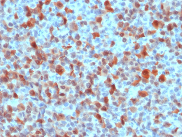
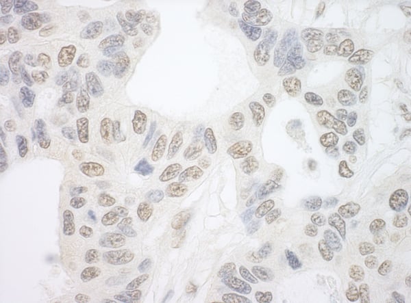
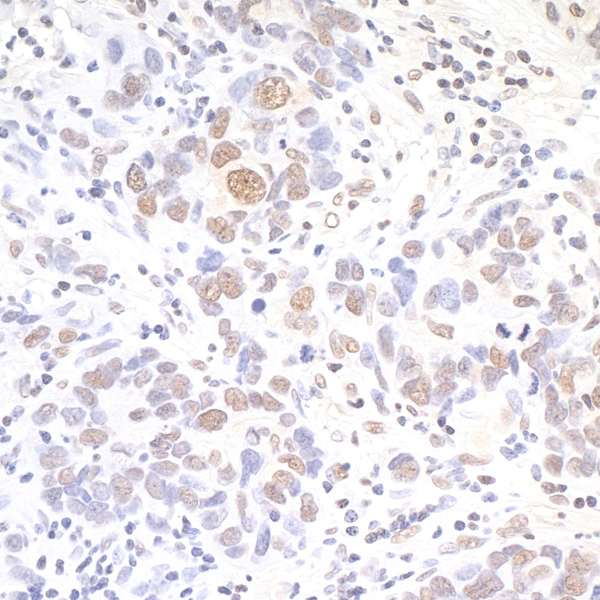
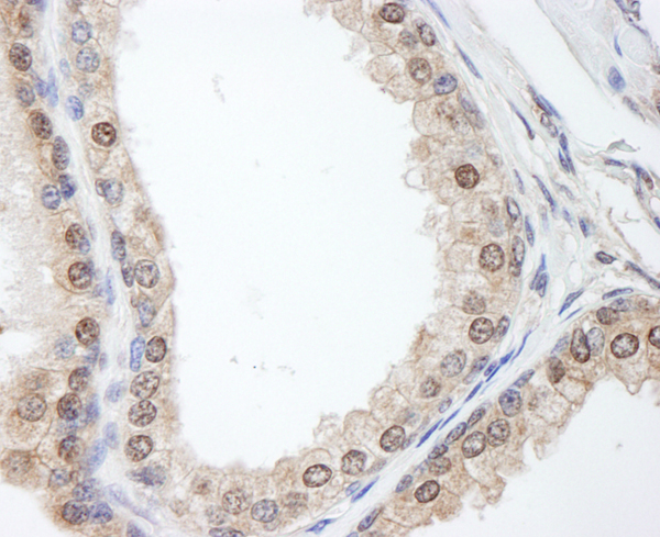
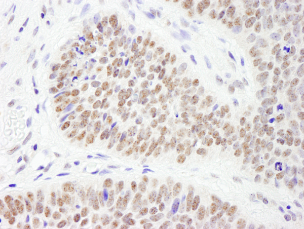
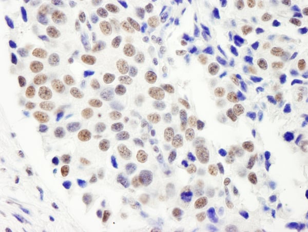
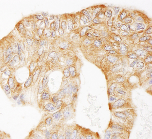
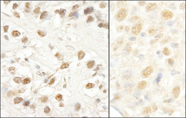
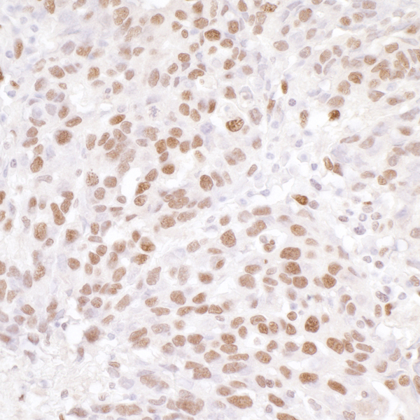
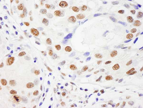
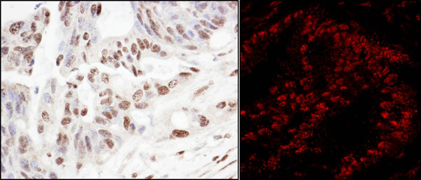
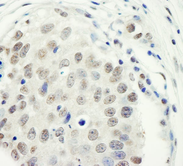
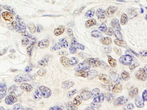
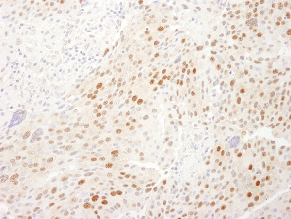
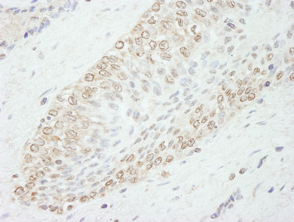
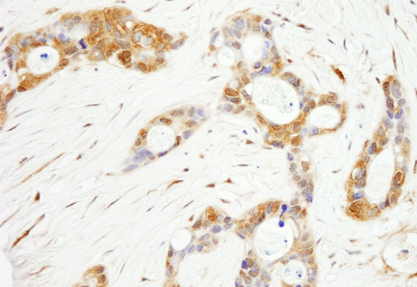
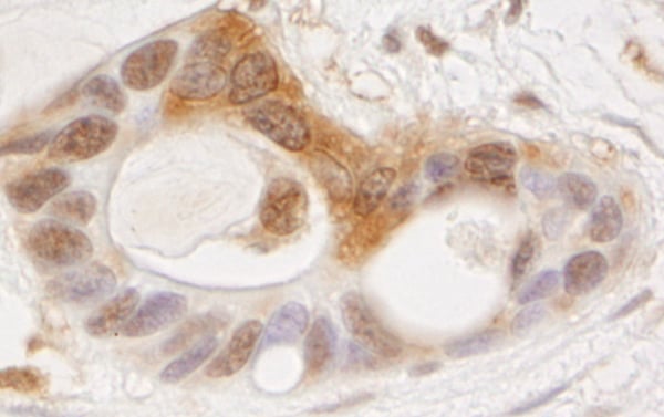
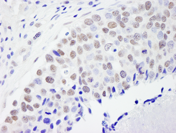
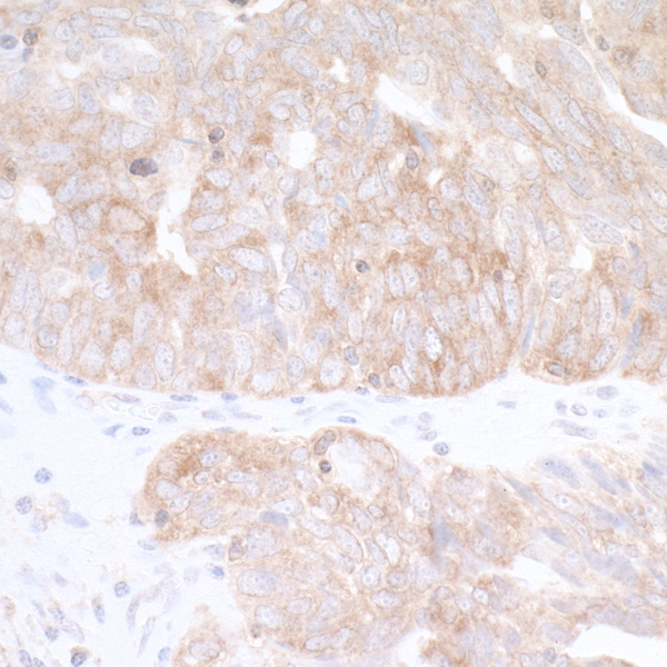
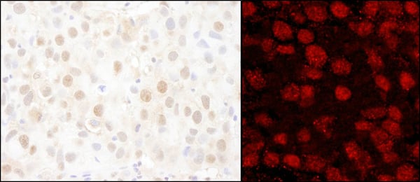
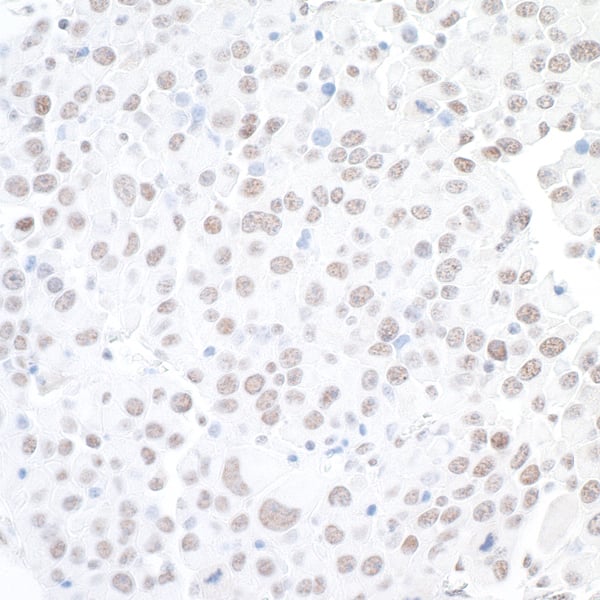
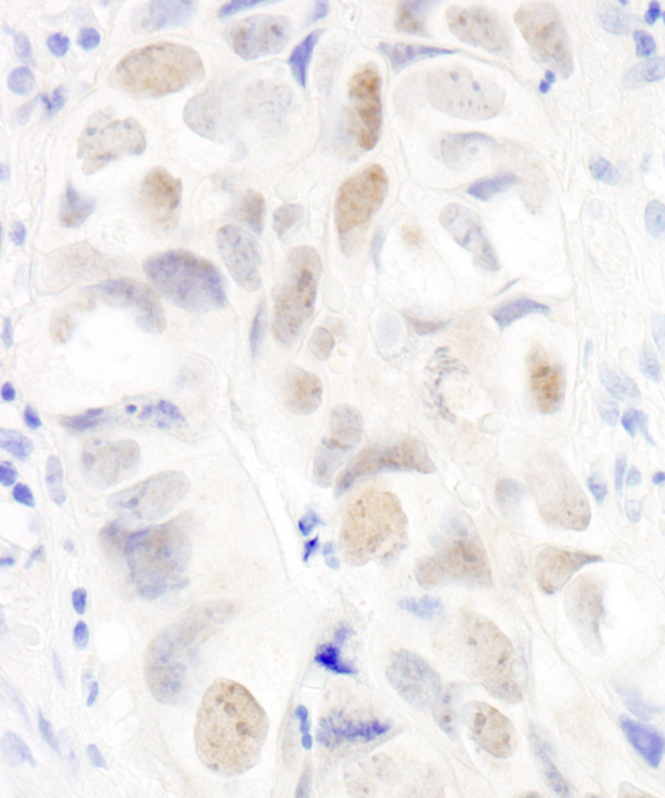
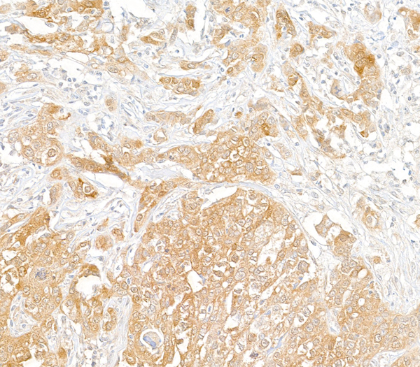
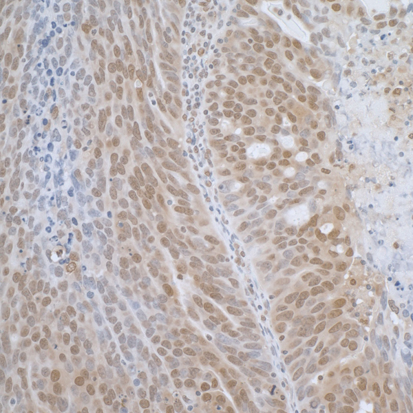
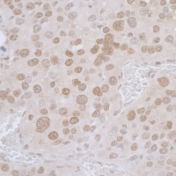
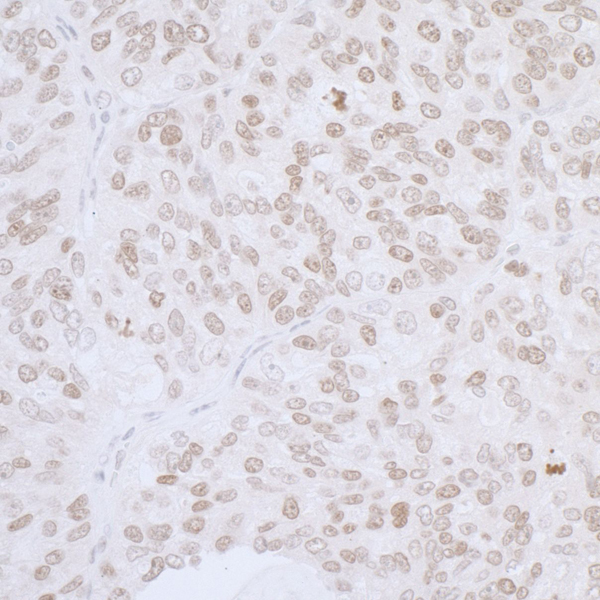
 IHC (Immunohiostchemistry)
(Formalin-fixed, paraffin-embedded human Schwanoma stained with S100B Rabbit Polyclonal Antibody.)
IHC (Immunohiostchemistry)
(Formalin-fixed, paraffin-embedded human Schwanoma stained with S100B Rabbit Polyclonal Antibody.)
S100B, Polyclonal Antibody (Cat# AAA215190)
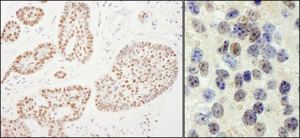
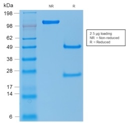 SDS-PAGE
(SDS-PAGE Analysis Purified p53 Rabbit Polyclonal Antibody. Confirmation of Purity and Integrity of Antibody.)
SDS-PAGE
(SDS-PAGE Analysis Purified p53 Rabbit Polyclonal Antibody. Confirmation of Purity and Integrity of Antibody.)
p53 Tumor Suppressor Protein, Polyclonal Antibody (Cat# AAA215232)
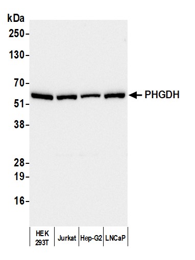
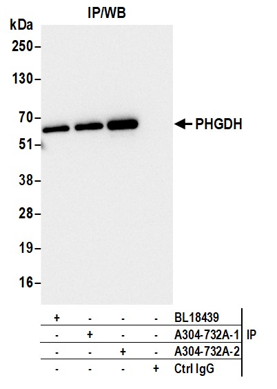
 WB (Western Blot)
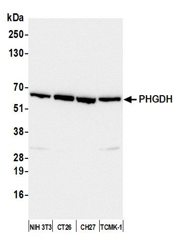
(Detection of mouse PHGDH by western blot. Samples: Whole cell lysate (10 ug) from NIH 3T3, CT26, CH27, and TCMK-1 cells prepared using NETN lysis buffer. Antibody: Affinity purified rabbit anti-PHGDH antibody (AAA212895-2 lot 2) used for WB at 0.1 ug/ml. Detection: Chemiluminescence with an exposure time of 30 seconds.)
WB (Western Blot)
(Detection of mouse PHGDH by western blot. Samples: Whole cell lysate (10 ug) from NIH 3T3, CT26, CH27, and TCMK-1 cells prepared using NETN lysis buffer. Antibody: Affinity purified rabbit anti-PHGDH antibody (AAA212895-2 lot 2) used for WB at 0.1 ug/ml. Detection: Chemiluminescence with an exposure time of 30 seconds.)
PHGDH, Polyclonal Antibody (Cat# AAA212895)


 WB (Western Blot)
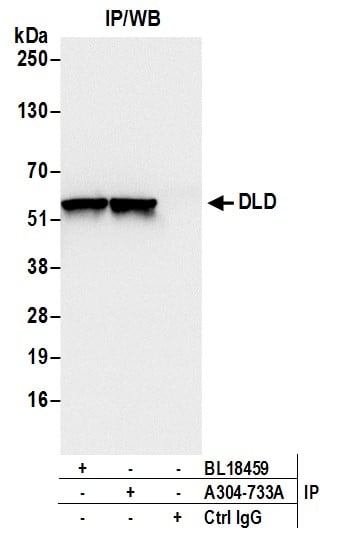
(Detection of human and mouse DLD by western blot. Samples: Whole cell lysate (50 ug) from HeLa, HEK293T, Jurkat, mouse TCMK-1, and mouse NIH 3T3 cells prepared using NETN lysis buffer. Antibodies: Affinity purified rabbit anti-DLD antibody AAA212896 (lot AAA212896-1) used for WB at 0.4 ug/ml. Detection: Chemiluminescence with an exposure time of 1 second.)
WB (Western Blot)
(Detection of human and mouse DLD by western blot. Samples: Whole cell lysate (50 ug) from HeLa, HEK293T, Jurkat, mouse TCMK-1, and mouse NIH 3T3 cells prepared using NETN lysis buffer. Antibodies: Affinity purified rabbit anti-DLD antibody AAA212896 (lot AAA212896-1) used for WB at 0.4 ug/ml. Detection: Chemiluminescence with an exposure time of 1 second.)
DLD, Polyclonal Antibody (Cat# AAA212896)
 WB (Western Blot)
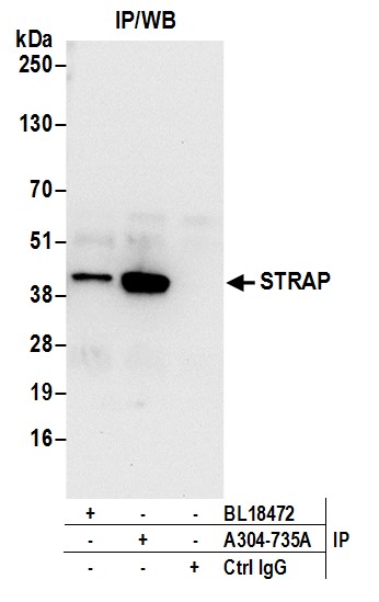
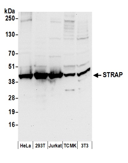
(Detection of human and mouse STRAP by western blot. Samples: Whole cell lysate (50 ug) from HeLa, HEK293T, Jurkat, mouse TCMK-1, and mouse NIH 3T3 cells prepared using NETN lysis buffer. Antibodies: Affinity purified rabbit anti-STRAP antibody AAA212898 (lot AAA212898-1) used for WB at 0.1 ug/ml. Detection: Chemiluminescence with an exposure time of 10 seconds.)
WB (Western Blot)
(Detection of human and mouse STRAP by western blot. Samples: Whole cell lysate (50 ug) from HeLa, HEK293T, Jurkat, mouse TCMK-1, and mouse NIH 3T3 cells prepared using NETN lysis buffer. Antibodies: Affinity purified rabbit anti-STRAP antibody AAA212898 (lot AAA212898-1) used for WB at 0.1 ug/ml. Detection: Chemiluminescence with an exposure time of 10 seconds.)
STRAP, Polyclonal Antibody (Cat# AAA212898)
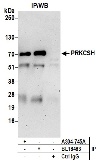
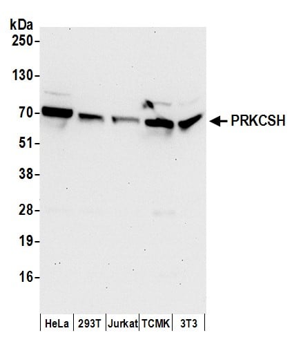 WB (Western Blot)
(Detection of human and mouse PRKCSH by western blot. Samples: Whole cell lysate (50 ug) from HeLa, HEK293T, Jurkat, mouse TCMK-1, and mouse NIH 3T3 cells prepared using NETN lysis buffer. Antibodies: Affinity purified rabbit anti-PRKCSH antibody AAA212904 (lot AAA212904-1) used for WB at 0.1 ug/ml. Detection: Chemiluminescence with an exposure time of 30 seconds.)
WB (Western Blot)
(Detection of human and mouse PRKCSH by western blot. Samples: Whole cell lysate (50 ug) from HeLa, HEK293T, Jurkat, mouse TCMK-1, and mouse NIH 3T3 cells prepared using NETN lysis buffer. Antibodies: Affinity purified rabbit anti-PRKCSH antibody AAA212904 (lot AAA212904-1) used for WB at 0.1 ug/ml. Detection: Chemiluminescence with an exposure time of 30 seconds.)
PRKCSH, Polyclonal Antibody (Cat# AAA212904)
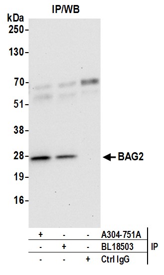
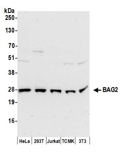 WB (Western Blot)
(Detection of human and mouse BAG2 by western blot. Samples: Whole cell lysate (50 ug) from HeLa, HEK293T, Jurkat, mouse TCMK-1, and mouse NIH 3T3 cells prepared using NETN lysis buffer. Antibodies: Affinity purified rabbit anti-BAG2 antibody AAA212910 (lot AAA212910-1) used for WB at 0.4 ug/ml. Detection: Chemiluminescence with an exposure time of 30 seconds.)
WB (Western Blot)
(Detection of human and mouse BAG2 by western blot. Samples: Whole cell lysate (50 ug) from HeLa, HEK293T, Jurkat, mouse TCMK-1, and mouse NIH 3T3 cells prepared using NETN lysis buffer. Antibodies: Affinity purified rabbit anti-BAG2 antibody AAA212910 (lot AAA212910-1) used for WB at 0.4 ug/ml. Detection: Chemiluminescence with an exposure time of 30 seconds.)
BAG2, Polyclonal Antibody (Cat# AAA212910)
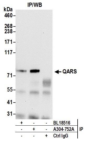
 WB (Western Blot)
(Detection of human and mouse QARS by western blot. Samples: Whole cell lysate (50 ug) from HeLa, HEK293T, Jurkat, mouse TCMK-1, and mouse NIH 3T3 cells prepared using NETN lysis buffer. Antibodies: Affinity purified rabbit anti-QARS antibody AAA212911 (lot AAA212911-1) used for WB at 0.1 ug/ml. Detection: Chemiluminescence with an exposure time of 3 minutes.)
WB (Western Blot)
(Detection of human and mouse QARS by western blot. Samples: Whole cell lysate (50 ug) from HeLa, HEK293T, Jurkat, mouse TCMK-1, and mouse NIH 3T3 cells prepared using NETN lysis buffer. Antibodies: Affinity purified rabbit anti-QARS antibody AAA212911 (lot AAA212911-1) used for WB at 0.1 ug/ml. Detection: Chemiluminescence with an exposure time of 3 minutes.)
QARS, Polyclonal Antibody (Cat# AAA212911)
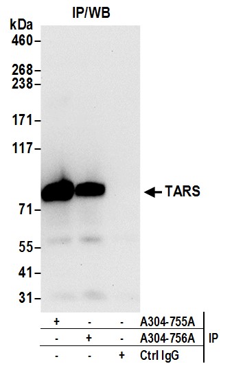
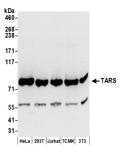 WB (Western Blot)
(Detection of human and mouse TARS by western blot. Samples: Whole cell lysate (50 ug) from HeLa, HEK293T, Jurkat, mouse TCMK-1, and mouse NIH 3T3 cells prepared using NETN lysis buffer. Antibodies: Affinity purified rabbit anti-TARS antibody AAA212915 (lot AAA212915-1) used for WB at 0.1 ug/ml. Detection: Chemiluminescence with an exposure time of 10 seconds.)
WB (Western Blot)
(Detection of human and mouse TARS by western blot. Samples: Whole cell lysate (50 ug) from HeLa, HEK293T, Jurkat, mouse TCMK-1, and mouse NIH 3T3 cells prepared using NETN lysis buffer. Antibodies: Affinity purified rabbit anti-TARS antibody AAA212915 (lot AAA212915-1) used for WB at 0.1 ug/ml. Detection: Chemiluminescence with an exposure time of 10 seconds.)
TARS, Polyclonal Antibody (Cat# AAA212915)
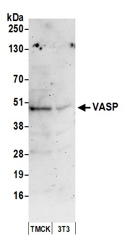
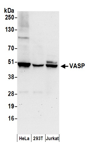 WB (Western Blot)
(Detection of human VASP by western blot. Samples: Whole cell lysate (50 ug) from HeLa, HEK293T, and Jurkat cells prepared using NETN lysis buffer. Antibody: Affinity purified rabbit anti-VASP antibody AAA212924 (lot AAA212924-1) used for WB at 0.4 ug/ml. Detection: Chemiluminescence with an exposure time of 30 seconds.)
WB (Western Blot)
(Detection of human VASP by western blot. Samples: Whole cell lysate (50 ug) from HeLa, HEK293T, and Jurkat cells prepared using NETN lysis buffer. Antibody: Affinity purified rabbit anti-VASP antibody AAA212924 (lot AAA212924-1) used for WB at 0.4 ug/ml. Detection: Chemiluminescence with an exposure time of 30 seconds.)
VASP, Polyclonal Antibody (Cat# AAA212924)
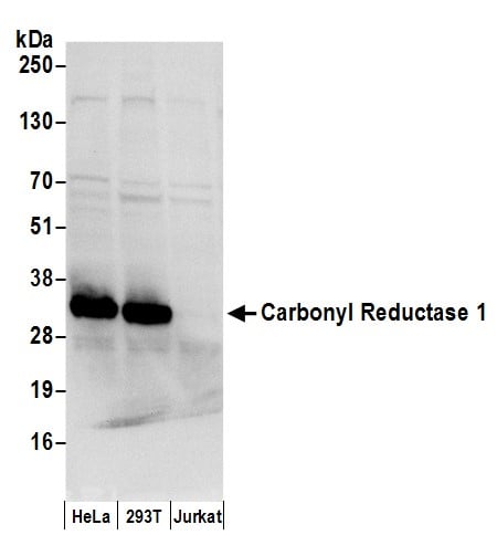 WB (Western Blot)
(Detection of human Carbonyl Reductase 1/CBR1 by western blot. Samples: Whole cell lysate (50 ug) from HeLa, HEK293T, and Jurkat cells prepared using NETN lysis buffer. Antibody: Affinity purified rabbit anti-Carbonyl Reductase 1/CBR1 antibody AAA212928 (lot AAA212928-1) used for WB at 0.1 ug/ml. Detection: Chemiluminescence with an exposure time of 3 seconds.)
WB (Western Blot)
(Detection of human Carbonyl Reductase 1/CBR1 by western blot. Samples: Whole cell lysate (50 ug) from HeLa, HEK293T, and Jurkat cells prepared using NETN lysis buffer. Antibody: Affinity purified rabbit anti-Carbonyl Reductase 1/CBR1 antibody AAA212928 (lot AAA212928-1) used for WB at 0.1 ug/ml. Detection: Chemiluminescence with an exposure time of 3 seconds.)
Carbonyl Reductase 1/CBR1, Polyclonal Antibody (Cat# AAA212928)
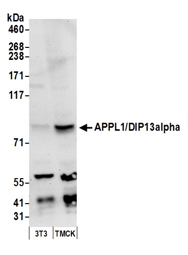
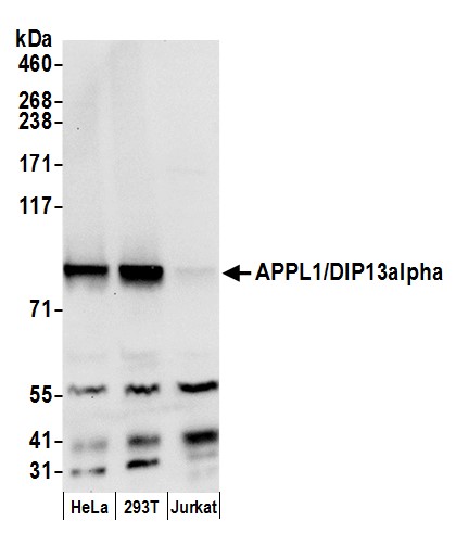 WB (Western Blot)
(Detection of human APPL1/DIP13alpha by western blot. Samples: Whole cell lysate (50 ug) from HeLa, HEK293T, and Jurkat cells prepared using NETN lysis buffer. Antibody: Affinity purified rabbit anti-APPL1/DIP13alpha antibody AAA212936 (lot AAA212936-1) used for WB at 0.1 ug/ml. Detection: Chemiluminescence with an exposure time of 10 seconds.)
WB (Western Blot)
(Detection of human APPL1/DIP13alpha by western blot. Samples: Whole cell lysate (50 ug) from HeLa, HEK293T, and Jurkat cells prepared using NETN lysis buffer. Antibody: Affinity purified rabbit anti-APPL1/DIP13alpha antibody AAA212936 (lot AAA212936-1) used for WB at 0.1 ug/ml. Detection: Chemiluminescence with an exposure time of 10 seconds.)
APPL1/DIP13alpha, Polyclonal Antibody (Cat# AAA212936)
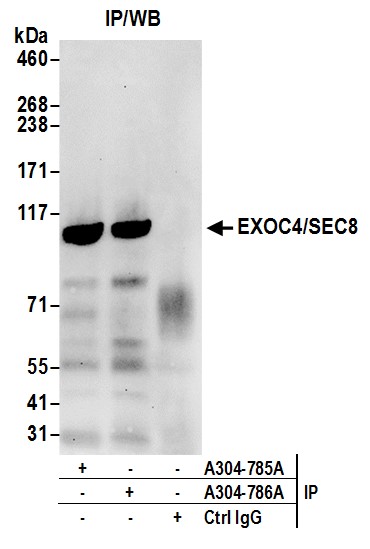
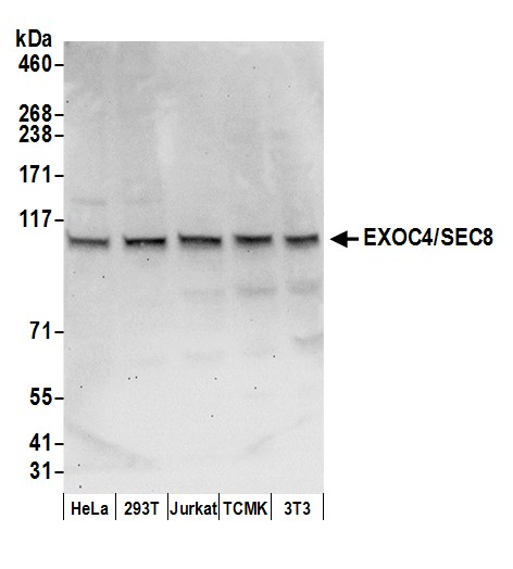 WB (Western Blot)
(Detection of human and mouse EXOC4/SEC8 by western blot. Samples: Whole cell lysate (50 ug) from HeLa, HEK293T, Jurkat, mouse TCMK-1, and mouse NIH 3T3 cells prepared using NETN lysis buffer. Antibody: Affinity purified rabbit anti-EXOC4/SEC8 antibody AAA212939 (lot AAA212939-1) used for WB at 0.1 ug/ml. Detection: Chemiluminescence with an exposure time of 30 seconds.)
WB (Western Blot)
(Detection of human and mouse EXOC4/SEC8 by western blot. Samples: Whole cell lysate (50 ug) from HeLa, HEK293T, Jurkat, mouse TCMK-1, and mouse NIH 3T3 cells prepared using NETN lysis buffer. Antibody: Affinity purified rabbit anti-EXOC4/SEC8 antibody AAA212939 (lot AAA212939-1) used for WB at 0.1 ug/ml. Detection: Chemiluminescence with an exposure time of 30 seconds.)
EXOC4/SEC8, Polyclonal Antibody (Cat# AAA212939)

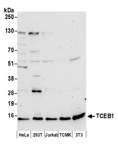 WB (Western Blot)
(Detection of human and mouse TCEB1 by western blot. Samples: Whole cell lysate (50 ug) from HeLa, HEK293T, Jurkat, mouse TCMK-1, and mouse NIH 3T3 cells prepared using NETN lysis buffer. Antibody: Affinity purified rabbit anti-TCEB1 antibody AAA212940 (lot AAA212940-1) used for WB at 0.1 ug/ml. Detection: Chemiluminescence with an exposure time of 3 minutes.)
WB (Western Blot)
(Detection of human and mouse TCEB1 by western blot. Samples: Whole cell lysate (50 ug) from HeLa, HEK293T, Jurkat, mouse TCMK-1, and mouse NIH 3T3 cells prepared using NETN lysis buffer. Antibody: Affinity purified rabbit anti-TCEB1 antibody AAA212940 (lot AAA212940-1) used for WB at 0.1 ug/ml. Detection: Chemiluminescence with an exposure time of 3 minutes.)
TCEB1, Polyclonal Antibody (Cat# AAA212940)

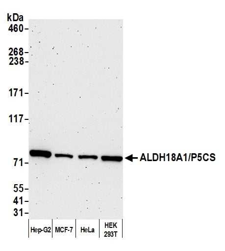 WB (Western Blot)
(Detection of human ALDH18A1/P5CS by western blot. Samples: Whole cell lysate (10 ug) from Hep-G2, MCF-7, HeLa, and HEK293T cells prepared using NETN lysis buffer. Antibody: Affinity purified rabbit anti-ALDH18A1/P5CS antibody AAA212946 Lot 1 used for WB at 0.04 ug/ml. Detection: Chemiluminescence with an exposure time of 3 minutes.)
WB (Western Blot)
(Detection of human ALDH18A1/P5CS by western blot. Samples: Whole cell lysate (10 ug) from Hep-G2, MCF-7, HeLa, and HEK293T cells prepared using NETN lysis buffer. Antibody: Affinity purified rabbit anti-ALDH18A1/P5CS antibody AAA212946 Lot 1 used for WB at 0.04 ug/ml. Detection: Chemiluminescence with an exposure time of 3 minutes.)
ALDH18A1/P5CS, Polyclonal Antibody (Cat# AAA212946)
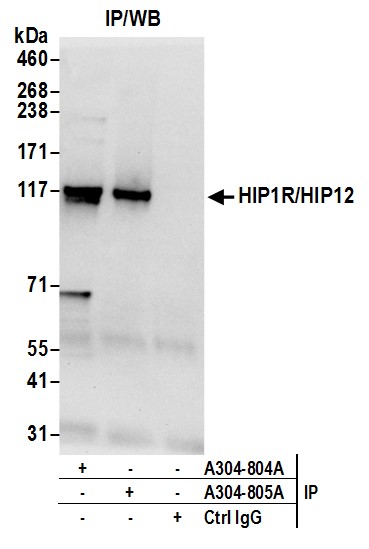
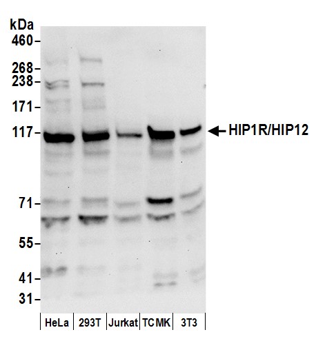 WB (Western Blot)
(Detection of human and mouse HIP1R/HIP12 by western blot. Samples: Whole cell lysate (50 ug) from HeLa, HEK293T, Jurkat, mouse TCMK-1, and mouse NIH 3T3 cells prepared using NETN lysis buffer. Antibody: Affinity purified rabbit anti-HIP1R/HIP12 antibody AAA212947 (lot AAA212947-1) used for WB at 0.1 ug/ml. Detection: Chemiluminescence with an exposure time of 10 seconds.)
WB (Western Blot)
(Detection of human and mouse HIP1R/HIP12 by western blot. Samples: Whole cell lysate (50 ug) from HeLa, HEK293T, Jurkat, mouse TCMK-1, and mouse NIH 3T3 cells prepared using NETN lysis buffer. Antibody: Affinity purified rabbit anti-HIP1R/HIP12 antibody AAA212947 (lot AAA212947-1) used for WB at 0.1 ug/ml. Detection: Chemiluminescence with an exposure time of 10 seconds.)
HIP1R/HIP12, Polyclonal Antibody (Cat# AAA212947)
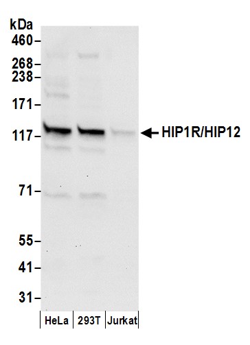 WB (Western Blot)
(Detection of human HIP1R/HIP12 by western blot. Samples: Whole cell lysate (50 ug) from HeLa, HEK293T, and Jurkat cells prepared using NETN lysis buffer. Antibody: Affinity purified rabbit anti-HIP1R/HIP12 antibody AAA212949 (lot AAA212949-1) used for WB at 0.4 ug/ml. Detection: Chemiluminescence with an exposure time of 10 seconds.)
WB (Western Blot)
(Detection of human HIP1R/HIP12 by western blot. Samples: Whole cell lysate (50 ug) from HeLa, HEK293T, and Jurkat cells prepared using NETN lysis buffer. Antibody: Affinity purified rabbit anti-HIP1R/HIP12 antibody AAA212949 (lot AAA212949-1) used for WB at 0.4 ug/ml. Detection: Chemiluminescence with an exposure time of 10 seconds.)
HIP1R/HIP12, Polyclonal Antibody (Cat# AAA212949)
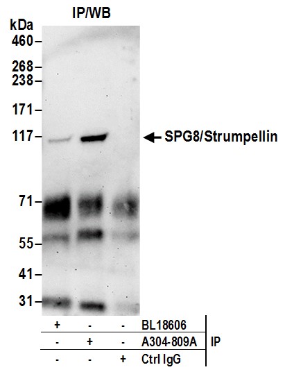
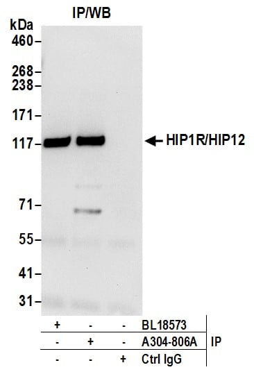
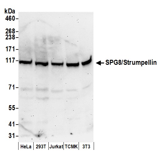 WB (Western Blot)
(Detection of human and mouse SPG8/Strumpellin by western blot. Samples: Whole cell lysate (50 ug) from HeLa, HEK293T, Jurkat, mouse TCMK-1, and mouse NIH 3T3 cells prepared using NETN lysis buffer. Antibody: Affinity purified rabbit anti-SPG8/Strumpellin antibody AAA212951 (lot AAA212951-1) used for WB at 0.1 ug/ml. Detection: Chemiluminescence with an exposure time of 3 minutes.)
WB (Western Blot)
(Detection of human and mouse SPG8/Strumpellin by western blot. Samples: Whole cell lysate (50 ug) from HeLa, HEK293T, Jurkat, mouse TCMK-1, and mouse NIH 3T3 cells prepared using NETN lysis buffer. Antibody: Affinity purified rabbit anti-SPG8/Strumpellin antibody AAA212951 (lot AAA212951-1) used for WB at 0.1 ug/ml. Detection: Chemiluminescence with an exposure time of 3 minutes.)
SPG8/Strumpellin, Polyclonal Antibody (Cat# AAA212951)
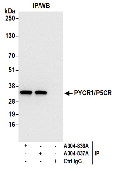
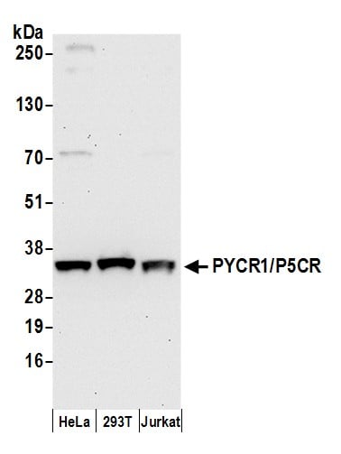 WB (Western Blot)
(Detection of human PYCR1/P5CR by western blot. Samples: Whole cell lysate (50 ug) from HeLa, HEK293T, and Jurkat cells prepared using NETN lysis buffer. Antibody: Affinity purified rabbit anti-PYCR1/P5CR antibody AAA212968 (lot AAA212968-1) used for WB at 0.1 ug/ml. Detection: Chemiluminescence with an exposure time of 30 seconds.)
WB (Western Blot)
(Detection of human PYCR1/P5CR by western blot. Samples: Whole cell lysate (50 ug) from HeLa, HEK293T, and Jurkat cells prepared using NETN lysis buffer. Antibody: Affinity purified rabbit anti-PYCR1/P5CR antibody AAA212968 (lot AAA212968-1) used for WB at 0.1 ug/ml. Detection: Chemiluminescence with an exposure time of 30 seconds.)
PYCR1/P5CR, Polyclonal Antibody (Cat# AAA212968)
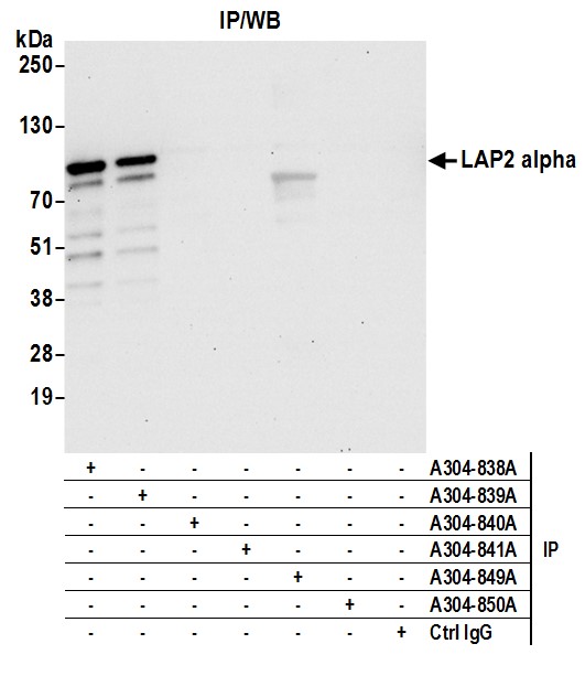
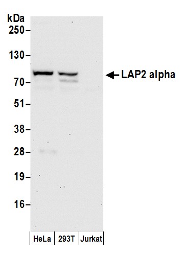 WB (Western Blot)
(Detection of human LAP2 alpha by western blot. Samples: Whole cell lysate (50 ug) from HeLa, HEK293T, and Jurkat cells prepared using NETN lysis buffer. Antibody: Affinity purified rabbit anti-LAP2 alpha antibody AAA212970 (lot AAA212970-1) used for WB at 0.1 ug/ml. Detection: Chemiluminescence with an exposure time of 30 seconds.)
WB (Western Blot)
(Detection of human LAP2 alpha by western blot. Samples: Whole cell lysate (50 ug) from HeLa, HEK293T, and Jurkat cells prepared using NETN lysis buffer. Antibody: Affinity purified rabbit anti-LAP2 alpha antibody AAA212970 (lot AAA212970-1) used for WB at 0.1 ug/ml. Detection: Chemiluminescence with an exposure time of 30 seconds.)
LAP2 alpha/TMPO, Polyclonal Antibody (Cat# AAA212970)
What are Polyclonal Antibodies?
Polyclonal antibodies are antibodies that come from multiple B cell clones of a host animal. The typical hosts used for the majority of polyclonal antibody production are rabbits, goats, sheep, and donkeys. These polyclonal antibodies, once having identified their target, will bind to different epitopes located at different regions or sequences on the same protein/antigen. As a result, they are ideal at locating and binding to the target, even if the target is in very low concentrations (due to many different antibodies being able to bind to the same target molecule, which allows for significant amplification of a downstream signal).
Polyclonal antibodies are typically produced by injecting an antigen into a host animal, which causes the animal’s immune system to attack the foreign antigen by mass generating antibodies against it. After a period of time, serum is collected from the animal and purified using physicochemical fractionation, class-specific affinity purification, and/or antigen-affinity purification.
Key Uses of Polyclonal Antibodies
- Western Blotting: This method is used to find specific proteins in biological samples after separating them by size.
- Immunohistochemistry: IHC helps visualize the location of proteins in tissue sections using various staining techniques.
- ELISA: (Enzyme-Linked Immunosorbent Assay) is typically used to identify specific protein quantities in a sample. ELISAs can be either “Quantitative” or “Qualitative”.
- Flow Cytometry: technique that identifies and measures the specific protein on the surface or inside the cells in a fluid suspension.
- Immunoprecipitation: IP isolates and studies a specific protein from a complex mixture using antibodies.
Why Buy Polyclonal Antibodies from AAA Biotech?
1. Ideal for Various Applications
Our antibodies are generally going to be validated for use in multiple types of assays, including ELISA, Western Blotting, Immunohistochemistry, Immunoprecipitation, amongst others. They are ideal for a wide range of research applications.
2. Rigorous Quality Control
All of the antibodies in our catalog undergo strict quality testing to ensure specificity, sensitivity, and consistent performance. We are confident in the ability of our antibodies to provide you with accurate results.
3. Wide Assortment of Antibodies
Antibodies in are catalog can be found for both common and exotic species, and these antibodies are also available in both conjugated and recombinant forms to suit many diverse experimental needs.
4. Highly Purified
Our antibodies are available in purified forms with over 85% purity, as confirmed by SDS-PAGE. They are also available with tags such as His, Flag, GST, or MBP. We cater to customers worldwide.
FAQ
1. How are polyclonal antibodies produced?
Traditionally, polyclonal antibodies are produced by injecting an antigen into a host animal (such as a rabbit or goat), which then triggers an immune response from the host animal. The animal’s B cells produce antibodies that will recognize different parts of the injected antigen. These antibodies are then collected from the animal’s blood and purified for use.
2. How do polyclonal antibodies differ from monoclonal antibodies?
Polyclonal antibodies are a mix of antibodies that bind to different locations (epitopes) of the same antigen, while monoclonal antibodies are identical and bind to just one specific epitope. This makes polyclonal antibodies more versatile and better at detecting proteins that may be present in low quantities or in altered/modified forms.
3. How should I store polyclonal antibodies?
Polyclonal antibodies should be stored at 4°C for short-term use (up to a few weeks) and at -20°C or -80°C for long-term storage. Avoid repeated freeze-thaw cycles by dividing them into small aliquots. Always check the datasheet for specific storage instructions.