Filters
▼Clonality
▼Type
▼Reactivity
▼Gene Name
▼Isotype
▼Host
▼Application
▼Clone
▼Polyclonal Antibodies
At AAA Biotech also known as AAA Bio or AAABio, we provide a broad range of purified polyclonal antibodies (pAbs) that are able to all be browsed online through our website. Due to their high specificity and strong binding affinity, these antibodies are ideal for wide swathes of research and experimental applications.
Our polyclonal antibodies can easily support your work, whether you use them for Western Blotting, Immunocytochemistry (with or without Immunofluorescence used in conjunction), Immunohistochemistry, Immunoprecipitation, and ELISA tests. We highly encourage you to browse our range of pAbs and choose the one that best suits your experimental model.
Viewing 9800-9850 of 96805 product results
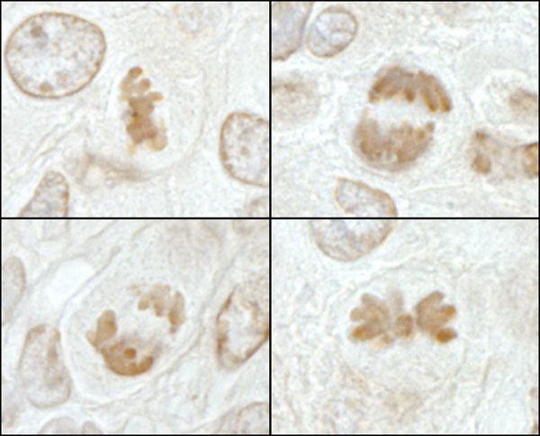
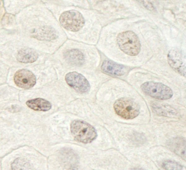
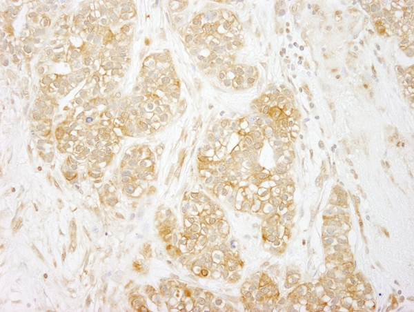
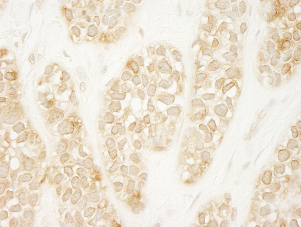
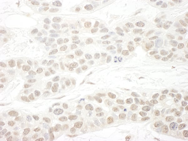
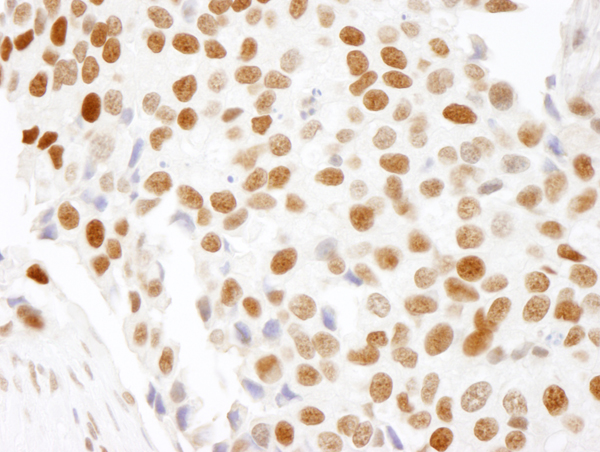
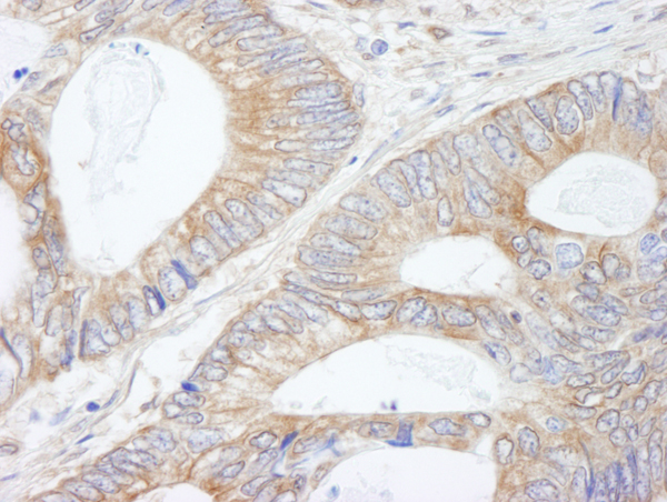
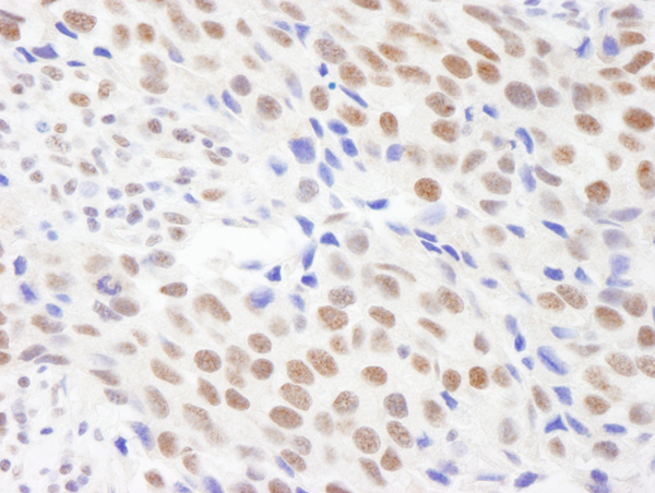
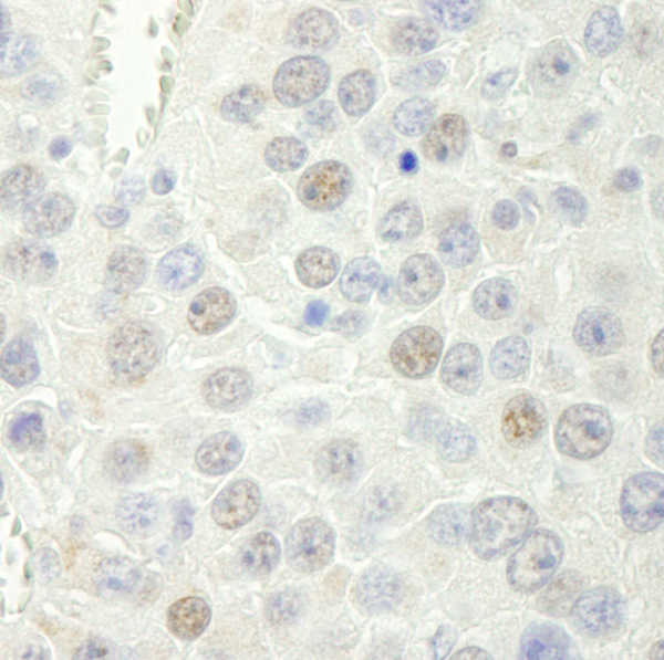
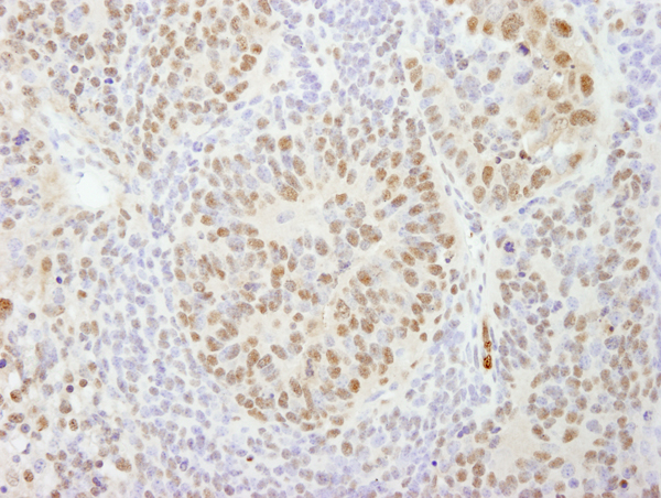
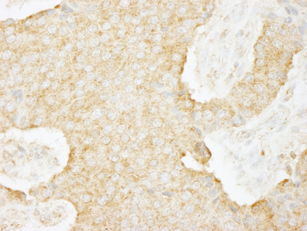
 IHC (Immunohiostchemistry)
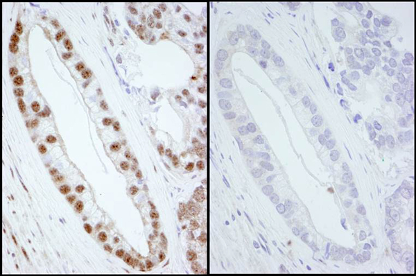
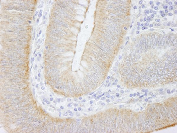
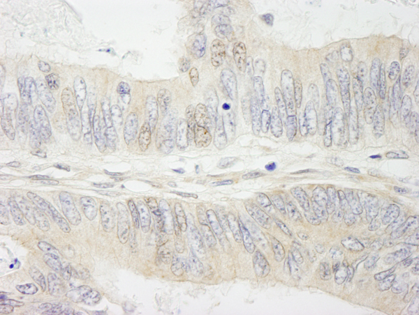
(Detection of mouse Phospho SMC1 (Ser 966) by immunohistochemistry. Samples: FFPE sections of mouse squamous cell carcinoma. Mock treatment (left panel) or lambda phosphatase-treatment (right panel). Antibody: Affinity purified rabbit anti-Phospho-SMC1 (Ser 966) (Cat. No. AAA213732) used at a dilution of 1:250. Detection: DAB)
IHC (Immunohiostchemistry)
(Detection of mouse Phospho SMC1 (Ser 966) by immunohistochemistry. Samples: FFPE sections of mouse squamous cell carcinoma. Mock treatment (left panel) or lambda phosphatase-treatment (right panel). Antibody: Affinity purified rabbit anti-Phospho-SMC1 (Ser 966) (Cat. No. AAA213732) used at a dilution of 1:250. Detection: DAB)
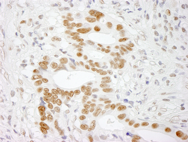
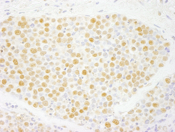
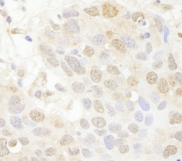
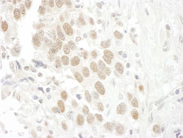
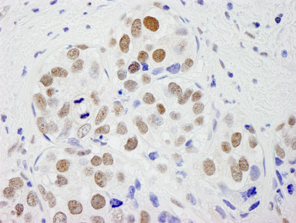
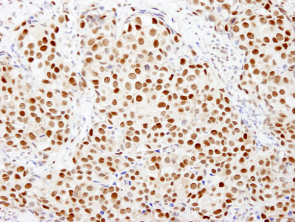
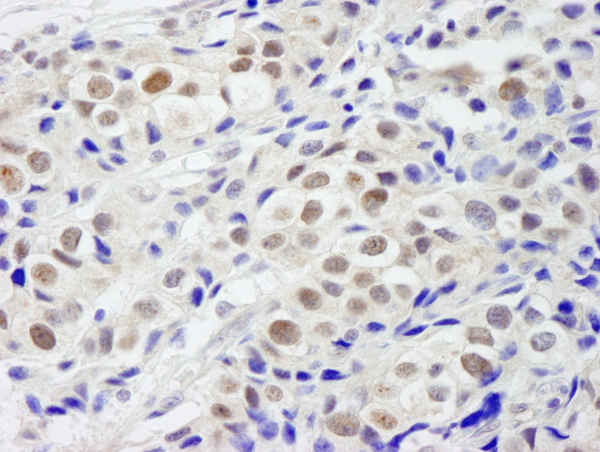
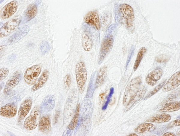
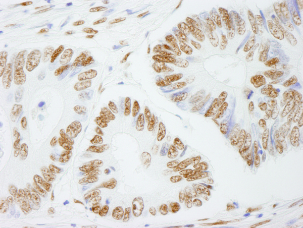
SMC1, Polyclonal Antibody (Cat# AAA213732)
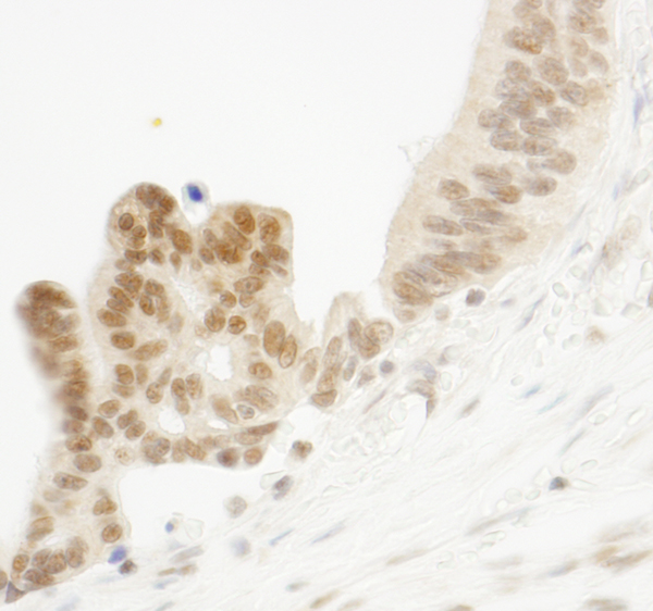
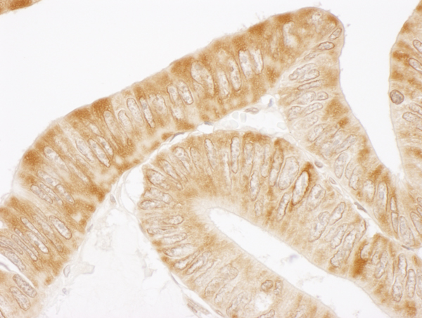
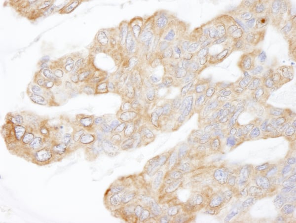
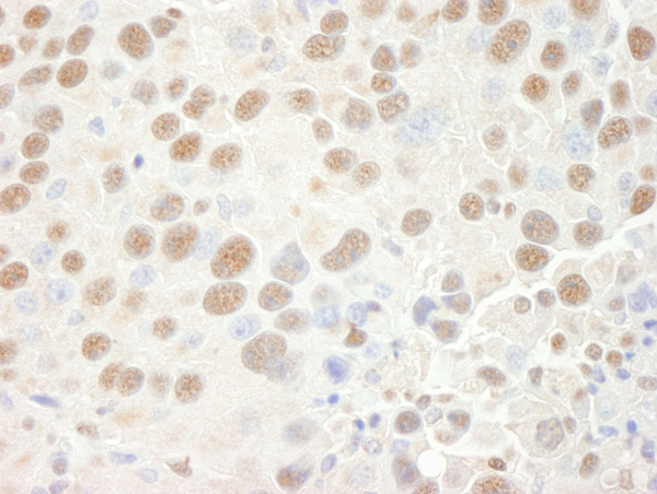
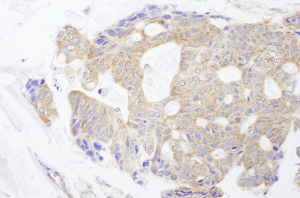
 IHC (Immunohistochemisry)
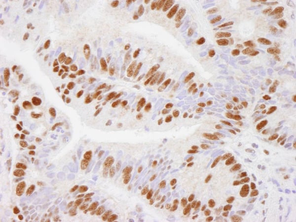
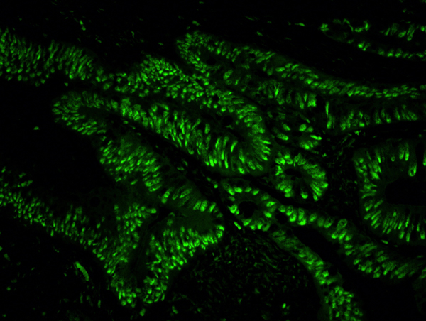
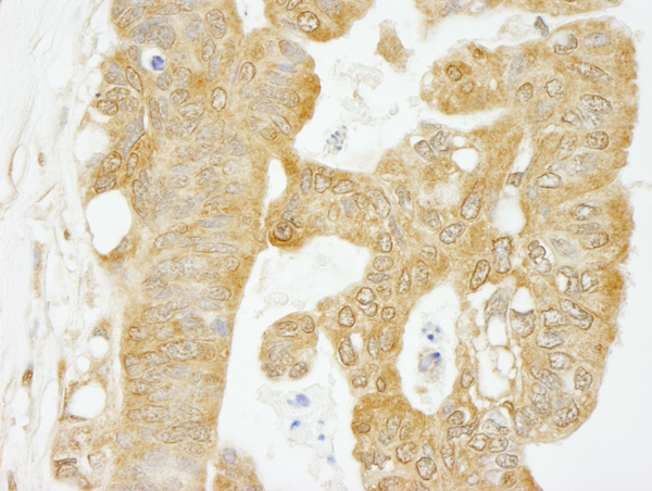
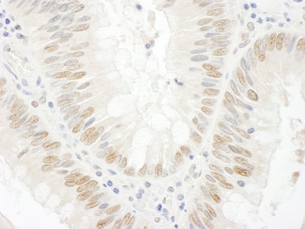
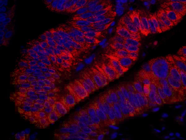
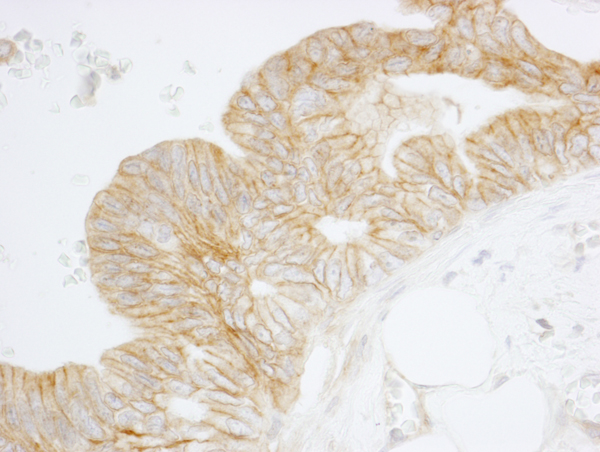
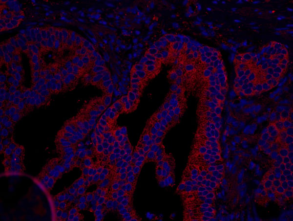
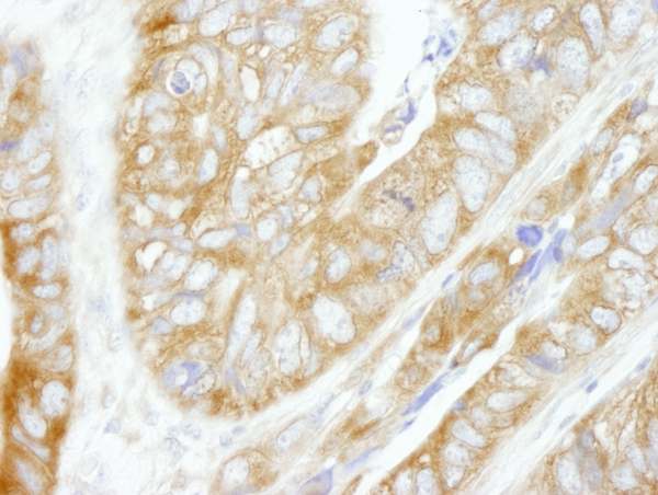
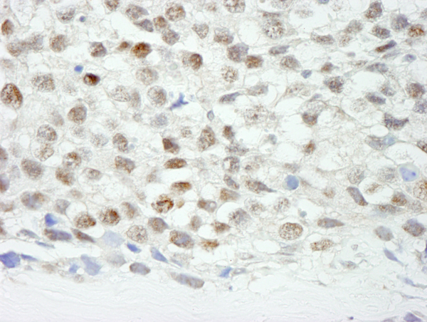
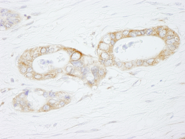
(Detection of human USP7 by immunohistochemistry. Sample: FFPE section of human colon adenocarcinoma. Antibody: Affinity purified rabbit anti-USP7 (Cat. No. AAA213734) used at a dilution of 1:100. Detection: Green-fluorescent Anti-rabbit IgG FITC conjugated (Cat. No. used at a dilution of 1:100.)
IHC (Immunohistochemisry)
(Detection of human USP7 by immunohistochemistry. Sample: FFPE section of human colon adenocarcinoma. Antibody: Affinity purified rabbit anti-USP7 (Cat. No. AAA213734) used at a dilution of 1:100. Detection: Green-fluorescent Anti-rabbit IgG FITC conjugated (Cat. No. used at a dilution of 1:100.)
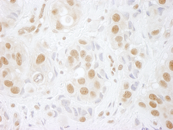
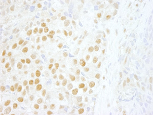
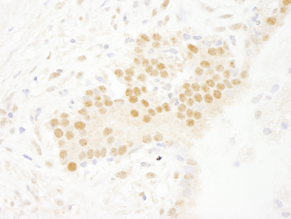
USP7, Polyclonal Antibody (Cat# AAA213734)
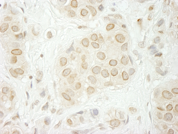
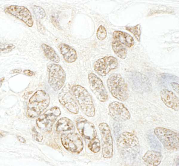
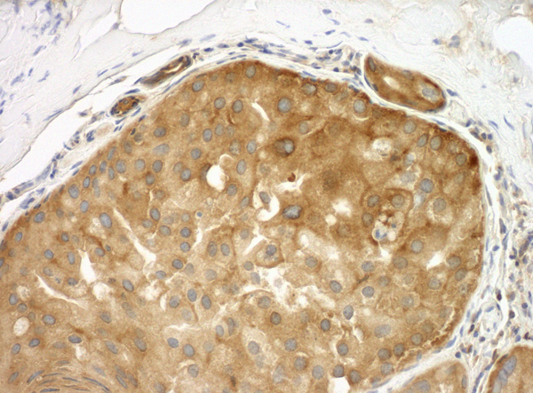
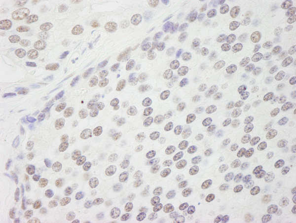
 IHC (Immunohiostchemistry)
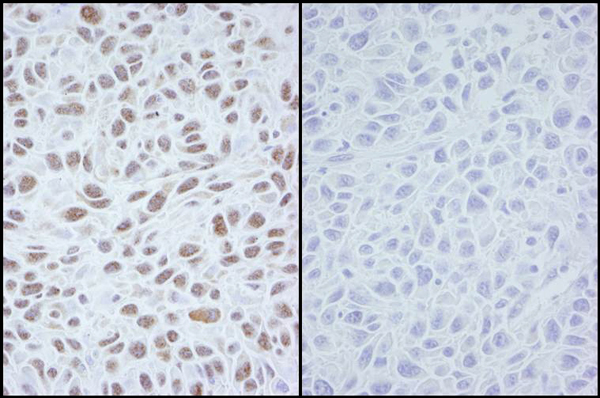
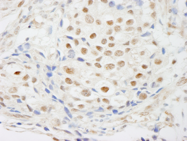
(Detection of mouse MCAK by immunohistochemistry. Sample: FFPE section of mouse hybridoma tumor. Antibody: Affinity purified rabbit anti-MCAK (Cat. No. AAA214057) used at a dilution of 1:250. Detection: DAB)
IHC (Immunohiostchemistry)
(Detection of mouse MCAK by immunohistochemistry. Sample: FFPE section of mouse hybridoma tumor. Antibody: Affinity purified rabbit anti-MCAK (Cat. No. AAA214057) used at a dilution of 1:250. Detection: DAB)
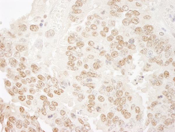
MCAK, Polyclonal Antibody (Cat# AAA214057)
 IHC (Immunohiostchemistry)
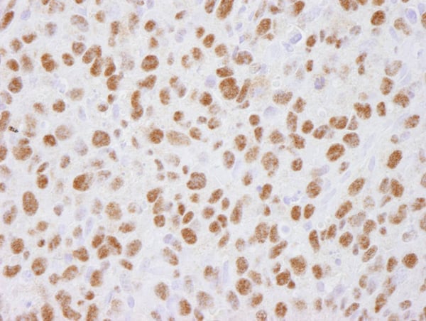
(Detection of human DHX33 by immunohistochemistry. Sample: FFPE section of human breast carcinoma. Antibody: Affinity purified rabbit anti-DHX33 (Cat. No. AAA214058) used at a dilution of 1:250. Detection: DAB)
IHC (Immunohiostchemistry)
(Detection of human DHX33 by immunohistochemistry. Sample: FFPE section of human breast carcinoma. Antibody: Affinity purified rabbit anti-DHX33 (Cat. No. AAA214058) used at a dilution of 1:250. Detection: DAB)
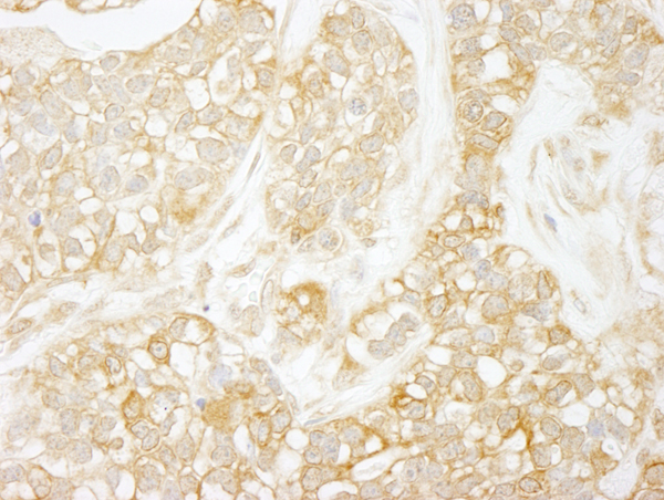
DHX33, Polyclonal Antibody (Cat# AAA214058)
 IHC (Immunohiostchemistry)
(Detection of human USP37 by immunohistochemistry. Sample: FFPE section of human ovarian carcinoma. Antibody: Affinity purified rabbit anti-USP37 (Cat. No. AAA214060) used at a dilution of 1:250. Detection: DAB)
IHC (Immunohiostchemistry)
(Detection of human USP37 by immunohistochemistry. Sample: FFPE section of human ovarian carcinoma. Antibody: Affinity purified rabbit anti-USP37 (Cat. No. AAA214060) used at a dilution of 1:250. Detection: DAB)
USP37, Polyclonal Antibody (Cat# AAA214060)
 IHC (Immunohiostchemistry)
(Detection of human RALY immunohistochemistry. Sample: FFPE section of human stomach carcinoma. Antibody: Affinity purified rabbit anti-RALY (Cat. No. AAA214063) used at a dilution of 1:500. Detection: DAB)
IHC (Immunohiostchemistry)
(Detection of human RALY immunohistochemistry. Sample: FFPE section of human stomach carcinoma. Antibody: Affinity purified rabbit anti-RALY (Cat. No. AAA214063) used at a dilution of 1:500. Detection: DAB)
RALY, Polyclonal Antibody (Cat# AAA214063)
 IHC (Immunohiostchemistry)
(Detection of mouse hPrp3p immunohistochemistry. Sample: FFPE section of mouse teratoma. Antibody: Affinity purified rabbit anti-hPrp3p (Cat. No. AAA214064) used at a dilution of 1:100. Detection: DAB)
IHC (Immunohiostchemistry)
(Detection of mouse hPrp3p immunohistochemistry. Sample: FFPE section of mouse teratoma. Antibody: Affinity purified rabbit anti-hPrp3p (Cat. No. AAA214064) used at a dilution of 1:100. Detection: DAB)
hPrp3p, Polyclonal Antibody (Cat# AAA214064)
 IHC (Immunohiostchemistry)
(Detection of human RelA immunohistochemistry. Sample: FFPE section of human breast carcinoma. Antibody: Affinity purified rabbit anti-RelA (Cat. No. AAA214065) used at a dilution of 1:250. Detection: DAB)
IHC (Immunohiostchemistry)
(Detection of human RelA immunohistochemistry. Sample: FFPE section of human breast carcinoma. Antibody: Affinity purified rabbit anti-RelA (Cat. No. AAA214065) used at a dilution of 1:250. Detection: DAB)
RelA, Polyclonal Antibody (Cat# AAA214065)
 IHC (Immunohiostchemistry)
(Detection of human ELF1 immunohistochemistry. Sample: FFPE section of human Hodgkin's Lymphoma. Antibody: Affinity purified rabbit anti-ELF1 (Cat. No. AAA214069) used at a dilution of 1:250. Detection: DAB)
IHC (Immunohiostchemistry)
(Detection of human ELF1 immunohistochemistry. Sample: FFPE section of human Hodgkin's Lymphoma. Antibody: Affinity purified rabbit anti-ELF1 (Cat. No. AAA214069) used at a dilution of 1:250. Detection: DAB)
ELF1, Polyclonal Antibody (Cat# AAA214069)
 IHC (Immunohiostchemistry)
(Detection of human LAP1B immunohistochemistry. Sample: FFPE section of human colon carcinoma. Antibody: Affinity purified rabbit anti-LAP1B (Cat. No. AAA214070) used at a dilution of 1:250. Detection: DAB)
IHC (Immunohiostchemistry)
(Detection of human LAP1B immunohistochemistry. Sample: FFPE section of human colon carcinoma. Antibody: Affinity purified rabbit anti-LAP1B (Cat. No. AAA214070) used at a dilution of 1:250. Detection: DAB)
LAP1B, Polyclonal Antibody (Cat# AAA214070)
 IHC (Immunohiostchemistry)
(Detection of mouse CPSF100 immunohistochemistry. Sample: FFPE section of mouse teratoma. Antibody: Affinity purified rabbit anti-CPSF100 (Cat. No. AAA214074) used at a dilution of 1:250. Detection: DAB)
IHC (Immunohiostchemistry)
(Detection of mouse CPSF100 immunohistochemistry. Sample: FFPE section of mouse teratoma. Antibody: Affinity purified rabbit anti-CPSF100 (Cat. No. AAA214074) used at a dilution of 1:250. Detection: DAB)
CPSF100, Polyclonal Antibody (Cat# AAA214074)
 IHC (Immunohiostchemistry)
(Detection of human THOC5 by immunohistochemistry. Sample: FFPE section of human stomach carcinoma. Antibody: Affinity purified rabbit anti-THOC5 (Cat. No. AAA214078) used at a dilution of 1:250. Detection: DAB)
IHC (Immunohiostchemistry)
(Detection of human THOC5 by immunohistochemistry. Sample: FFPE section of human stomach carcinoma. Antibody: Affinity purified rabbit anti-THOC5 (Cat. No. AAA214078) used at a dilution of 1:250. Detection: DAB)
THOC5, Polyclonal Antibody (Cat# AAA214078)
 IHC (Immunohiostchemistry)
(Detection of mouse eIF4G1/eIF4GI by immunohistochemistry. Sample: FFPE section of mouse teratoma. Antibody: Affinity purified rabbit anti-eIF4G1/eIF4GI (Cat. No. AAA214079) used at a dilution of 1:500. Detection: DAB)
IHC (Immunohiostchemistry)
(Detection of mouse eIF4G1/eIF4GI by immunohistochemistry. Sample: FFPE section of mouse teratoma. Antibody: Affinity purified rabbit anti-eIF4G1/eIF4GI (Cat. No. AAA214079) used at a dilution of 1:500. Detection: DAB)
eIF4G1/eIF4GI, Polyclonal Antibody (Cat# AAA214079)

 IHC (Immunohiostchemistry)
(Detection of mouse Menin by immunohistochemistry. Sample: FFPE section of mouse renal cell carcinoma. Antibody: Affinity purified rabbit anti-Menin (Cat. No. AAA214081) used at a dilution of 1:250. Detection: DAB)
IHC (Immunohiostchemistry)
(Detection of mouse Menin by immunohistochemistry. Sample: FFPE section of mouse renal cell carcinoma. Antibody: Affinity purified rabbit anti-Menin (Cat. No. AAA214081) used at a dilution of 1:250. Detection: DAB)
Menin, Polyclonal Antibody (Cat# AAA214081)
 IHC (Immunohiostchemistry)
(Detection of human ARHGEF16 by immunohistochemistry. Sample: FFPE section of human colon carcinoma. Antibody: Affinity purified rabbit anti-ARHGEF16 (Cat. No. AAA214088) used at a dilution of 1:250. Detection: DAB)
IHC (Immunohiostchemistry)
(Detection of human ARHGEF16 by immunohistochemistry. Sample: FFPE section of human colon carcinoma. Antibody: Affinity purified rabbit anti-ARHGEF16 (Cat. No. AAA214088) used at a dilution of 1:250. Detection: DAB)
ARHGEF16, Polyclonal Antibody (Cat# AAA214088)
 IHC (Immunohiostchemistry)
(Detection of human GBF1 by immunohistochemistry. Sample: FFPE section of human prostate carcinoma. Antibody: Affinity purified rabbit anti-GBF1 (Cat. No. AAA214095) used at a dilution of 1:250. Detection: DAB)
IHC (Immunohiostchemistry)
(Detection of human GBF1 by immunohistochemistry. Sample: FFPE section of human prostate carcinoma. Antibody: Affinity purified rabbit anti-GBF1 (Cat. No. AAA214095) used at a dilution of 1:250. Detection: DAB)
GBF1, Polyclonal Antibody (Cat# AAA214095)
 IHC (Immunohistochemisry)
(Detection of human ZNF198 by immunohistochemistry. Sample: FFPE section of human breast carcinoma. Antibody: Affinity purified rabbit anti-ZNF198 (Cat. No. AAA214104) used at a dilution of 1:250. Detection: DAB)
IHC (Immunohistochemisry)
(Detection of human ZNF198 by immunohistochemistry. Sample: FFPE section of human breast carcinoma. Antibody: Affinity purified rabbit anti-ZNF198 (Cat. No. AAA214104) used at a dilution of 1:250. Detection: DAB)
ZNF198, Polyclonal Antibody (Cat# AAA214104)
 IHC (Immunohiostchemistry)
(Detection of human BRD2 by immunohistochemistry. Sample: FFPE section of human stomach carcinoma. Antibody: Affinity purified rabbit anti-BRD2 (Cat. No. AAA214106 lot 3) used at a dilution of 1:250. Detection: DAB)
IHC (Immunohiostchemistry)
(Detection of human BRD2 by immunohistochemistry. Sample: FFPE section of human stomach carcinoma. Antibody: Affinity purified rabbit anti-BRD2 (Cat. No. AAA214106 lot 3) used at a dilution of 1:250. Detection: DAB)
BRD2, Polyclonal Antibody (Cat# AAA214106)
 IHC (Immunohiostchemistry)
(Detection of human SET7 by immunohistochemistry. Sample: FFPE section of human prostate carcinoma. Antibody: Affinity purified rabbit anti-SET7 (Cat. No. AAA214109) used at a dilution of 1:250 Detection: DAB)
IHC (Immunohiostchemistry)
(Detection of human SET7 by immunohistochemistry. Sample: FFPE section of human prostate carcinoma. Antibody: Affinity purified rabbit anti-SET7 (Cat. No. AAA214109) used at a dilution of 1:250 Detection: DAB)
SET7, Polyclonal Antibody (Cat# AAA214109)
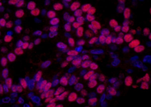
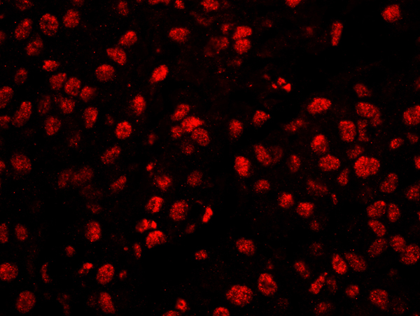
 IHC (Immunohiostchemistry)
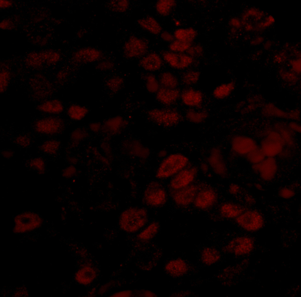
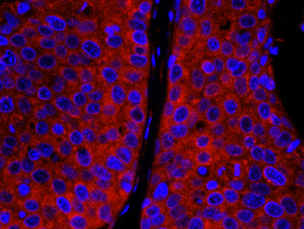
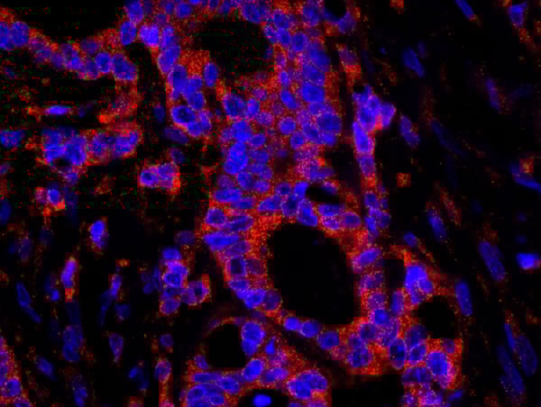
(Detection of human CNOT3 by immunohistochemistry. Sample: FFPE section of human prostate carcinoma. Antibody: Affinity purified rabbit anti-CNOT3 (Cat. No. AAA214113) used at a dilution of 1:100. Detection: Red-fluorescent goat anti-rabbit IgG-heavy and light chain cross-adsorbed Antibody DyLight 594 Conjugated used at a dilution of 1:100.)
IHC (Immunohiostchemistry)
(Detection of human CNOT3 by immunohistochemistry. Sample: FFPE section of human prostate carcinoma. Antibody: Affinity purified rabbit anti-CNOT3 (Cat. No. AAA214113) used at a dilution of 1:100. Detection: Red-fluorescent goat anti-rabbit IgG-heavy and light chain cross-adsorbed Antibody DyLight 594 Conjugated used at a dilution of 1:100.)
CNOT3, Polyclonal Antibody (Cat# AAA214113)
 IHC (Immunohistochemisry)
(Detection of human BCCIP by immunohistochemistry. Sample: FFPE section of human breast carcinoma. Antibody: Affinity purified rabbit anti-BCCIP (Cat. No. AAA214117) used at a dilution of 1:100. Detection: Red-fluorescent goat anti-rabbit IgG highly cross-adsorbed Antibody used at a dilution of 1:100.)
IHC (Immunohistochemisry)
(Detection of human BCCIP by immunohistochemistry. Sample: FFPE section of human breast carcinoma. Antibody: Affinity purified rabbit anti-BCCIP (Cat. No. AAA214117) used at a dilution of 1:100. Detection: Red-fluorescent goat anti-rabbit IgG highly cross-adsorbed Antibody used at a dilution of 1:100.)
BCCIP, Polyclonal Antibody (Cat# AAA214117)
 IHC (Immunohiostchemistry)
(Detection of human IP3R3 by immunohistochemistry. Sample: FFPE section of human stomach carcinoma. Antibody: Affinity purified rabbit anti-IP3R3 (Cat. No. AAA214119) used at a dilution of 1:100. Detection: Red-fluorescent goat anti-rabbit IgG-heavy and light chain cross-adsorbed Antibody DyLight 594 Conjugated used at a dilution of 1:100.)
IHC (Immunohiostchemistry)
(Detection of human IP3R3 by immunohistochemistry. Sample: FFPE section of human stomach carcinoma. Antibody: Affinity purified rabbit anti-IP3R3 (Cat. No. AAA214119) used at a dilution of 1:100. Detection: Red-fluorescent goat anti-rabbit IgG-heavy and light chain cross-adsorbed Antibody DyLight 594 Conjugated used at a dilution of 1:100.)
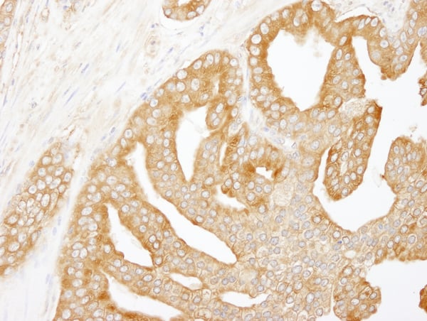
IP3R3, Polyclonal Antibody (Cat# AAA214119)
 IHC (Immunohiostchemistry)
(Detection of human hSET1B by immunohistochemistry. Sample: FFPE section of human ovarian carcinoma. Antibody: Affinity purified rabbit anti-hSET1B (Cat. No. AAA214121) used at a dilution of 1:250. Detection: DAB)
IHC (Immunohiostchemistry)
(Detection of human hSET1B by immunohistochemistry. Sample: FFPE section of human ovarian carcinoma. Antibody: Affinity purified rabbit anti-hSET1B (Cat. No. AAA214121) used at a dilution of 1:250. Detection: DAB)
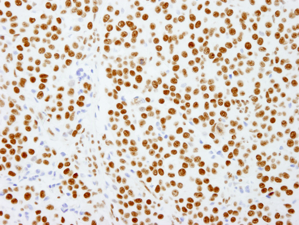
hSET1B, Polyclonal Antibody (Cat# AAA214121)
 IHC (Immunohiostchemistry)
(Detection of human ARHGEF5 by immunohistochemistry. Sample: FFPE section of human colon carcinoma. Antibody: Affinity purified rabbit anti-ARHGEF5 (Cat. No. AAA214127) used at a dilution of 1:100. Detection: Red-fluorescent goat anti-rabbit IgG-heavy and light chain cross-adsorbed Antibody DyLight 594 Conjugated used at a dilution of 1:100.)
IHC (Immunohiostchemistry)
(Detection of human ARHGEF5 by immunohistochemistry. Sample: FFPE section of human colon carcinoma. Antibody: Affinity purified rabbit anti-ARHGEF5 (Cat. No. AAA214127) used at a dilution of 1:100. Detection: Red-fluorescent goat anti-rabbit IgG-heavy and light chain cross-adsorbed Antibody DyLight 594 Conjugated used at a dilution of 1:100.)
ARHGEF5, Polyclonal Antibody (Cat# AAA214127)
 IHC (Immunohiostchemistry)
(Detection of human YAP1 by immunohistochemistry. Sample: FFPE section of human ovarian carcinoma. Antibody: Affinity purified rabbit anti-YAP1 (Cat. No. AAA214134) used at a dilution of 1:100. Detection: Red-fluorescent goat anti-rabbit IgG-heavy and light chain cross-adsorbed Antibody DyLight 594 Conjugated used at a dilution of 1:100.)
IHC (Immunohiostchemistry)
(Detection of human YAP1 by immunohistochemistry. Sample: FFPE section of human ovarian carcinoma. Antibody: Affinity purified rabbit anti-YAP1 (Cat. No. AAA214134) used at a dilution of 1:100. Detection: Red-fluorescent goat anti-rabbit IgG-heavy and light chain cross-adsorbed Antibody DyLight 594 Conjugated used at a dilution of 1:100.)
YAP1, Polyclonal Antibody (Cat# AAA214134)
 IHC (Immunohiostchemistry)
(Detection of mouse Pc2 by immunohistochemistry. Sample: FFPE section of mouse renal cell carcinoma. Antibody: Affinity purified rabbit anti-Pc2 (Cat. No. AAA214135) used at a dilution of 1:250. Detection: DAB)
IHC (Immunohiostchemistry)
(Detection of mouse Pc2 by immunohistochemistry. Sample: FFPE section of mouse renal cell carcinoma. Antibody: Affinity purified rabbit anti-Pc2 (Cat. No. AAA214135) used at a dilution of 1:250. Detection: DAB)
Pc2, Polyclonal Antibody (Cat# AAA214135)
 IHC (Immunohiostchemistry)
(Detection of mouse p66alpha by immunohistochemistry. Sample: FFPE section of mouse teratoma. Antibody: Affinity purified rabbit anti-p66alpha (Cat. No. AAA214136) used at a dilution of 1:250. Detection: DAB)
IHC (Immunohiostchemistry)
(Detection of mouse p66alpha by immunohistochemistry. Sample: FFPE section of mouse teratoma. Antibody: Affinity purified rabbit anti-p66alpha (Cat. No. AAA214136) used at a dilution of 1:250. Detection: DAB)
p66alpha, Polyclonal Antibody (Cat# AAA214136)
 IHC (Immunohistochemisry)
(Detection of human HMGN1 by immunohistochemistry. Sample: FFPE section of human breast carcinoma. Antibody: Affinity purified rabbit anti-HMGN1 (Cat. No. AAA214137) used at a dilution of 1:100. Detection: Red-fluorescent goat anti-rabbit IgG highly cross-adsorbed Antibody used at a dilution of 1:100.)
IHC (Immunohistochemisry)
(Detection of human HMGN1 by immunohistochemistry. Sample: FFPE section of human breast carcinoma. Antibody: Affinity purified rabbit anti-HMGN1 (Cat. No. AAA214137) used at a dilution of 1:100. Detection: Red-fluorescent goat anti-rabbit IgG highly cross-adsorbed Antibody used at a dilution of 1:100.)
HMGN1, Polyclonal Antibody (Cat# AAA214137)
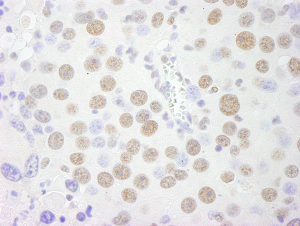
 IHC (Immunohistochemisry)
(Detection of human Septin 9 by immunohistochemistry. Sample: FFPE section of human breast carcinoma. Antibody: Affinity purified rabbit anti-Septin 9 (Cat. No. AAA214138) used at a dilution of 1:250. Detection: DAB)
IHC (Immunohistochemisry)
(Detection of human Septin 9 by immunohistochemistry. Sample: FFPE section of human breast carcinoma. Antibody: Affinity purified rabbit anti-Septin 9 (Cat. No. AAA214138) used at a dilution of 1:250. Detection: DAB)
Septin 9, Polyclonal Antibody (Cat# AAA214138)
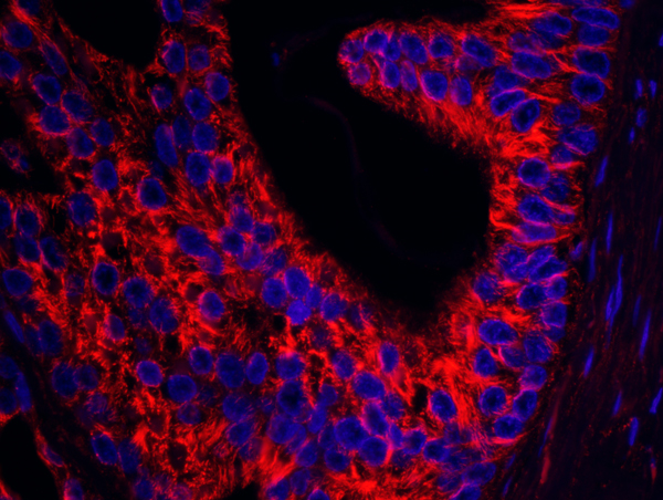
 IHC (Immunohistochemisry)
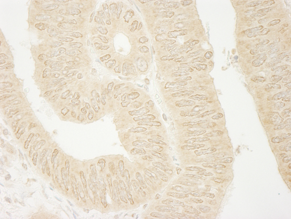
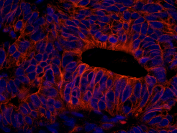
(Detection of human SPTAN1/Alpha II-spectrin by immunohistochemistry. Sample: FFPE section of human colon carcinoma. Antibody: Affinity purified rabbit anti-SPTAN1/Alpha II-spectrin (Cat. No. AAA213903) used at a dilution of 1:250. Detection: Red-fluorescent goat anti-rabbit IgG highly cross-adsorbed IHC Antibody Hilyte Plus 555 used at a dilution of 1:100)
IHC (Immunohistochemisry)
(Detection of human SPTAN1/Alpha II-spectrin by immunohistochemistry. Sample: FFPE section of human colon carcinoma. Antibody: Affinity purified rabbit anti-SPTAN1/Alpha II-spectrin (Cat. No. AAA213903) used at a dilution of 1:250. Detection: Red-fluorescent goat anti-rabbit IgG highly cross-adsorbed IHC Antibody Hilyte Plus 555 used at a dilution of 1:100)
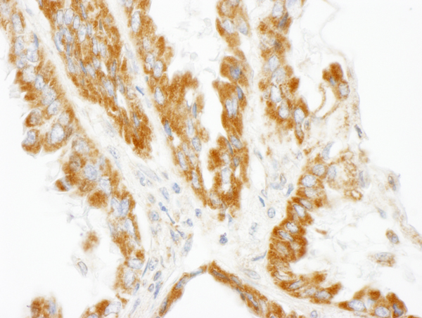
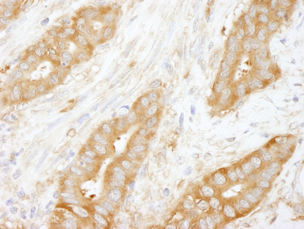
SPTAN1/Alpha II-spectrin, Polyclonal Antibody (Cat# AAA213903)
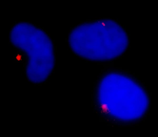
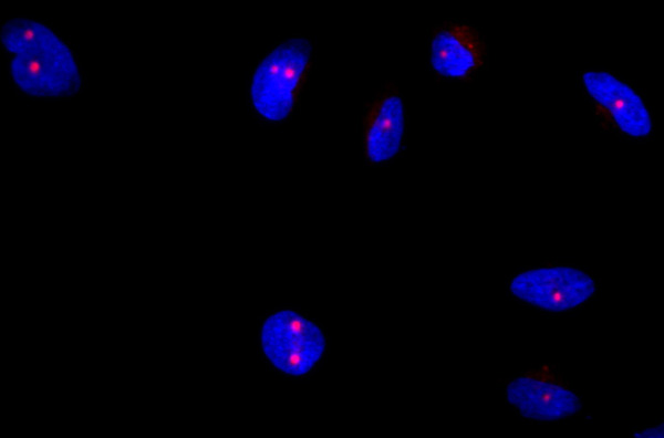
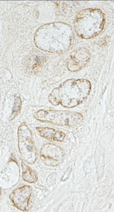
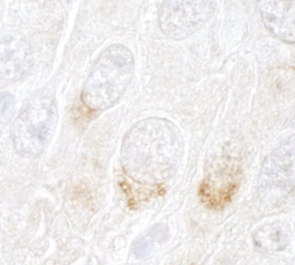
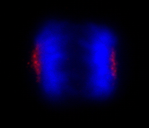
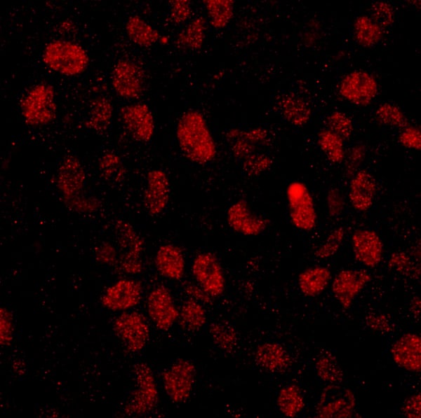
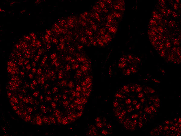
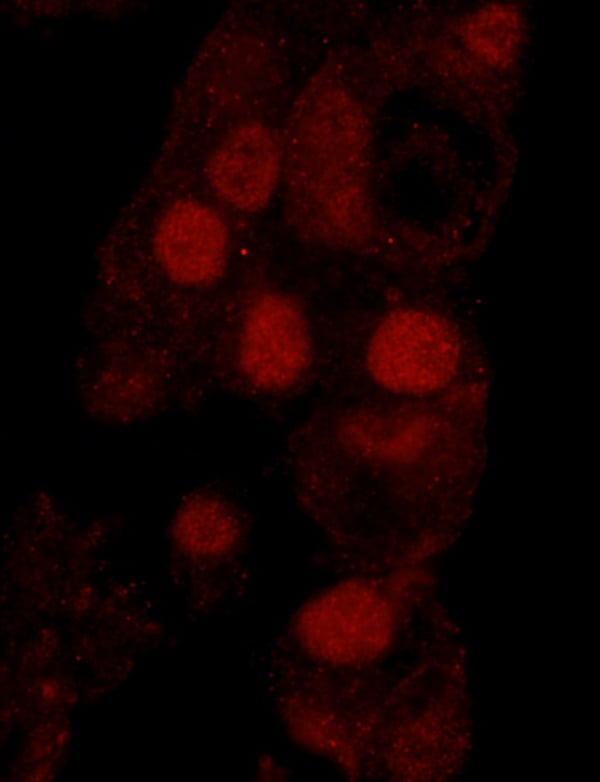
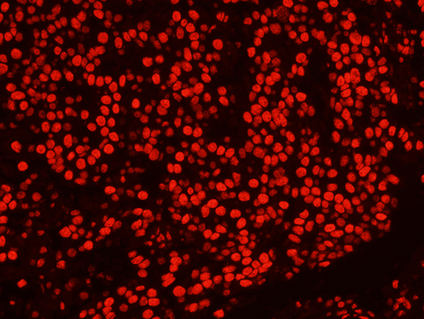
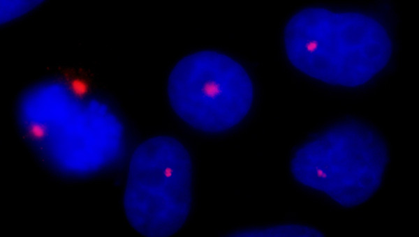 ICC (Immunocytochemistry)
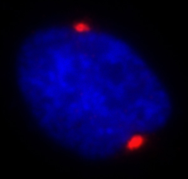
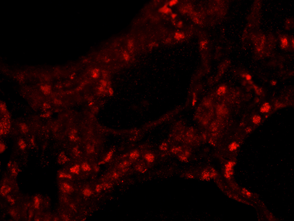
(Detection of human Pericentrin/Kendrin by immunocytochemistry. Sample: NBF-fixed asynchronous HeLa cells. Antibody: Affinity purified rabbit anti-Pericentrin/Kendrin (Cat. No. AAA213906) used at a dilution of 1:100. Detection: Red-fluorescent Alexa Fluor 594 goat anti-rabbit IgG (Invitrogen).)
ICC (Immunocytochemistry)
(Detection of human Pericentrin/Kendrin by immunocytochemistry. Sample: NBF-fixed asynchronous HeLa cells. Antibody: Affinity purified rabbit anti-Pericentrin/Kendrin (Cat. No. AAA213906) used at a dilution of 1:100. Detection: Red-fluorescent Alexa Fluor 594 goat anti-rabbit IgG (Invitrogen).)
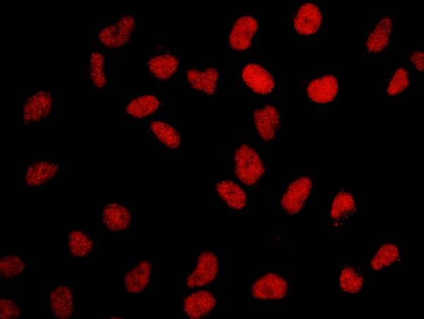
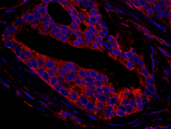
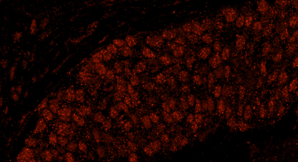
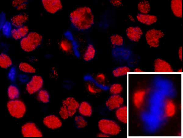
Pericentrin/Kendrin, Polyclonal Antibody (Cat# AAA213906)
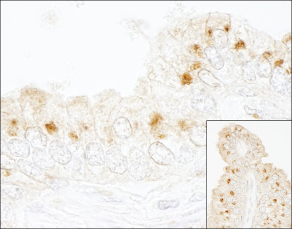
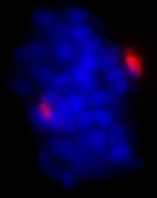
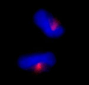 ICC (Immunocytochemistry)
(Detection of human CEP170 by immunocytochemistry. Sample: NBF-fixed asynchronous HeLa cells. Antibody: Affinity purified rabbit anti-CEP170 (Cat. No. AAA213907) used at a dilution of 1:100. Detection: Red-fluorescent Alexa Fluor 594 goat anti-rabbit IgG (Invitrogen).)
ICC (Immunocytochemistry)
(Detection of human CEP170 by immunocytochemistry. Sample: NBF-fixed asynchronous HeLa cells. Antibody: Affinity purified rabbit anti-CEP170 (Cat. No. AAA213907) used at a dilution of 1:100. Detection: Red-fluorescent Alexa Fluor 594 goat anti-rabbit IgG (Invitrogen).)
CEP170, Polyclonal Antibody (Cat# AAA213907)
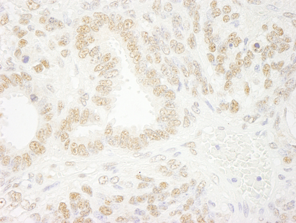
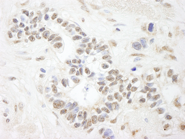
 IHC (Immunohistochemisry)
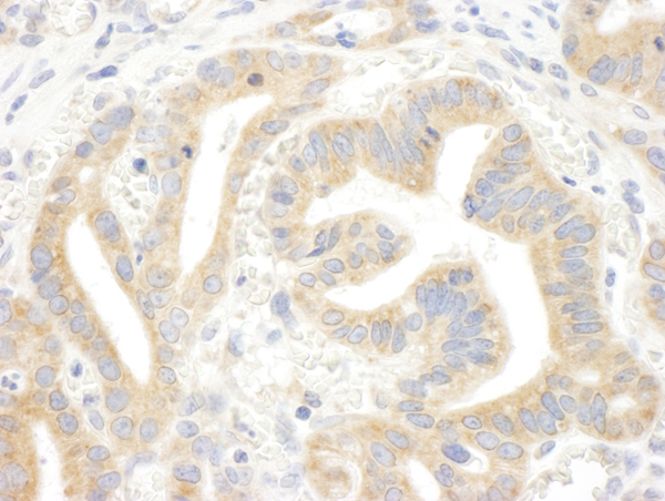
(Detection of human CIP2A by immunohistochemistry. Sample: FFPE section of human colon carcinoma. Antibody: Affinity purified rabbit anti-CIP2A (Cat. No. AAA213908) used at a dilution of 1:100. Detection: Red-fluorescent goat anti-rabbit IgG highly cross-adsorbed Antibody used at a dilution of 1:100.)
IHC (Immunohistochemisry)
(Detection of human CIP2A by immunohistochemistry. Sample: FFPE section of human colon carcinoma. Antibody: Affinity purified rabbit anti-CIP2A (Cat. No. AAA213908) used at a dilution of 1:100. Detection: Red-fluorescent goat anti-rabbit IgG highly cross-adsorbed Antibody used at a dilution of 1:100.)
CIP2A, Polyclonal Antibody (Cat# AAA213908)
 IHC (Immunohistochemisry)
(Detection of human CPSF68 by immunohistochemistry. Sample: FFPE section of human colon carcinoma. Antibody: Affinity purified rabbit anti-CPSF68 (Cat. No. AAA213909) used at a dilution of 1:100. Detection: Red-fluorescent goat anti-rabbit IgG highly cross-adsorbed Antibody used at a dilution of 1:100.)
IHC (Immunohistochemisry)
(Detection of human CPSF68 by immunohistochemistry. Sample: FFPE section of human colon carcinoma. Antibody: Affinity purified rabbit anti-CPSF68 (Cat. No. AAA213909) used at a dilution of 1:100. Detection: Red-fluorescent goat anti-rabbit IgG highly cross-adsorbed Antibody used at a dilution of 1:100.)
CPSF68, Polyclonal Antibody (Cat# AAA213909)
 IHC (Immunohiostchemistry)
(Detection of human CPSF59 by immunohistochemistry. Sample: FFPE section of human breast carcinoma. Antibody: Affinity purified rabbit anti-CPSF59 (Cat. No. AAA213910) used at a dilution of 1:100. Detection: Red-fluorescent goat anti-rabbit IgG highly cross-adsorbed Antibody used at a dilution of 1:100.)
IHC (Immunohiostchemistry)
(Detection of human CPSF59 by immunohistochemistry. Sample: FFPE section of human breast carcinoma. Antibody: Affinity purified rabbit anti-CPSF59 (Cat. No. AAA213910) used at a dilution of 1:100. Detection: Red-fluorescent goat anti-rabbit IgG highly cross-adsorbed Antibody used at a dilution of 1:100.)
CPSF59, Polyclonal Antibody (Cat# AAA213910)
 IHC (Immunohistochemisry)
(Detection of human Coronin 2 by immunohistochemistry. Sample: FFPE section of human prostate carcinoma. Antibody: Affinity purified rabbit anti-Coronin 2 (Cat. No. AAA213911) used at a dilution of 1:100. Detection: Red-fluorescent goat anti-rabbit IgG highly cross-adsorbed Antibody used at a dilution of 1:100.)
IHC (Immunohistochemisry)
(Detection of human Coronin 2 by immunohistochemistry. Sample: FFPE section of human prostate carcinoma. Antibody: Affinity purified rabbit anti-Coronin 2 (Cat. No. AAA213911) used at a dilution of 1:100. Detection: Red-fluorescent goat anti-rabbit IgG highly cross-adsorbed Antibody used at a dilution of 1:100.)
Coronin 2, Polyclonal Antibody (Cat# AAA213911)
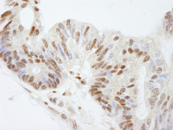
 IHC (Immunohiostchemistry)
(Detection of human RBM27 by immunohistochemistry. Sample: FFPE section of human prostate carcinoma. Antibody: Affinity purified rabbit anti-RBM27 (Cat. No. AAA213913) used at a dilution of 1:250. Detection: DAB)
IHC (Immunohiostchemistry)
(Detection of human RBM27 by immunohistochemistry. Sample: FFPE section of human prostate carcinoma. Antibody: Affinity purified rabbit anti-RBM27 (Cat. No. AAA213913) used at a dilution of 1:250. Detection: DAB)
RBM27, Polyclonal Antibody (Cat# AAA213913)
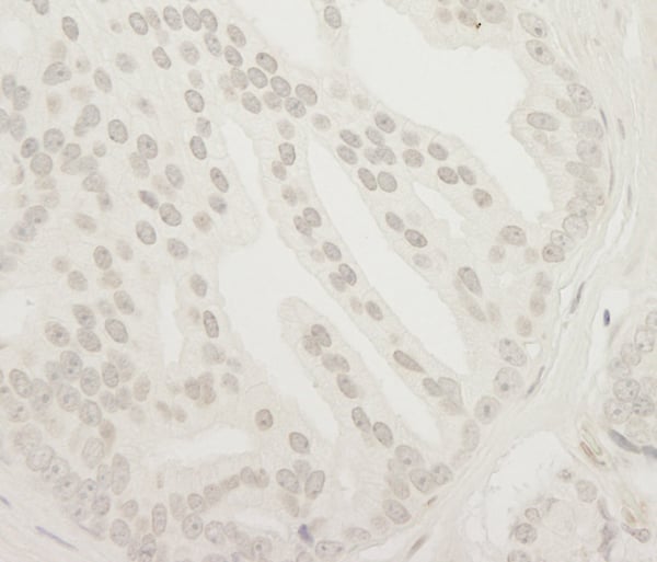
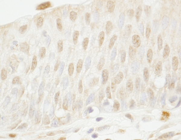
 IHC (Immunohistochemisry)
(Detection of human Rtf1 by immunohistochemistry. Sample: FFPE section of human skin carcinoma. Antibody: Affinity purified rabbit anti-Rtf1 (Cat. No. AAA213914) used at a dilution of 1:100. Detection: Red-fluorescent goat anti-rabbit IgG highly cross-adsorbed Antibody used at a dilution of 1:100.)
IHC (Immunohistochemisry)
(Detection of human Rtf1 by immunohistochemistry. Sample: FFPE section of human skin carcinoma. Antibody: Affinity purified rabbit anti-Rtf1 (Cat. No. AAA213914) used at a dilution of 1:100. Detection: Red-fluorescent goat anti-rabbit IgG highly cross-adsorbed Antibody used at a dilution of 1:100.)
Rtf1, Polyclonal Antibody (Cat# AAA213914)

 IHC (Immunohiostchemistry)
(Detection of human BRCA1 by immunohistochemistry. Sample: FFPE section of human breast carcinoma. Antibody: Affinity purified rabbit anti-BRCA1 (Cat. No. AAA213918) used at a dilution of 1:100. Detection: Red-fluorescent goat anti-rabbit IgG highly cross-adsorbed Antibody used at a dilution of 1:100.)
IHC (Immunohiostchemistry)
(Detection of human BRCA1 by immunohistochemistry. Sample: FFPE section of human breast carcinoma. Antibody: Affinity purified rabbit anti-BRCA1 (Cat. No. AAA213918) used at a dilution of 1:100. Detection: Red-fluorescent goat anti-rabbit IgG highly cross-adsorbed Antibody used at a dilution of 1:100.)
BRCA1, Polyclonal Antibody (Cat# AAA213918)
 IHC (Immunohistochemisry)
(Detection of human CAD by immunohistochemistry. Sample: FFPE section of human breast carcinoma. Antibody: Affinity purified rabbit anti-CAD (Cat. No. AAA213920) used at a dilution of 1:100. Detection: Red-fluorescent goat anti-rabbit IgG highly cross-adsorbed IHC Antibody Hilyte Plus 555 used at a dilution of 1:100.)
IHC (Immunohistochemisry)
(Detection of human CAD by immunohistochemistry. Sample: FFPE section of human breast carcinoma. Antibody: Affinity purified rabbit anti-CAD (Cat. No. AAA213920) used at a dilution of 1:100. Detection: Red-fluorescent goat anti-rabbit IgG highly cross-adsorbed IHC Antibody Hilyte Plus 555 used at a dilution of 1:100.)
CAD, Polyclonal Antibody (Cat# AAA213920)
 IHC (Immunohistochemisry)
(Detection of human WRNIP1 by immunohistochemistry. Sample: FFPE section of human breast carcinoma. Antibody: Affinity purified rabbit anti-WRNIP1 (Cat. No. AAA213922) used at a dilution of 1:100. Detection: Red-fluorescent goat anti-rabbit IgG highly cross-adsorbed Antibody used at a dilution of 1:100.)
IHC (Immunohistochemisry)
(Detection of human WRNIP1 by immunohistochemistry. Sample: FFPE section of human breast carcinoma. Antibody: Affinity purified rabbit anti-WRNIP1 (Cat. No. AAA213922) used at a dilution of 1:100. Detection: Red-fluorescent goat anti-rabbit IgG highly cross-adsorbed Antibody used at a dilution of 1:100.)
WRNIP1, Polyclonal Antibody (Cat# AAA213922)
 IHC (Immunohiostchemistry)
(Detection of mouse ANKS3 by immunohistochemistry. Sample: FFPE section of mouse teratoma. Antibody: Affinity purified rabbit anti-ANKS3 (Cat. No. AAA213925) used at a dilution of 1:250. Detection: DAB)
IHC (Immunohiostchemistry)
(Detection of mouse ANKS3 by immunohistochemistry. Sample: FFPE section of mouse teratoma. Antibody: Affinity purified rabbit anti-ANKS3 (Cat. No. AAA213925) used at a dilution of 1:250. Detection: DAB)
ANKS3, Polyclonal Antibody (Cat# AAA213925)
 IHC (Immunohiostchemistry)
(Detection of human FKBP5/FKBP51 by immunohistochemistry. Sample: FFPE section of human breast carcinoma. Antibody: Affinity purified rabbit anti-FKBP5/FKBP51 (Cat. No. AAA213927) used at a dilution of 1:500. Detection: DAB)
IHC (Immunohiostchemistry)
(Detection of human FKBP5/FKBP51 by immunohistochemistry. Sample: FFPE section of human breast carcinoma. Antibody: Affinity purified rabbit anti-FKBP5/FKBP51 (Cat. No. AAA213927) used at a dilution of 1:500. Detection: DAB)
FKBP5/FKBP51, Polyclonal Antibody (Cat# AAA213927)
 IHC (Immunohistochemisry)
(Detection of human PHF6 by immunohistochemistry. Sample: FFPE section of human skin carcinoma. Antibody: Affinity purified rabbit anti-PHF6 (Cat. No. AAA213930) used at a dilution of 1:100. Detection: Red-fluorescent goat anti-rabbit IgG highly cross-adsorbed Antibody used at a dilution of 1:100.)
IHC (Immunohistochemisry)
(Detection of human PHF6 by immunohistochemistry. Sample: FFPE section of human skin carcinoma. Antibody: Affinity purified rabbit anti-PHF6 (Cat. No. AAA213930) used at a dilution of 1:100. Detection: Red-fluorescent goat anti-rabbit IgG highly cross-adsorbed Antibody used at a dilution of 1:100.)
PHF6, Polyclonal Antibody (Cat# AAA213930)
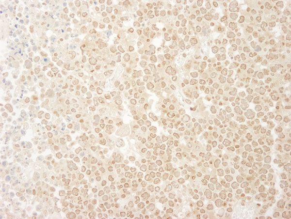
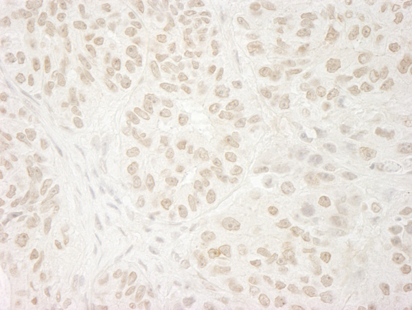
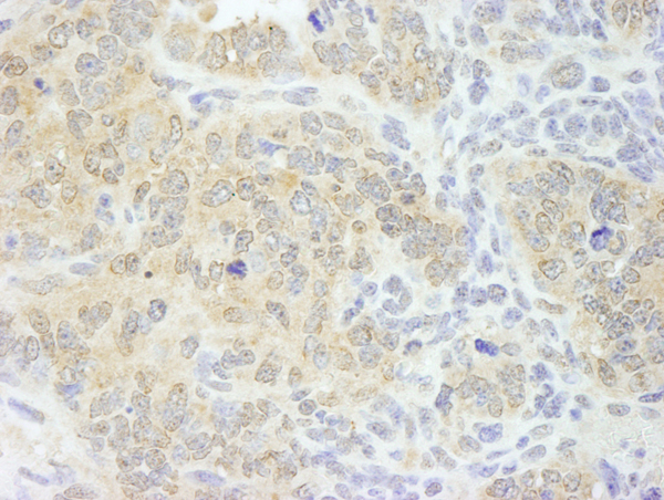
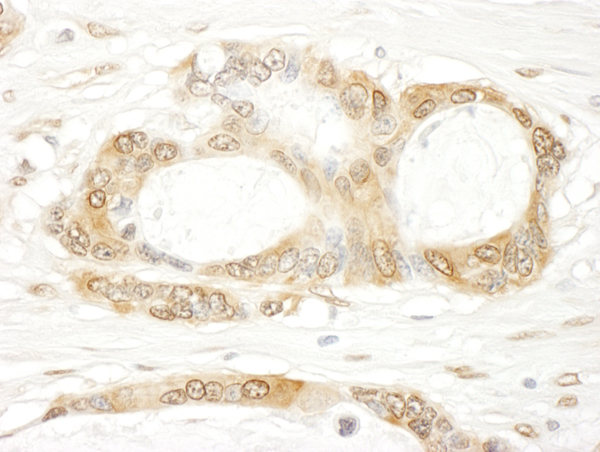
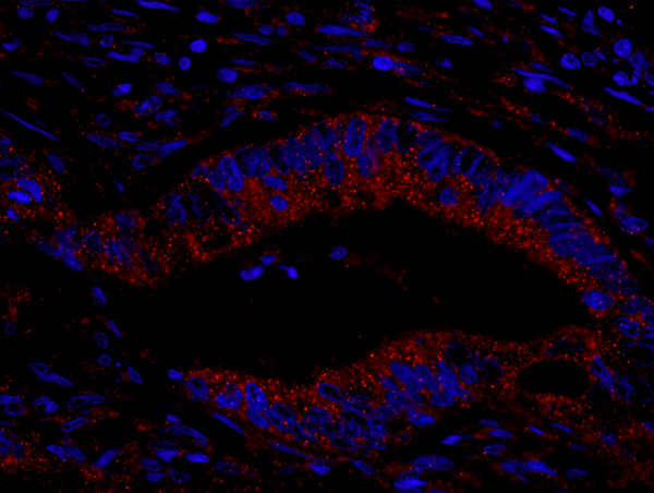
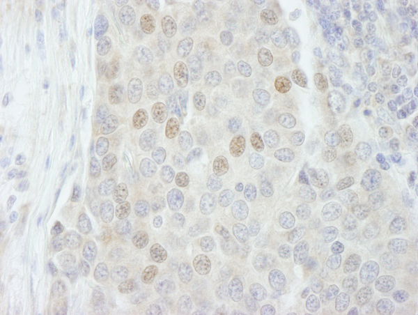
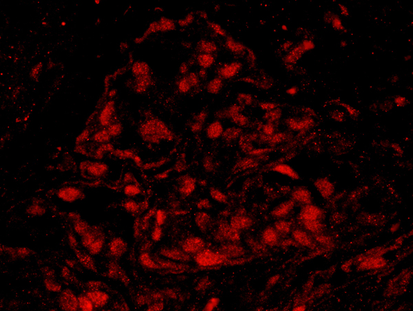
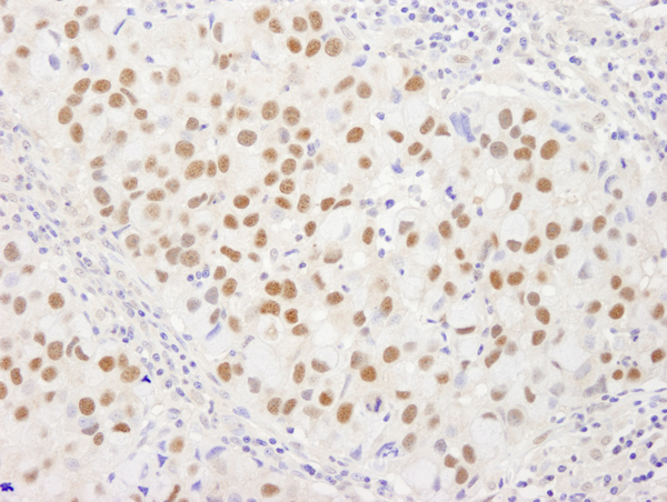
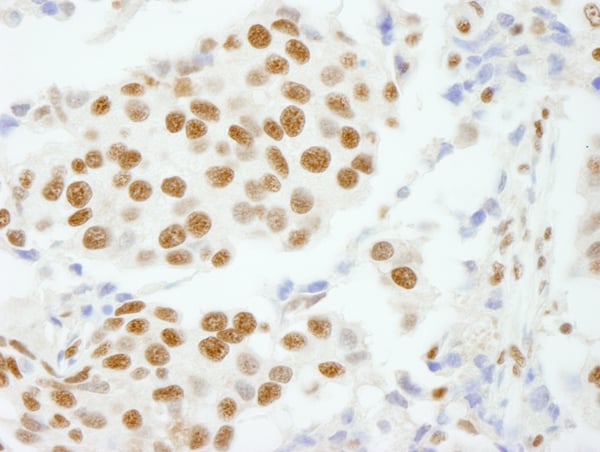
 IHC (Immunohistochemistry)
(Detection of human NUMA by immunohistochemistry. Sample: FFPE section of human breast carcinoma. Antibody: Affinity purified rabbit anti-NUMA (Cat. No. AAA213937) used at a dilution of 1:100. Detection: Red-fluorescent goat anti-rabbit IgG highly cross-adsorbed Antibody Hilyte Plus 555 used at a dilution of 1:100.)
IHC (Immunohistochemistry)
(Detection of human NUMA by immunohistochemistry. Sample: FFPE section of human breast carcinoma. Antibody: Affinity purified rabbit anti-NUMA (Cat. No. AAA213937) used at a dilution of 1:100. Detection: Red-fluorescent goat anti-rabbit IgG highly cross-adsorbed Antibody Hilyte Plus 555 used at a dilution of 1:100.)
NUMA, Polyclonal Antibody (Cat# AAA213937)
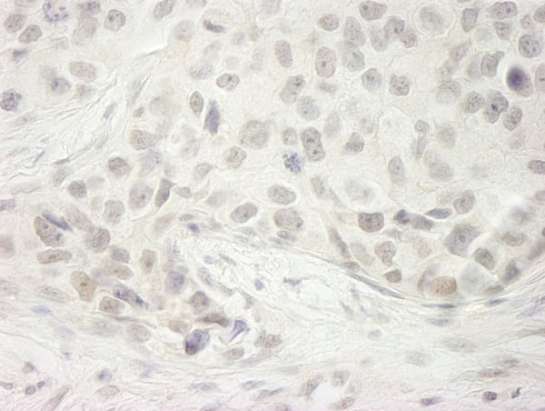
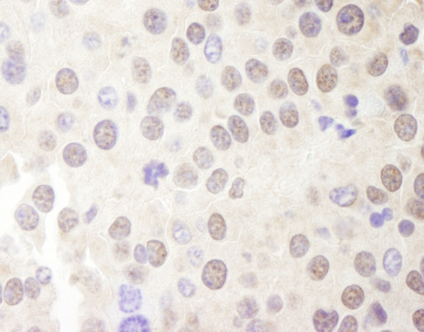
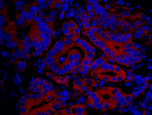
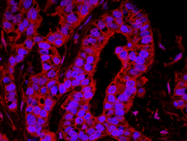
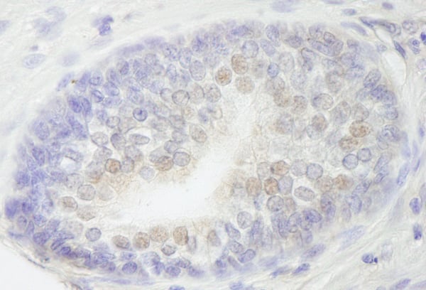
 IHC (Immunohistochemistry)
(Detection of human PSF/SFPQ by IHC-IF. Sample: FFPE section of human breast carcinoma. Antibody: Affinity purified rabbit anti-PSF/SFPQ (Cat. No. AAA213939) used at a dilution of 1:100. Detection: Red-fluorescent goat anti-rabbit IgG highly cross-adsorbed Antibody Hilyte Plus 555 used at a dilution of 1:100.)
IHC (Immunohistochemistry)
(Detection of human PSF/SFPQ by IHC-IF. Sample: FFPE section of human breast carcinoma. Antibody: Affinity purified rabbit anti-PSF/SFPQ (Cat. No. AAA213939) used at a dilution of 1:100. Detection: Red-fluorescent goat anti-rabbit IgG highly cross-adsorbed Antibody Hilyte Plus 555 used at a dilution of 1:100.)
PSF/SFPQ, Polyclonal Antibody (Cat# AAA213939)
 IHC (Immunohistochemistry)
(Detection of human CPSF160 by IHC-IF. Sample: FFPE section of human breast carcinoma. Antibody: Affinity purified rabbit anti-CPSF160 (Cat. No. AAA213941) used at a dilution of 1:100. Detection: Red-fluorescent goat anti-rabbit IgG highly cross-adsorbed Antibody Hilyte Plus 555 used at a dilution of 1:100.)
IHC (Immunohistochemistry)
(Detection of human CPSF160 by IHC-IF. Sample: FFPE section of human breast carcinoma. Antibody: Affinity purified rabbit anti-CPSF160 (Cat. No. AAA213941) used at a dilution of 1:100. Detection: Red-fluorescent goat anti-rabbit IgG highly cross-adsorbed Antibody Hilyte Plus 555 used at a dilution of 1:100.)
CPSF160, Polyclonal Antibody (Cat# AAA213941)
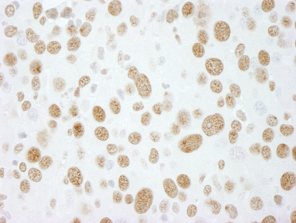
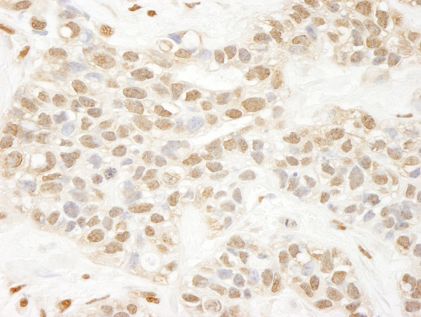
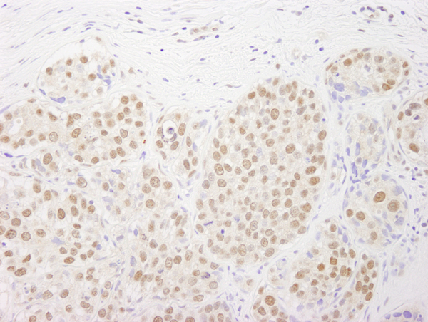
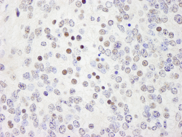
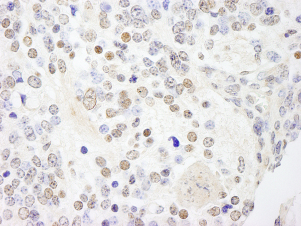
 IHC (Immunohiostchemistry)
(Detection of human MYBBP1a by immunohistochemistry. Sample: FFPE section of human colon carcinoma. Antibody: Affinity purified rabbit anti-MYBBP1a (Cat. No. AAA213942) used at a dilution of 1:250. Detection: DAB)
IHC (Immunohiostchemistry)
(Detection of human MYBBP1a by immunohistochemistry. Sample: FFPE section of human colon carcinoma. Antibody: Affinity purified rabbit anti-MYBBP1a (Cat. No. AAA213942) used at a dilution of 1:250. Detection: DAB)
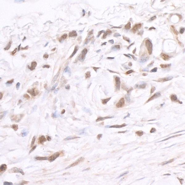
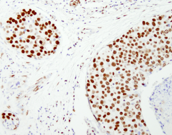
MYBBP1A, Polyclonal Antibody (Cat# AAA213942)
 IHC (Immunohistochemisry)
(Detection of human ZNF318/TZF by IHC-IF. Sample: FFPE section of human anaplastic thyroid carcinoma Antibody: Affinity purified rabbit anti-ZNF318/TZF (Cat. No. AAA213945) used at a dilution of 1:100. Detection: Red-fluorescent goat anti-rabbit IgG highly cross-adsorbed Antibody used at a dilution of 1:100.)
IHC (Immunohistochemisry)
(Detection of human ZNF318/TZF by IHC-IF. Sample: FFPE section of human anaplastic thyroid carcinoma Antibody: Affinity purified rabbit anti-ZNF318/TZF (Cat. No. AAA213945) used at a dilution of 1:100. Detection: Red-fluorescent goat anti-rabbit IgG highly cross-adsorbed Antibody used at a dilution of 1:100.)
ZNF318/TZF, Polyclonal Antibody (Cat# AAA213945)
 IHC (Immunohistochemisry)
(Detection of human USP3 by IHC-IF. Sample: FFPE section of human ovarian carcinoma. Antibody: Affinity purified rabbit anti-USP3 (Cat. No. AAA213951) used at a dilution of 1:100. Detection: Red-fluorescent goat anti-rabbit IgG highly cross-adsorbed Antibody Hilyte Plus 555 used at a dilution of 1:100.)
IHC (Immunohistochemisry)
(Detection of human USP3 by IHC-IF. Sample: FFPE section of human ovarian carcinoma. Antibody: Affinity purified rabbit anti-USP3 (Cat. No. AAA213951) used at a dilution of 1:100. Detection: Red-fluorescent goat anti-rabbit IgG highly cross-adsorbed Antibody Hilyte Plus 555 used at a dilution of 1:100.)
USP3, Polyclonal Antibody (Cat# AAA213951)
What are Polyclonal Antibodies?
Polyclonal antibodies are antibodies that come from multiple B cell clones of a host animal. The typical hosts used for the majority of polyclonal antibody production are rabbits, goats, sheep, and donkeys. These polyclonal antibodies, once having identified their target, will bind to different epitopes located at different regions or sequences on the same protein/antigen. As a result, they are ideal at locating and binding to the target, even if the target is in very low concentrations (due to many different antibodies being able to bind to the same target molecule, which allows for significant amplification of a downstream signal).
Polyclonal antibodies are typically produced by injecting an antigen into a host animal, which causes the animal’s immune system to attack the foreign antigen by mass generating antibodies against it. After a period of time, serum is collected from the animal and purified using physicochemical fractionation, class-specific affinity purification, and/or antigen-affinity purification.
Key Uses of Polyclonal Antibodies
- Western Blotting: This method is used to find specific proteins in biological samples after separating them by size.
- Immunohistochemistry: IHC helps visualize the location of proteins in tissue sections using various staining techniques.
- ELISA: (Enzyme-Linked Immunosorbent Assay) is typically used to identify specific protein quantities in a sample. ELISAs can be either “Quantitative” or “Qualitative”.
- Flow Cytometry: technique that identifies and measures the specific protein on the surface or inside the cells in a fluid suspension.
- Immunoprecipitation: IP isolates and studies a specific protein from a complex mixture using antibodies.
Why Buy Polyclonal Antibodies from AAA Biotech?
1. Ideal for Various Applications
Our antibodies are generally going to be validated for use in multiple types of assays, including ELISA, Western Blotting, Immunohistochemistry, Immunoprecipitation, amongst others. They are ideal for a wide range of research applications.
2. Rigorous Quality Control
All of the antibodies in our catalog undergo strict quality testing to ensure specificity, sensitivity, and consistent performance. We are confident in the ability of our antibodies to provide you with accurate results.
3. Wide Assortment of Antibodies
Antibodies in are catalog can be found for both common and exotic species, and these antibodies are also available in both conjugated and recombinant forms to suit many diverse experimental needs.
4. Highly Purified
Our antibodies are available in purified forms with over 85% purity, as confirmed by SDS-PAGE. They are also available with tags such as His, Flag, GST, or MBP. We cater to customers worldwide.
FAQ
1. How are polyclonal antibodies produced?
Traditionally, polyclonal antibodies are produced by injecting an antigen into a host animal (such as a rabbit or goat), which then triggers an immune response from the host animal. The animal’s B cells produce antibodies that will recognize different parts of the injected antigen. These antibodies are then collected from the animal’s blood and purified for use.
2. How do polyclonal antibodies differ from monoclonal antibodies?
Polyclonal antibodies are a mix of antibodies that bind to different locations (epitopes) of the same antigen, while monoclonal antibodies are identical and bind to just one specific epitope. This makes polyclonal antibodies more versatile and better at detecting proteins that may be present in low quantities or in altered/modified forms.
3. How should I store polyclonal antibodies?
Polyclonal antibodies should be stored at 4°C for short-term use (up to a few weeks) and at -20°C or -80°C for long-term storage. Avoid repeated freeze-thaw cycles by dividing them into small aliquots. Always check the datasheet for specific storage instructions.