Filters
▼Clonality
▼Type
▼Reactivity
▼Gene Name
▼Isotype
▼Host
▼Application
▼Clone
▼Polyclonal Antibodies
At AAA Biotech also known as AAA Bio or AAABio, we provide a broad range of purified polyclonal antibodies (pAbs) that are able to all be browsed online through our website. Due to their high specificity and strong binding affinity, these antibodies are ideal for wide swathes of research and experimental applications.
Our polyclonal antibodies can easily support your work, whether you use them for Western Blotting, Immunocytochemistry (with or without Immunofluorescence used in conjunction), Immunohistochemistry, Immunoprecipitation, and ELISA tests. We highly encourage you to browse our range of pAbs and choose the one that best suits your experimental model.
Viewing 9450-9500 of 96805 product results
 WB (Western Blot)
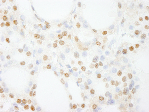
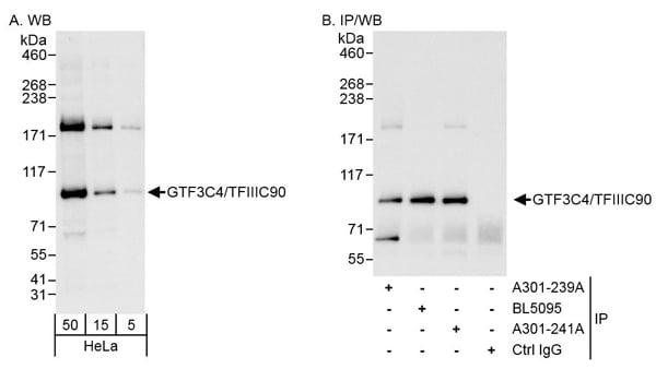
(Detection of human GTF3C4/TFIIIC90 by western blot and immunoprecipitation. Samples: Whole cell lysate (5, 15 and 50 ug for WB; 1 mg for IP, 20% of IP loaded) from HeLa cells. Antibodies: Affinity purified rabbit anti-GTF3C4/TFIIIC90 antibody AAA211372 used for WB at 0.04 ug/ml (A) and 1 ug/ml (B) and used for IP at 3 ug/mg lysate. GTF3C4/TFIIIC90 was also immunoprecipitated by rabbit anti-GTF3C4/TFIIIC90 antibodies BL5095 and which recognize downstream epitopes. Detection: Chemiluminescence with exposure times of 10 seconds (A) and 3 seconds (B).)
WB (Western Blot)
(Detection of human GTF3C4/TFIIIC90 by western blot and immunoprecipitation. Samples: Whole cell lysate (5, 15 and 50 ug for WB; 1 mg for IP, 20% of IP loaded) from HeLa cells. Antibodies: Affinity purified rabbit anti-GTF3C4/TFIIIC90 antibody AAA211372 used for WB at 0.04 ug/ml (A) and 1 ug/ml (B) and used for IP at 3 ug/mg lysate. GTF3C4/TFIIIC90 was also immunoprecipitated by rabbit anti-GTF3C4/TFIIIC90 antibodies BL5095 and which recognize downstream epitopes. Detection: Chemiluminescence with exposure times of 10 seconds (A) and 3 seconds (B).)
GTF3C4/TFIIIC90, Polyclonal Antibody (Cat# AAA211372)
 WB (Western Blot)
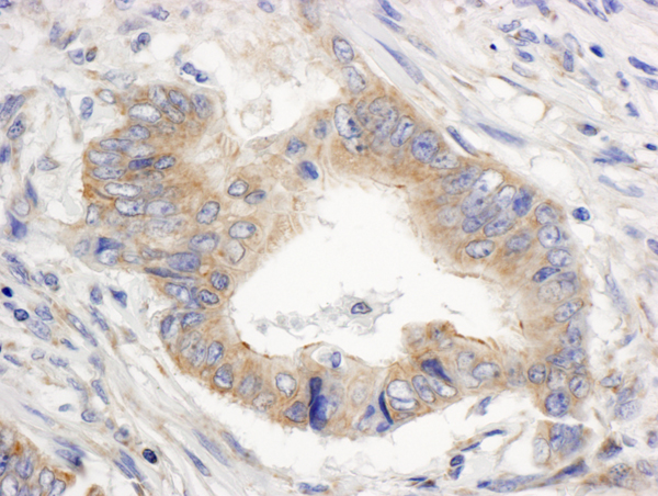
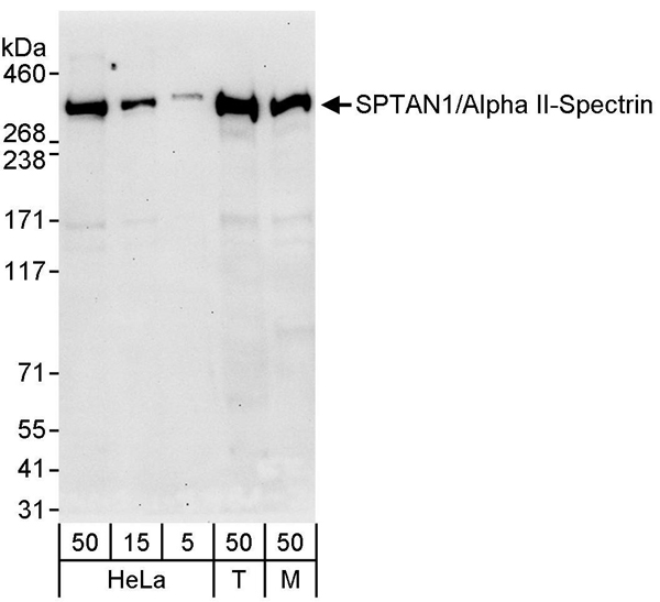
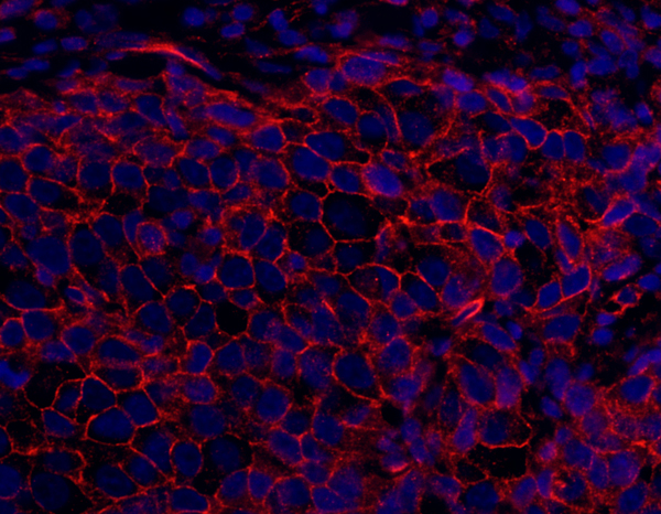
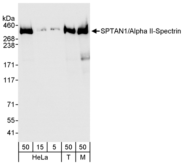
(Detection of human and mouse SPTAN1/Alpha II-Spectrin by western blot. Samples: Whole cell lysate from HeLa (5, 15 and 50 ug), HEK293T (T; 50 ug), and mouse NIH 3T3 (M; 50 ug) cells. Antibody: Affinity purified rabbit anti-SPTAN1/Alpha II-Spectrin antibody AAA211380 (lot AAA211380-2) used for WB at 0.2 ug/ml. Detection: Chemiluminescence with an exposure time of 30 seconds.)
WB (Western Blot)
(Detection of human and mouse SPTAN1/Alpha II-Spectrin by western blot. Samples: Whole cell lysate from HeLa (5, 15 and 50 ug), HEK293T (T; 50 ug), and mouse NIH 3T3 (M; 50 ug) cells. Antibody: Affinity purified rabbit anti-SPTAN1/Alpha II-Spectrin antibody AAA211380 (lot AAA211380-2) used for WB at 0.2 ug/ml. Detection: Chemiluminescence with an exposure time of 30 seconds.)
SPTAN1/Alpha II-spectrin, Polyclonal Antibody (Cat# AAA211380)
 WB (Western Blot)
(Detection of human and mouse SPTAN1/Alpha II-Spectrin by western blot. Samples: Whole cell lysate from HeLa (5, 15 and 50 ug), HEK293T (T; 50 ug), and mouse NIH 3T3 (M; 50 ug) cells. Antibody: Affinity purified rabbit anti-SPTAN1/Alpha II-Spectrin antibody AAA211381 used for WB at 0.04 ug/ml Detection: Chemiluminescence with an exposure time of 3 seconds.)
WB (Western Blot)
(Detection of human and mouse SPTAN1/Alpha II-Spectrin by western blot. Samples: Whole cell lysate from HeLa (5, 15 and 50 ug), HEK293T (T; 50 ug), and mouse NIH 3T3 (M; 50 ug) cells. Antibody: Affinity purified rabbit anti-SPTAN1/Alpha II-Spectrin antibody AAA211381 used for WB at 0.04 ug/ml Detection: Chemiluminescence with an exposure time of 3 seconds.)
SPTAN1/Alpha II-spectrin, Polyclonal Antibody (Cat# AAA211381)
 WB (Western Blot)
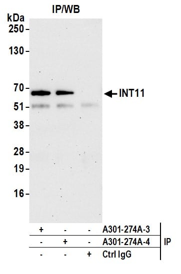
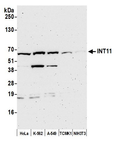
(Detection of human and mouse INT11 by western blot. Samples: Whole cell lysate (50 ug) from HeLa, K-562, A-549, TCMK-1, and NIH 3T3 cells prepared using NETN lysis buffer. Antibody: Affinity purified rabbit anti-INT11 antibody AAA211388 (lot AAA211388-4) used for WB at 0.1 ug/ml. Detection: Chemiluminescence with an exposure time of 3 minutes.)
WB (Western Blot)
(Detection of human and mouse INT11 by western blot. Samples: Whole cell lysate (50 ug) from HeLa, K-562, A-549, TCMK-1, and NIH 3T3 cells prepared using NETN lysis buffer. Antibody: Affinity purified rabbit anti-INT11 antibody AAA211388 (lot AAA211388-4) used for WB at 0.1 ug/ml. Detection: Chemiluminescence with an exposure time of 3 minutes.)
INT11, Polyclonal Antibody (Cat# AAA211388)
 WB (Western Blot)
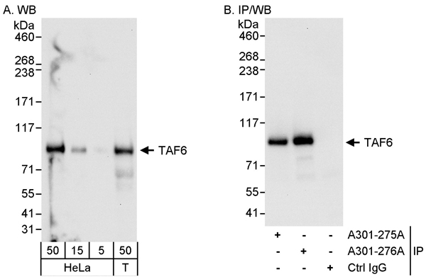
(Detection of human TAF6 by western blot and immunoprecipitation. Samples: Whole cell lysate from HeLa (5, 15 and 50 ug for WB; 1 mg for IP, 20% of IP loaded) and HEK293T (T; 50 ug) cells. Antibodies: Affinity purified rabbit anti-TAF6 antibody AAA211389 used for WB at 0.04 ug/ml (A) and 1 ug/ml (B) and used for IP at 3 ug/mg lysate. TAF6 was also immunoprecipitated by rabbit anti-TAF6 antibody which recognizes an upstream epitope. Detection: Chemiluminescence with exposure times of 10 seconds (A and B).)
WB (Western Blot)
(Detection of human TAF6 by western blot and immunoprecipitation. Samples: Whole cell lysate from HeLa (5, 15 and 50 ug for WB; 1 mg for IP, 20% of IP loaded) and HEK293T (T; 50 ug) cells. Antibodies: Affinity purified rabbit anti-TAF6 antibody AAA211389 used for WB at 0.04 ug/ml (A) and 1 ug/ml (B) and used for IP at 3 ug/mg lysate. TAF6 was also immunoprecipitated by rabbit anti-TAF6 antibody which recognizes an upstream epitope. Detection: Chemiluminescence with exposure times of 10 seconds (A and B).)
TAF6, Polyclonal Antibody (Cat# AAA211389)
 WB (Western Blot)
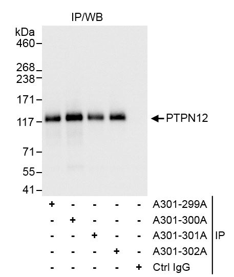
(Detection of human PTPN12 by western blot. Samples: Whole cell lysate (5, 15, and 50 ug) from HeLa cells. Antibody: Affinity purified rabbit anti-PTPN12 antibody AAA211405 used at 0.04 ug/ml. Detection: Chemiluminescence with an exposure time of 10 seconds.)
WB (Western Blot)
(Detection of human PTPN12 by western blot. Samples: Whole cell lysate (5, 15, and 50 ug) from HeLa cells. Antibody: Affinity purified rabbit anti-PTPN12 antibody AAA211405 used at 0.04 ug/ml. Detection: Chemiluminescence with an exposure time of 10 seconds.)
PTPN12, Polyclonal Antibody (Cat# AAA211405)
 WB (Western Blot)
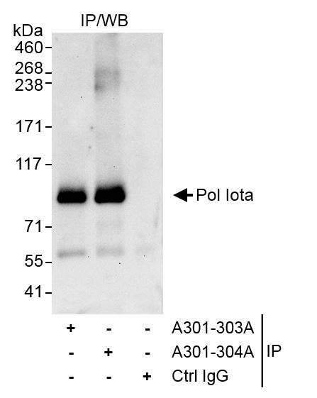
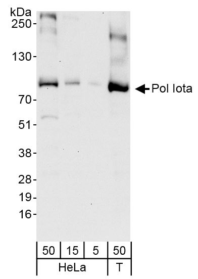
(Detection of human Pol Iota by western blot. Samples: Whole cell lysate from HeLa (5, 15 and 50 ug) and HEK293T (T; 50 ug) cells. Antibodies: Affinity purified rabbit anti-Pol Iota antibody AAA211408 (lot AAA211408-1) used for WB at 0.04 ug/ml. Detection: Chemiluminescence with exposure time of 30 seconds.)
WB (Western Blot)
(Detection of human Pol Iota by western blot. Samples: Whole cell lysate from HeLa (5, 15 and 50 ug) and HEK293T (T; 50 ug) cells. Antibodies: Affinity purified rabbit anti-Pol Iota antibody AAA211408 (lot AAA211408-1) used for WB at 0.04 ug/ml. Detection: Chemiluminescence with exposure time of 30 seconds.)
Pol Iota, Polyclonal Antibody (Cat# AAA211408)
 WB (Western Blot)
(Detection of human DOCK10 by western blot. Samples: Whole cell lysate (50 ug) from Jurkat, LNCaP, and GaMG cells prepared using NETN lysis buffer. Antibody: Affinity purified rabbit anti-DOCK10 antibody AAA211409 (lot AAA211409-2) used for WB at 0.1 ug/ml. Detection: Chemiluminescence with an exposure time of 10 seconds.)
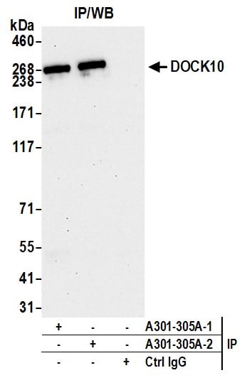
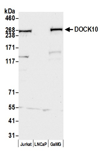
WB (Western Blot)
(Detection of human DOCK10 by western blot. Samples: Whole cell lysate (50 ug) from Jurkat, LNCaP, and GaMG cells prepared using NETN lysis buffer. Antibody: Affinity purified rabbit anti-DOCK10 antibody AAA211409 (lot AAA211409-2) used for WB at 0.1 ug/ml. Detection: Chemiluminescence with an exposure time of 10 seconds.)
DOCK10, Polyclonal Antibody (Cat# AAA211409)
 WB (Western Blot)
(Detection of human DDX54 by western blot and immunoprecipitation. Samples: Whole cell lysate (5, 15 and 50 ug for WB; 1 mg for IP, 20% of IP loaded) from HeLa cells. Antibodies: Affinity purified rabbit anti-DDX54 antibody AAA211412 used for WB at 0.04 ug/ml (A) and 1 ug/ml (B) and used for IP at 3 ug/mg lysate. DDX54 was also immunoprecipitated by rabbit anti-DDX54 antibody which recognizes a downstream epitope. Detection: Chemiluminescence with exposure times of 10 seconds (A) and 3 seconds (B).)
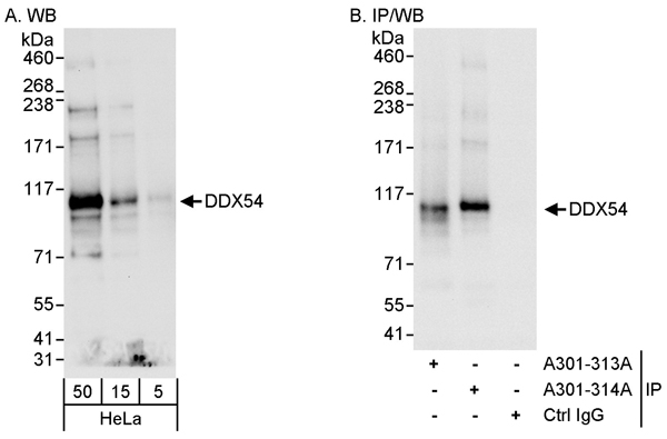
WB (Western Blot)
(Detection of human DDX54 by western blot and immunoprecipitation. Samples: Whole cell lysate (5, 15 and 50 ug for WB; 1 mg for IP, 20% of IP loaded) from HeLa cells. Antibodies: Affinity purified rabbit anti-DDX54 antibody AAA211412 used for WB at 0.04 ug/ml (A) and 1 ug/ml (B) and used for IP at 3 ug/mg lysate. DDX54 was also immunoprecipitated by rabbit anti-DDX54 antibody which recognizes a downstream epitope. Detection: Chemiluminescence with exposure times of 10 seconds (A) and 3 seconds (B).)
DDX54, Polyclonal Antibody (Cat# AAA211412)
 WB (Western Blot)
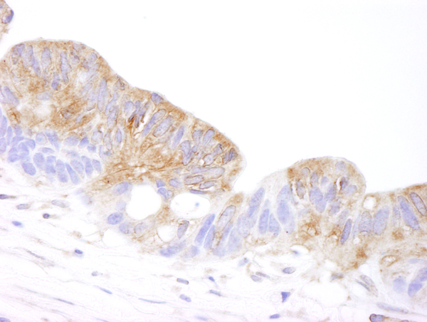
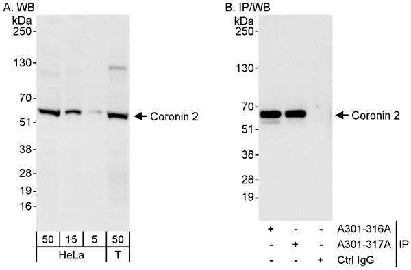
(Detection of human Coronin 2 by western blot and immunoprecipitation. Samples: Whole cell lysate from HeLa (5, 15 and 50 ug for WB; 1 mg for IP, 20% of IP loaded) and HEK293T (T; 50 ug) cells. Antibodies: Affinity purified rabbit anti-Coronin 2 antibody AAA211414 used for WB at 0.04 ug/ml (A) and 1 ug/ml (B) and used for IP at 3 ug/mg lysate. Coronin 2 was also immunoprecipitated by rabbit anti-Coronin 2 antibody which recognizes a downstream epitope. Detection: Chemiluminescence with exposure times of 10 seconds (A) and 3 seconds (B).)
WB (Western Blot)
(Detection of human Coronin 2 by western blot and immunoprecipitation. Samples: Whole cell lysate from HeLa (5, 15 and 50 ug for WB; 1 mg for IP, 20% of IP loaded) and HEK293T (T; 50 ug) cells. Antibodies: Affinity purified rabbit anti-Coronin 2 antibody AAA211414 used for WB at 0.04 ug/ml (A) and 1 ug/ml (B) and used for IP at 3 ug/mg lysate. Coronin 2 was also immunoprecipitated by rabbit anti-Coronin 2 antibody which recognizes a downstream epitope. Detection: Chemiluminescence with exposure times of 10 seconds (A) and 3 seconds (B).)
Coronin 2, Polyclonal Antibody (Cat# AAA211414)
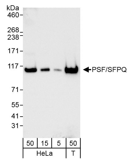 WB (Western Blot)
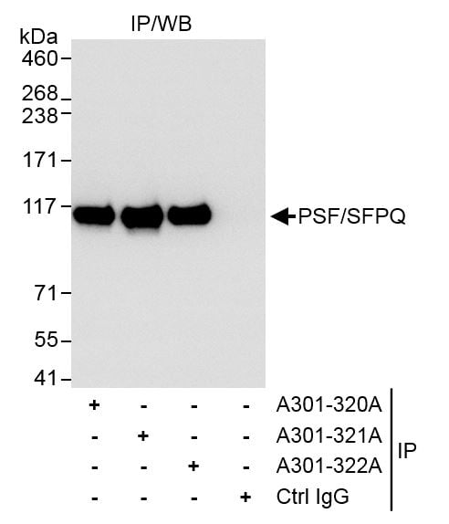
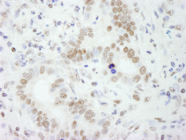
(Detection of human PSF/SFPQ by western blot. Samples: Whole cell lysate from HeLa (5, 15 and 50 ug) and HEK293T (T; 50 ug) cells. Antibodies: Affinity purified rabbit anti-PSF/SFPQ antibody AAA211417 (lot AAA211417-1) used for WB at 0.04 ug/ml. Detection: Chemiluminescence with exposure time of 5 seconds.)
WB (Western Blot)
(Detection of human PSF/SFPQ by western blot. Samples: Whole cell lysate from HeLa (5, 15 and 50 ug) and HEK293T (T; 50 ug) cells. Antibodies: Affinity purified rabbit anti-PSF/SFPQ antibody AAA211417 (lot AAA211417-1) used for WB at 0.04 ug/ml. Detection: Chemiluminescence with exposure time of 5 seconds.)
PSF/SFPQ, Polyclonal Antibody (Cat# AAA211417)
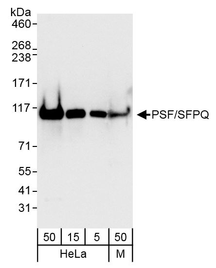 WB (Western Blot)
(Detection of human and mouse PSF/SFPQ by western blot. Samples: Whole cell lysate from HeLa (5, 15 and 50 ug) and mouse NIH 3T3 (M; 50 ug) cells. Antibodies: Affinity purified rabbit anti-PSF/SFPQ antibody AAA211419 (lot AAA211419-1) used for WB at 0.04 ug/ml. Detection: Chemiluminescence with exposure time of 3 seconds.)
WB (Western Blot)
(Detection of human and mouse PSF/SFPQ by western blot. Samples: Whole cell lysate from HeLa (5, 15 and 50 ug) and mouse NIH 3T3 (M; 50 ug) cells. Antibodies: Affinity purified rabbit anti-PSF/SFPQ antibody AAA211419 (lot AAA211419-1) used for WB at 0.04 ug/ml. Detection: Chemiluminescence with exposure time of 3 seconds.)
PSF/SFPQ, Polyclonal Antibody (Cat# AAA211419)

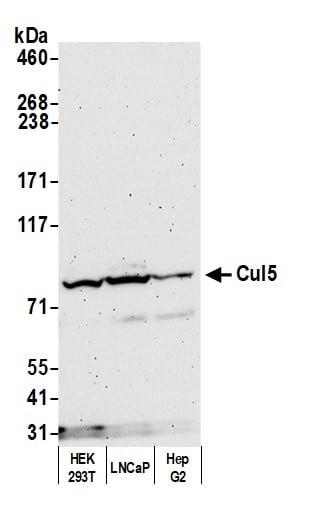 WB (Western Blot)
(Detection of human Cul5 by western blot. Samples: Whole cell lysate (50 ug) from HEK293T, LNCaP, and Hep-G2 cells prepared using NETN lysis buffer. Antibody: Affinity purified rabbit anti-Cul5 antibody AAA211790 (lot AAA211790-2) used for WB at 0.1 ug/ml. Detection: Chemiluminescence with an exposure time of 3 minutes.)
WB (Western Blot)
(Detection of human Cul5 by western blot. Samples: Whole cell lysate (50 ug) from HEK293T, LNCaP, and Hep-G2 cells prepared using NETN lysis buffer. Antibody: Affinity purified rabbit anti-Cul5 antibody AAA211790 (lot AAA211790-2) used for WB at 0.1 ug/ml. Detection: Chemiluminescence with an exposure time of 3 minutes.)
Cul5, Polyclonal Antibody (Cat# AAA211790)
 WB (Western Blot)
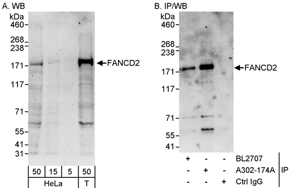
(Detection of human FANCD2 by western blot and immunoprecipitation. Samples: Whole cell lysate from HeLa (5, 15 and 50 ug for WB) and HEK293T (T; 50 ug for WB; 1 mg for IP, 20% of IP loaded) cells. Antibodies: Affinity purified rabbit anti-FANCD2 antibody AAA211791 used for WB at 0.04 ug/ml (A) and 1 ug/ml (B) and used for IP at 10 ug/mg lysate. FANCD2 was also immunoprecipitated by rabbit anti-FANCD2 antibody BL2707, which recognizes an upstream epitope. Detection: Chemiluminescence with exposure times of 3 minutes (A) and 30 seconds (B).)
WB (Western Blot)
(Detection of human FANCD2 by western blot and immunoprecipitation. Samples: Whole cell lysate from HeLa (5, 15 and 50 ug for WB) and HEK293T (T; 50 ug for WB; 1 mg for IP, 20% of IP loaded) cells. Antibodies: Affinity purified rabbit anti-FANCD2 antibody AAA211791 used for WB at 0.04 ug/ml (A) and 1 ug/ml (B) and used for IP at 10 ug/mg lysate. FANCD2 was also immunoprecipitated by rabbit anti-FANCD2 antibody BL2707, which recognizes an upstream epitope. Detection: Chemiluminescence with exposure times of 3 minutes (A) and 30 seconds (B).)
FANCD2, Polyclonal Antibody (Cat# AAA211791)
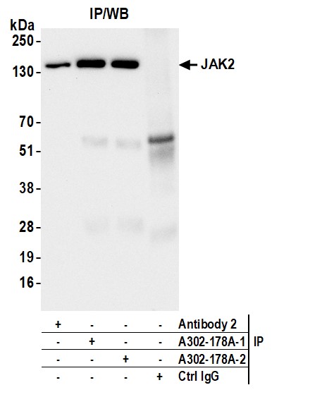
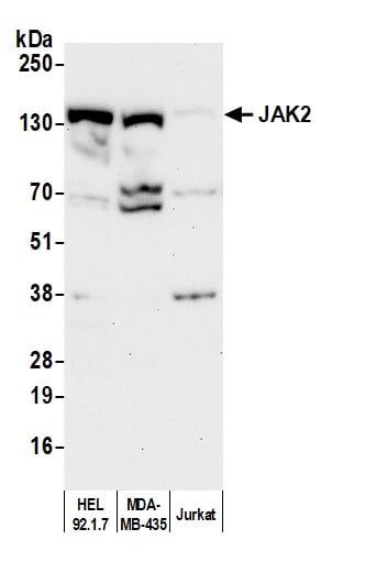 WB (Western Blot)
(Detection of human JAK2 by western blot. Samples: Whole cell lysate (50 ug) from HEL 92.1.7, MDA-MB-435, and Jurkat cells prepared using NETN lysis buffer. Antibody: Affinity purified rabbit anti-JAK2 antibody (AAA211792 lot 2) used for WB at 0.4 ug/ml. Detection: Chemiluminescence with an exposure time of 3 minutes.)
WB (Western Blot)
(Detection of human JAK2 by western blot. Samples: Whole cell lysate (50 ug) from HEL 92.1.7, MDA-MB-435, and Jurkat cells prepared using NETN lysis buffer. Antibody: Affinity purified rabbit anti-JAK2 antibody (AAA211792 lot 2) used for WB at 0.4 ug/ml. Detection: Chemiluminescence with an exposure time of 3 minutes.)
JAK2, Polyclonal Antibody (Cat# AAA211792)
 WB (Western Blot)
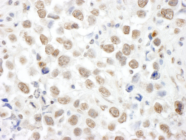
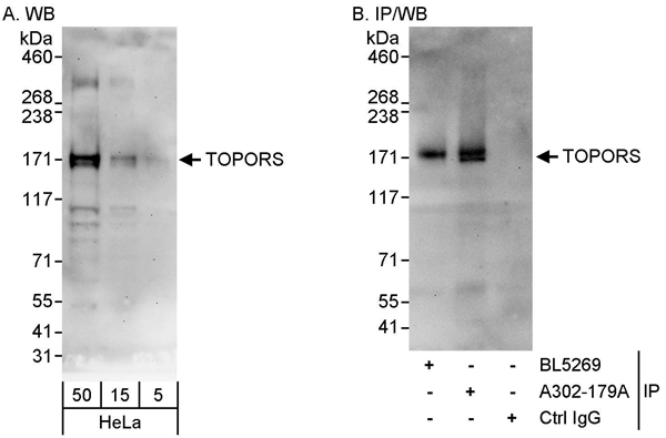
(Detection of human TOPORS by western blot and immunoprecipitation. Samples: Whole cell lysate (5, 15 and 50 ug for WB; 1 mg for IP, 20% of IP loaded) from HeLa cells. Antibodies: Affinity purified rabbit anti-TOPORS antibody AAA211793 used for WB at 0.04 ug/ml (A) and 1 ug/ml (B) and used for IP at 10 ug/mg lysate. TOPORS was also immunoprecipitated by rabbit anti-TOPORS antibody BL5269, which recognizes an upstream epitope. Detection: Chemiluminescence with exposure times of 1 minute (A) and 30 seconds (B).)
WB (Western Blot)
(Detection of human TOPORS by western blot and immunoprecipitation. Samples: Whole cell lysate (5, 15 and 50 ug for WB; 1 mg for IP, 20% of IP loaded) from HeLa cells. Antibodies: Affinity purified rabbit anti-TOPORS antibody AAA211793 used for WB at 0.04 ug/ml (A) and 1 ug/ml (B) and used for IP at 10 ug/mg lysate. TOPORS was also immunoprecipitated by rabbit anti-TOPORS antibody BL5269, which recognizes an upstream epitope. Detection: Chemiluminescence with exposure times of 1 minute (A) and 30 seconds (B).)
TOPORS, Polyclonal Antibody (Cat# AAA211793)
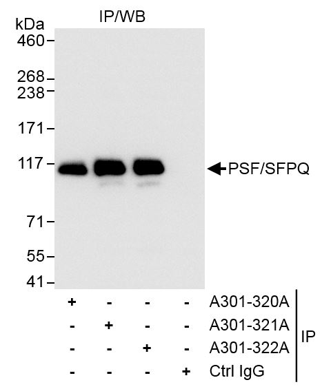
 IP (Immunoprecipitation)
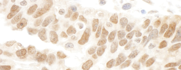
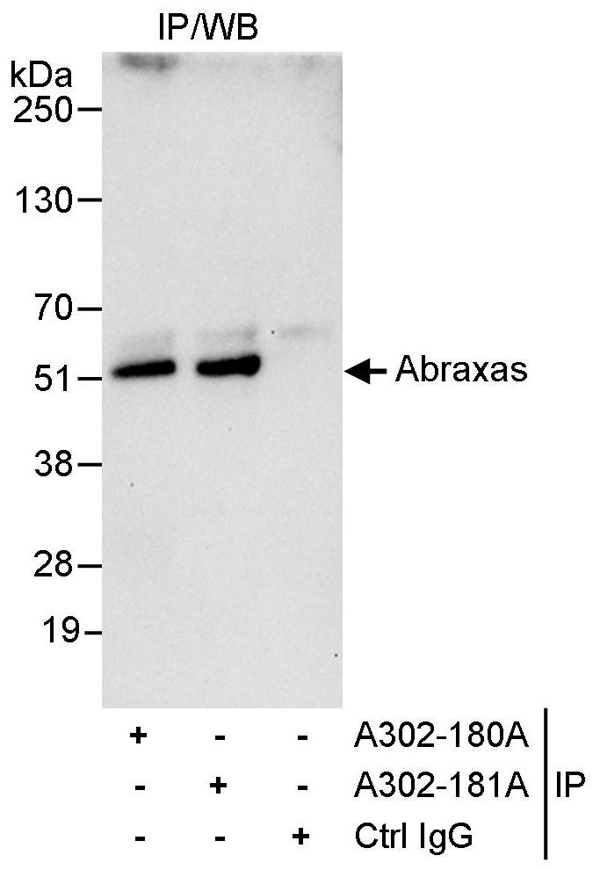
(Detection of human Abraxas by western blot of immunoprecipitates. Samples: Whole cell lysate (1 mg for IP, 20% of IP loaded) from HeLa cells. Antibodies: Affinity purified rabbit anti-Abraxas antibody AAA211794 used for IP at 10 ug/mg lysate. Abraxas was also immunoprecipitated by rabbit anti-Abraxas antibody which recognizes an upstream epitope. For blotting immunoprecipitated Abraxas, was used at 1 ug/ml. Detection: Chemiluminescence with an exposure time of 10 seconds.)
IP (Immunoprecipitation)
(Detection of human Abraxas by western blot of immunoprecipitates. Samples: Whole cell lysate (1 mg for IP, 20% of IP loaded) from HeLa cells. Antibodies: Affinity purified rabbit anti-Abraxas antibody AAA211794 used for IP at 10 ug/mg lysate. Abraxas was also immunoprecipitated by rabbit anti-Abraxas antibody which recognizes an upstream epitope. For blotting immunoprecipitated Abraxas, was used at 1 ug/ml. Detection: Chemiluminescence with an exposure time of 10 seconds.)
Abraxas, Polyclonal Antibody (Cat# AAA211794)
 WB (Western Blot)
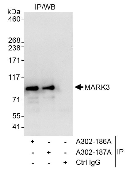
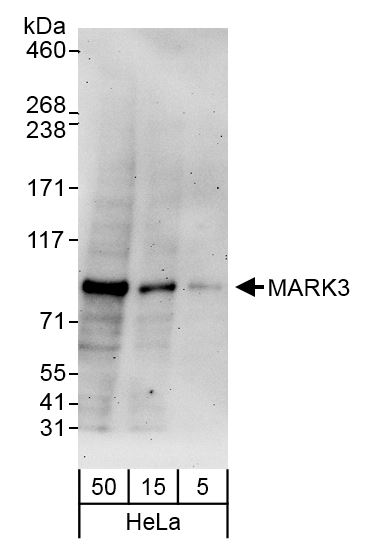
(Detection of human MARK3 by western blot. Samples: Whole cell lysate (5, 15, and 50 ug) from HeLa cells. Antibody: Affinity purified rabbit anti-MARK3 antibody AAA211796 (lot AAA211796-1) used at 0.04 ug/ml. Detection: Chemiluminescence with an exposure time of 3 minutes.)
WB (Western Blot)
(Detection of human MARK3 by western blot. Samples: Whole cell lysate (5, 15, and 50 ug) from HeLa cells. Antibody: Affinity purified rabbit anti-MARK3 antibody AAA211796 (lot AAA211796-1) used at 0.04 ug/ml. Detection: Chemiluminescence with an exposure time of 3 minutes.)
MARK3, Polyclonal Antibody (Cat# AAA211796)
 IP (Immunoprecipitation)
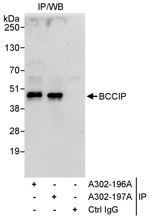
(Detection of human BCCIP by western blot of immunoprecipitates. Samples: Whole cell lysate (1 mg for IP, 20% of IP loaded) from HeLa cells. Antibodies: Affinity purified rabbit anti-BCCIP antibody AAA211799 used for IP at 3 ug/mg lysate. BCCIP was also immunoprecipitated by rabbit anti-BCCIP antibody which recognizes an upstream epitope. For blotting immunoprecipitated BCCIP, was used at 1 ug/ml. Detection: Chemiluminescence with an exposure time of 10 seconds.)
IP (Immunoprecipitation)
(Detection of human BCCIP by western blot of immunoprecipitates. Samples: Whole cell lysate (1 mg for IP, 20% of IP loaded) from HeLa cells. Antibodies: Affinity purified rabbit anti-BCCIP antibody AAA211799 used for IP at 3 ug/mg lysate. BCCIP was also immunoprecipitated by rabbit anti-BCCIP antibody which recognizes an upstream epitope. For blotting immunoprecipitated BCCIP, was used at 1 ug/ml. Detection: Chemiluminescence with an exposure time of 10 seconds.)
BCCIP, Polyclonal Antibody (Cat# AAA211799)
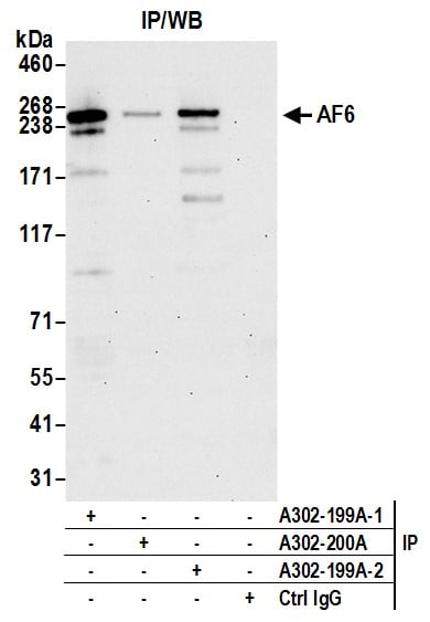
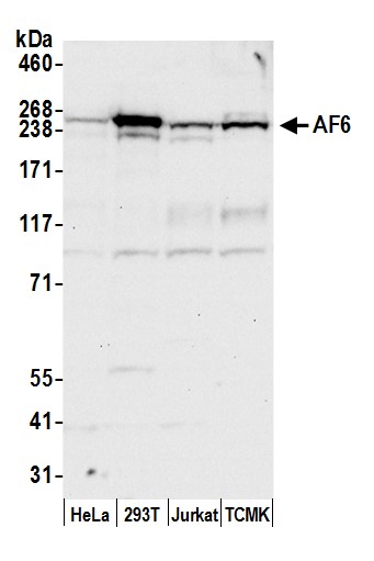 WB (Western Blot)
(Detection of human and mouse AF6 by western blot. Samples: Whole cell lysate (50 ug) from HeLa, HEK293T, Jurkat, and mouse TCMK-1 cells prepared using NETN lysis buffer. Antibody: Affinity purified rabbit anti-AF6 antibody AAA211800 (lot AAA211800-2) used for WB at 0.1 ug/ml. Detection: Chemiluminescence with an exposure time of 10 seconds.)
WB (Western Blot)
(Detection of human and mouse AF6 by western blot. Samples: Whole cell lysate (50 ug) from HeLa, HEK293T, Jurkat, and mouse TCMK-1 cells prepared using NETN lysis buffer. Antibody: Affinity purified rabbit anti-AF6 antibody AAA211800 (lot AAA211800-2) used for WB at 0.1 ug/ml. Detection: Chemiluminescence with an exposure time of 10 seconds.)
AF6, Polyclonal Antibody (Cat# AAA211800)
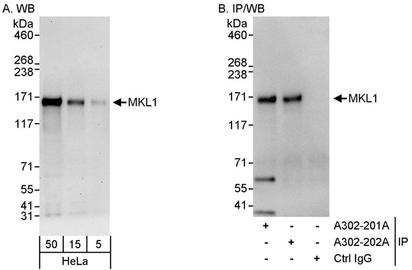 WB (Western Blot)
(Detection of human MKL1 by western blot and immunoprecipitation. Samples: Whole cell lysate (5, 15 and 50 ug for WB; 1 mg for IP, 20% of IP loaded) from HeLa cells. Antibodies: Affinity purified rabbit anti-MKL1 antibody AAA211801 used for WB at 0.04 ug/ml (A) and 0.4 ug/ml (B) and used for IP at 3 ug/mg lysate. MKL1 was also immunoprecipitated by rabbit anti-MKL1 antibody which recognizes a downstream epitope. Detection: Chemiluminescence with exposure times of 30 seconds (A) and 10 seconds (B).)
WB (Western Blot)
(Detection of human MKL1 by western blot and immunoprecipitation. Samples: Whole cell lysate (5, 15 and 50 ug for WB; 1 mg for IP, 20% of IP loaded) from HeLa cells. Antibodies: Affinity purified rabbit anti-MKL1 antibody AAA211801 used for WB at 0.04 ug/ml (A) and 0.4 ug/ml (B) and used for IP at 3 ug/mg lysate. MKL1 was also immunoprecipitated by rabbit anti-MKL1 antibody which recognizes a downstream epitope. Detection: Chemiluminescence with exposure times of 30 seconds (A) and 10 seconds (B).)
MKL1, Polyclonal Antibody (Cat# AAA211801)
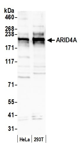 WB (Western Blot)
(Detection of human ARID4A by western blot. Samples: Whole cell lysate (50 ug) from HeLa and HEK293T cells prepared using NETN lysis buffer. Antibody: Affinity purified rabbit anti-ARID4A antibody AAA211810 (lot AAA211810-2) used for WB at 0.1 ug/ml. Detection: Chemiluminescence with an exposure time of 3 minutes.)
WB (Western Blot)
(Detection of human ARID4A by western blot. Samples: Whole cell lysate (50 ug) from HeLa and HEK293T cells prepared using NETN lysis buffer. Antibody: Affinity purified rabbit anti-ARID4A antibody AAA211810 (lot AAA211810-2) used for WB at 0.1 ug/ml. Detection: Chemiluminescence with an exposure time of 3 minutes.)
ARID4A, Polyclonal Antibody (Cat# AAA211810)
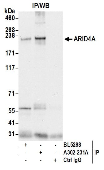
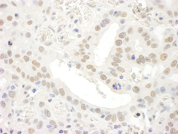
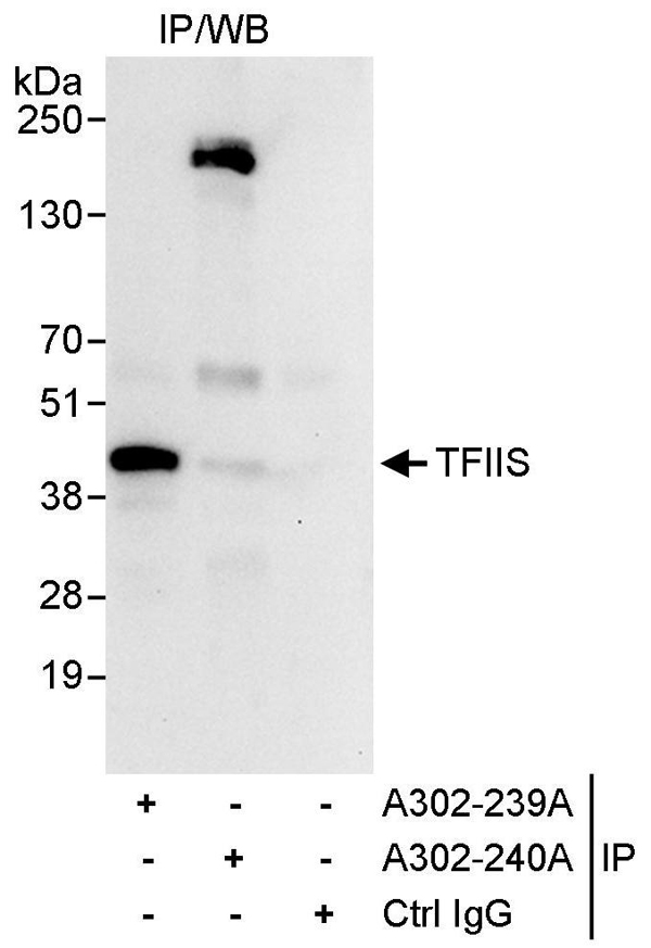 IP (Immunoprecipitation)
(Detection of human TFIIS by western blot of immunoprecipitates. Samples: Whole cell lysate (1 mg for IP, 20% of IP loaded) from HeLa cells. Antibodies: Affinity purified rabbit anti-TFIIS antibody AAA211812 used for IP at 10 ug/mg lysate. TFIIS was unsuccessfully immunoprecipitated by rabbit anti-TFIIS antibody which recognizes a downstream epitope. For blotting immunoprecipitated TFIIS, was used at 1 ug/ml. Detection: Chemiluminescence with an exposure time of 10 seconds.)
IP (Immunoprecipitation)
(Detection of human TFIIS by western blot of immunoprecipitates. Samples: Whole cell lysate (1 mg for IP, 20% of IP loaded) from HeLa cells. Antibodies: Affinity purified rabbit anti-TFIIS antibody AAA211812 used for IP at 10 ug/mg lysate. TFIIS was unsuccessfully immunoprecipitated by rabbit anti-TFIIS antibody which recognizes a downstream epitope. For blotting immunoprecipitated TFIIS, was used at 1 ug/ml. Detection: Chemiluminescence with an exposure time of 10 seconds.)
TFIIS, Polyclonal Antibody (Cat# AAA211812)
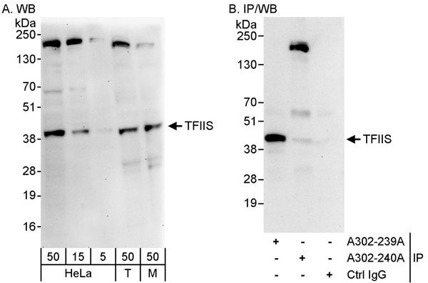 WB (Western Blot)
(Detection of human and mouse TFIIS by western blot (h&m) and immunoprecipitation (h). Samples: Whole cell lysate from HeLa (5, 15 and 50 ug for WB; 1 mg for IP, 20% of IP loaded), HEK293T (T; 50 ug) and mouse NIH 3T3 (M; 50ug) cells. Antibodies: Affinity purified rabbit anti-TFIIS antibody AAA211813 used for WB at 0.04 ug/ml (A) and 1 ug/ml (B) and used for IP at 10 ug/mg lysate. TFIIS was successfully immunoprecipitated by rabbit anti-TFIIS antibody which recognizes an upstream epitope. Detection: Chemiluminescence with exposure times of 3 minutes (A) and 10 seconds (B).)
WB (Western Blot)
(Detection of human and mouse TFIIS by western blot (h&m) and immunoprecipitation (h). Samples: Whole cell lysate from HeLa (5, 15 and 50 ug for WB; 1 mg for IP, 20% of IP loaded), HEK293T (T; 50 ug) and mouse NIH 3T3 (M; 50ug) cells. Antibodies: Affinity purified rabbit anti-TFIIS antibody AAA211813 used for WB at 0.04 ug/ml (A) and 1 ug/ml (B) and used for IP at 10 ug/mg lysate. TFIIS was successfully immunoprecipitated by rabbit anti-TFIIS antibody which recognizes an upstream epitope. Detection: Chemiluminescence with exposure times of 3 minutes (A) and 10 seconds (B).)
TFIIS, Polyclonal Antibody (Cat# AAA211813)
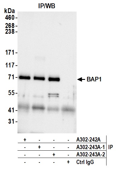
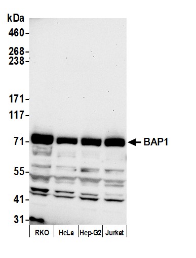 WB (Western Blot)
(Detection of human BAP1 by western blot. Samples: Whole cell lysate (50 ug) from RKO, HeLa, Hep-G2, and Jurkat cells prepared using NETN lysis buffer. Antibody: Affinity purified rabbit anti-BAP1 antibody (AAA211816 lot 2) used for WB at 0.1 ug/ml. Detection: Chemiluminescence with an exposure time of 30 seconds.)
WB (Western Blot)
(Detection of human BAP1 by western blot. Samples: Whole cell lysate (50 ug) from RKO, HeLa, Hep-G2, and Jurkat cells prepared using NETN lysis buffer. Antibody: Affinity purified rabbit anti-BAP1 antibody (AAA211816 lot 2) used for WB at 0.1 ug/ml. Detection: Chemiluminescence with an exposure time of 30 seconds.)
BAP1, Polyclonal Antibody (Cat# AAA211816)
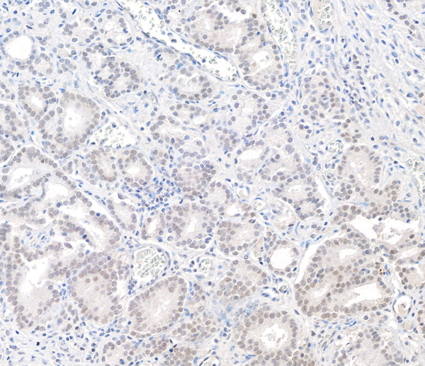
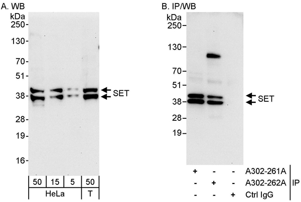 WB (Western Blot)
(Detection of human SET by western blot and immunoprecipitation. Samples: Whole cell lysate from HeLa (5, 15 and 50 ug for WB; 1 mg for IP, 20% of IP loaded) and HEK293T (T; 50 ug) cells. Antibodies: Affinity purified rabbit anti-SET antibody AAA211820 used for WB at 0.04 ug/ml (A) and 1 ug/ml (B) and used for IP at 3 ug/mg lysate. SET was also immunoprecipitated by rabbit anti-SET antibody which recognizes an upstream epitope. Detection: Chemiluminescence with exposure times of 30 seconds (A) and 10 seconds (B).)
WB (Western Blot)
(Detection of human SET by western blot and immunoprecipitation. Samples: Whole cell lysate from HeLa (5, 15 and 50 ug for WB; 1 mg for IP, 20% of IP loaded) and HEK293T (T; 50 ug) cells. Antibodies: Affinity purified rabbit anti-SET antibody AAA211820 used for WB at 0.04 ug/ml (A) and 1 ug/ml (B) and used for IP at 3 ug/mg lysate. SET was also immunoprecipitated by rabbit anti-SET antibody which recognizes an upstream epitope. Detection: Chemiluminescence with exposure times of 30 seconds (A) and 10 seconds (B).)
SET, Polyclonal Antibody (Cat# AAA211820)
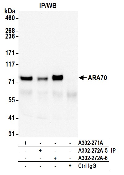
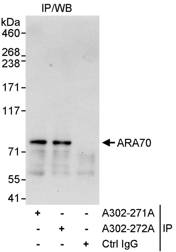 IP (Immunoprecipitation)
(Detection of human ARA70 by western blot of immunoprecipitates. Samples: Whole cell lysate (1 mg for IP, 20% of IP loaded) from HeLa cells. Antibodies: Affinity purified rabbit anti-ARA70 antibody AAA211826 used for IP at 10 ug/mg lysate. ARA70 was also immunoprecipitated by rabbit anti-ARA70 antibody which recognizes a downstream epitope. For blotting immunoprecipitated ARA70, was used at 1 ug/ml. Detection: Chemiluminescence with an exposure time of 30 seconds.)
IP (Immunoprecipitation)
(Detection of human ARA70 by western blot of immunoprecipitates. Samples: Whole cell lysate (1 mg for IP, 20% of IP loaded) from HeLa cells. Antibodies: Affinity purified rabbit anti-ARA70 antibody AAA211826 used for IP at 10 ug/mg lysate. ARA70 was also immunoprecipitated by rabbit anti-ARA70 antibody which recognizes a downstream epitope. For blotting immunoprecipitated ARA70, was used at 1 ug/ml. Detection: Chemiluminescence with an exposure time of 30 seconds.)
ARA70, Polyclonal Antibody (Cat# AAA211826)
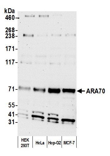 WB (Western Blot)
(Detection of human ARA70 by western blot. Samples: Whole cell lysate (25 ug) from HEK293T, HeLa, Hep-G2, and MCF-7 cells prepared using NETN lysis buffer. Antibody: Affinity purified rabbit anti-ARA70 antibody (AAA211827 lot 6) used for WB at 0.4 ug/ml. Detection: Chemiluminescence with an exposure time of 30 seconds.)
WB (Western Blot)
(Detection of human ARA70 by western blot. Samples: Whole cell lysate (25 ug) from HEK293T, HeLa, Hep-G2, and MCF-7 cells prepared using NETN lysis buffer. Antibody: Affinity purified rabbit anti-ARA70 antibody (AAA211827 lot 6) used for WB at 0.4 ug/ml. Detection: Chemiluminescence with an exposure time of 30 seconds.)
ARA70, Polyclonal Antibody (Cat# AAA211827)
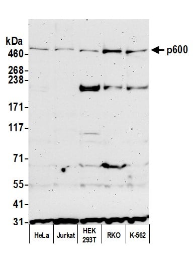
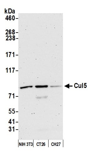
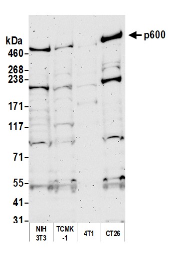 WB (Western Blot)
(Detection of mouse p600 by western blot. Samples: Whole cell lysate (50 ug) from NIH 3T3, TCMK-1, 4T1, and CT26 cells prepared using NETN lysis buffer. Antibody: Affinity purified rabbit anti-p600 antibody (AAA211831 lot 2) used for WB at 0.1 ug/ml. Detection: Chemiluminescence with an exposure time of 3 minutes.)
WB (Western Blot)
(Detection of mouse p600 by western blot. Samples: Whole cell lysate (50 ug) from NIH 3T3, TCMK-1, 4T1, and CT26 cells prepared using NETN lysis buffer. Antibody: Affinity purified rabbit anti-p600 antibody (AAA211831 lot 2) used for WB at 0.1 ug/ml. Detection: Chemiluminescence with an exposure time of 3 minutes.)
p600, Polyclonal Antibody (Cat# AAA211831)
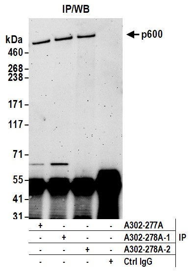
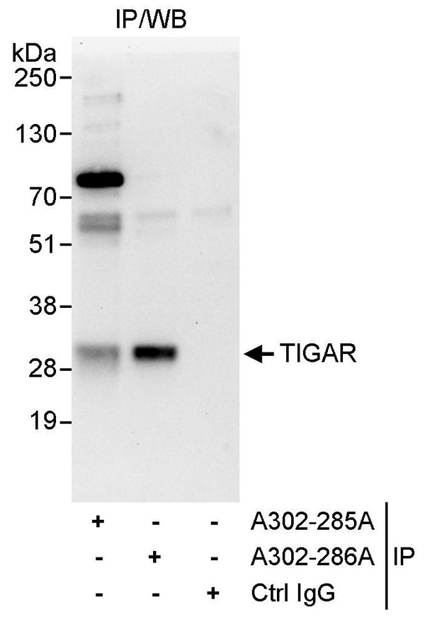 IP (Immunoprecipitation)
(Detection of human TIGAR by western blot of immunoprecipitates. Samples: Whole cell lysate (1 mg for IP, 20% of IP loaded) from HeLa cells. Antibodies: Affinity purified rabbit anti-TIGAR antibody AAA211834 used for IP at 10 ug/mg lysate. TIGAR was less efficiently immunoprecipitated by rabbit anti-TIGAR antibody which recognizes an upstream epitope. For blotting immunoprecipitated TIGAR, was used at 1 ug/ml. Detection: Chemiluminescence with an exposure time of 30 seconds.)
IP (Immunoprecipitation)
(Detection of human TIGAR by western blot of immunoprecipitates. Samples: Whole cell lysate (1 mg for IP, 20% of IP loaded) from HeLa cells. Antibodies: Affinity purified rabbit anti-TIGAR antibody AAA211834 used for IP at 10 ug/mg lysate. TIGAR was less efficiently immunoprecipitated by rabbit anti-TIGAR antibody which recognizes an upstream epitope. For blotting immunoprecipitated TIGAR, was used at 1 ug/ml. Detection: Chemiluminescence with an exposure time of 30 seconds.)
TIGAR, Polyclonal Antibody (Cat# AAA211834)
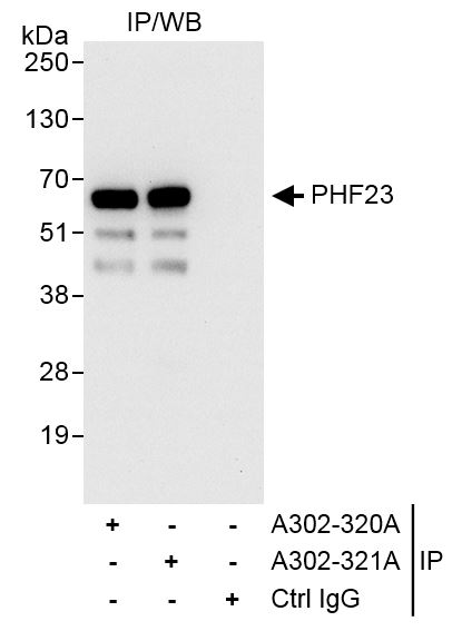
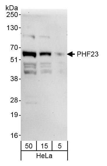 WB (Western Blot)
(Detection of human PHF23 by western blot. Samples: Whole cell lysate (5, 15, and 50 ug) from HeLa cells. Antibody: Affinity purified rabbit anti-PHF23 antibody AAA211845 (lot AAA211845-1) used at 0.1 ug/ml. Detection: Chemiluminescence with an exposure time of 30 seconds.)
WB (Western Blot)
(Detection of human PHF23 by western blot. Samples: Whole cell lysate (5, 15, and 50 ug) from HeLa cells. Antibody: Affinity purified rabbit anti-PHF23 antibody AAA211845 (lot AAA211845-1) used at 0.1 ug/ml. Detection: Chemiluminescence with an exposure time of 30 seconds.)
PHF23, Polyclonal Antibody (Cat# AAA211845)
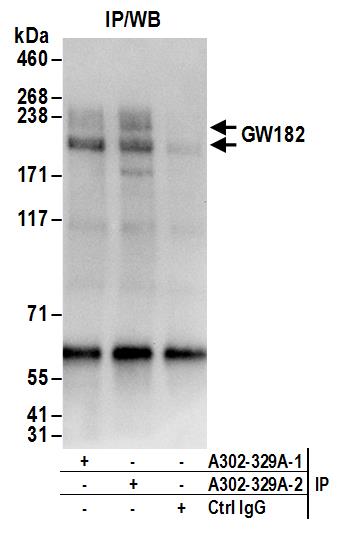
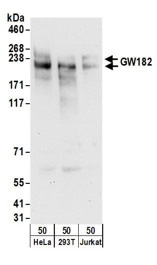 WB (Western Blot)
(Detection of human GW182 by western blot. Samples: Whole cell lysate (50 ug) from HeLa, HEK293T, and Jurkat cells. Antibodies: Affinity purified rabbit anti-GW182 antibody AAA211846 (lot AAA211846-2) used for WB at 0.1 ug/ml. Detection: Chemiluminescence with an exposure time of 30 seconds.)
WB (Western Blot)
(Detection of human GW182 by western blot. Samples: Whole cell lysate (50 ug) from HeLa, HEK293T, and Jurkat cells. Antibodies: Affinity purified rabbit anti-GW182 antibody AAA211846 (lot AAA211846-2) used for WB at 0.1 ug/ml. Detection: Chemiluminescence with an exposure time of 30 seconds.)
GW182, Polyclonal Antibody (Cat# AAA211846)
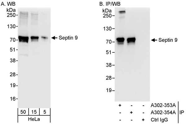 WB (Western Blot)
(Detection of human Septin 9 by western blot and immunoprecipitation. Samples: Whole cell lysate (5, 15 and 50 ug for WB; 1 mg for IP, 20% of IP loaded) from HeLa cells. Antibodies: Affinity purified rabbit anti-Septin 9 antibody AAA211850 used for WB at 0.04 ug/ml (A) and 0.4 ug/ml (B) and used for IP at 3 ug/mg lysate. Septin 9 was also immunoprecipitated by rabbit anti-Septin 9 antibody which recognizes a downstream epitope. Detection: Chemiluminescence with exposure times of 3 seconds (A) and 1 second (B).)
WB (Western Blot)
(Detection of human Septin 9 by western blot and immunoprecipitation. Samples: Whole cell lysate (5, 15 and 50 ug for WB; 1 mg for IP, 20% of IP loaded) from HeLa cells. Antibodies: Affinity purified rabbit anti-Septin 9 antibody AAA211850 used for WB at 0.04 ug/ml (A) and 0.4 ug/ml (B) and used for IP at 3 ug/mg lysate. Septin 9 was also immunoprecipitated by rabbit anti-Septin 9 antibody which recognizes a downstream epitope. Detection: Chemiluminescence with exposure times of 3 seconds (A) and 1 second (B).)
Septin 9, Polyclonal Antibody (Cat# AAA211850)
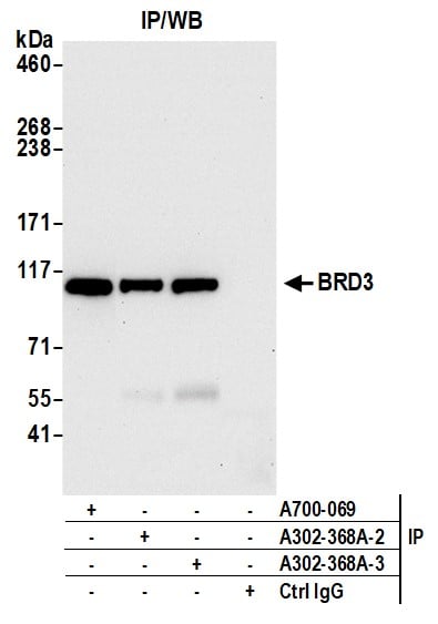
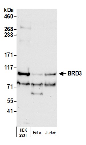 WB (Western Blot)
(Detection of human BRD3 by western blot. Samples: Whole cell lysate (50 ug) from HEK293T, HeLa, and Jurkat cells prepared using NETN lysis buffer. Antibody: Affinity purified rabbit anti-BRD3 antibody AAA211856 (lot AAA211856-3) used for WB at 0.04 ug/ml. Detection: Chemiluminescence with an exposure time of 30 seconds.)
WB (Western Blot)
(Detection of human BRD3 by western blot. Samples: Whole cell lysate (50 ug) from HEK293T, HeLa, and Jurkat cells prepared using NETN lysis buffer. Antibody: Affinity purified rabbit anti-BRD3 antibody AAA211856 (lot AAA211856-3) used for WB at 0.04 ug/ml. Detection: Chemiluminescence with an exposure time of 30 seconds.)
BRD3, Polyclonal Antibody (Cat# AAA211856)
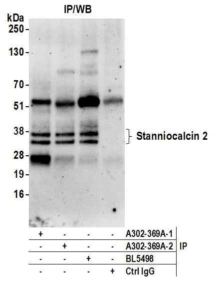
 WB (Western Blot)
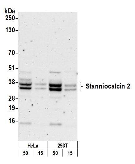
(Detection of human Stanniocalcin 2 by western blot. Samples: Whole cell lysate (15 and 50 ug) from HeLa and HEK293T cells prepared using NETN lysis buffer. Antibody: Affinity purified rabbit anti-Stanniocalcin 2 antibody AAA211857 (lot AAA211857-2) used for WB at 0.1 ug/ml. Detection: Chemiluminescence with an exposure time of 3 minutes.)
WB (Western Blot)
(Detection of human Stanniocalcin 2 by western blot. Samples: Whole cell lysate (15 and 50 ug) from HeLa and HEK293T cells prepared using NETN lysis buffer. Antibody: Affinity purified rabbit anti-Stanniocalcin 2 antibody AAA211857 (lot AAA211857-2) used for WB at 0.1 ug/ml. Detection: Chemiluminescence with an exposure time of 3 minutes.)
Stanniocalcin 2, Polyclonal Antibody (Cat# AAA211857)
 WB (Western Blot)
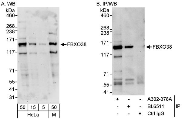
(Detection of human and mouse FBXO38 by western blot (h & m) and immunoprecipitation (h). Samples: Whole cell lysate from HeLa (5, 15 and 50 ug for WB; 1 mg for IP, 20% of IP loaded) and mouse NIH 3T3 (M; 50 ug) cells. Antibodies: Affinity purified rabbit anti-FBXO38 antibody AAA211860 used for WB at 0.1 ug/ml (A) and 1 ug/ml (B) and used for IP at 10 ug/mg lysate. FBXO38 was also immunoprecipitated by rabbit anti-FBXO38 antibody BL6511, which recognizes a downstream epitope. Detection: Chemiluminescence with exposure times of 3 minutes (A) and 30 seconds (B).)
WB (Western Blot)
(Detection of human and mouse FBXO38 by western blot (h & m) and immunoprecipitation (h). Samples: Whole cell lysate from HeLa (5, 15 and 50 ug for WB; 1 mg for IP, 20% of IP loaded) and mouse NIH 3T3 (M; 50 ug) cells. Antibodies: Affinity purified rabbit anti-FBXO38 antibody AAA211860 used for WB at 0.1 ug/ml (A) and 1 ug/ml (B) and used for IP at 10 ug/mg lysate. FBXO38 was also immunoprecipitated by rabbit anti-FBXO38 antibody BL6511, which recognizes a downstream epitope. Detection: Chemiluminescence with exposure times of 3 minutes (A) and 30 seconds (B).)
FBXO38, Polyclonal Antibody (Cat# AAA211860)
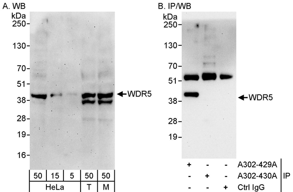
 WB (Western Blot)
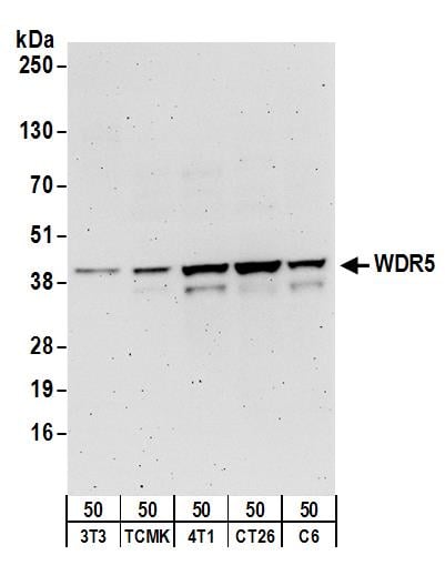
(Detection of mouse and rat WDR5 by western blot. Samples: Whole cell lysate (50 ug) from NIH 3T3, TCMK-1, 4T1, CT26.WT, and rat C6 cells. Antibodies: Affinity purified rabbit anti-WDR5 antibody AAA211872 (lot AAA211872-1) used for WB at 0.1 ug/ml. Detection: Chemiluminescence with an exposure time of 3 minutes.)
WB (Western Blot)
(Detection of mouse and rat WDR5 by western blot. Samples: Whole cell lysate (50 ug) from NIH 3T3, TCMK-1, 4T1, CT26.WT, and rat C6 cells. Antibodies: Affinity purified rabbit anti-WDR5 antibody AAA211872 (lot AAA211872-1) used for WB at 0.1 ug/ml. Detection: Chemiluminescence with an exposure time of 3 minutes.)
WDR5, Polyclonal Antibody (Cat# AAA211872)
 WB (Western Blot)
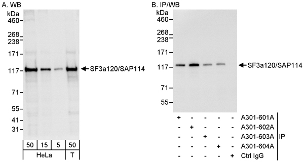
(Detection of human SF3a120/SAP114 by western blot and immunoprecipitation. Samples: Whole cell lysate from HeLa (5, 15 and 50 ug for WB; 1 mg for IP, 20% of IP loaded) and HEK293T (T; 50 ug) cells. Antibodies: Affinity purified rabbit anti-SF3a120/SAP114 antibody AAA211548 used for WB at 0.04 ug/ml (A) and 0.1 ug/ml (B) and used for IP at 3 ug/mg lysate. SF3a120/SAP114 was also immunoprecipitated by rabbit anti-SF3a120/SAP114 antibodies)
WB (Western Blot)
(Detection of human SF3a120/SAP114 by western blot and immunoprecipitation. Samples: Whole cell lysate from HeLa (5, 15 and 50 ug for WB; 1 mg for IP, 20% of IP loaded) and HEK293T (T; 50 ug) cells. Antibodies: Affinity purified rabbit anti-SF3a120/SAP114 antibody AAA211548 used for WB at 0.04 ug/ml (A) and 0.1 ug/ml (B) and used for IP at 3 ug/mg lysate. SF3a120/SAP114 was also immunoprecipitated by rabbit anti-SF3a120/SAP114 antibodies)
SF3a120/SAP114, Polyclonal Antibody (Cat# AAA211548)
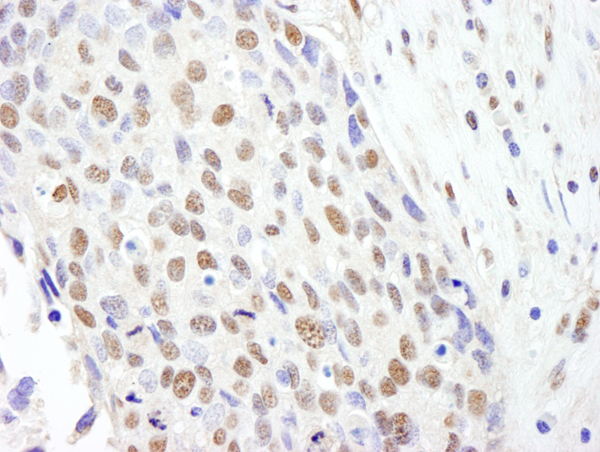
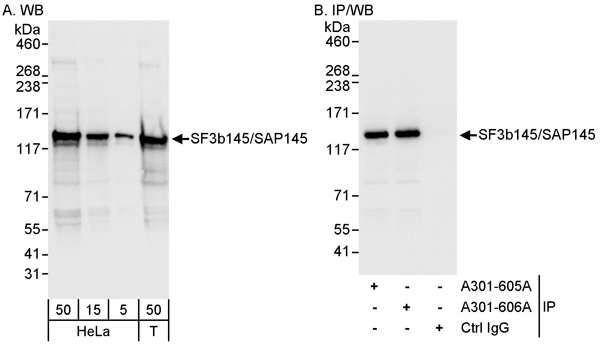 WB (Western Blot)
(Detection of human SF3b145/SAP145 by western blot and immunoprecipitation. Samples: Whole cell lysate from HeLa (5, 15 and 50 ug for WB; 1 mg for IP, 20% of IP loaded) and HEK293T (T; 50 ug) cells. Antibodies: Affinity purified rabbit anti-SF3b145/SAP145 antibody AAA211549 used for WB at 0.04 ug/ml (A) and 0.1 ug/ml (B) and used for IP at 3 ug/mg lysate. SF3b145/SAP145 was also immunoprecipitated by rabbit anti-SF3b145/SAP145 antibody which recognizes a downstream epitope. Detection: Chemiluminescence with exposure times of 1 second (A and B).)
WB (Western Blot)
(Detection of human SF3b145/SAP145 by western blot and immunoprecipitation. Samples: Whole cell lysate from HeLa (5, 15 and 50 ug for WB; 1 mg for IP, 20% of IP loaded) and HEK293T (T; 50 ug) cells. Antibodies: Affinity purified rabbit anti-SF3b145/SAP145 antibody AAA211549 used for WB at 0.04 ug/ml (A) and 0.1 ug/ml (B) and used for IP at 3 ug/mg lysate. SF3b145/SAP145 was also immunoprecipitated by rabbit anti-SF3b145/SAP145 antibody which recognizes a downstream epitope. Detection: Chemiluminescence with exposure times of 1 second (A and B).)
SF3b145/SAP145, Polyclonal Antibody (Cat# AAA211549)
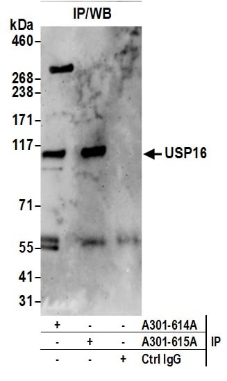
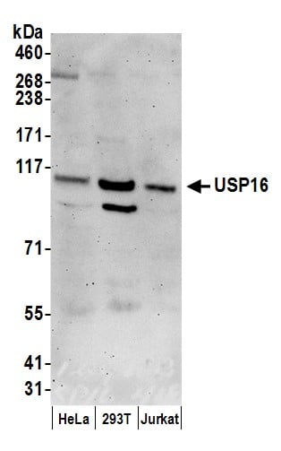 WB (Western Blot)
(Detection of human USP16 by western blot. Samples: Whole cell lysate (50 ug) from HeLa, HEK293T, and Jurkat cells prepared using NETN lysis buffer. Antibodies: Affinity purified rabbit anti-USP16 antibody AAA211554 (lot AAA211554-2) used for WB at 0.1 ug/ml. Detection: Chemiluminescence with an exposure time of 3 minutes.)
WB (Western Blot)
(Detection of human USP16 by western blot. Samples: Whole cell lysate (50 ug) from HeLa, HEK293T, and Jurkat cells prepared using NETN lysis buffer. Antibodies: Affinity purified rabbit anti-USP16 antibody AAA211554 (lot AAA211554-2) used for WB at 0.1 ug/ml. Detection: Chemiluminescence with an exposure time of 3 minutes.)
USP16, Polyclonal Antibody (Cat# AAA211554)
 WB (Western Blot)
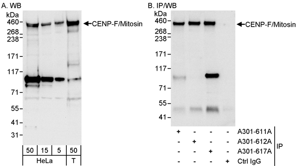
(Detection of human CENP-F/Mitosin by western blot and immunoprecipitation. Samples: Whole cell lysate from HeLa (5, 15 and 50 ug for WB; 1 mg for IP, 20% of IP loaded) and HEK293T (T; 50 ug) cells. Antibodies: Affinity purified rabbit anti-CENP-F/Mitosin antibody AAA211555 used for WB at 0.04 ug/ml (A) and 1 ug/ml (B) and used for IP at 3 ug/mg lysate. CENP-F/Mitosin was also immunoprecipitated by rabbit anti-CENP-F/Mitosin antibodies and which recognize other epitopes. Detection: Chemiluminescence with exposure times of 10 seconds (A) and 3 seconds (B).)
WB (Western Blot)
(Detection of human CENP-F/Mitosin by western blot and immunoprecipitation. Samples: Whole cell lysate from HeLa (5, 15 and 50 ug for WB; 1 mg for IP, 20% of IP loaded) and HEK293T (T; 50 ug) cells. Antibodies: Affinity purified rabbit anti-CENP-F/Mitosin antibody AAA211555 used for WB at 0.04 ug/ml (A) and 1 ug/ml (B) and used for IP at 3 ug/mg lysate. CENP-F/Mitosin was also immunoprecipitated by rabbit anti-CENP-F/Mitosin antibodies and which recognize other epitopes. Detection: Chemiluminescence with exposure times of 10 seconds (A) and 3 seconds (B).)
CENP-F/Mitosin, Polyclonal Antibody (Cat# AAA211555)
 WB (Western Blot)
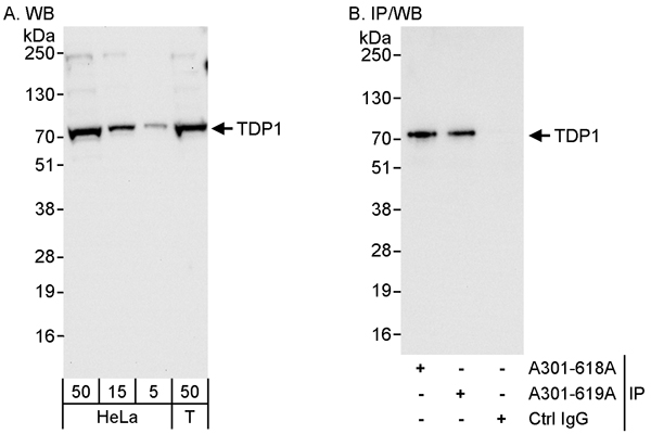
(Detection of human TDP1 by western blot and immunoprecipitation. Samples: Whole cell lysate from HeLa (5, 15 and 50 ug for WB; 1 mg for IP, 20% of IP loaded) and HEK293T (T; 50 ug) cells. Antibodies: Affinity purified rabbit anti-TDP1 antibody AAA211556 used for WB at 0.04 ug/ml (A) and 0.4 ug/ml (B) and used for IP at 3 ug/mg lysate. TDP1 was also immunoprecipitated by rabbit anti-TDP1 antibody which recognizes a downstream epitope. Detection: Chemiluminescence with exposure times of 10 seconds (A and B).)
WB (Western Blot)
(Detection of human TDP1 by western blot and immunoprecipitation. Samples: Whole cell lysate from HeLa (5, 15 and 50 ug for WB; 1 mg for IP, 20% of IP loaded) and HEK293T (T; 50 ug) cells. Antibodies: Affinity purified rabbit anti-TDP1 antibody AAA211556 used for WB at 0.04 ug/ml (A) and 0.4 ug/ml (B) and used for IP at 3 ug/mg lysate. TDP1 was also immunoprecipitated by rabbit anti-TDP1 antibody which recognizes a downstream epitope. Detection: Chemiluminescence with exposure times of 10 seconds (A and B).)
TDP1, Polyclonal Antibody (Cat# AAA211556)
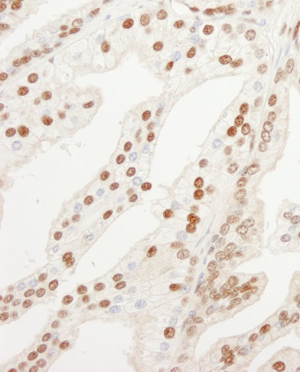
 WB (Western Blot)
(Detection of human KPNA4 by western blot and immunoprecipitation. Samples: Whole cell lysate from HeLa (5, 15 and 50 ug for WB; 1 mg for IP, 20% of IP loaded) and HEK293T (T; 50 ug) cells. Antibodies: Affinity purified rabbit anti-KPNA4 antibody AAA211558 used for WB at 0.04 ug/ml (A) and 0.4 ug/ml (B) and used for IP at 3 ug/mg lysate. KPNA4 was also immunoprecipitated by rabbit anti-KPNA4 antibody BL6207, which recognizes an upstream epitope. Detection: Chemiluminescence with exposure times of 3 seconds (A) and 0.5 seconds (B).)
WB (Western Blot)
(Detection of human KPNA4 by western blot and immunoprecipitation. Samples: Whole cell lysate from HeLa (5, 15 and 50 ug for WB; 1 mg for IP, 20% of IP loaded) and HEK293T (T; 50 ug) cells. Antibodies: Affinity purified rabbit anti-KPNA4 antibody AAA211558 used for WB at 0.04 ug/ml (A) and 0.4 ug/ml (B) and used for IP at 3 ug/mg lysate. KPNA4 was also immunoprecipitated by rabbit anti-KPNA4 antibody BL6207, which recognizes an upstream epitope. Detection: Chemiluminescence with exposure times of 3 seconds (A) and 0.5 seconds (B).)
KPNA4, Polyclonal Antibody (Cat# AAA211558)
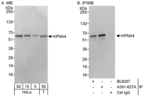
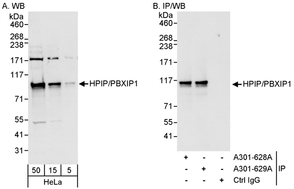 WB (Western Blot)
(Detection of human HPIP/PBXIP1 by western blot and immunoprecipitation. Samples: Whole cell lysate (5, 15 and 50 ug for WB; 1 mg for IP, 20% of IP loaded) from HeLa cells. Antibodies: Affinity purified rabbit anti-HPIP/PBXIP1 antibody AAA211559 used for WB at 0.04 ug/ml (A) and 0.1 ug/ml (B) and used for IP at 3 ug/mg lysate. HPIP/PBXIP1 was also immunoprecipitated by rabbit anti-HPIP/PBXIP1 antibody which recognizes a downstream epitope. Detection: Chemiluminescence with exposure times of 10 seconds (A and B).)
WB (Western Blot)
(Detection of human HPIP/PBXIP1 by western blot and immunoprecipitation. Samples: Whole cell lysate (5, 15 and 50 ug for WB; 1 mg for IP, 20% of IP loaded) from HeLa cells. Antibodies: Affinity purified rabbit anti-HPIP/PBXIP1 antibody AAA211559 used for WB at 0.04 ug/ml (A) and 0.1 ug/ml (B) and used for IP at 3 ug/mg lysate. HPIP/PBXIP1 was also immunoprecipitated by rabbit anti-HPIP/PBXIP1 antibody which recognizes a downstream epitope. Detection: Chemiluminescence with exposure times of 10 seconds (A and B).)
HPIP, Polyclonal Antibody (Cat# AAA211559)
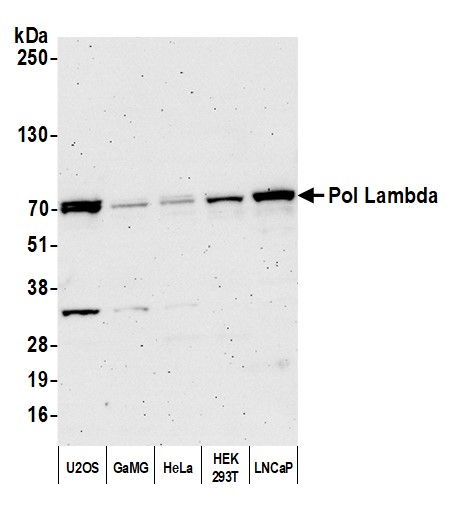 WB (Western Blot)
(Detection of human Pol Lambda by western blot. Samples: Whole cell lysate (50 ug) from HeLa, 293T, and Jurkat cells prepared using NETN lysis buffer. Antibody: Affinity purified rabbit anti-Pol Lambda antibody (AAA211564) used for WB at 0.1 ug/ml. Detection: Chemiluminescence with an exposure time of 3 minutes.)
WB (Western Blot)
(Detection of human Pol Lambda by western blot. Samples: Whole cell lysate (50 ug) from HeLa, 293T, and Jurkat cells prepared using NETN lysis buffer. Antibody: Affinity purified rabbit anti-Pol Lambda antibody (AAA211564) used for WB at 0.1 ug/ml. Detection: Chemiluminescence with an exposure time of 3 minutes.)
Pol Lambda, Polyclonal Antibody (Cat# AAA211564)
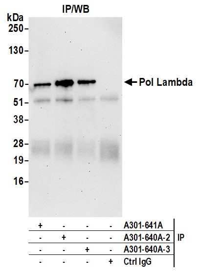
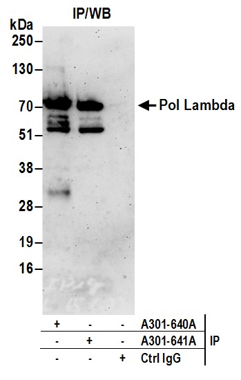 IP (Immunoprecipitation)
(Detection of human Pol Lambda by western blot of immunoprecipitates. Samples: Whole cell lysate (0.5 or 1.0 mg per IP reaction; 20% of IP loaded) from HeLa cells prepared using NETN lysis buffer. Antibodies: Affinity purified rabbit anti-Pol Lambda antibody AAA211565 (lot AAA211565-2) used for IP at 6 ug per reaction. Pol Lambda was also immunoprecipitated by rabbit anti-Pol Lambda antibody For blotting immunoprecipitated Pol Lambda, was used at 1 ug/ml. Detection: Chemiluminescence with an exposure time of 3 minutes.)
IP (Immunoprecipitation)
(Detection of human Pol Lambda by western blot of immunoprecipitates. Samples: Whole cell lysate (0.5 or 1.0 mg per IP reaction; 20% of IP loaded) from HeLa cells prepared using NETN lysis buffer. Antibodies: Affinity purified rabbit anti-Pol Lambda antibody AAA211565 (lot AAA211565-2) used for IP at 6 ug per reaction. Pol Lambda was also immunoprecipitated by rabbit anti-Pol Lambda antibody For blotting immunoprecipitated Pol Lambda, was used at 1 ug/ml. Detection: Chemiluminescence with an exposure time of 3 minutes.)
Pol Lambda, Polyclonal Antibody (Cat# AAA211565)
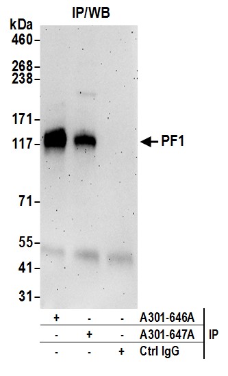
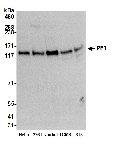 WB (Western Blot)
(Detection of human and mouse PF1 by western blot. Samples: Whole cell lysate (50 ug) from HeLa, HEK293T, Jurkat, mouse TCMK-1, and mouse NIH 3T3 cells prepared using NETN lysis buffer. Antibody: Affinity purified rabbit anti-PF1 antibody AAA211567 (lot AAA211567-2) used for WB at 0.1 ug/ml. Detection: Chemiluminescence with an exposure time of 3 minutes.)
WB (Western Blot)
(Detection of human and mouse PF1 by western blot. Samples: Whole cell lysate (50 ug) from HeLa, HEK293T, Jurkat, mouse TCMK-1, and mouse NIH 3T3 cells prepared using NETN lysis buffer. Antibody: Affinity purified rabbit anti-PF1 antibody AAA211567 (lot AAA211567-2) used for WB at 0.1 ug/ml. Detection: Chemiluminescence with an exposure time of 3 minutes.)
PF1, Polyclonal Antibody (Cat# AAA211567)
 WB (Western Blot)
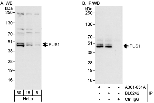
(Detection of human PUS1 by western blot and immunoprecipitation. Samples: Whole cell lysate (5, 15 and 50 ug for WB; 1 mg for IP, 20% of IP loaded) from HeLa cells. Antibodies: Affinity purified rabbit anti-PUS1 antibody AAA211569 used for WB at 0.04 ug/ml (A) and 1 ug/ml (B) and used for IP at 3 ug/mg lysate. PUS1 was also immunoprecipitated by rabbit anti-PUS1 antibody BL6242, which recognizes an upstream epitope. Detection: Chemiluminescence with exposure times of 10 seconds (A and B).)
WB (Western Blot)
(Detection of human PUS1 by western blot and immunoprecipitation. Samples: Whole cell lysate (5, 15 and 50 ug for WB; 1 mg for IP, 20% of IP loaded) from HeLa cells. Antibodies: Affinity purified rabbit anti-PUS1 antibody AAA211569 used for WB at 0.04 ug/ml (A) and 1 ug/ml (B) and used for IP at 3 ug/mg lysate. PUS1 was also immunoprecipitated by rabbit anti-PUS1 antibody BL6242, which recognizes an upstream epitope. Detection: Chemiluminescence with exposure times of 10 seconds (A and B).)
PUS1, Polyclonal Antibody (Cat# AAA211569)
 WB (Western Blot)
(Detection of human CEP290 by western blot and immunoprecipitation. Samples: Whole cell lysate from HeLa (5, 15 and 50 ug for WB; 1 mg for IP, 20% of IP loaded) and HEK293T (T; 50 ug) cells. Antibodies: Affinity purified rabbit anti-CEP290 antibody AAA211575 used for WB at 0.1 ug/ml (A) and 1 ug/ml (B) and used for IP at 3 ug/mg lysate. CEP290 was also immunoprecipitated by rabbit anti-CEP290 antibody BL6289, which recognizes an upstream epitope. Detection: Chemiluminescence with exposure times of 30 seconds (A) and 10 second (B).)
WB (Western Blot)
(Detection of human CEP290 by western blot and immunoprecipitation. Samples: Whole cell lysate from HeLa (5, 15 and 50 ug for WB; 1 mg for IP, 20% of IP loaded) and HEK293T (T; 50 ug) cells. Antibodies: Affinity purified rabbit anti-CEP290 antibody AAA211575 used for WB at 0.1 ug/ml (A) and 1 ug/ml (B) and used for IP at 3 ug/mg lysate. CEP290 was also immunoprecipitated by rabbit anti-CEP290 antibody BL6289, which recognizes an upstream epitope. Detection: Chemiluminescence with exposure times of 30 seconds (A) and 10 second (B).)
CEP290, Polyclonal Antibody (Cat# AAA211575)
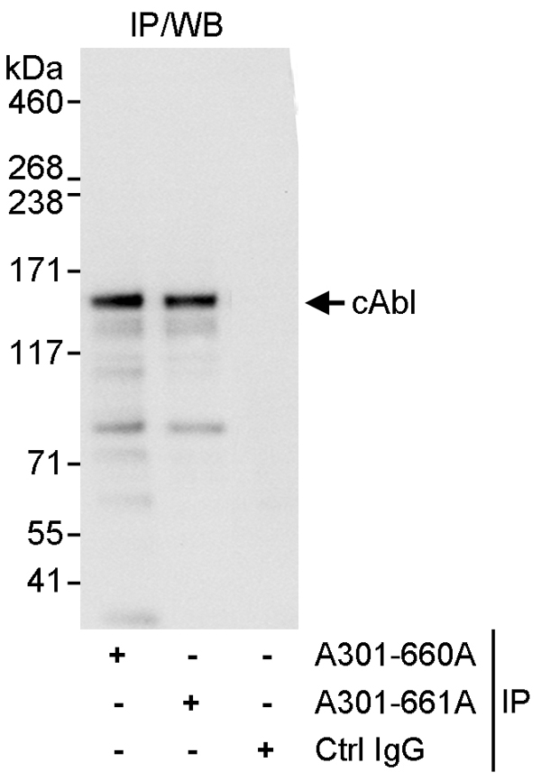 IP (Immunoprecipitation)
(Detection of human cAbl by western blot of immunoprecipitates. Samples: Whole cell lysate (1 mg for IP, 20% of IP loaded) from HeLa cells. Antibodies: Affinity purified rabbit anti-cAbl antibody AAA211577 used for IP at 3 ug/mg lysate. cAbl was also immunoprecipitated by rabbit anti-cAbl antibody which recognizes an upstream epitope. For blotting immunoprecipitated cAbl, was used at 1 ug/ml. Detection: Chemiluminescence with an exposure time of 3 second.)
IP (Immunoprecipitation)
(Detection of human cAbl by western blot of immunoprecipitates. Samples: Whole cell lysate (1 mg for IP, 20% of IP loaded) from HeLa cells. Antibodies: Affinity purified rabbit anti-cAbl antibody AAA211577 used for IP at 3 ug/mg lysate. cAbl was also immunoprecipitated by rabbit anti-cAbl antibody which recognizes an upstream epitope. For blotting immunoprecipitated cAbl, was used at 1 ug/ml. Detection: Chemiluminescence with an exposure time of 3 second.)
cAbl, Polyclonal Antibody (Cat# AAA211577)
What are Polyclonal Antibodies?
Polyclonal antibodies are antibodies that come from multiple B cell clones of a host animal. The typical hosts used for the majority of polyclonal antibody production are rabbits, goats, sheep, and donkeys. These polyclonal antibodies, once having identified their target, will bind to different epitopes located at different regions or sequences on the same protein/antigen. As a result, they are ideal at locating and binding to the target, even if the target is in very low concentrations (due to many different antibodies being able to bind to the same target molecule, which allows for significant amplification of a downstream signal).
Polyclonal antibodies are typically produced by injecting an antigen into a host animal, which causes the animal’s immune system to attack the foreign antigen by mass generating antibodies against it. After a period of time, serum is collected from the animal and purified using physicochemical fractionation, class-specific affinity purification, and/or antigen-affinity purification.
Key Uses of Polyclonal Antibodies
- Western Blotting: This method is used to find specific proteins in biological samples after separating them by size.
- Immunohistochemistry: IHC helps visualize the location of proteins in tissue sections using various staining techniques.
- ELISA: (Enzyme-Linked Immunosorbent Assay) is typically used to identify specific protein quantities in a sample. ELISAs can be either “Quantitative” or “Qualitative”.
- Flow Cytometry: technique that identifies and measures the specific protein on the surface or inside the cells in a fluid suspension.
- Immunoprecipitation: IP isolates and studies a specific protein from a complex mixture using antibodies.
Why Buy Polyclonal Antibodies from AAA Biotech?
1. Ideal for Various Applications
Our antibodies are generally going to be validated for use in multiple types of assays, including ELISA, Western Blotting, Immunohistochemistry, Immunoprecipitation, amongst others. They are ideal for a wide range of research applications.
2. Rigorous Quality Control
All of the antibodies in our catalog undergo strict quality testing to ensure specificity, sensitivity, and consistent performance. We are confident in the ability of our antibodies to provide you with accurate results.
3. Wide Assortment of Antibodies
Antibodies in are catalog can be found for both common and exotic species, and these antibodies are also available in both conjugated and recombinant forms to suit many diverse experimental needs.
4. Highly Purified
Our antibodies are available in purified forms with over 85% purity, as confirmed by SDS-PAGE. They are also available with tags such as His, Flag, GST, or MBP. We cater to customers worldwide.
FAQ
1. How are polyclonal antibodies produced?
Traditionally, polyclonal antibodies are produced by injecting an antigen into a host animal (such as a rabbit or goat), which then triggers an immune response from the host animal. The animal’s B cells produce antibodies that will recognize different parts of the injected antigen. These antibodies are then collected from the animal’s blood and purified for use.
2. How do polyclonal antibodies differ from monoclonal antibodies?
Polyclonal antibodies are a mix of antibodies that bind to different locations (epitopes) of the same antigen, while monoclonal antibodies are identical and bind to just one specific epitope. This makes polyclonal antibodies more versatile and better at detecting proteins that may be present in low quantities or in altered/modified forms.
3. How should I store polyclonal antibodies?
Polyclonal antibodies should be stored at 4°C for short-term use (up to a few weeks) and at -20°C or -80°C for long-term storage. Avoid repeated freeze-thaw cycles by dividing them into small aliquots. Always check the datasheet for specific storage instructions.