Filters
▼Clonality
▼Type
▼Reactivity
▼Gene Name
▼Isotype
▼Host
▼Application
▼Clone
▼Monoclonal Antibodies
Get accurate results in your research with our Monoclonal Antibodies, which are specially made to target exactly what you require for your research, and will produce consistent, reliable performance in lab tests.
Viewing 1050-1100 of 27560 product results
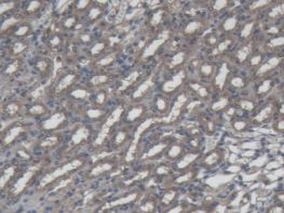
 IHC (Immunohiostchemistry)
(DAB staining on IHC-P; Samples: Rat Kidney Tissue))
IHC (Immunohiostchemistry)
(DAB staining on IHC-P; Samples: Rat Kidney Tissue))
Interleukin 1 Beta, Monoclonal Antibody (Cat# AAA141328)
 WB (Western Blot)
(Western Blot;Sample: Human Serum;Primary Ab: 2ug/ml Mouse Anti-Human IgG1 Antibody;Second Ab: 0.2ug/mL HRP-Linked Caprine Anti-Mouse IgG Polyclonal Antibody (Catalog: ))
WB (Western Blot)
(Western Blot;Sample: Human Serum;Primary Ab: 2ug/ml Mouse Anti-Human IgG1 Antibody;Second Ab: 0.2ug/mL HRP-Linked Caprine Anti-Mouse IgG Polyclonal Antibody (Catalog: ))
Immunoglobulin G1, Monoclonal Antibody (Cat# AAA141349)
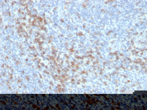 IHC (Immunohistochemistry)
(Formalin-fixed, paraffin-embedded human tonsil stained with CD5 Recombinant Rabbit Monoclonal Antibody (C5/6438R).)
IHC (Immunohistochemistry)
(Formalin-fixed, paraffin-embedded human tonsil stained with CD5 Recombinant Rabbit Monoclonal Antibody (C5/6438R).)
CD5, Monoclonal Antibody (Cat# AAA216039)
 IHC (Immunohistochemistry)
(Formalin-fixed, paraffin-embedded human tonsil stained with CD8 Recombinant Mouse Monoclonal Antibody (rCD8/6590).)
IHC (Immunohistochemistry)
(Formalin-fixed, paraffin-embedded human tonsil stained with CD8 Recombinant Mouse Monoclonal Antibody (rCD8/6590).)
CCD8A, Monoclonal Antibody (Cat# AAA216044)
 IHC (Immunohistochemistry)
(Formalin-fixed, paraffin-embedded human tonsil stained with CD14 Rabbit Recombinant Monoclonal Antibody (LPSR/4180R).)
IHC (Immunohistochemistry)
(Formalin-fixed, paraffin-embedded human tonsil stained with CD14 Rabbit Recombinant Monoclonal Antibody (LPSR/4180R).)
CD14, Monoclonal Antibody (Cat# AAA216046)
 IHC (Immunohistochemistry)
(Formalin-fixed, paraffin-embedded human lymph node stained with CD19 Recombinant Mouse Monoclonal Antibody (rCD19/4591).)
IHC (Immunohistochemistry)
(Formalin-fixed, paraffin-embedded human lymph node stained with CD19 Recombinant Mouse Monoclonal Antibody (rCD19/4591).)
CD19, Monoclonal Antibody (Cat# AAA216047)
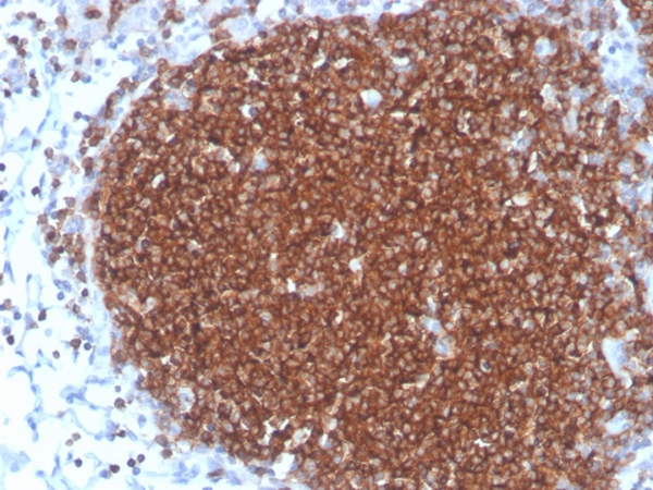
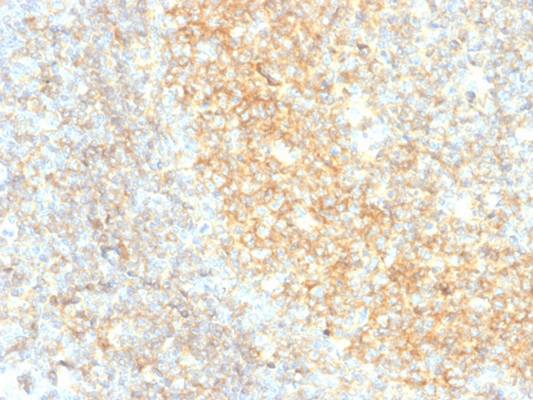
 IHC (Immunohistochemistry)
(Formalin-fixed, paraffin-embedded human tonsil stained with CD20 Mouse Monoclonal Antibody (MS4A1/4655).)
IHC (Immunohistochemistry)
(Formalin-fixed, paraffin-embedded human tonsil stained with CD20 Mouse Monoclonal Antibody (MS4A1/4655).)
CD20/MS4A1, Monoclonal Antibody (Cat# AAA216048)
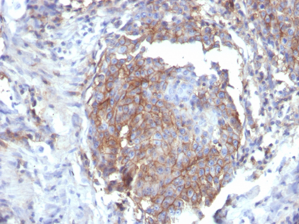
 IHC (Immunohistochemistry)
(Formalin-fixed, paraffin-embedded human tonsil stained with CD27 Mouse Monoclonal Antibody (LPFS2/4177).)
IHC (Immunohistochemistry)
(Formalin-fixed, paraffin-embedded human tonsil stained with CD27 Mouse Monoclonal Antibody (LPFS2/4177).)
CD27, Monoclonal Antibody (Cat# AAA216057)
 IHC (Immunohistochemistry)
(Formalin-fixed, paraffin-embedded human lung carcinoma stained with CD86-Monospecific Mouse Monoclonal Antibody (C86/3711).)
IHC (Immunohistochemistry)
(Formalin-fixed, paraffin-embedded human lung carcinoma stained with CD86-Monospecific Mouse Monoclonal Antibody (C86/3711).)
CD86, Monoclonal Antibody (Cat# AAA216061)
 IHC (Immunohistochemistry)
(Formalin-fixed, paraffin-embedded human tonsil stained with CD40 Recombinant Rabbit Monoclonal Antibody (C40/4826R).)
IHC (Immunohistochemistry)
(Formalin-fixed, paraffin-embedded human tonsil stained with CD40 Recombinant Rabbit Monoclonal Antibody (C40/4826R).)
CD40/TNFRSF5/CD40L-Receptor, Monoclonal Antibody (Cat# AAA216068)
 IHC (Immunohistochemistry)
(Formalin-fixed, paraffin-embedded human breast stained with CD44 Recombinant Mouse Monoclonal Antibody (rHCAM/6449).)
IHC (Immunohistochemistry)
(Formalin-fixed, paraffin-embedded human breast stained with CD44 Recombinant Mouse Monoclonal Antibody (rHCAM/6449).)
CD44/HCAM Std., Monoclonal Antibody (Cat# AAA216069)
 IHC (Immunohistochemistry)
(Formalin-fixed, paraffin-embedded human esophagus stained with CD44 Recombinant Rabbit Monoclonal Antibody (HCAM/6459R).)
IHC (Immunohistochemistry)
(Formalin-fixed, paraffin-embedded human esophagus stained with CD44 Recombinant Rabbit Monoclonal Antibody (HCAM/6459R).)
CD44/HCAM Std., Monoclonal Antibody (Cat# AAA216071)
 IHC (Immunohistochemistry)
(Formalin-fixed, paraffin-embedded human kidney stained with E-Cadherin Mouse Monoclonal Antibody (CDH1/4585).)
IHC (Immunohistochemistry)
(Formalin-fixed, paraffin-embedded human kidney stained with E-Cadherin Mouse Monoclonal Antibody (CDH1/4585).)
E-Cadherin(CDH1)/CD324, Monoclonal Antibody (Cat# AAA216082)
 SDS-PAGE
(SDS-PAGE Analysis Purified Penicillin Mouse Monoclonal Antibody (Pen-9) Confirmation of Integrity and Purity of Antibody.)
SDS-PAGE
(SDS-PAGE Analysis Purified Penicillin Mouse Monoclonal Antibody (Pen-9) Confirmation of Integrity and Purity of Antibody.)
Penicillin, Monoclonal Antibody (Cat# AAA216084)
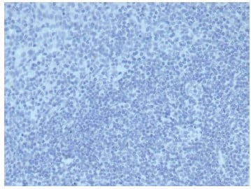
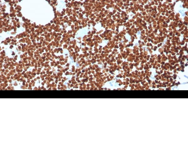 IHC (Immunohistochemistry)
(Formalin-fixed, paraffin-embedded human lymph node stained with dsDNA Recombinant Mouse Monoclonal Antibody (rDSD/4565).)
IHC (Immunohistochemistry)
(Formalin-fixed, paraffin-embedded human lymph node stained with dsDNA Recombinant Mouse Monoclonal Antibody (rDSD/4565).)
Double Stranded DNA (dsDNA), Monoclonal Antibody (Cat# AAA216093)
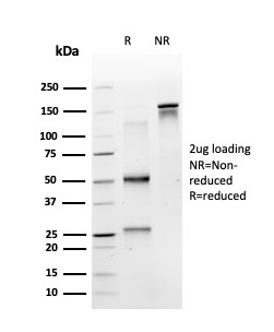
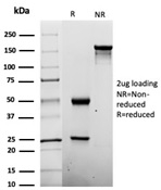
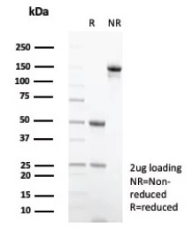 SDS-PAGE
(SDS-PAGE Analysis of Purified Negative Control for Mouse Monoclonal Abs(IGG2a/6723). Confirmation of Purity and Integrity of Antibody.)
SDS-PAGE
(SDS-PAGE Analysis of Purified Negative Control for Mouse Monoclonal Abs(IGG2a/6723). Confirmation of Purity and Integrity of Antibody.)
IgG2a, Monoclonal Isotype Control (Cat# AAA216099)

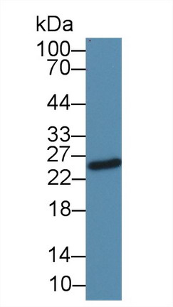
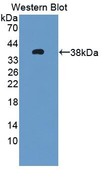
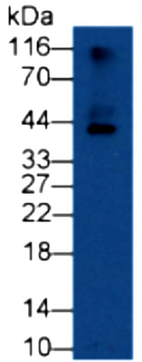
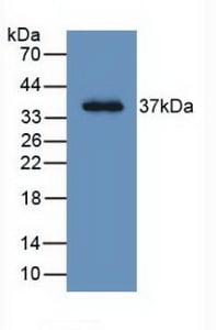 WB (Western Blot)
(Western Blot: Sample: Recombinant LRG1, Human.)
WB (Western Blot)
(Western Blot: Sample: Recombinant LRG1, Human.)
Leucine Rich Alpha-2-Glycoprotein 1 (LRG1), Monoclonal Antibody (Cat# AAA147918)
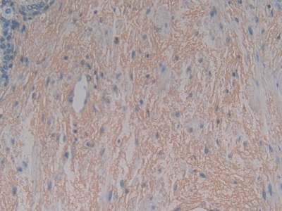
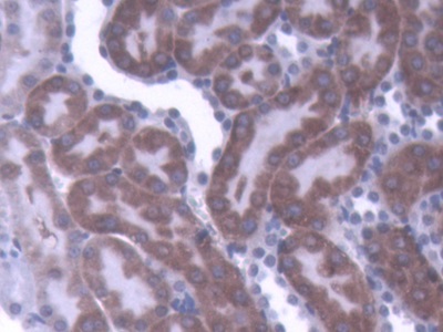
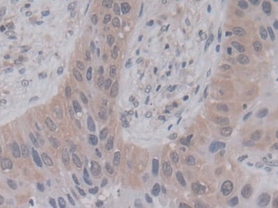
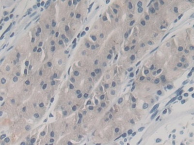
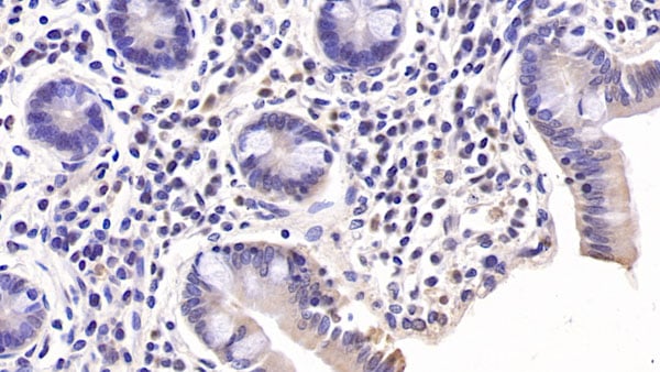
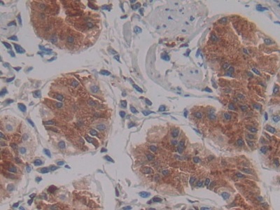 IHC (Immunohiostchemistry)
(DAB staining on IHC-P; Samples: Human Stomach Tissue)
IHC (Immunohiostchemistry)
(DAB staining on IHC-P; Samples: Human Stomach Tissue)
Left/Right Determination Factor 1 (LEFTY1), Monoclonal Antibody (Cat# AAA147923)
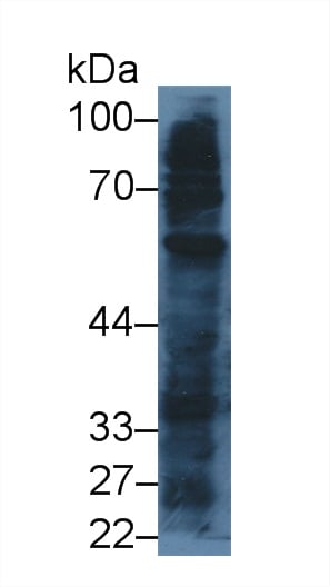
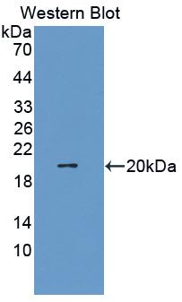
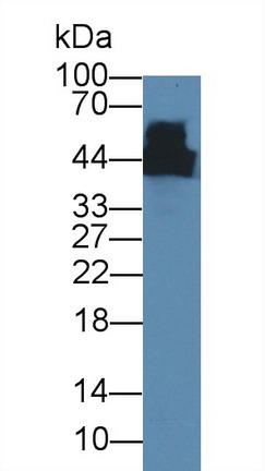
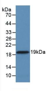
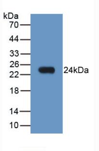
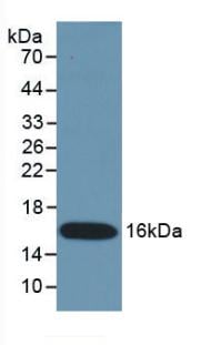
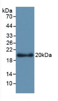
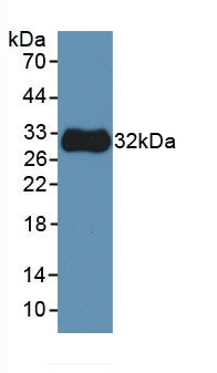
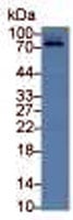
 WB (Western Blot)
(Western Blot: Sample: Recombinant protein.)
WB (Western Blot)
(Western Blot: Sample: Recombinant protein.)
Toll Like Receptor 4 (TLR4), Monoclonal Antibody (Cat# AAA147925)
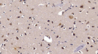
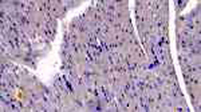
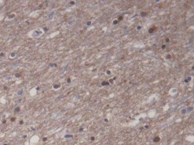 IHC (Immunohiostchemistry)
(DAB staining on IHC-P; Samples: Human Cerebrum Tissue))
IHC (Immunohiostchemistry)
(DAB staining on IHC-P; Samples: Human Cerebrum Tissue))
Creatine Kinase, Muscle (CKM), Monoclonal Antibody (Cat# AAA147926)
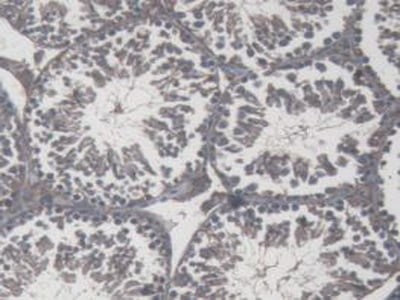
 IHC (Immunohistochemistry)
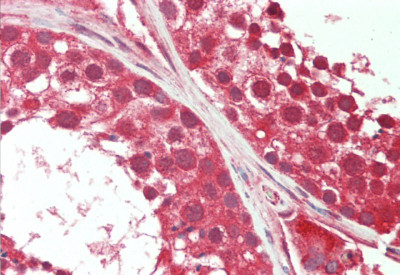
(Vector Red staining on IHC-PSamples: Human Testis Tissue;Primary Ab: 10ug/ml Mouse Anti-Human DHEA Antibody Second Ab: 2ug/mL HRP-Linked Caprine Anti-Mouse IgG Monoclonal Antibody)
IHC (Immunohistochemistry)
(Vector Red staining on IHC-PSamples: Human Testis Tissue;Primary Ab: 10ug/ml Mouse Anti-Human DHEA Antibody Second Ab: 2ug/mL HRP-Linked Caprine Anti-Mouse IgG Monoclonal Antibody)
Dehydroepiandrosterone (DHEA), Monoclonal Antibody (Cat# AAA147929)
 IHC (Immunohistochemisry)
(DAB staining on IHC-P; Samples: Human Lung cancer Tissue))
IHC (Immunohistochemisry)
(DAB staining on IHC-P; Samples: Human Lung cancer Tissue))
Cyclophilin A (CYPA), Monoclonal Antibody (Cat# AAA147930)
 IHC (Immunohiostchemistry)
(DAB staining on IHC-P; Samples: Human Stomach Tissue))
IHC (Immunohiostchemistry)
(DAB staining on IHC-P; Samples: Human Stomach Tissue))
Neutrophil Gelatinase Associated Lipocalin (NGAL), Monoclonal Antibody (Cat# AAA147939)
 IHC (Immunohistochemistry)
(DAB staining on IHC-P; Samples: Mouse Testis Tissue.)
IHC (Immunohistochemistry)
(DAB staining on IHC-P; Samples: Mouse Testis Tissue.)
Interferon Gamma (IFNg), Monoclonal Antibody (Cat# AAA147950)
Connective Tissue Growth Factor (CTGF), Monoclonal Antibody (Cat# AAA147953)
 IHC (Immunohistochemisry)
(DAB staining on IHC-P; Samples: Human Cerebrum Tissue; Primary Ab: 10ug/ml Mouse Anti-Human APOL2 Antibody Second Ab: 2ug/mL HRP Linked Caprine Anti-Mouse IgG Polyclonal Antibody .)
IHC (Immunohistochemisry)
(DAB staining on IHC-P; Samples: Human Cerebrum Tissue; Primary Ab: 10ug/ml Mouse Anti-Human APOL2 Antibody Second Ab: 2ug/mL HRP Linked Caprine Anti-Mouse IgG Polyclonal Antibody .)
Apolipoprotein L2 (APOL2), Monoclonal Antibody (Cat# AAA147963)
Immunoglobulin G (IgG), Monoclonal Antibody (Cat# AAA147965)
FMS Like Tyrosine Kinase 3 Ligand (Flt3L), Monoclonal Antibody (Cat# AAA147977)
Interferon Gamma (IFNg), Monoclonal Antibody (Cat# AAA147978)
Matrix Metalloproteinase 3 (MMP3), Monoclonal Antibody (Cat# AAA147984)
Transforming Growth Factor Alpha (TGFa), Monoclonal Antibody (Cat# AAA147985)
Procollagen III N-Terminal Propeptide (PIIINP), Monoclonal Antibody (Cat# AAA147993)
Kidney Injury Molecule 1 (Kim1), Monoclonal Antibody (Cat# AAA147999)
Urocortin 3 (UCN3), Monoclonal Antibody (Cat# AAA148010)
 WB (Western Blot)
(Figure 1. Western Blot: Lane1: Human Hela Cells)
WB (Western Blot)
(Figure 1. Western Blot: Lane1: Human Hela Cells)
Tubulin Beta (TUBb), Monoclonal Antibody (Cat# AAA149306)
Connective Tissue Growth Factor (CTGF), Monoclonal Antibody (Cat# AAA149309)
Platelet Factor 4 (PF4), Monoclonal Antibody (Cat# AAA149333)
Estrone Sulfate (E1S), Monoclonal Antibody (Cat# AAA149335)
Bovine Serum Albumin (BSA), Monoclonal Antibody (Cat# AAA149337)
Immunoglobulin E (IgE), Monoclonal Antibody (Cat# AAA149352)
Interleukin 1 Beta (IL1b), Monoclonal Antibody (Cat# AAA149354)
Procollagen III N-Terminal Propeptide (PIIINP), Monoclonal Antibody (Cat# AAA149356)
Procollagen III N-Terminal Propeptide (PIIINP), Monoclonal Antibody (Cat# AAA149357)
Procollagen III N-Terminal Propeptide (PIIINP), Monoclonal Antibody (Cat# AAA149358)
Prostaglandin D2 Synthase (PGD2S), Monoclonal Antibody (Cat# AAA149362)
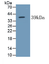
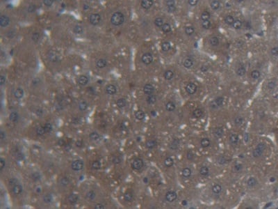
 WB (Western Blot)
(Western Blot;Sample: Porcine Liver lysate)
WB (Western Blot)
(Western Blot;Sample: Porcine Liver lysate)
Alkaline Phosphatase, Tissue-nonspecific (ALPL), Monoclonal Antibody (Cat# AAA148992)
Selenoprotein P1 (SEPP1), Monoclonal Antibody (Cat# AAA149377)
Troponin C Type 1 (TNNC1), Monoclonal Antibody (Cat# AAA149393)
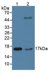
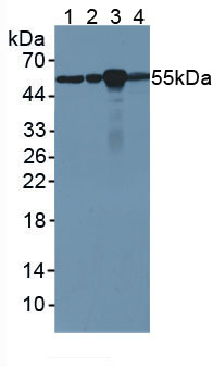
 WB (Western Blot)
(Western Blot: Sample:Lane1: Rabbit Liver lysate;Lane2: Rat Liver lysate;Lane3: 293T cell lysate;Lane4: THP1 cell lysate Primary Ab: 2ug/ml Mouse Anti-Rabbit IL10 Antibody Second Ab: 0.2ug/mL HRP-Linked Rabbit Anti-Mouse IgG Polyclonal Antibody)
WB (Western Blot)
(Western Blot: Sample:Lane1: Rabbit Liver lysate;Lane2: Rat Liver lysate;Lane3: 293T cell lysate;Lane4: THP1 cell lysate Primary Ab: 2ug/ml Mouse Anti-Rabbit IL10 Antibody Second Ab: 0.2ug/mL HRP-Linked Rabbit Anti-Mouse IgG Polyclonal Antibody)
Interleukin 10 (IL10), Monoclonal Antibody (Cat# AAA149418)
 IHC (Immunohistochemistry)
(DAB staining on IHC-P; Samples: Rabbit Cardiac Muscle TissuePrimary Ab: 40ug/ml Mouse Anti-Rabbit IL1b AntibodySecond Ab: 2ug/mL HRP-Linked Caprine Anti-Mouse IgG Polyclonal Antibody)
IHC (Immunohistochemistry)
(DAB staining on IHC-P; Samples: Rabbit Cardiac Muscle TissuePrimary Ab: 40ug/ml Mouse Anti-Rabbit IL1b AntibodySecond Ab: 2ug/mL HRP-Linked Caprine Anti-Mouse IgG Polyclonal Antibody)
Interleukin 1 Beta (IL1b), Monoclonal Antibody (Cat# AAA149433)
What are Monoclonal Antibodies?
Monoclonal antibodies are specialized laboratory-produced proteins developed for binding to specific biological antigens or other molecular targets. Since they come from a single cell (or clone), they are especially consistent and accurate in the data they are involved in producing.
This type of antibody material has been shown to be a powerful tool in finding and subsequently destroying harmful cells in an organism, such as those found in cancers or various autoimmune diseases. This makes them excellent aids in medical testing and research, which is why they are so widely used.
AAA Biotech offers a comprehensive range of high-quality monoclonal antibodies that perform effectively in various laboratory tests, including (amongst others) ELISA, western blotting, immunohistochemistry, and flow cytometry. All of the products in our catalog are thoroughly quality tested to make sure that they are reliable and will consistently perform well in your research.
What Are The Uses of Monoclonal Antibodies
Monoclonal antibodies are used in many lab tests, including (amongst others) ELISA, western blotting, immunohistochemistry, and flow cytometry.
ELISA is a test that helps detect a specific substance/analyte in a sample. It uses antibodies (often monoclonal) bound to a solid surface (such as the well of a microplate) to “capture” the substance/analyte in the sample and immobilize it so that the detection antibody component can then bind to it and produce a signal, which can then be measured.
Western blotting identifies specific proteins in a sample. The sample is first separated on a gel, and then antibodies are applied that will typically bind to the target, which will all be localized to a single band in a lane.
Immunohistochemistry helps locate specific proteins in cells or tissue samples using antibodies.
Flow cytometry looks at and sorts cells. It uses antibodies that are conjugated to reporter molecules called “fluorophores”, which, under special lights, emit light themselves, which can then be measured by a detector instrument.
How Monoclonal Antibodies Are Used as Medicine?
Please note that all of the products listed in AAA Biotech’s also known as AAA Bio or AAABio catalog are strictly for research-use only (RUO).
Monoclonal antibodies can also be used as therapeutic/medical treatments, particularly in the context of cancers. They are designed to find and bind to specific cells or proteins, helping the immune system recognize and attack the cancer. These treatments work in different ways, such as:
- Radioimmunotherapy attaches a small amount of radioactive molecule to the antibody, so it delivers the radiation directly to the cancer cells that the antibody is specifically binding to.
- Antibody-directed enzyme prodrug therapy uses antibodies that are specifically bound to special enzymes. These enzymes activate a harmless drug in the body and turn it into a cancer-killing drug only near the cancer cells—this helps avoid harming healthy cells.
- Immunoliposomes are tiny “bubbles” filled with medicine/drug and coated with antibodies. They carry the drug straight to the cancer cells.
Why Buy Monoclonal Antibodies From Us?
At AAA Biotech, we provide high-performance monoclonal antibodies designed to support a wide range of research needs.
1. Validated for Versatile Applications
The antibodies in our catalog are extensively validated and compatible with multiple techniques, including (but not limited to) ELISA, flow cytometry (FC), immunocytochemistry (ICC), immunofluorescence (IF), immunohistochemistry (IHC), immunoprecipitation (IP), and western blotting (WB).
2. Wide Selection & Specialized Options
We offer antibodies for common and rare species, that are available in various conjugated forms, and also in recombinant formats. Essentially, there is almost anything one might need to meet their experimental model’s requirements.
3. High-Quality Proteins
Our proteins meet high purity standards—90% or more as confirmed by SDS-PAGE. Many are available with tags like His, Flag, GST, or MBP, and we also supply native and biologically active proteins for functional studies.
Frequently Asked Questions
1. Are your monoclonal antibodies validated for specific applications?
Yes, our antibodies are tested and validated for use in methods such as ELISA, western blot, IHC, flow cytometry, and more. Refer to specific product pages or datasheets for individual product information.
2. How do I choose the right monoclonal antibody for my application?
Review the product details directly for application validation, species reactivity, and target information. You may also contact our support team at any time for help.
3. How quickly can I receive my order?
Most orders are processed and shipped within 1–3 business days, depending on product availability and your shipping location.