Filters
▼Clonality
▼Type
▼Reactivity
▼Gene Name
▼Isotype
▼Host
▼Application
▼Clone
▼Monoclonal Antibodies
Get accurate results in your research with our Monoclonal Antibodies, which are specially made to target exactly what you require for your research, and will produce consistent, reliable performance in lab tests.
Viewing 900-950 of 27597 product results
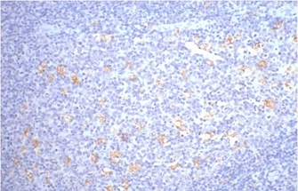
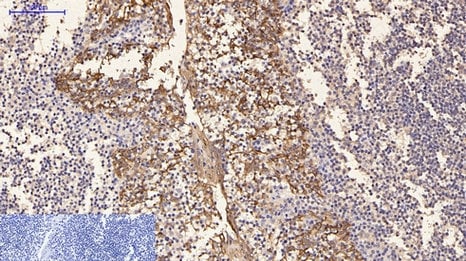
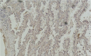
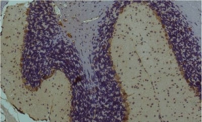
 IHC (Immunohistochemistry)
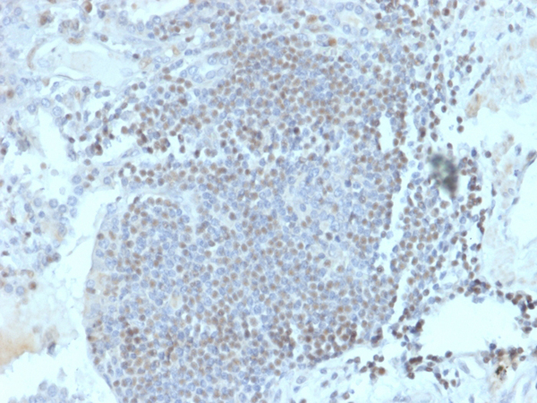
(Formalin-fixed, paraffin-embedded human lymph node stained with HOMEZ Mouse Monoclonal Antibody (PCRP-HOMEZ-1A5).)
IHC (Immunohistochemistry)
(Formalin-fixed, paraffin-embedded human lymph node stained with HOMEZ Mouse Monoclonal Antibody (PCRP-HOMEZ-1A5).)
Homeobox and Leucine Zipper Encoding/HOMEZ, Monoclonal Antibody (Cat# AAA215867)
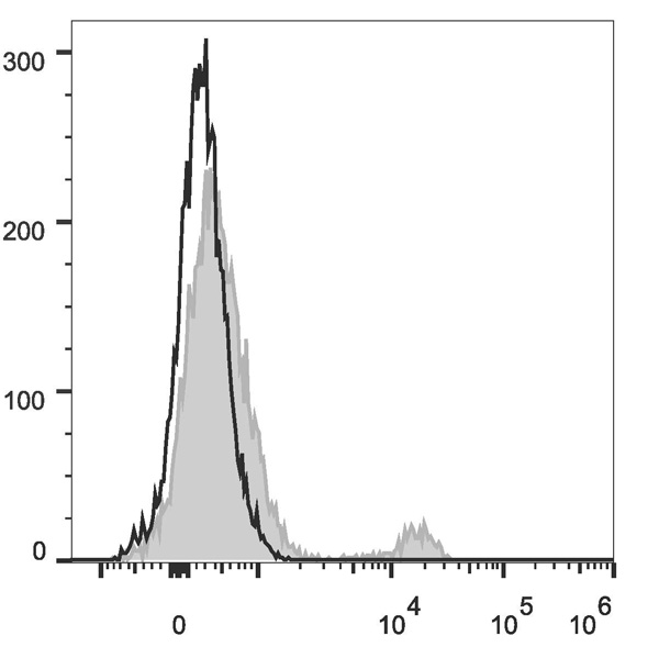
 FCM/FACS (Flow Cytometry)
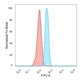
(Flow cytometric analysis of PFA-fixed HeLa cells. OVOL2 Mouse Monoclonal Antibody (PCRP-OVOL2-2A1) followed by goat anti-mouse IgG-CF488 (blue); unstained cells (red).)
FCM/FACS (Flow Cytometry)
(Flow cytometric analysis of PFA-fixed HeLa cells. OVOL2 Mouse Monoclonal Antibody (PCRP-OVOL2-2A1) followed by goat anti-mouse IgG-CF488 (blue); unstained cells (red).)
OVOL2/CRE-BPa, Monoclonal Antibody (Cat# AAA215870)
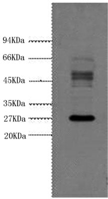
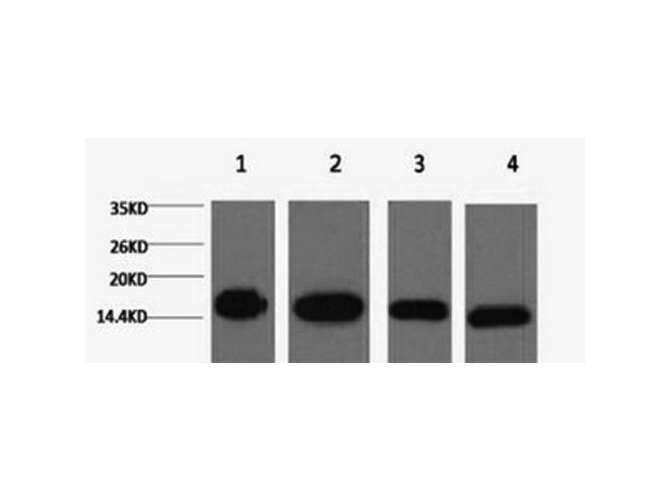
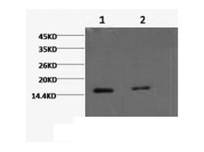
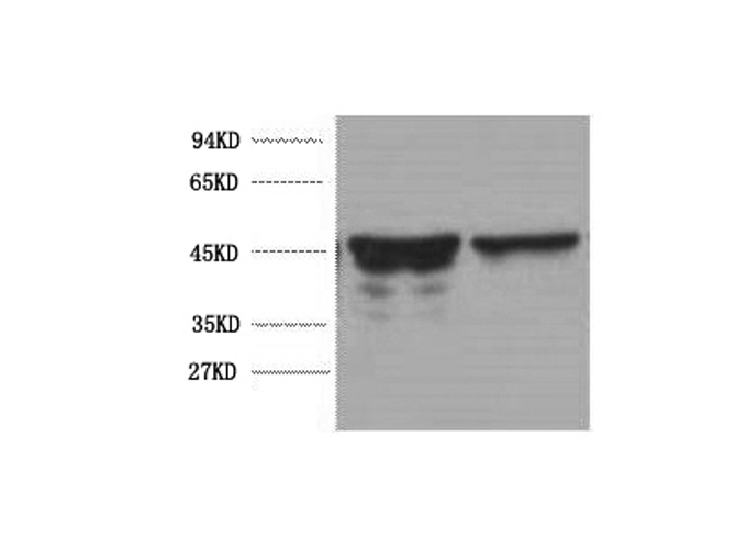
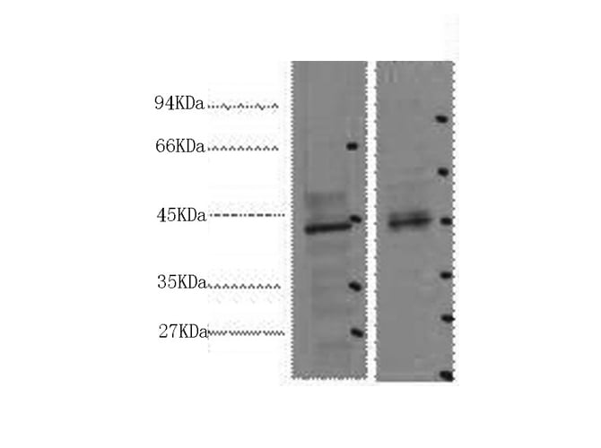
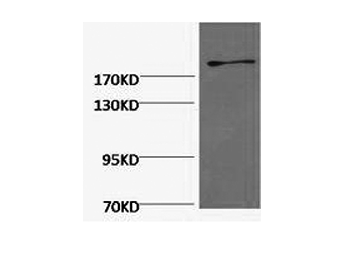
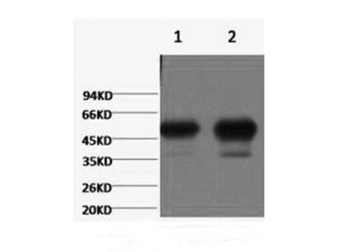
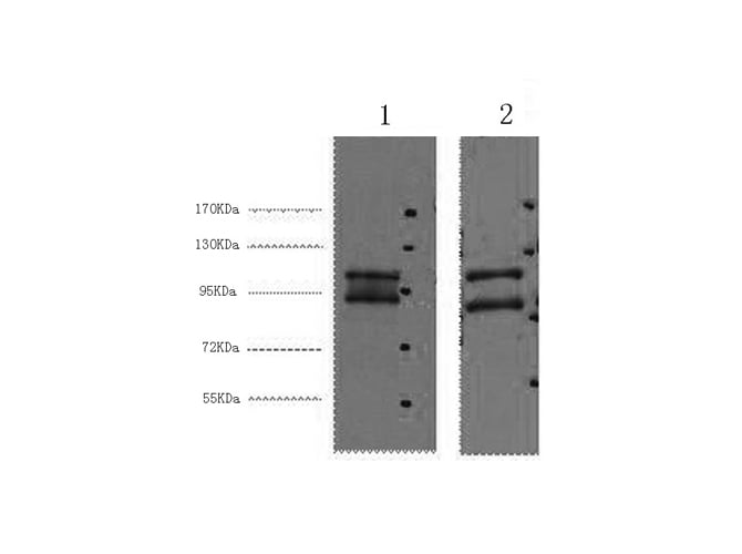
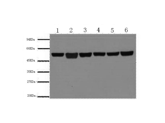
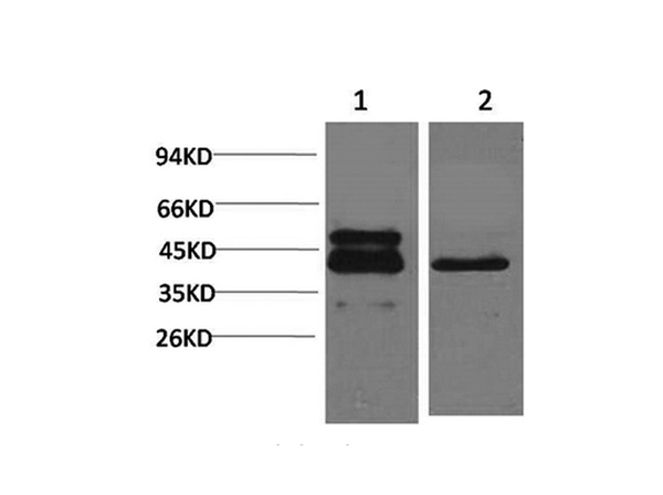
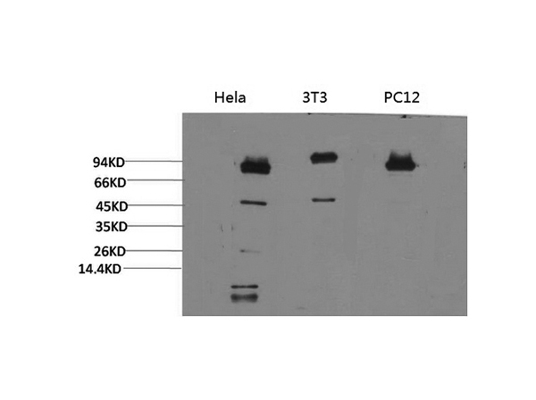
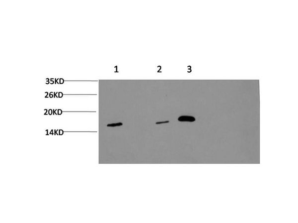
 SDS-PAGE
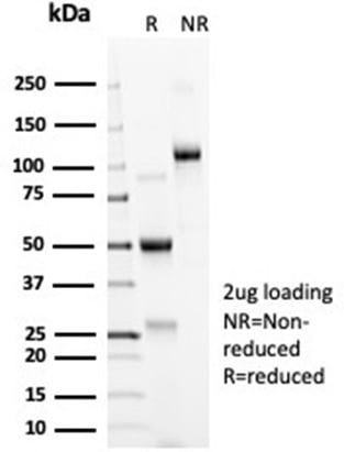
(SDS-PAGE Analysis Purified ACE2/CD143 Rabbit Monoclonal Antibody (ACE2/6788R). Confirmation of Integrity and Purity of Antibody.)
SDS-PAGE
(SDS-PAGE Analysis Purified ACE2/CD143 Rabbit Monoclonal Antibody (ACE2/6788R). Confirmation of Integrity and Purity of Antibody.)
ACE2/Angiotensin I Converting Enzyme 2, Monoclonal Antibody (Cat# AAA215871)
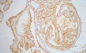
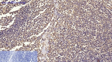
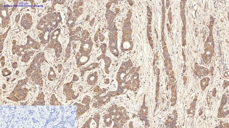
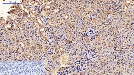
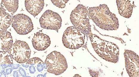
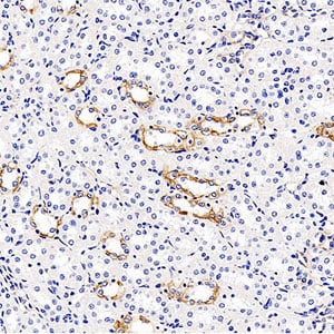
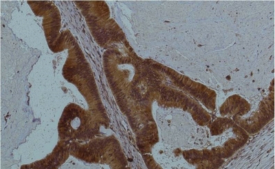
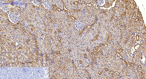
 IHC (Immunohistochemistry)
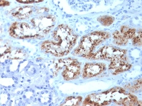
(Formalin-fixed, paraffin-embedded human kidney stained with RBP4 Mouse Monoclonal Antibody (RBP4/4051). Inset: PBS was used instead of the primary antibody as the negative control.)
IHC (Immunohistochemistry)
(Formalin-fixed, paraffin-embedded human kidney stained with RBP4 Mouse Monoclonal Antibody (RBP4/4051). Inset: PBS was used instead of the primary antibody as the negative control.)
RBP4/Retinol Binding Protein 4, Monoclonal Antibody (Cat# AAA215873)
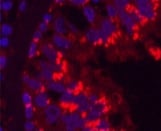
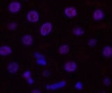
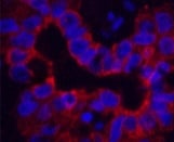
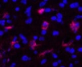
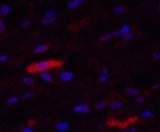
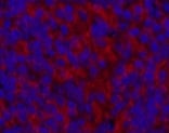
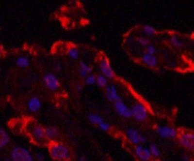 IF (Immunofluorescence)
(Immunofluorescence analysis of Human lung cancer tissue using Galectin-3 Monoclonal Antibody at dilution of 1:200.)
IF (Immunofluorescence)
(Immunofluorescence analysis of Human lung cancer tissue using Galectin-3 Monoclonal Antibody at dilution of 1:200.)
Galectin-3, Monoclonal Antibody (Cat# AAA171592)
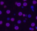
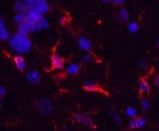
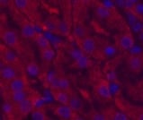
 IF (Immunofluorescence)
(Immunofluorescence analysis of Human lung cancer tissue using CD68 Monoclonal Antibody at dilution of 1:200.)
IF (Immunofluorescence)
(Immunofluorescence analysis of Human lung cancer tissue using CD68 Monoclonal Antibody at dilution of 1:200.)
CD68, Monoclonal Antibody (Cat# AAA171596)

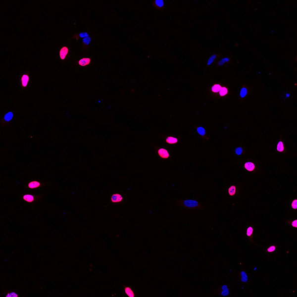
 IF (Immunofluorescence)
(Immunofluorescence analysis of Rat liver tissue using Histone H3 Monoclonal Antibody at dilution of 1:200.)
IF (Immunofluorescence)
(Immunofluorescence analysis of Rat liver tissue using Histone H3 Monoclonal Antibody at dilution of 1:200.)
Histone H3, Monoclonal Antibody (Cat# AAA171603)

 IF (Immunofluorescence)
(Immunofluorescence analysis of Rat liver tissue using Histone H3 (Tri Methyl Lys79) Monoclonal Antibody at dilution of 1:200.)
IF (Immunofluorescence)
(Immunofluorescence analysis of Rat liver tissue using Histone H3 (Tri Methyl Lys79) Monoclonal Antibody at dilution of 1:200.)
Histone H3, Monoclonal Antibody (Cat# AAA171607)
 IF (Immunofluorescence)
(Immunofluorescence analysis of Hela tissue using Aquaporin 4 Monoclonal Antibody at dilution of 1:100.)
IF (Immunofluorescence)
(Immunofluorescence analysis of Hela tissue using Aquaporin 4 Monoclonal Antibody at dilution of 1:100.)
Aquaporin 4, Monoclonal Antibody (Cat# AAA171621)
 IF (Immunofluorescence)
(Immunofluorescence analysis of Mouse brain tissue using GFAP Monoclonal Antibody at dilution of 1:200.)
IF (Immunofluorescence)
(Immunofluorescence analysis of Mouse brain tissue using GFAP Monoclonal Antibody at dilution of 1:200.)
GFAP, Monoclonal Antibody (Cat# AAA171558)

 IF (Immunofluorescence)
(Immunofluorescence analysis of Human liver cancer tissue using CK16 Monoclonal Antibody at dilution of 1:200.)
IF (Immunofluorescence)
(Immunofluorescence analysis of Human liver cancer tissue using CK16 Monoclonal Antibody at dilution of 1:200.)
CK16, Monoclonal Antibody (Cat# AAA171562)
 IF (Immunofluorescence)
(Immunofluorescence analysis of Mouse kidney tissue using CDX2 Monoclonal Antibody at dilution of 1:200.)
IF (Immunofluorescence)
(Immunofluorescence analysis of Mouse kidney tissue using CDX2 Monoclonal Antibody at dilution of 1:200.)
CDX2, Monoclonal Antibody (Cat# AAA171564)
 IF (Immunofluorescence)
(Immunofluorescence analysis of Human appendix tissue using Fibronectin Monoclonal Antibody at dilution of 1:200.)
IF (Immunofluorescence)
(Immunofluorescence analysis of Human appendix tissue using Fibronectin Monoclonal Antibody at dilution of 1:200.)
Fibronectin, Monoclonal Antibody (Cat# AAA171573)
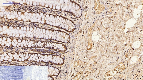
 IHC (Immunohiostchemistry)
(Immunohistochemistry of paraffin-embedded Human colon tissue using ? II tubulin Monoclonal Antibody at dilution of 1:200.)
IHC (Immunohiostchemistry)
(Immunohistochemistry of paraffin-embedded Human colon tissue using ? II tubulin Monoclonal Antibody at dilution of 1:200.)
beta II tubulin, Monoclonal Antibody (Cat# AAA171582)
 IF (Immunofluorescence)
(Immunofluorescence analysis of Human breast tissue using IDE Monoclonal Antibody at dilution of 1:200.)
IF (Immunofluorescence)
(Immunofluorescence analysis of Human breast tissue using IDE Monoclonal Antibody at dilution of 1:200.)
IDE, Monoclonal Antibody (Cat# AAA171586)
 IF (Immunofluorescence)
(Immunofluorescence analysis of Hela tissue using ?-Tubulin Monoclonal Antibody at dilution of 1:100.)
IF (Immunofluorescence)
(Immunofluorescence analysis of Hela tissue using ?-Tubulin Monoclonal Antibody at dilution of 1:100.)
beta-Tubulin, Monoclonal Antibody (Cat# AAA171588)
IgG2a, kappa, Monoclonal Isotype Control (Cat# AAA174726)
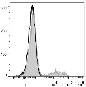
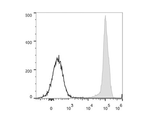
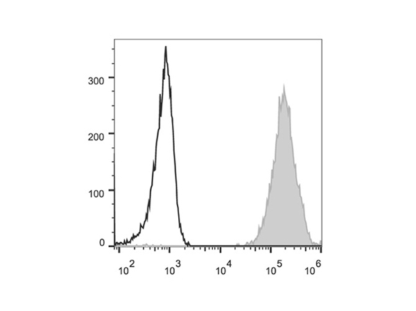
 FCM/FACS (Flow Cytometry)
(Each lot of this antibody is quality control tested by flow cytometric analysis. The amount of the reagent is suggested to be used 5 uL of antibody per test (million cells in 100 uL staining volume or per 100 uL of whole blood). Please check your vial before the experiment. Since applications vary, the appropriate dilutions must be determined for individual use. Human peripheral blood lymphocytes are stained with PerCP/Cyanine5.5 Anti-Human CD40 Monoclonal Antibody (filled gray histogram). Unstained lymphocytes (empty black histogram) are used as control.)
FCM/FACS (Flow Cytometry)
(Each lot of this antibody is quality control tested by flow cytometric analysis. The amount of the reagent is suggested to be used 5 uL of antibody per test (million cells in 100 uL staining volume or per 100 uL of whole blood). Please check your vial before the experiment. Since applications vary, the appropriate dilutions must be determined for individual use. Human peripheral blood lymphocytes are stained with PerCP/Cyanine5.5 Anti-Human CD40 Monoclonal Antibody (filled gray histogram). Unstained lymphocytes (empty black histogram) are used as control.)
CD40, Monoclonal Antibody (Cat# AAA174739)
 FCM/FACS (Flow Cytometry)
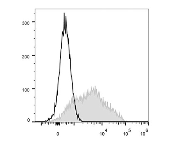
(Human peripheral blood lymphocytes are stained with Anti-Human CD24 Monoclonal Antibody(FITC Conjugated)(filled gray histogram). Unstained lymphocytes (empty black histogram) are used as control.)
FCM/FACS (Flow Cytometry)
(Human peripheral blood lymphocytes are stained with Anti-Human CD24 Monoclonal Antibody(FITC Conjugated)(filled gray histogram). Unstained lymphocytes (empty black histogram) are used as control.)
CD24, Monoclonal Antibody (Cat# AAA174753)
 FCM/FACS (Flow Cytometry)
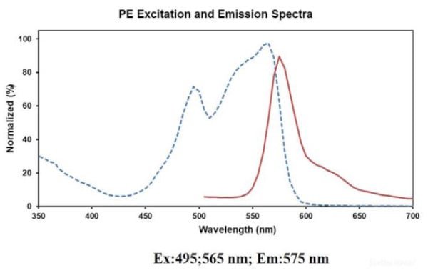
(Conjugation: PE)
FCM/FACS (Flow Cytometry)
(Conjugation: PE)
CD134, Monoclonal Antibody (Cat# AAA174754)
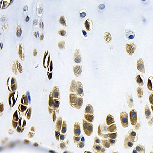 IHC (Immunohistochemistry)
(Immunohistochemistry analysis of paraffin-embedded rat bone using BMP2 Monoclonal Antibody at dilution of 1:400.)
IHC (Immunohistochemistry)
(Immunohistochemistry analysis of paraffin-embedded rat bone using BMP2 Monoclonal Antibody at dilution of 1:400.)
BMP2, Monoclonal Antibody (Cat# AAA174554)

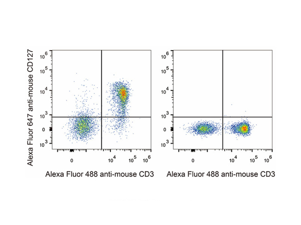 FCM/FACS (Flow Cytometry)
(C57BL/6 murine splenocytes are stained with Anti-Mouse CD127 Monoclonal Antibody(Alexa Fluor 647 Conjuaged)(left). Splenocytes stained with Anti-Mouse CD3 Monoclonal Antibody(Alexa Fluor 488 Conjuaged)(right) are used as control.)
FCM/FACS (Flow Cytometry)
(C57BL/6 murine splenocytes are stained with Anti-Mouse CD127 Monoclonal Antibody(Alexa Fluor 647 Conjuaged)(left). Splenocytes stained with Anti-Mouse CD3 Monoclonal Antibody(Alexa Fluor 488 Conjuaged)(right) are used as control.)
CD127, Monoclonal Antibody (Cat# AAA174611)
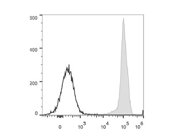
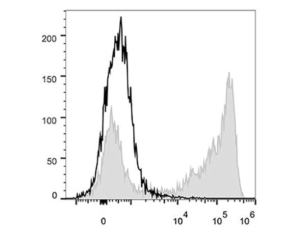
 FCM/FACS (Flow Cytometry)
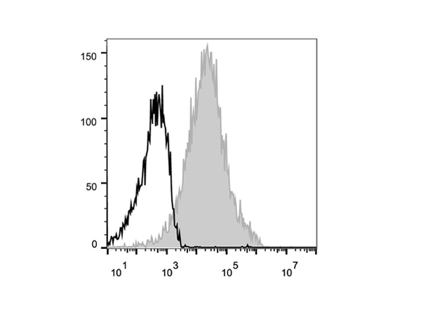
(Human peripheral blood lymphocytes are stained with Anti-Human CD44 Monoclonal Antibody(PerCP/Cy5.5 Conjugated)(filled gray histogram). Unstained lymphocytes (empty black histogram) are used as control.)
FCM/FACS (Flow Cytometry)
(Human peripheral blood lymphocytes are stained with Anti-Human CD44 Monoclonal Antibody(PerCP/Cy5.5 Conjugated)(filled gray histogram). Unstained lymphocytes (empty black histogram) are used as control.)
CD44, Monoclonal Antibody (Cat# AAA174617)
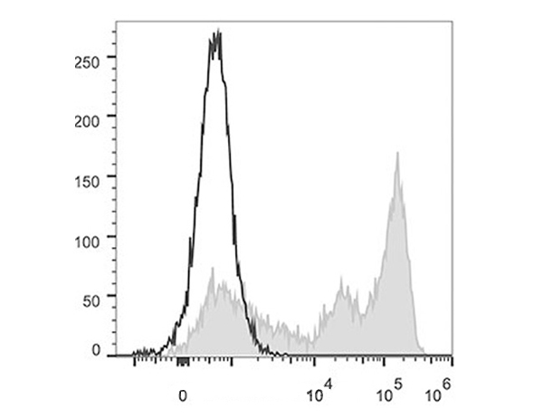
 FCM/FACS (Flow Cytometry)
(Human peripheral blood lymphocytes are stained with Anti-Human CD38 Monoclonal Antibody(PerCP/Cy5.5 Conjugated)(filled gray histogram). Unstained lymphocytes (empty black histogram) are used as control.)
FCM/FACS (Flow Cytometry)
(Human peripheral blood lymphocytes are stained with Anti-Human CD38 Monoclonal Antibody(PerCP/Cy5.5 Conjugated)(filled gray histogram). Unstained lymphocytes (empty black histogram) are used as control.)
CD38, Monoclonal Antibody (Cat# AAA174626)
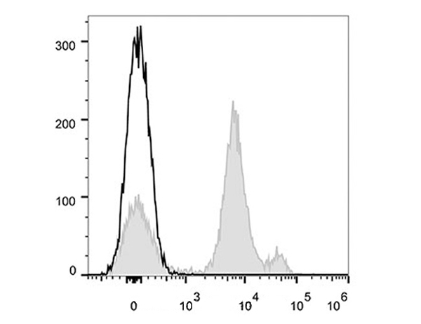
 FCM/FACS (Flow Cytometry)
(Human peripheral blood lymphocytes are stained with Anti-Human CD38 Monoclonal Antibody(Alexa Fluor 647 Conjuaged)(filled gray histogram). Unstained lymphocytes (empty black histogram) are used as control.)
FCM/FACS (Flow Cytometry)
(Human peripheral blood lymphocytes are stained with Anti-Human CD38 Monoclonal Antibody(Alexa Fluor 647 Conjuaged)(filled gray histogram). Unstained lymphocytes (empty black histogram) are used as control.)
CD38, Monoclonal Antibody (Cat# AAA174627)

 FCM/FACS (Flow Cytometry)
(Human peripheral blood lymphocytes are stained with Anti-Human CD57 Monoclonal Antibody(PerCP/Cy5.5 Conjugated)(filled gray histogram). Unstained lymphocytes (empty black histogram) are used as control.)
FCM/FACS (Flow Cytometry)
(Human peripheral blood lymphocytes are stained with Anti-Human CD57 Monoclonal Antibody(PerCP/Cy5.5 Conjugated)(filled gray histogram). Unstained lymphocytes (empty black histogram) are used as control.)
CD57, Monoclonal Antibody (Cat# AAA174629)

 FCM/FACS (Flow Cytometry)
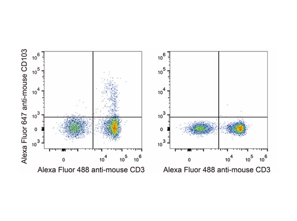
(C57BL/6 murine splenocytes are stained with Anti-Mouse CD103 Monoclonal Antibody(Alexa Fluor 647 Conjuaged)(left). Splenocytes stained with Anti-Mouse CD3 Monoclonal Antibody(Alexa Fluor 488 Conjuaged)(right) are used as control.)
FCM/FACS (Flow Cytometry)
(C57BL/6 murine splenocytes are stained with Anti-Mouse CD103 Monoclonal Antibody(Alexa Fluor 647 Conjuaged)(left). Splenocytes stained with Anti-Mouse CD3 Monoclonal Antibody(Alexa Fluor 488 Conjuaged)(right) are used as control.)
CD103, Monoclonal Antibody (Cat# AAA174635)
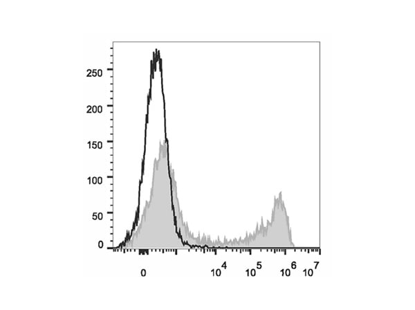
 FCM/FACS (Flow Cytometry)
(C57BL/6 murine bone marrow cells are stained with Anti-Mouse Gr-1 Monoclonal Antibody(PerCP/Cy5.5 Conjugated)(filled gray histogram). Unstained bone marrow cells (empty black histogram) are used as control.)
FCM/FACS (Flow Cytometry)
(C57BL/6 murine bone marrow cells are stained with Anti-Mouse Gr-1 Monoclonal Antibody(PerCP/Cy5.5 Conjugated)(filled gray histogram). Unstained bone marrow cells (empty black histogram) are used as control.)
Gr-1, Monoclonal Antibody (Cat# AAA174642)
 FCM/FACS (Flow Cytometry)
(C57BL/6 murine bone marrow cells are stained with Anti-Mouse Gr-1 Monoclonal Antibody(Alexa Fluor 488 Conjugated)(filled gray histogram). Unstained bone marrow cells (empty black histogram) are used as control.)
FCM/FACS (Flow Cytometry)
(C57BL/6 murine bone marrow cells are stained with Anti-Mouse Gr-1 Monoclonal Antibody(Alexa Fluor 488 Conjugated)(filled gray histogram). Unstained bone marrow cells (empty black histogram) are used as control.)
Gr-1, Monoclonal Antibody (Cat# AAA174643)

 FCM/FACS (Flow Cytometry)
(C57BL/6 murine bone marrow cells are stained with Anti-Mouse Ly6C Monoclonal Antibody(APC Conjugated)(filled gray histogram). Unstained bone marrow cells (empty black histogram) are used as control.)
FCM/FACS (Flow Cytometry)
(C57BL/6 murine bone marrow cells are stained with Anti-Mouse Ly6C Monoclonal Antibody(APC Conjugated)(filled gray histogram). Unstained bone marrow cells (empty black histogram) are used as control.)
Ly6C, Monoclonal Antibody (Cat# AAA174646)

 FCM/FACS (Flow Cytometry)
(Human peripheral blood lymphocytes are stained with Anti-Human HLA-A,B,C Monoclonal Antibody(Alexa Fluor 488 Conjugated)(filled gray histogram). Unstained lymphocytes (empty black histogram) are used as control.)
FCM/FACS (Flow Cytometry)
(Human peripheral blood lymphocytes are stained with Anti-Human HLA-A,B,C Monoclonal Antibody(Alexa Fluor 488 Conjugated)(filled gray histogram). Unstained lymphocytes (empty black histogram) are used as control.)
HLA-A,B,C, Monoclonal Antibody (Cat# AAA174663)
 IHC (Immunohistochemisry)
(Immunohistochemistry analysis of paraffin-embedded rat kidney using MMP9 Monoclonal Antibody at dilution of 1:300.)
IHC (Immunohistochemisry)
(Immunohistochemistry analysis of paraffin-embedded rat kidney using MMP9 Monoclonal Antibody at dilution of 1:300.)
MMP9, Monoclonal Antibody (Cat# AAA174504)

 IF (Immunofluorescence)
(Immunofluorescence analysis of Hela cells(treated with 0.03mg/ml BrdU for 40 min) using Brdu Monoclonal Antibody at dilution of 1:100.)
IF (Immunofluorescence)
(Immunofluorescence analysis of Hela cells(treated with 0.03mg/ml BrdU for 40 min) using Brdu Monoclonal Antibody at dilution of 1:100.)
Brdu, Monoclonal Antibody (Cat# AAA174505)
CD162, Monoclonal Antibody (Cat# AAA174048)
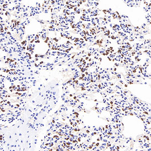
 IHC (Immunohiostchemistry)
(Immunohistochemistry of paraffin-embedded Human lung carcinoma tissue with CREB-1 Monoclonal Antibody.)
IHC (Immunohiostchemistry)
(Immunohistochemistry of paraffin-embedded Human lung carcinoma tissue with CREB-1 Monoclonal Antibody.)
CREB-1, Monoclonal Antibody (Cat# AAA173653)
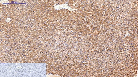
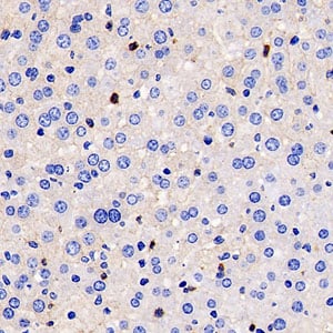
 IHC (Immunohistochemisry)
(Immunohistochemistry of paraffin-embedded Mouse brain tissue using STAT3 Monoclonal Antibody at dilution of 1:200.)
IHC (Immunohistochemisry)
(Immunohistochemistry of paraffin-embedded Mouse brain tissue using STAT3 Monoclonal Antibody at dilution of 1:200.)
STAT3, Monoclonal Antibody (Cat# AAA173658)
 IF (Immunofluorescence)
(Immunofluorescence analysis of Mouse spleen tissue using LC3A Monoclonal Antibody at dilution of 1:200.)
IF (Immunofluorescence)
(Immunofluorescence analysis of Mouse spleen tissue using LC3A Monoclonal Antibody at dilution of 1:200.)
LC3A, Monoclonal Antibody (Cat# AAA173672)
Enrofloxacin, Monoclonal Antibody (Cat# AAA117775)
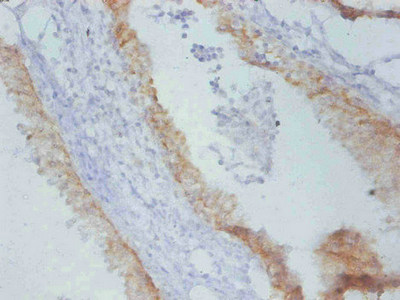
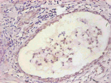 IHC (Immunohistochemistry)
(Immunohistochemical of paraffin-embedded human prostate tissue using AAA117778 at dilution of 1:200)
IHC (Immunohistochemistry)
(Immunohistochemical of paraffin-embedded human prostate tissue using AAA117778 at dilution of 1:200)
Interleukin-6, Monoclonal Antibody (Cat# AAA117778)
 Application Data
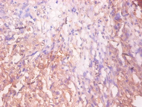
(Immunohistochemical of paraffin-embedded Human prostate tissue at dilution of 1:200)
Application Data
(Immunohistochemical of paraffin-embedded Human prostate tissue at dilution of 1:200)
Epidermal growth factor receptor, Monoclonal Antibody (Cat# AAA117780)
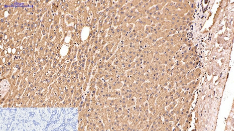
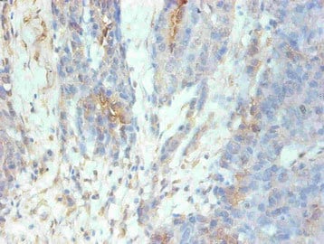
 IHC (Immunohistochemistry)
(Immunohistochemistry of paraffin-embedded Rat prostate cancer using AAA117784 at dilution of 1:100.)
IHC (Immunohistochemistry)
(Immunohistochemistry of paraffin-embedded Rat prostate cancer using AAA117784 at dilution of 1:100.)
Timp1, Monoclonal Antibody (Cat# AAA117784)
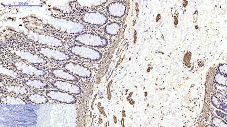
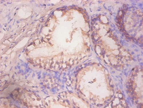
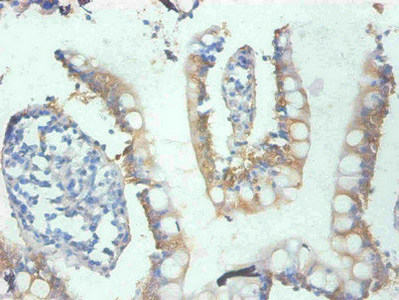 IHC (Immunohiostchemistry)
(Immunohistochemical of paraffin-embedded human small Intestinal tissue using AAA117786 at dilution of 1:200)
IHC (Immunohiostchemistry)
(Immunohistochemical of paraffin-embedded human small Intestinal tissue using AAA117786 at dilution of 1:200)
Galectin 3, Monoclonal Antibody (Cat# AAA117786)
TGFB1/TGF-beta-1 & LRRC32/GARP, Monoclonal Antibody (Cat# AAA120160)
CD4, Monoclonal Antibody (Cat# AAA120161)
CD333/FGFR3, Monoclonal Antibody (Cat# AAA120169)
Vibrio cholerae ctxB/Cholera Toxin Subunit B, Monoclonal Recombinant Antibody (Cat# AAA120275)
IL31RA, Monoclonal Antibody (Cat# AAA120295)
Protein A/G purified from cell culture supernatant.
MOG, Monoclonal Antibody (Cat# AAA120297)
CXCL12/SDF-1, Monoclonal Antibody (Cat# AAA120306)
What are Monoclonal Antibodies?
Monoclonal antibodies are specialized laboratory-produced proteins developed for binding to specific biological antigens or other molecular targets. Since they come from a single cell (or clone), they are especially consistent and accurate in the data they are involved in producing.
This type of antibody material has been shown to be a powerful tool in finding and subsequently destroying harmful cells in an organism, such as those found in cancers or various autoimmune diseases. This makes them excellent aids in medical testing and research, which is why they are so widely used.
AAA Biotech offers a comprehensive range of high-quality monoclonal antibodies that perform effectively in various laboratory tests, including (amongst others) ELISA, western blotting, immunohistochemistry, and flow cytometry. All of the products in our catalog are thoroughly quality tested to make sure that they are reliable and will consistently perform well in your research.
What Are The Uses of Monoclonal Antibodies
Monoclonal antibodies are used in many lab tests, including (amongst others) ELISA, western blotting, immunohistochemistry, and flow cytometry.
ELISA is a test that helps detect a specific substance/analyte in a sample. It uses antibodies (often monoclonal) bound to a solid surface (such as the well of a microplate) to “capture” the substance/analyte in the sample and immobilize it so that the detection antibody component can then bind to it and produce a signal, which can then be measured.
Western blotting identifies specific proteins in a sample. The sample is first separated on a gel, and then antibodies are applied that will typically bind to the target, which will all be localized to a single band in a lane.
Immunohistochemistry helps locate specific proteins in cells or tissue samples using antibodies.
Flow cytometry looks at and sorts cells. It uses antibodies that are conjugated to reporter molecules called “fluorophores”, which, under special lights, emit light themselves, which can then be measured by a detector instrument.
How Monoclonal Antibodies Are Used as Medicine?
Please note that all of the products listed in AAA Biotech’s also known as AAA Bio or AAABio catalog are strictly for research-use only (RUO).
Monoclonal antibodies can also be used as therapeutic/medical treatments, particularly in the context of cancers. They are designed to find and bind to specific cells or proteins, helping the immune system recognize and attack the cancer. These treatments work in different ways, such as:
- Radioimmunotherapy attaches a small amount of radioactive molecule to the antibody, so it delivers the radiation directly to the cancer cells that the antibody is specifically binding to.
- Antibody-directed enzyme prodrug therapy uses antibodies that are specifically bound to special enzymes. These enzymes activate a harmless drug in the body and turn it into a cancer-killing drug only near the cancer cells—this helps avoid harming healthy cells.
- Immunoliposomes are tiny “bubbles” filled with medicine/drug and coated with antibodies. They carry the drug straight to the cancer cells.
Why Buy Monoclonal Antibodies From Us?
At AAA Biotech, we provide high-performance monoclonal antibodies designed to support a wide range of research needs.
1. Validated for Versatile Applications
The antibodies in our catalog are extensively validated and compatible with multiple techniques, including (but not limited to) ELISA, flow cytometry (FC), immunocytochemistry (ICC), immunofluorescence (IF), immunohistochemistry (IHC), immunoprecipitation (IP), and western blotting (WB).
2. Wide Selection & Specialized Options
We offer antibodies for common and rare species, that are available in various conjugated forms, and also in recombinant formats. Essentially, there is almost anything one might need to meet their experimental model’s requirements.
3. High-Quality Proteins
Our proteins meet high purity standards—90% or more as confirmed by SDS-PAGE. Many are available with tags like His, Flag, GST, or MBP, and we also supply native and biologically active proteins for functional studies.
Frequently Asked Questions
1. Are your monoclonal antibodies validated for specific applications?
Yes, our antibodies are tested and validated for use in methods such as ELISA, western blot, IHC, flow cytometry, and more. Refer to specific product pages or datasheets for individual product information.
2. How do I choose the right monoclonal antibody for my application?
Review the product details directly for application validation, species reactivity, and target information. You may also contact our support team at any time for help.
3. How quickly can I receive my order?
Most orders are processed and shipped within 1–3 business days, depending on product availability and your shipping location.