Filters
▼Clonality
▼Type
▼Reactivity
▼Gene Name
▼Isotype
▼Host
▼Application
▼Clone
▼Monoclonal Antibodies
Get accurate results in your research with our Monoclonal Antibodies, which are specially made to target exactly what you require for your research, and will produce consistent, reliable performance in lab tests.
Viewing 1100-1150 of 27597 product results
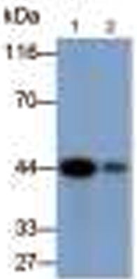 WB (Western Blot)
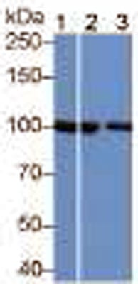
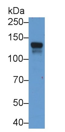
(Western Blot;Samples:Lane 1: Jurkat cell lysate;Lane 2: HUVEC cell lysate;Primary Ab: 0.3ug/ml Mouse Anti-Rabbit vWF AntibodySecond Ab: 0.2ug/ml HRP-Linked Caprine Anti-Mouse IgG Polyclonal Antibody)
WB (Western Blot)
(Western Blot;Samples:Lane 1: Jurkat cell lysate;Lane 2: HUVEC cell lysate;Primary Ab: 0.3ug/ml Mouse Anti-Rabbit vWF AntibodySecond Ab: 0.2ug/ml HRP-Linked Caprine Anti-Mouse IgG Polyclonal Antibody)
Von Willebrand Factor (vWF), Monoclonal Antibody (Cat# AAA149441)
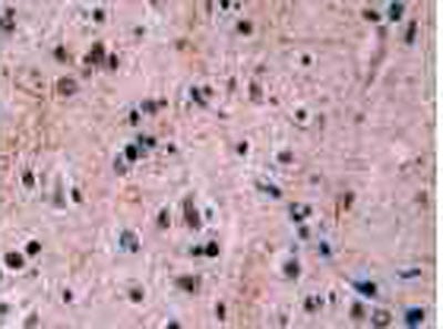
 IHC (Immunohistochemistry)
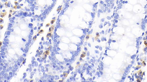
(DAB staining on IHC-P;Samples: Human Prostate Tissue;Primary Ab: 30ug/ml Mouse Anti-Multi-species Ab1-42 AntibodySecond Ab: 2ug/mL HRP-Linked Caprine Anti-Mouse IgG Polyclonal Antibody)
IHC (Immunohistochemistry)
(DAB staining on IHC-P;Samples: Human Prostate Tissue;Primary Ab: 30ug/ml Mouse Anti-Multi-species Ab1-42 AntibodySecond Ab: 2ug/mL HRP-Linked Caprine Anti-Mouse IgG Polyclonal Antibody)
Amyloid Beta Peptide 1-42 (Ab1-42), Monoclonal Antibody (Cat# AAA149442)
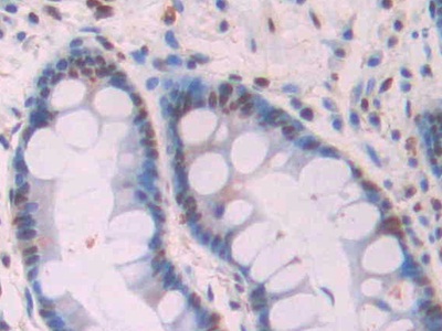
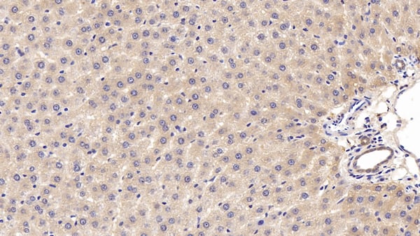
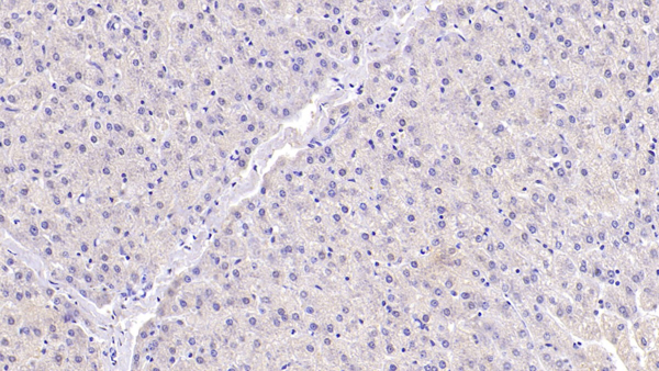
 IHC (Immunohiostchemistry)
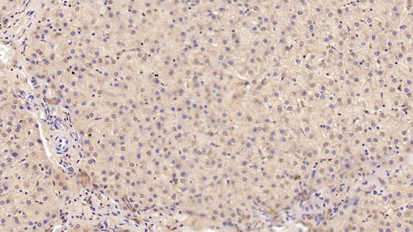
(DAB staining on IHC-P; Samples: Human Liver Tissue)
IHC (Immunohiostchemistry)
(DAB staining on IHC-P; Samples: Human Liver Tissue)
Catenin Beta 1 (CTNNb1), Monoclonal Antibody (Cat# AAA149443)
 IHC (Immunohiostchemistry)
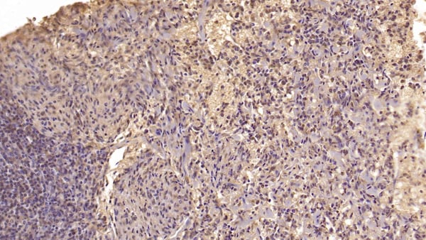
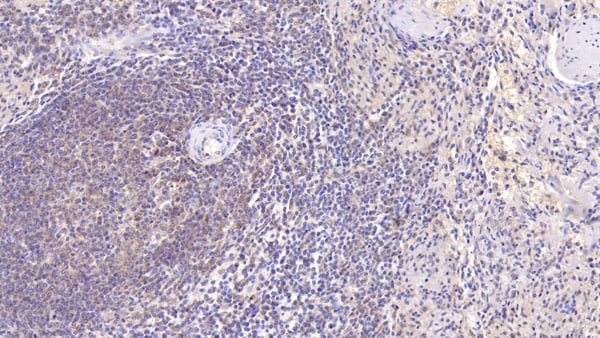
(DAB staining on IHC-P; Samples: Human Spleen Tissue)
IHC (Immunohiostchemistry)
(DAB staining on IHC-P; Samples: Human Spleen Tissue)
Tetraspanin 30 (TSPAN30), Monoclonal Antibody (Cat# AAA149448)
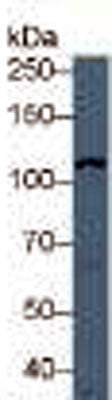
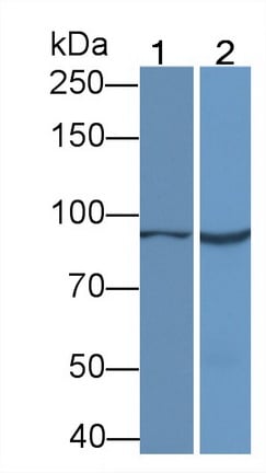
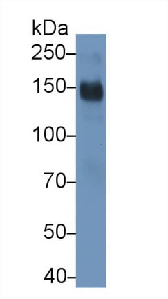
 WB (Western Blot)
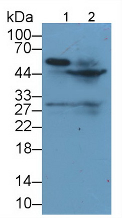
(Western Blot: Sample: Lane1: Human Lung lysate; Lane2: 293T cell lysate;)
WB (Western Blot)
(Western Blot: Sample: Lane1: Human Lung lysate; Lane2: 293T cell lysate;)
TATA Binding Protein (TBP), Monoclonal Antibody (Cat# AAA149451)

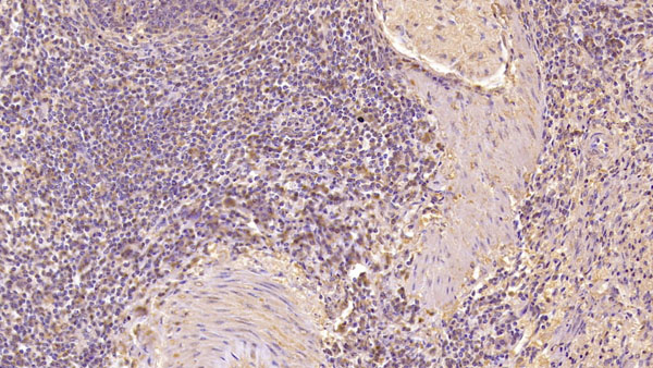
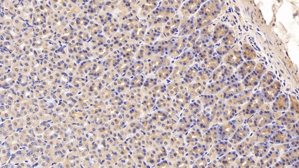
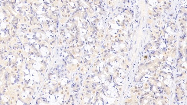
 IHC (Immunohistochemisry)
(DAB staining on IHC-P;Sample: Human Liver Tissue;Primary Ab: 20ug/ml Mouse Anti-Human Plg AntibodySecond Ab: 2ug/mL HRP-Linked Caprine Anti-Mouse IgG Polyclonal Antibody)
IHC (Immunohistochemisry)
(DAB staining on IHC-P;Sample: Human Liver Tissue;Primary Ab: 20ug/ml Mouse Anti-Human Plg AntibodySecond Ab: 2ug/mL HRP-Linked Caprine Anti-Mouse IgG Polyclonal Antibody)
Plasminogen (Plg), Monoclonal Antibody (Cat# AAA149546)

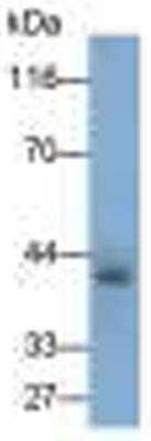
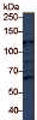
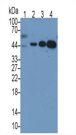
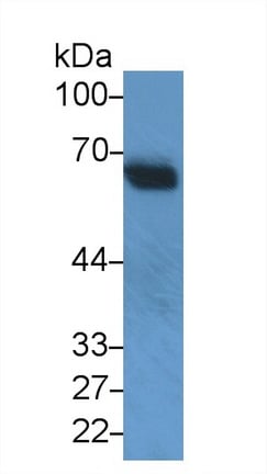
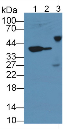
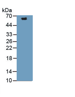
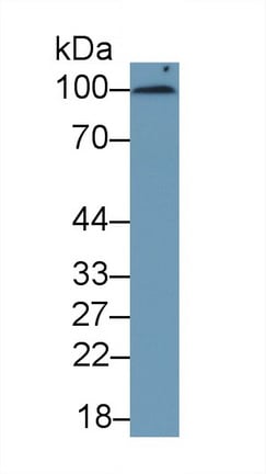
 WB (Western Blot)
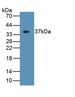
(Western Blot; Sample: Human Leukocyte lysatePrimary Ab: 3ug/ml Mouse Anti-Human AZU1 Antibody Second Ab: 0.2ug/mL HRP-Linked Rabbit Anti-Mouse IgG Polyclonal Antibody)
WB (Western Blot)
(Western Blot; Sample: Human Leukocyte lysatePrimary Ab: 3ug/ml Mouse Anti-Human AZU1 Antibody Second Ab: 0.2ug/mL HRP-Linked Rabbit Anti-Mouse IgG Polyclonal Antibody)
Azurocidin (AZU), Monoclonal Antibody (Cat# AAA149551)
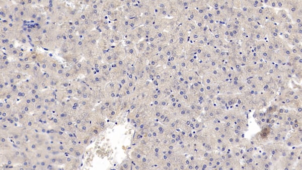
 IHC (Immunohiostchemistry)
(DAB staining on IHC-P; Samples: Human Cerebrum Tissue; Primary Ab: 30ug/ml Mouse Anti-Human DMD Antibody Second Ab: 2ug/mL HRP-Linked Caprine Anti-Mouse IgG Polyclonal Antibody)
IHC (Immunohiostchemistry)
(DAB staining on IHC-P; Samples: Human Cerebrum Tissue; Primary Ab: 30ug/ml Mouse Anti-Human DMD Antibody Second Ab: 2ug/mL HRP-Linked Caprine Anti-Mouse IgG Polyclonal Antibody)
Dystrophin (DMD), Monoclonal Antibody (Cat# AAA149553)
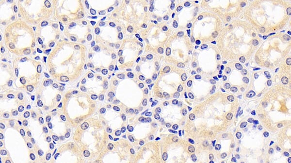
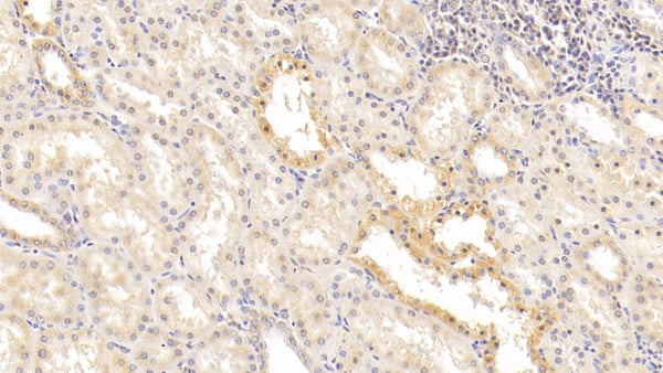
 IHC (Immunohistochemistry)
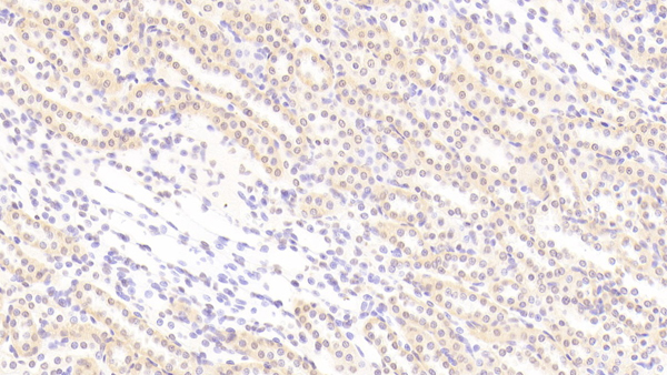
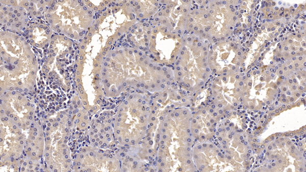
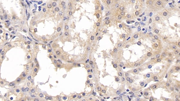
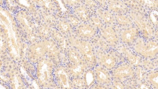
(DAB staining on IHC-P; Samples: Human Kidney Tissue; Primary Ab: 20ug/ml Mouse Anti-Human IDO AntibodySecond Ab: 2ug/mL HRP-Linked Caprine Anti-Mouse IgG Polyclonal Antibody)
IHC (Immunohistochemistry)
(DAB staining on IHC-P; Samples: Human Kidney Tissue; Primary Ab: 20ug/ml Mouse Anti-Human IDO AntibodySecond Ab: 2ug/mL HRP-Linked Caprine Anti-Mouse IgG Polyclonal Antibody)
Indoleamine-2,3-Dioxygenase (IDO), Monoclonal Antibody (Cat# AAA149555)
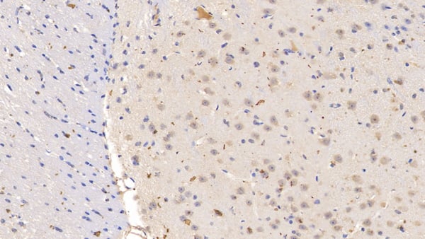 IHC (Immunohiostchemistry)
(DAB staining on IHC-P; Samples: Mouse Cerebrum Tissue; Primary Ab: 10ug/ml Mouse Anti-Mouse IL10Ra Antibody Second Ab: 2ug/mL HRP-Linked Caprine Anti-Mouse IgG Polyclonal Antibody)
IHC (Immunohiostchemistry)
(DAB staining on IHC-P; Samples: Mouse Cerebrum Tissue; Primary Ab: 10ug/ml Mouse Anti-Mouse IL10Ra Antibody Second Ab: 2ug/mL HRP-Linked Caprine Anti-Mouse IgG Polyclonal Antibody)
Interleukin 10 Receptor Alpha (IL10Ra), Monoclonal Antibody (Cat# AAA149556)
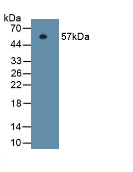
 WB (Western Blot)
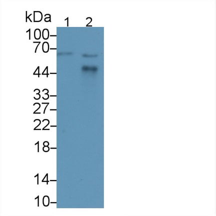
(Western Blot; Sample: Lane1: Human Serum; Lane2: Human PlasmaPrimary Ab: 2?g/ml Mouse Anti-Human GYPA AntibodySecond Ab: 0.2ug/mL HRP-Linked Rabbit Anti-Mouse IgG Polyclonal Antibody)
WB (Western Blot)
(Western Blot; Sample: Lane1: Human Serum; Lane2: Human PlasmaPrimary Ab: 2?g/ml Mouse Anti-Human GYPA AntibodySecond Ab: 0.2ug/mL HRP-Linked Rabbit Anti-Mouse IgG Polyclonal Antibody)
Glycophorin A (GYPA), Monoclonal Antibody (Cat# AAA149558)
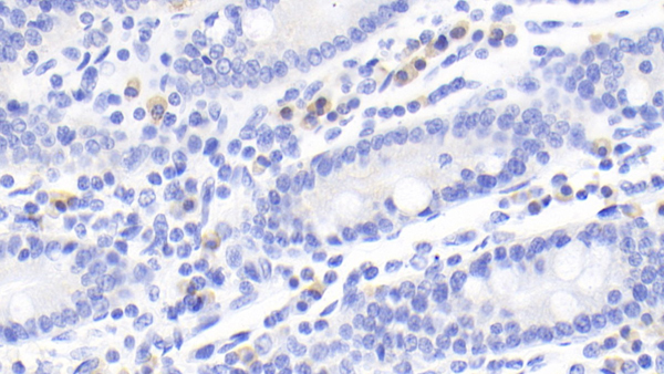
 IHC (Immunohistochemistry)
(DAB staining on IHC-P; Samples: Rat Stomach Tissue; Primary Ab: 10ug/ml Mouse Anti-Rat CD163 AntibodySecond Ab: 2ug/mL HRP-Linked Caprine Anti-Mouse IgG Polyclonal Antibody)
IHC (Immunohistochemistry)
(DAB staining on IHC-P; Samples: Rat Stomach Tissue; Primary Ab: 10ug/ml Mouse Anti-Rat CD163 AntibodySecond Ab: 2ug/mL HRP-Linked Caprine Anti-Mouse IgG Polyclonal Antibody)
Cluster of Differentiation (CD163), Monoclonal Antibody (Cat# AAA149559)
 IHC (Immunohiostchemistry)
(DAB staining on IHC-P; Samples: Human Cerebrum Tissue; Primary Ab: 30ug/ml Mouse Anti-Human IL22 Antibody Second Ab: 2ug/mL HRP-Linked Caprine Anti-Mouse IgG Polyclonal Antibody)
IHC (Immunohiostchemistry)
(DAB staining on IHC-P; Samples: Human Cerebrum Tissue; Primary Ab: 30ug/ml Mouse Anti-Human IL22 Antibody Second Ab: 2ug/mL HRP-Linked Caprine Anti-Mouse IgG Polyclonal Antibody)
Interleukin 22 (IL22), Monoclonal Antibody (Cat# AAA149563)

 IHC (Immunohistochemistry)
(DAB staining on IHC-P; Samples: Rat Kidney Tissue; Primary Ab: 30?g/ml Mouse Anti-Bovine BMP3 AntibodySecond Ab: 2ug/mL HRP-Linked Caprine Anti-Mouse IgG Polyclonal Antibody)
IHC (Immunohistochemistry)
(DAB staining on IHC-P; Samples: Rat Kidney Tissue; Primary Ab: 30?g/ml Mouse Anti-Bovine BMP3 AntibodySecond Ab: 2ug/mL HRP-Linked Caprine Anti-Mouse IgG Polyclonal Antibody)
Bone Morphogenetic Protein 3 (BMP3), Monoclonal Antibody (Cat# AAA149566)
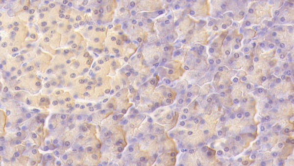
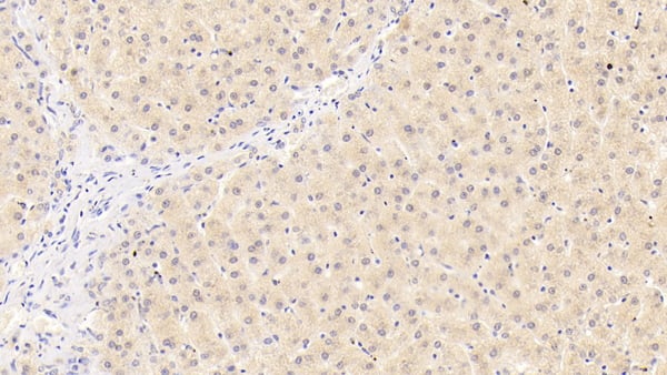
 IHC (Immunohistochemisry)
(DAB staining on IHC-P; Samples: Human Liver Tissue; Primary Ab: 20ug/ml Mouse Anti-Human MANF AntibodySecond Ab: 2ug/mL HRP-Linked Caprine Anti-Mouse IgG Polyclonal Antibody)
IHC (Immunohistochemisry)
(DAB staining on IHC-P; Samples: Human Liver Tissue; Primary Ab: 20ug/ml Mouse Anti-Human MANF AntibodySecond Ab: 2ug/mL HRP-Linked Caprine Anti-Mouse IgG Polyclonal Antibody)
Mesencephalic Astrocyte Derived Neurotrophic Factor (MANF), Monoclonal Antibody (Cat# AAA149568)


 IHC (Immunohistochemisry)
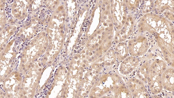
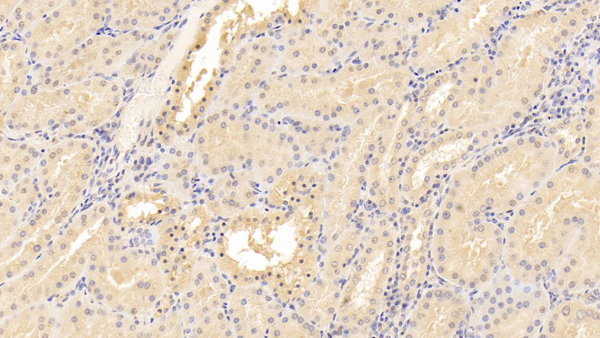
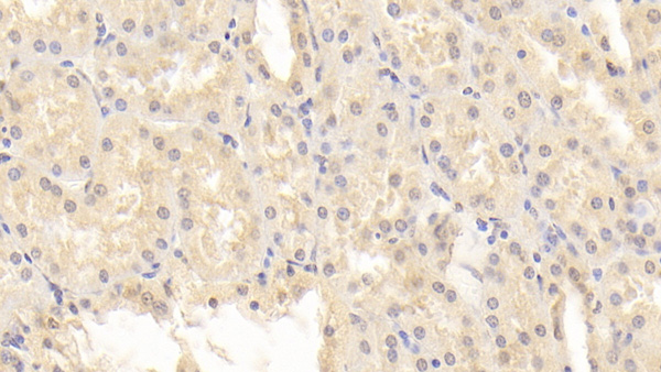
(DAB staining on IHC-P; Samples: Human Kidney Tissue; Primary Ab: 10ug/ml Mouse Anti-Human ANXA3 AntibodySecond Ab: 2ug/mL HRP-Linked Caprine Anti-Mouse IgG Polyclonal Antibody)
IHC (Immunohistochemisry)
(DAB staining on IHC-P; Samples: Human Kidney Tissue; Primary Ab: 10ug/ml Mouse Anti-Human ANXA3 AntibodySecond Ab: 2ug/mL HRP-Linked Caprine Anti-Mouse IgG Polyclonal Antibody)
Annexin A3 (ANXA3), Monoclonal Antibody (Cat# AAA149578)
 IHC (Immunohiostchemistry)
(DAB staining on IHC-P; Samples: Human Kidney Tissue; Primary Ab: 30ug/ml Mouse Anti-Human CRYl1 Antibody Second Ab: 2ug/mL HRP-Linked Caprine Anti-Mouse IgG Polyclonal Antibody)
IHC (Immunohiostchemistry)
(DAB staining on IHC-P; Samples: Human Kidney Tissue; Primary Ab: 30ug/ml Mouse Anti-Human CRYl1 Antibody Second Ab: 2ug/mL HRP-Linked Caprine Anti-Mouse IgG Polyclonal Antibody)
Crystallin Lambda 1 (CRYl1), Monoclonal Antibody (Cat# AAA149580)
 IHC (Immunohiostchemistry)
(DAB staining on IHC-P; Samples: Human Kidney Tissue; Primary Ab: 10ug/ml Mouse Anti-Human PARK7 Antibody Second Ab: 2ug/mL HRP-Linked Caprine Anti-Mouse IgG Polyclonal Antibody)
IHC (Immunohiostchemistry)
(DAB staining on IHC-P; Samples: Human Kidney Tissue; Primary Ab: 10ug/ml Mouse Anti-Human PARK7 Antibody Second Ab: 2ug/mL HRP-Linked Caprine Anti-Mouse IgG Polyclonal Antibody)
Parkinson Disease Protein 7 (PARK7), Monoclonal Antibody (Cat# AAA149585)

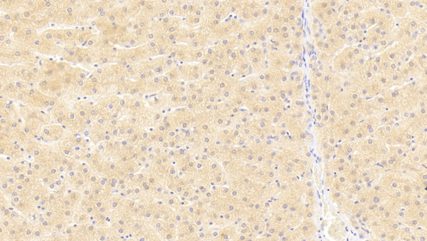
 IHC (Immunohistochemisry)
(DAB staining on IHC-P; Samples: Human Liver Tissue; Primary Ab: 40ug/ml Mouse Anti-Human ANGPTL8 Antibody Second Ab: 2ug/mL HRP-Linked Caprine Anti-Mouse IgG Polyclonal Antibody)
IHC (Immunohistochemisry)
(DAB staining on IHC-P; Samples: Human Liver Tissue; Primary Ab: 40ug/ml Mouse Anti-Human ANGPTL8 Antibody Second Ab: 2ug/mL HRP-Linked Caprine Anti-Mouse IgG Polyclonal Antibody)
Angiopoietin Like Protein 8 (ANGPTL8), Monoclonal Antibody (Cat# AAA149588)
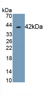
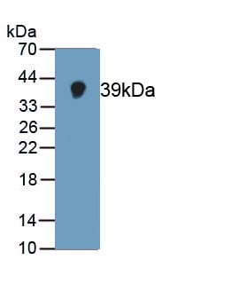
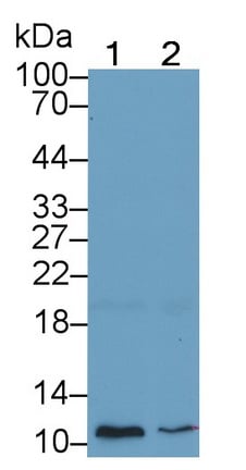
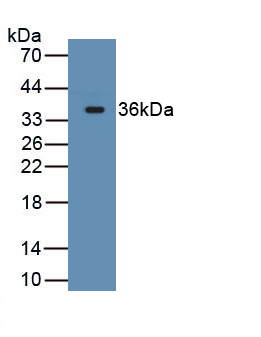
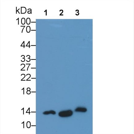
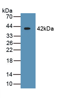
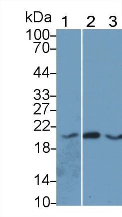
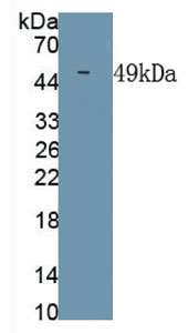
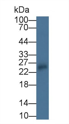
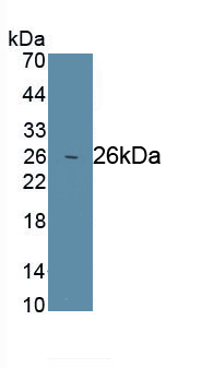
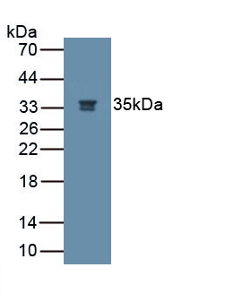
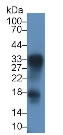
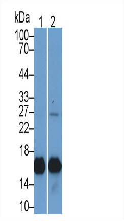
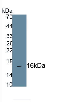
 WB (Western Blot)
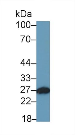
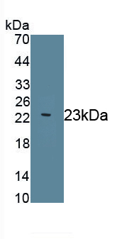
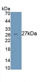
(Western Blot:Sample: Recombinant MT1E, Human)
WB (Western Blot)
(Western Blot:Sample: Recombinant MT1E, Human)
Metallothionein 1E (MT1E), Monoclonal Antibody (Cat# AAA149458)
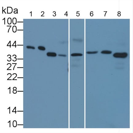
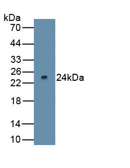
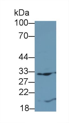
 WB (Western Blot)
(Sample: Porcine Liver lysatePrimary Ab: 2 ug/ml Mouse Anti-Human IFNa21 AntibodySecondary Ab: 0.2 ug/ml HRP-Linked Rabbit Anti-Mouse IgG Polyclonal Antibody)
WB (Western Blot)
(Sample: Porcine Liver lysatePrimary Ab: 2 ug/ml Mouse Anti-Human IFNa21 AntibodySecondary Ab: 0.2 ug/ml HRP-Linked Rabbit Anti-Mouse IgG Polyclonal Antibody)
Interferon Alpha 21 (IFNa21), Monoclonal Antibody (Cat# AAA149462)
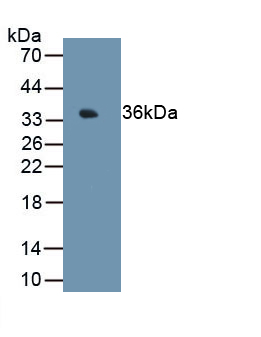
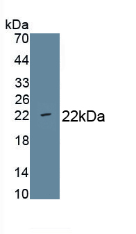
 WB (Western Blot)
(Western Blot;Sample: Recombinant CHIKV, Human.)
WB (Western Blot)
(Western Blot;Sample: Recombinant CHIKV, Human.)
Chikungunya Virus (CHIKV), Monoclonal Antibody (Cat# AAA149489)
Interferon Alpha (IFNa), Monoclonal Antibody (Cat# AAA149502)

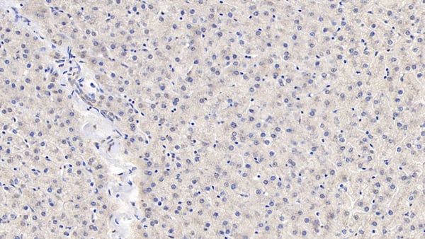
 IHC (Immunohistochemisry)
(DAB staining on IHC-P; Samples: Human Liver Tissue; Primary Ab: 20?g/ml Mouse Anti-Human Flt3L Antibody Second Ab: 2ug/mL HRP-Linked Caprine Anti-Mouse IgG Polyclonal Antibody)
IHC (Immunohistochemisry)
(DAB staining on IHC-P; Samples: Human Liver Tissue; Primary Ab: 20?g/ml Mouse Anti-Human Flt3L Antibody Second Ab: 2ug/mL HRP-Linked Caprine Anti-Mouse IgG Polyclonal Antibody)
FMS Like Tyrosine Kinase 3 Ligand (Flt3L), Monoclonal Antibody (Cat# AAA149503)

 IHC (Immunohistochemistry)
(DAB staining on IHC-P; Sample: Rabbit Spleen Tissue; Primary Ab: 20ug/ml Mouse Anti-Rabbit IL18 Antibody Second Ab: 2ug/mL HRP-Linked Caprine Anti-Mouse IgG Polyclonal Antibody)
IHC (Immunohistochemistry)
(DAB staining on IHC-P; Sample: Rabbit Spleen Tissue; Primary Ab: 20ug/ml Mouse Anti-Rabbit IL18 Antibody Second Ab: 2ug/mL HRP-Linked Caprine Anti-Mouse IgG Polyclonal Antibody)
Interleukin 18 (IL18), Monoclonal Antibody (Cat# AAA149504)
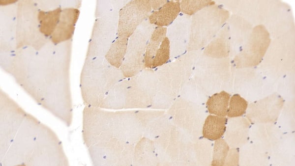
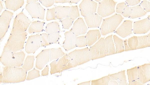 IHC (Immunohistochemistry)
(DAB staining on IHC-P; Samples: Human Skeletal muscle Tissue; Primary Ab: 30?g/ml Mouse Anti-Human LIF AntibodySecond Ab: 2ug/mL HRP-Linked Caprine Anti-Mouse IgG Polyclonal Antibody)
IHC (Immunohistochemistry)
(DAB staining on IHC-P; Samples: Human Skeletal muscle Tissue; Primary Ab: 30?g/ml Mouse Anti-Human LIF AntibodySecond Ab: 2ug/mL HRP-Linked Caprine Anti-Mouse IgG Polyclonal Antibody)
Leukemia Inhibitory Factor (LIF), Monoclonal Antibody (Cat# AAA149505)
 IHC (Immunohiostchemistry)
(DAB staining on IHC-P; Samples: Human Liver Tissue; Primary Ab: 10ug/ml Mouse Anti-Human MMP13 Antibody Second Ab: 2ug/mL HRP-Linked Caprine Anti-Mouse IgG Polyclonal Antibody)
IHC (Immunohiostchemistry)
(DAB staining on IHC-P; Samples: Human Liver Tissue; Primary Ab: 10ug/ml Mouse Anti-Human MMP13 Antibody Second Ab: 2ug/mL HRP-Linked Caprine Anti-Mouse IgG Polyclonal Antibody)
Matrix Metalloproteinase 13 (MMP13), Monoclonal Antibody (Cat# AAA149506)
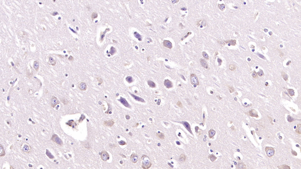 IHC (Immunohiostchemistry)
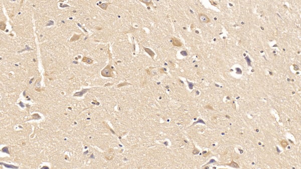
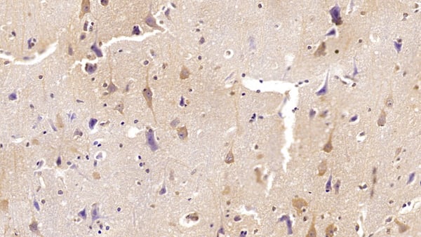
(DAB staining on IHC-P; Samples: Human Cerebrum Tissue; Primary Ab: 40ug/ml Mouse Anti-Human NGF Antibody Second Ab: 2ug/mL HRP-Linked Caprine Anti-Mouse IgG Polyclonal Antibody)
IHC (Immunohiostchemistry)
(DAB staining on IHC-P; Samples: Human Cerebrum Tissue; Primary Ab: 40ug/ml Mouse Anti-Human NGF Antibody Second Ab: 2ug/mL HRP-Linked Caprine Anti-Mouse IgG Polyclonal Antibody)
Nerve Growth Factor (NGF), Monoclonal Antibody (Cat# AAA149507)
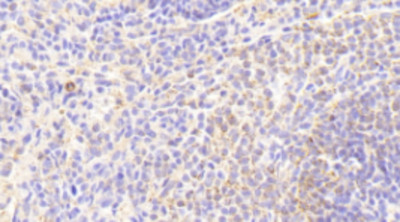
 IHC (Immunohistochemistry)
(DAB staining on IHC-P;Samples: Human Spleen Tissue;Primary Ab: 20ug/ml Mouse Anti-Human SOD3 AntibodySecond Ab: 2ug/mL HRP-Linked Caprine Anti-Mouse IgG Polyclonal Antibody)
IHC (Immunohistochemistry)
(DAB staining on IHC-P;Samples: Human Spleen Tissue;Primary Ab: 20ug/ml Mouse Anti-Human SOD3 AntibodySecond Ab: 2ug/mL HRP-Linked Caprine Anti-Mouse IgG Polyclonal Antibody)
Superoxide Dismutase 3 (SOD3), Monoclonal Antibody (Cat# AAA149508)
 IHC (Immunohistochemisry)
(DAB staining on IHC-P; Samples: Human Kidney Tissue; Primary Ab: 10ug/ml Mouse Anti-Human CD40L AntibodySecond Ab: 2ug/mL HRP-Linked Caprine Anti-Mouse IgG Polyclonal Antibody)
IHC (Immunohistochemisry)
(DAB staining on IHC-P; Samples: Human Kidney Tissue; Primary Ab: 10ug/ml Mouse Anti-Human CD40L AntibodySecond Ab: 2ug/mL HRP-Linked Caprine Anti-Mouse IgG Polyclonal Antibody)
Cluster of Differentiation 40 Ligand (CD40L), Monoclonal Antibody (Cat# AAA149509)
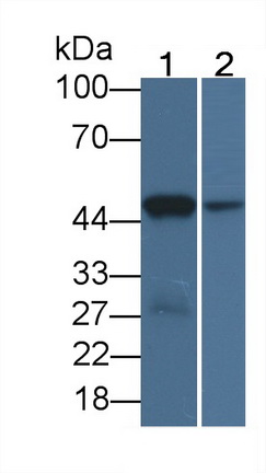
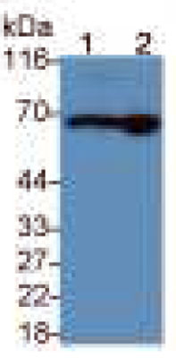 WB (Western Blot)
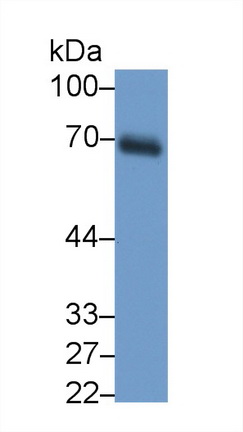
(Western Blot;Sample: Lane 1: Human Serum;Lane 2: Human Plasma Primary Ab: 0.2ug/ml Mouse Anti-Human a2PI AntibodySecond Ab: 0.2ug/mL HRP Linked Caprine Anti-Mouse IgG Polyclonal Antibody)
WB (Western Blot)
(Western Blot;Sample: Lane 1: Human Serum;Lane 2: Human Plasma Primary Ab: 0.2ug/ml Mouse Anti-Human a2PI AntibodySecond Ab: 0.2ug/mL HRP Linked Caprine Anti-Mouse IgG Polyclonal Antibody)
Alpha 2-Antiplasmin (a2PI), Monoclonal Antibody (Cat# AAA149513)
 IHC (Immunohistochemistry)
(DAB staining on IHC-P; Samples: Human Liver Tissue; Primary Ab: 30ug/ml Mouse Anti-Human APOH Antibody Second Ab: 2ug/mL HRP-Linked Caprine Anti-Mouse IgG Polyclonal Antibody)
IHC (Immunohistochemistry)
(DAB staining on IHC-P; Samples: Human Liver Tissue; Primary Ab: 30ug/ml Mouse Anti-Human APOH Antibody Second Ab: 2ug/mL HRP-Linked Caprine Anti-Mouse IgG Polyclonal Antibody)
Apolipoprotein H (APOH), Monoclonal Antibody (Cat# AAA149518)
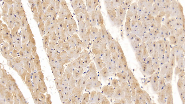
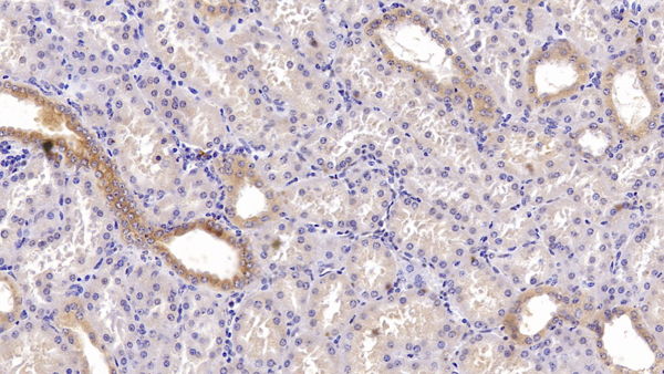
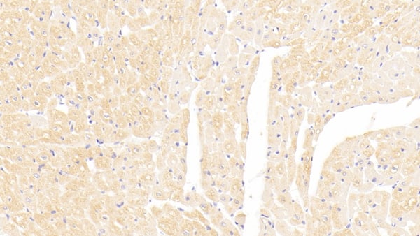
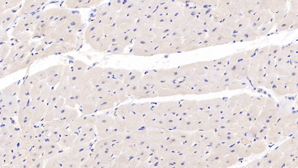
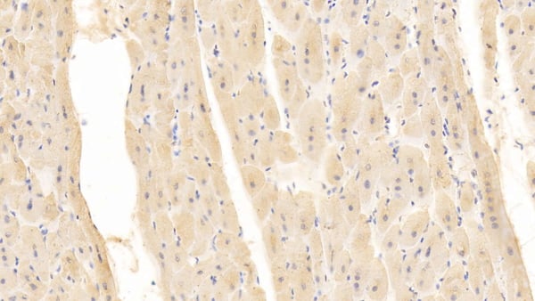
 IHC (Immunohistochemisry)
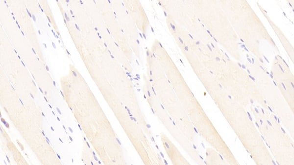
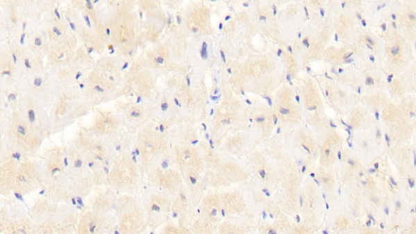
(DAB staining on IHC-P; Samples: Human Skeletal muscle Tissue; Primary Ab: 20ug/ml Mouse Anti-Human MYO AntibodySecond Ab: 2ug/mL HRP-Linked Caprine Anti-Mouse IgG Polyclonal Antibody)
IHC (Immunohistochemisry)
(DAB staining on IHC-P; Samples: Human Skeletal muscle Tissue; Primary Ab: 20ug/ml Mouse Anti-Human MYO AntibodySecond Ab: 2ug/mL HRP-Linked Caprine Anti-Mouse IgG Polyclonal Antibody)
Myoglobin (MYO), Monoclonal Antibody (Cat# AAA149524)

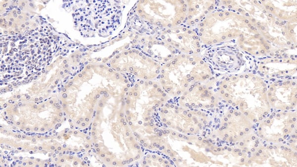
 IHC (Immunohiostchemistry)
(DAB staining on IHC-P;Samples: Human Kidney Tissue;Primary Ab: 20ug/ml Mouse Anti-Human PAI1 AntibodySecond Ab: 2ug/mL HRP Linked Caprine Anti-Mouse IgG Polyclonal Antibody)
IHC (Immunohiostchemistry)
(DAB staining on IHC-P;Samples: Human Kidney Tissue;Primary Ab: 20ug/ml Mouse Anti-Human PAI1 AntibodySecond Ab: 2ug/mL HRP Linked Caprine Anti-Mouse IgG Polyclonal Antibody)
Plasminogen Activator Inhibitor 1 (PAI1), Monoclonal Antibody (Cat# AAA149527)
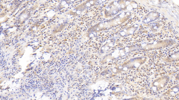
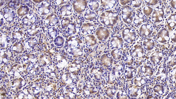
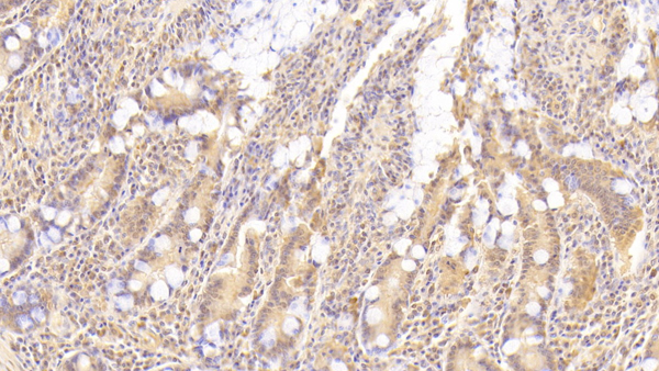
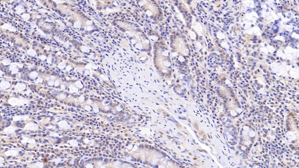
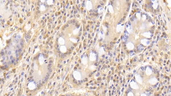
 IHC (Immunohistochemistry)
(DAB staining on IHC-P; Samples: Human Stomach Tissue; Primary Ab: 20?g/ml Mouse Anti-Human PCNA AntibodySecond Ab: 2ug/mL HRP-Linked Caprine Anti-Mouse IgG Polyclonal Antibody)
IHC (Immunohistochemistry)
(DAB staining on IHC-P; Samples: Human Stomach Tissue; Primary Ab: 20?g/ml Mouse Anti-Human PCNA AntibodySecond Ab: 2ug/mL HRP-Linked Caprine Anti-Mouse IgG Polyclonal Antibody)
Proliferating Cell Nuclear Antigen (PCNA), Monoclonal Antibody (Cat# AAA149530)
 WB (Western Blot)
(Western Blot;Sample: Recombinant CNP, Human.)
WB (Western Blot)
(Western Blot;Sample: Recombinant CNP, Human.)
C-Type Natriuretic Peptide (CNP), Monoclonal Antibody (Cat# AAA149535)
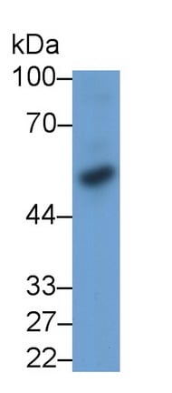
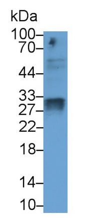
 WB (Western Blot)
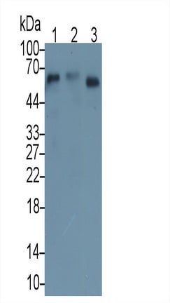
(Western Blot;Sample: Human SerumPrimary Ab: 2ug/ml Mouse Anti-Human CD276 AntibodySecond Ab: 0.2ug/mL HRP-Linked Caprine Anti-Mouse IgG Polyclonal Antibody)
WB (Western Blot)
(Western Blot;Sample: Human SerumPrimary Ab: 2ug/ml Mouse Anti-Human CD276 AntibodySecond Ab: 0.2ug/mL HRP-Linked Caprine Anti-Mouse IgG Polyclonal Antibody)
Cluster of Differentiation 276 (CD276), Monoclonal Antibody (Cat# AAA149536)
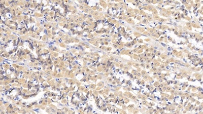
 IHC (Immunohistochemisry)
(DAB staining on IHC-P; Samples: Human Liver Tissue; Primary Ab: 30ug/ml Mouse Anti-Human ITGb1 AntibodySecond Ab: 2ug/mL HRP-Linked Caprine Anti-Mouse IgG Polyclonal Antibody)
IHC (Immunohistochemisry)
(DAB staining on IHC-P; Samples: Human Liver Tissue; Primary Ab: 30ug/ml Mouse Anti-Human ITGb1 AntibodySecond Ab: 2ug/mL HRP-Linked Caprine Anti-Mouse IgG Polyclonal Antibody)
Integrin Beta 1 (ITGb1), Monoclonal Antibody (Cat# AAA149541)
 WB (Western Blot)
(Western Blot; Sample: Raji cell lysatePrimary Ab: 2?g/ml Mouse Anti-Human SIGLEC2 AntibodySecond Ab: 0.2ug/mL HRP-Linked Rabbit Anti-Mouse IgG Polyclonal Antibody)
WB (Western Blot)
(Western Blot; Sample: Raji cell lysatePrimary Ab: 2?g/ml Mouse Anti-Human SIGLEC2 AntibodySecond Ab: 0.2ug/mL HRP-Linked Rabbit Anti-Mouse IgG Polyclonal Antibody)
Sialic Acid Binding Ig Like Lectin 2 (CD22), Monoclonal Antibody (Cat# AAA149543)
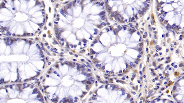
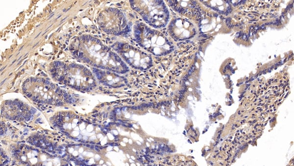
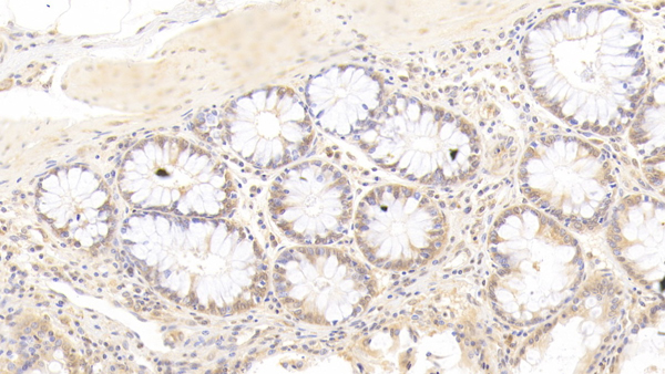
 IHC (Immunohistochemisry)
(DAB staining on IHC-P; Samples: Human Small intestine Tissue; Primary Ab: 20?g/ml Mouse Anti-Human CTLA4 AntibodySecond Ab: 2ug/mL HRP-Linked Caprine Anti-Mouse IgG Polyclonal Antibody)
IHC (Immunohistochemisry)
(DAB staining on IHC-P; Samples: Human Small intestine Tissue; Primary Ab: 20?g/ml Mouse Anti-Human CTLA4 AntibodySecond Ab: 2ug/mL HRP-Linked Caprine Anti-Mouse IgG Polyclonal Antibody)
Cytotoxic T-Lymphocyte Associated Antigen 4 (CTLA4), Monoclonal Antibody (Cat# AAA149545)
Major Basic Protein (MBP), Monoclonal Antibody (Cat# AAA149822)
Mucin 1 (MUC1), Monoclonal Antibody (Cat# AAA149824)
Triiodothyronine (T3), Monoclonal Antibody (Cat# AAA149825)
Cluster of Differentiation 276 (CD276), Monoclonal Antibody (Cat# AAA149828)
Fc Fragment Of IgG Low Affinity IIIb Receptor (FcgR3B), Monoclonal Antibody (Cat# AAA149831)
Azurocidin (AZU), Monoclonal Antibody (Cat# AAA149833)
Aspartate Beta Hydroxylase (ASPH), Monoclonal Antibody (Cat# AAA149849)
Semaphorin 5B (SEMA5B), Monoclonal Antibody (Cat# AAA149803)
Transient Receptor Potential Cation Channel Subfamily M, Member 4 (TRPM4), Monoclonal Antibody (Cat# AAA150816)
 IHC (Immunohistochemistry)
(DAB staining on IHCP;Sample: Human Spleen Tissue; Primary Ab: 20ug/ml Mouse AntiHuman SOD3 AntibodySecond Ab: 2ug/mL HRPLinked Caprine AntiMouse IgG Polyclonal Antibody(Catalog: SAA544Mu19))
IHC (Immunohistochemistry)
(DAB staining on IHCP;Sample: Human Spleen Tissue; Primary Ab: 20ug/ml Mouse AntiHuman SOD3 AntibodySecond Ab: 2ug/mL HRPLinked Caprine AntiMouse IgG Polyclonal Antibody(Catalog: SAA544Mu19))
Superoxide Dismutase 3, Extracellular (SOD3), Monoclonal Antibody (Cat# AAA151483)
What are Monoclonal Antibodies?
Monoclonal antibodies are specialized laboratory-produced proteins developed for binding to specific biological antigens or other molecular targets. Since they come from a single cell (or clone), they are especially consistent and accurate in the data they are involved in producing.
This type of antibody material has been shown to be a powerful tool in finding and subsequently destroying harmful cells in an organism, such as those found in cancers or various autoimmune diseases. This makes them excellent aids in medical testing and research, which is why they are so widely used.
AAA Biotech offers a comprehensive range of high-quality monoclonal antibodies that perform effectively in various laboratory tests, including (amongst others) ELISA, western blotting, immunohistochemistry, and flow cytometry. All of the products in our catalog are thoroughly quality tested to make sure that they are reliable and will consistently perform well in your research.
What Are The Uses of Monoclonal Antibodies
Monoclonal antibodies are used in many lab tests, including (amongst others) ELISA, western blotting, immunohistochemistry, and flow cytometry.
ELISA is a test that helps detect a specific substance/analyte in a sample. It uses antibodies (often monoclonal) bound to a solid surface (such as the well of a microplate) to “capture” the substance/analyte in the sample and immobilize it so that the detection antibody component can then bind to it and produce a signal, which can then be measured.
Western blotting identifies specific proteins in a sample. The sample is first separated on a gel, and then antibodies are applied that will typically bind to the target, which will all be localized to a single band in a lane.
Immunohistochemistry helps locate specific proteins in cells or tissue samples using antibodies.
Flow cytometry looks at and sorts cells. It uses antibodies that are conjugated to reporter molecules called “fluorophores”, which, under special lights, emit light themselves, which can then be measured by a detector instrument.
How Monoclonal Antibodies Are Used as Medicine?
Please note that all of the products listed in AAA Biotech’s also known as AAA Bio or AAABio catalog are strictly for research-use only (RUO).
Monoclonal antibodies can also be used as therapeutic/medical treatments, particularly in the context of cancers. They are designed to find and bind to specific cells or proteins, helping the immune system recognize and attack the cancer. These treatments work in different ways, such as:
- Radioimmunotherapy attaches a small amount of radioactive molecule to the antibody, so it delivers the radiation directly to the cancer cells that the antibody is specifically binding to.
- Antibody-directed enzyme prodrug therapy uses antibodies that are specifically bound to special enzymes. These enzymes activate a harmless drug in the body and turn it into a cancer-killing drug only near the cancer cells—this helps avoid harming healthy cells.
- Immunoliposomes are tiny “bubbles” filled with medicine/drug and coated with antibodies. They carry the drug straight to the cancer cells.
Why Buy Monoclonal Antibodies From Us?
At AAA Biotech, we provide high-performance monoclonal antibodies designed to support a wide range of research needs.
1. Validated for Versatile Applications
The antibodies in our catalog are extensively validated and compatible with multiple techniques, including (but not limited to) ELISA, flow cytometry (FC), immunocytochemistry (ICC), immunofluorescence (IF), immunohistochemistry (IHC), immunoprecipitation (IP), and western blotting (WB).
2. Wide Selection & Specialized Options
We offer antibodies for common and rare species, that are available in various conjugated forms, and also in recombinant formats. Essentially, there is almost anything one might need to meet their experimental model’s requirements.
3. High-Quality Proteins
Our proteins meet high purity standards—90% or more as confirmed by SDS-PAGE. Many are available with tags like His, Flag, GST, or MBP, and we also supply native and biologically active proteins for functional studies.
Frequently Asked Questions
1. Are your monoclonal antibodies validated for specific applications?
Yes, our antibodies are tested and validated for use in methods such as ELISA, western blot, IHC, flow cytometry, and more. Refer to specific product pages or datasheets for individual product information.
2. How do I choose the right monoclonal antibody for my application?
Review the product details directly for application validation, species reactivity, and target information. You may also contact our support team at any time for help.
3. How quickly can I receive my order?
Most orders are processed and shipped within 1–3 business days, depending on product availability and your shipping location.