Filters
▼Clonality
▼Type
▼Reactivity
▼Gene Name
▼Isotype
▼Host
▼Application
▼Clone
▼Monoclonal Antibodies
Get accurate results in your research with our Monoclonal Antibodies, which are specially made to target exactly what you require for your research, and will produce consistent, reliable performance in lab tests.
Viewing 1150-1200 of 27560 product results
 WB (Western Blot)
(Western Blot; Sample: Human Serum Primary Ab: 2ug/ml Mouse AntiHuman a1BG Antibody Second Ab: 0.2ug/mL HRPLinked Rabbit AntiMouse IgG Polyclonal Antibody (Catalog: SAA544Mu19))
WB (Western Blot)
(Western Blot; Sample: Human Serum Primary Ab: 2ug/ml Mouse AntiHuman a1BG Antibody Second Ab: 0.2ug/mL HRPLinked Rabbit AntiMouse IgG Polyclonal Antibody (Catalog: SAA544Mu19))
Alpha1BGlycoprotein (a1BG), Monoclonal Antibody (Cat# AAA151855)
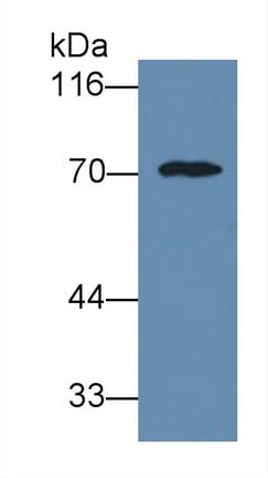
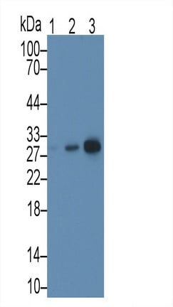 WB (Western Blot)
(Western Blot; Sample: Lane1: Rat Thymus lysate; Lane2: Hela cell lysate; Lane3: Jurkat cell lysate Primary Ab: 3ug/ml Mouse AntiHuman MAPRE1 Antibody Second Ab: 0.2ug/mL HRPLinked Caprine AntiMouse IgG Polyclonal Antibody (Catalog: SAA544Mu19))
WB (Western Blot)
(Western Blot; Sample: Lane1: Rat Thymus lysate; Lane2: Hela cell lysate; Lane3: Jurkat cell lysate Primary Ab: 3ug/ml Mouse AntiHuman MAPRE1 Antibody Second Ab: 0.2ug/mL HRPLinked Caprine AntiMouse IgG Polyclonal Antibody (Catalog: SAA544Mu19))
Microtubule Associated Protein RP/EB Family, Member 1 (MAPRE1), Monoclonal Antibody (Cat# AAA151877)
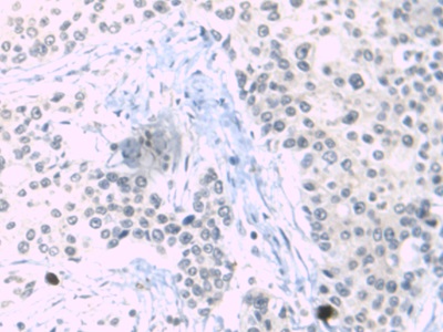
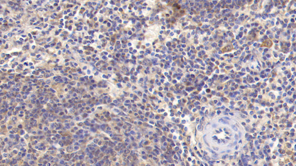
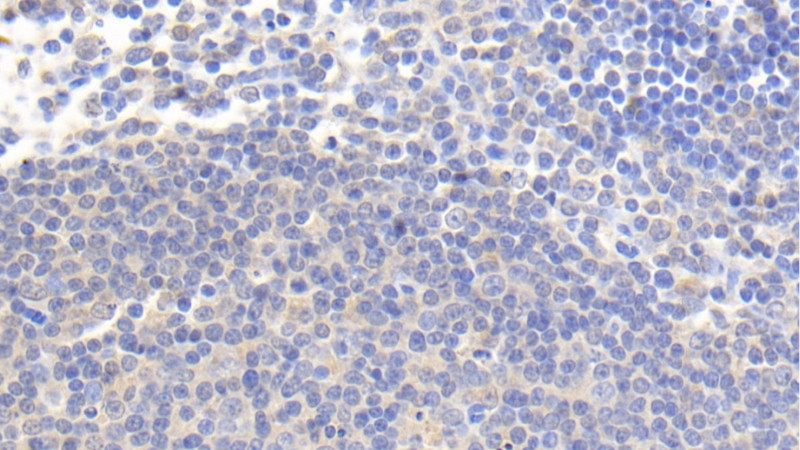 IHC (Immunohistochemistry)
(DAB staining on IHC-PSample: Human Lymph node TissuePrimary Ab: 20ug/ml Mouse Anti-Human IL4I1 AntibodySecond Ab: 2ug/mL HRP-Linked Caprine Anti-Mouse IgG Polyclonal Antibody)
IHC (Immunohistochemistry)
(DAB staining on IHC-PSample: Human Lymph node TissuePrimary Ab: 20ug/ml Mouse Anti-Human IL4I1 AntibodySecond Ab: 2ug/mL HRP-Linked Caprine Anti-Mouse IgG Polyclonal Antibody)
Interleukin 4 Induced Protein 1 (IL4I1), Monoclonal Antibody (Cat# AAA151884)
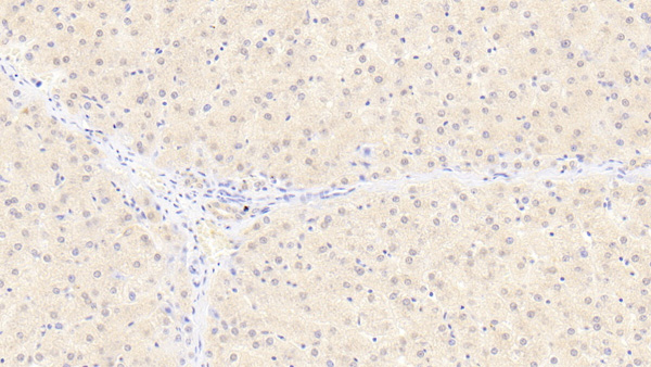 IHC (Immunohistochemistry)
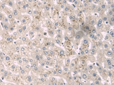
(DAB staining on IHC-P;Sample: Human Liver Tissue;Primary Ab: 20ug/ml Mouse Anti-Human FIS1 AntibodySecond Ab: 2ug/mL HRP-Linked Caprine Anti-Mouse IgG Polyclonal Antibody)
IHC (Immunohistochemistry)
(DAB staining on IHC-P;Sample: Human Liver Tissue;Primary Ab: 20ug/ml Mouse Anti-Human FIS1 AntibodySecond Ab: 2ug/mL HRP-Linked Caprine Anti-Mouse IgG Polyclonal Antibody)
Fission 1 (FIS1), Monoclonal Antibody (Cat# AAA151892)
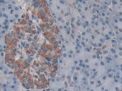 IHC (Immunohiostchemistry)
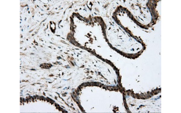
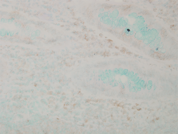
(DAB staining on IHCP;Sample: Human Pancreas Tissue; Primary Ab: 30ug/ml Mouse AntiHuman TMEM27 AntibodySecond Ab: 2ug/mL HRPLinked Caprine AntiMouse IgG Polyclonal Antibody(Catalog: SAA544Mu19))
IHC (Immunohiostchemistry)
(DAB staining on IHCP;Sample: Human Pancreas Tissue; Primary Ab: 30ug/ml Mouse AntiHuman TMEM27 AntibodySecond Ab: 2ug/mL HRPLinked Caprine AntiMouse IgG Polyclonal Antibody(Catalog: SAA544Mu19))
Transmembrane Protein 27 (TMEM27), Monoclonal Antibody (Cat# AAA151900)


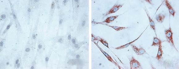
 ICC (Immunocytochemistry)
(Immunocytochemistry/Immunofluorescence analysis using Mouse Anti-Hsp60 Monoclonal Antibody, Clone LK1,. Tissue: skin Fibroblasts. Species: Human. Fixation: Cold 100% methanol for 30 minutes at -20 degree C . Primary Antibody: Mouse Anti-Hsp60 Monoclonal Antibody at 1:1000 for 1 hour at RT. Secondary Antibody: DAKO LSAB2 streptavidin-peroxidase system. Counterstain: Mayer Hematoxylin (purple/blue) nuclear stain. Left: control; Right: 24 hours after 7th passage of senescence. Courtesy of: Valentina di Felice, University of Palermo, Italy.)
ICC (Immunocytochemistry)
(Immunocytochemistry/Immunofluorescence analysis using Mouse Anti-Hsp60 Monoclonal Antibody, Clone LK1,. Tissue: skin Fibroblasts. Species: Human. Fixation: Cold 100% methanol for 30 minutes at -20 degree C . Primary Antibody: Mouse Anti-Hsp60 Monoclonal Antibody at 1:1000 for 1 hour at RT. Secondary Antibody: DAKO LSAB2 streptavidin-peroxidase system. Counterstain: Mayer Hematoxylin (purple/blue) nuclear stain. Left: control; Right: 24 hours after 7th passage of senescence. Courtesy of: Valentina di Felice, University of Palermo, Italy.)
HSP60, Monoclonal Antibody (Cat# AAA103232)
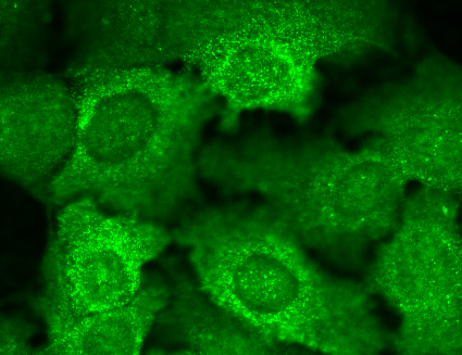
 ICC (Immunocytochemistry)
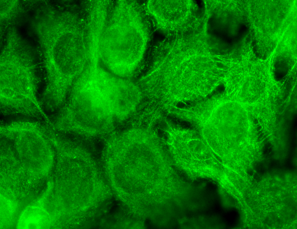
(Immunocytochemistry/Immunofluorescence analysis using Mouse Anti-TrpV3 Monoclonal Antibody, Clone S15-39. Tissue: HaCaT cells. Species: Human. Fixation: Cold 100% methanol for 10 minutes at -20 degree C. Primary Antibody: Mouse Anti-TrpV3 Monoclonal Antibody at 1:100 for 1 hour at RT. Secondary Antibody: FITC Goat Anti-Mouse (green) at 1:50 for 1 hour at RT. Localization: Dotty staining in all cells. Some intermediate filament-like staining in some cells.)
ICC (Immunocytochemistry)
(Immunocytochemistry/Immunofluorescence analysis using Mouse Anti-TrpV3 Monoclonal Antibody, Clone S15-39. Tissue: HaCaT cells. Species: Human. Fixation: Cold 100% methanol for 10 minutes at -20 degree C. Primary Antibody: Mouse Anti-TrpV3 Monoclonal Antibody at 1:100 for 1 hour at RT. Secondary Antibody: FITC Goat Anti-Mouse (green) at 1:50 for 1 hour at RT. Localization: Dotty staining in all cells. Some intermediate filament-like staining in some cells.)
TrpV3, Monoclonal Antibody (Cat# AAA103235)
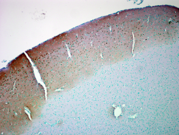
 IHC (Immunohistochemisry)
(Immunohistochemistry analysis using Mouse Anti-HCN4 Monoclonal Antibody, Clone S114-10. Tissue: hippocampus. Species: Human. Fixation: Bouin's Fixative and paraffin-embedded. Primary Antibody: Mouse Anti-HCN4 Monoclonal Antibody at 1:100 for 1 hour at RT. Secondary Antibody: FITC Goat Anti-Mouse (green) at 1:50 for 1 hour at RT.)
IHC (Immunohistochemisry)
(Immunohistochemistry analysis using Mouse Anti-HCN4 Monoclonal Antibody, Clone S114-10. Tissue: hippocampus. Species: Human. Fixation: Bouin's Fixative and paraffin-embedded. Primary Antibody: Mouse Anti-HCN4 Monoclonal Antibody at 1:100 for 1 hour at RT. Secondary Antibody: FITC Goat Anti-Mouse (green) at 1:50 for 1 hour at RT.)
HCN4, Monoclonal Antibody (Cat# AAA103242)
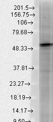
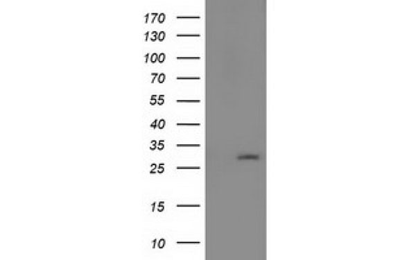
 WB (Western Blot)
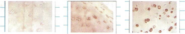
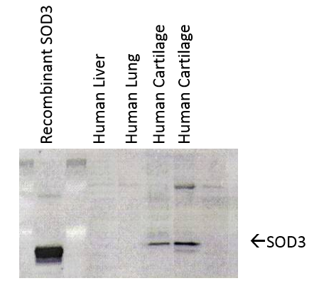
(Western Blot analysis of Human cartilage lysates showing detection of SOD3 protein using Mouse Anti-SOD3 Monoclonal Antibody, Clone 4GG11G6. Primary Antibody: Mouse Anti-SOD3 Monoclonal Antibody at 1:1000. Left: Control, Middle: Young cartilage, Right: Cartilage sample with osteoarthritis-arthritis.)
WB (Western Blot)
(Western Blot analysis of Human cartilage lysates showing detection of SOD3 protein using Mouse Anti-SOD3 Monoclonal Antibody, Clone 4GG11G6. Primary Antibody: Mouse Anti-SOD3 Monoclonal Antibody at 1:1000. Left: Control, Middle: Young cartilage, Right: Cartilage sample with osteoarthritis-arthritis.)
SOD (EC), Monoclonal Antibody (Cat# AAA103250)

 WB (Western Blot)
(Western Blot analysis of Human cartilage lysates showing detection of SOD3 protein using Mouse Anti-SOD3 Monoclonal Antibody, Clone 4GG11G6. Primary Antibody: Mouse Anti-SOD3 Monoclonal Antibody at 1:1000. Left: Control, Middle: Young cartilage, Right: Cartilage sample with osteoarthritis-arthritis.)
WB (Western Blot)
(Western Blot analysis of Human cartilage lysates showing detection of SOD3 protein using Mouse Anti-SOD3 Monoclonal Antibody, Clone 4GG11G6. Primary Antibody: Mouse Anti-SOD3 Monoclonal Antibody at 1:1000. Left: Control, Middle: Young cartilage, Right: Cartilage sample with osteoarthritis-arthritis.)
SOD (EC), Monoclonal Antibody (Cat# AAA103282)
 IHC (Immunohistochemisry)
(Immunohistochemistry analysis using Mouse Anti-HCN2 Monoclonal Antibody, Clone S71-37. Tissue: hippocampus. Species: Human. Fixation: Bouin's Fixative and paraffin-embedded. Primary Antibody: Mouse Anti-HCN2 Monoclonal Antibody at 1:100 for 1 hour at RT. Secondary Antibody: FITC Goat Anti-Mouse (green) at 1:50 for 1 hour at RT.)
IHC (Immunohistochemisry)
(Immunohistochemistry analysis using Mouse Anti-HCN2 Monoclonal Antibody, Clone S71-37. Tissue: hippocampus. Species: Human. Fixation: Bouin's Fixative and paraffin-embedded. Primary Antibody: Mouse Anti-HCN2 Monoclonal Antibody at 1:100 for 1 hour at RT. Secondary Antibody: FITC Goat Anti-Mouse (green) at 1:50 for 1 hour at RT.)
HCN2, Monoclonal Antibody (Cat# AAA103306)

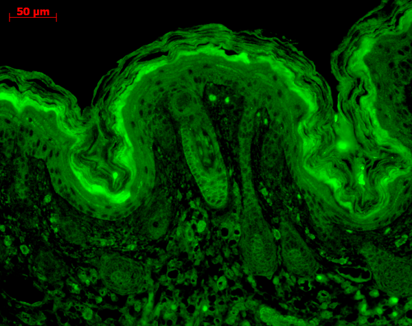
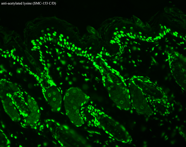
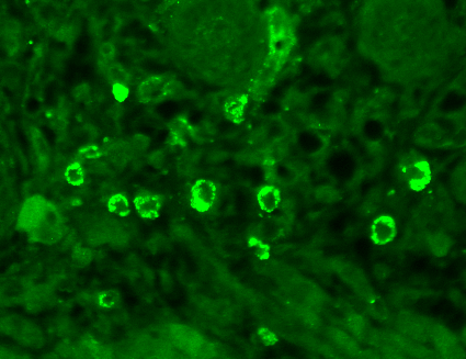
 IHC (Immunohistochemisry)
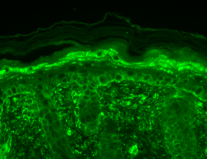
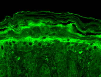
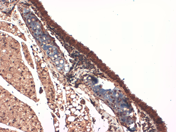
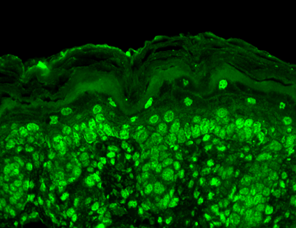
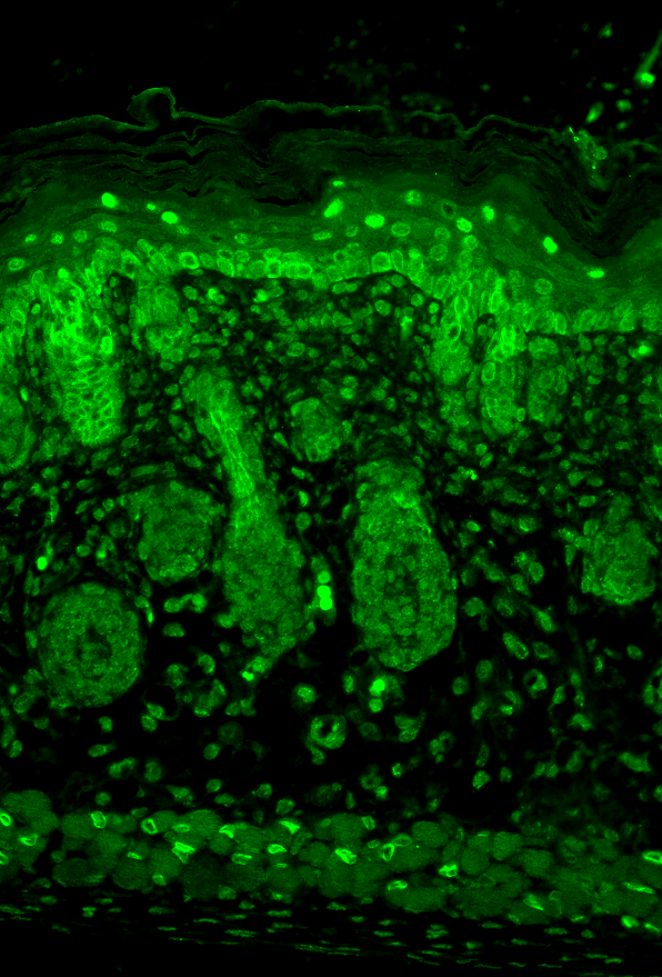
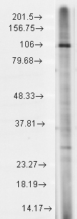
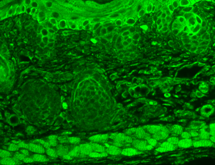
(Immunohistochemistry analysis using Mouse Anti-Slo2.2 Potassium Channel Monoclonal Antibody, Clone S3-26. Tissue: backskin. Species: Mouse. Fixation: Bouin's Fixative and paraffin-embedded. Primary Antibody: Mouse Anti-Slo2.2 Potassium Channel Monoclonal Antibody at 1:100 for 1 hour at RT. Secondary Antibody: FITC Goat Anti-Mouse (green) at 1:50 for 1 hour at RT. Localization: Suprabasal epidermal staining. Hair follicles negative.)
IHC (Immunohistochemisry)
(Immunohistochemistry analysis using Mouse Anti-Slo2.2 Potassium Channel Monoclonal Antibody, Clone S3-26. Tissue: backskin. Species: Mouse. Fixation: Bouin's Fixative and paraffin-embedded. Primary Antibody: Mouse Anti-Slo2.2 Potassium Channel Monoclonal Antibody at 1:100 for 1 hour at RT. Secondary Antibody: FITC Goat Anti-Mouse (green) at 1:50 for 1 hour at RT. Localization: Suprabasal epidermal staining. Hair follicles negative.)
Slo2.2, Monoclonal Antibody (Cat# AAA103307)


 IHC (Immunohistochemisry)
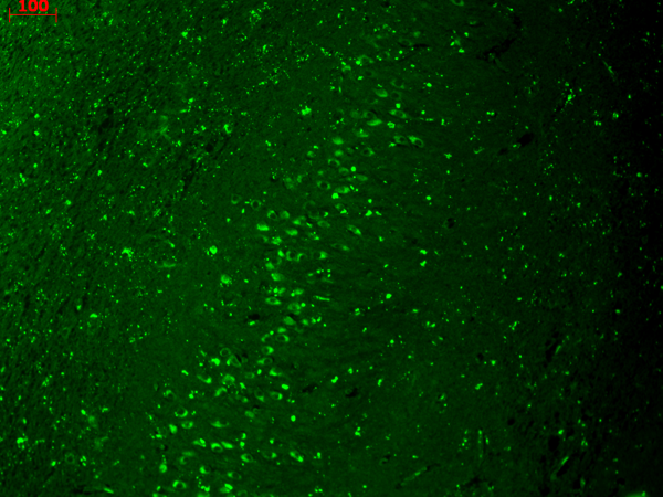
(Immunohistochemistry analysis using Mouse Anti-HCN4 Monoclonal Antibody, Clone S114-10. Tissue: hippocampus. Species: Human. Fixation: Bouin's Fixative and paraffin-embedded. Primary Antibody: Mouse Anti-HCN4 Monoclonal Antibody at 1:100 for 1 hour at RT. Secondary Antibody: FITC Goat Anti-Mouse (green) at 1:50 for 1 hour at RT.)
IHC (Immunohistochemisry)
(Immunohistochemistry analysis using Mouse Anti-HCN4 Monoclonal Antibody, Clone S114-10. Tissue: hippocampus. Species: Human. Fixation: Bouin's Fixative and paraffin-embedded. Primary Antibody: Mouse Anti-HCN4 Monoclonal Antibody at 1:100 for 1 hour at RT. Secondary Antibody: FITC Goat Anti-Mouse (green) at 1:50 for 1 hour at RT.)
HCN4, Monoclonal Antibody (Cat# AAA103317)
 IHC (Immunohistochemistry)
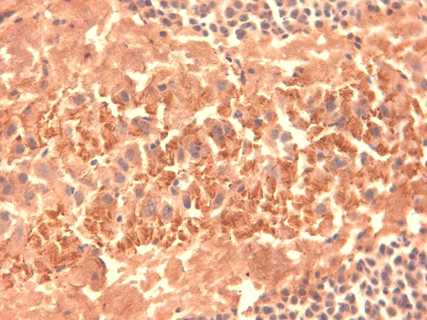
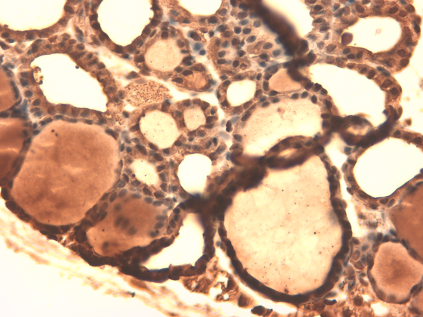
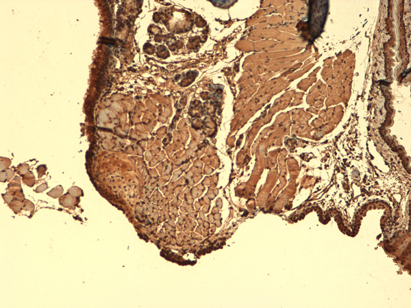
(Immunohistochemistry analysis using Mouse Anti-Sodium Iodide Symporter Monoclonal Antibody, Clone 14F. Tissue: Thyroid. Species: Mouse. Fixation: 10% Formalin Solution for 12-24 hours at RT. Primary Antibody: Mouse Anti-Sodium Iodide Symporter Monoclonal Antibody at 1:1000 for 1 hour at RT. Secondary Antibody: HRP/DAB Detection System: Biotinylated Goat Anti-Mouse, Streptavidin Peroxidase, DAB Chromogen (brown) for 30 minutes at RT. Counterstain: Mayer Hematoxylin (purple/blue) nuclear stain at 250-500 ul for 5 minutes at RT.)
IHC (Immunohistochemistry)
(Immunohistochemistry analysis using Mouse Anti-Sodium Iodide Symporter Monoclonal Antibody, Clone 14F. Tissue: Thyroid. Species: Mouse. Fixation: 10% Formalin Solution for 12-24 hours at RT. Primary Antibody: Mouse Anti-Sodium Iodide Symporter Monoclonal Antibody at 1:1000 for 1 hour at RT. Secondary Antibody: HRP/DAB Detection System: Biotinylated Goat Anti-Mouse, Streptavidin Peroxidase, DAB Chromogen (brown) for 30 minutes at RT. Counterstain: Mayer Hematoxylin (purple/blue) nuclear stain at 250-500 ul for 5 minutes at RT.)
Sodium-Iodide Symporter, Monoclonal Antibody (Cat# AAA103084)
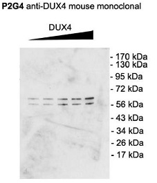 WB (Western Blot)
(Western Blot analysis of Mouse C2C12 cell lysate showing detection of DUX4 protein using Mouse Anti-DUX4 Monoclonal Antibody, Clone P2B1. Primary Antibody: Mouse Anti-DUX4 Monoclonal Antibody at 1:1000. Cells transfected with pCS2+DUX4 which, contains an additional upstream start site.)
WB (Western Blot)
(Western Blot analysis of Mouse C2C12 cell lysate showing detection of DUX4 protein using Mouse Anti-DUX4 Monoclonal Antibody, Clone P2B1. Primary Antibody: Mouse Anti-DUX4 Monoclonal Antibody at 1:1000. Cells transfected with pCS2+DUX4 which, contains an additional upstream start site.)
DUX4, Monoclonal Antibody (Cat# AAA103117)
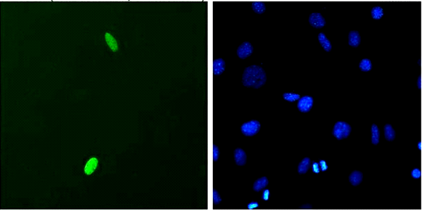
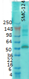
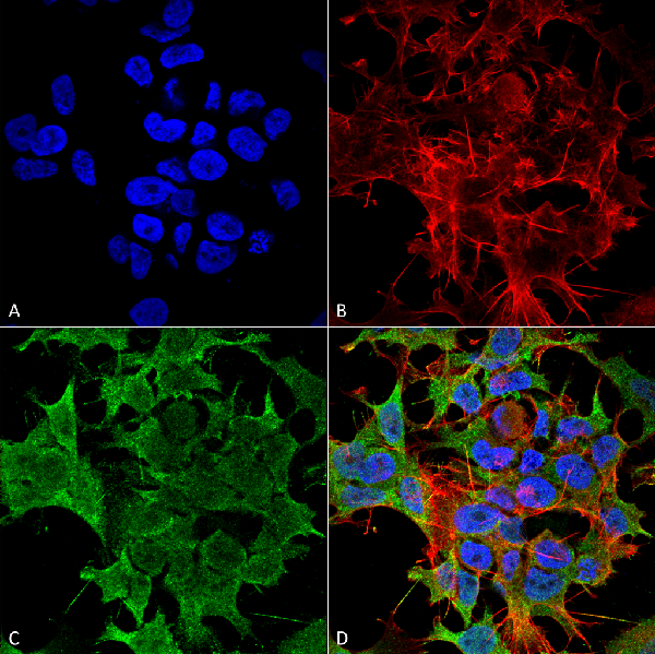
 ICC (Immunocytochemistry)
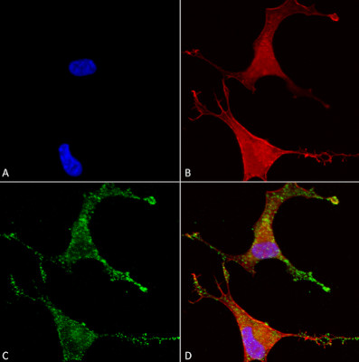
(Immunocytochemistry/Immunofluorescence analysis using Mouse Anti-Cav beta 2 Monoclonal Antibody, Clone N8b/1 (SMC-332). Tissue: Neuroblastoma cells (SH-SY5Y). Species: Human. Fixation: 4% PFA for 15 min. Primary Antibody: Mouse Anti-Cav beta 2 Monoclonal Antibody (SMC-332) at 1:50 for overnight at 4°C with slow rocking. Secondary Antibody: AlexaFluor 488 at 1:1000 for 1 hour at RT. Counterstain: Phalloidin-iFluor 647 (red) F-Actin stain; Hoechst (blue) nuclear stain at 1:800, 1.6mM for 20 min at RT. (A) Hoechst (blue) nuclear stain. (B) Phalloidin-iFluor 647 (red) F-Actin stain. (C) Cav beta 2 Antibody (D) Composite.)
ICC (Immunocytochemistry)
(Immunocytochemistry/Immunofluorescence analysis using Mouse Anti-Cav beta 2 Monoclonal Antibody, Clone N8b/1 (SMC-332). Tissue: Neuroblastoma cells (SH-SY5Y). Species: Human. Fixation: 4% PFA for 15 min. Primary Antibody: Mouse Anti-Cav beta 2 Monoclonal Antibody (SMC-332) at 1:50 for overnight at 4°C with slow rocking. Secondary Antibody: AlexaFluor 488 at 1:1000 for 1 hour at RT. Counterstain: Phalloidin-iFluor 647 (red) F-Actin stain; Hoechst (blue) nuclear stain at 1:800, 1.6mM for 20 min at RT. (A) Hoechst (blue) nuclear stain. (B) Phalloidin-iFluor 647 (red) F-Actin stain. (C) Cav beta 2 Antibody (D) Composite.)
Cavbeta2, Monoclonal Antibody (Cat# AAA103475)
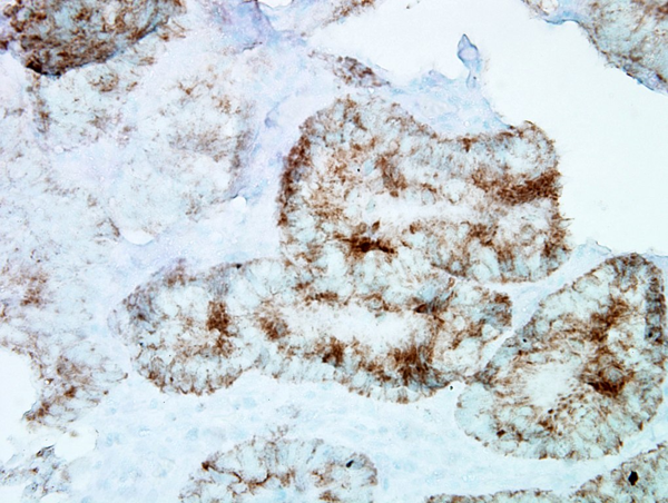
 WB (Western Blot)
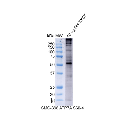
(Western Blot analysis of Human SH-SY5Y showing detection of Copper Transporting ATPase 1 protein using Mouse Anti-Copper Transporting ATPase 1 Monoclonal Antibody, Clone S60-4 . Lane 1: MW Ladder. Lane 2: 10 ug SH-SY5Y. Load: 10 ug. Block: 5% Skim Milk powder in TBST. Primary Antibody: Mouse Anti-Copper Transporting ATPase 1 Monoclonal Antibody at 1:500 for 2 hours at RT with shaking. Secondary Antibody: Goat anti-mouse IgG:HRP at 1:4000 for 1 hour at RT with shaking. Color Development: Chemiluminescent for HRP (Moss) for 5 min in RT.)
WB (Western Blot)
(Western Blot analysis of Human SH-SY5Y showing detection of Copper Transporting ATPase 1 protein using Mouse Anti-Copper Transporting ATPase 1 Monoclonal Antibody, Clone S60-4 . Lane 1: MW Ladder. Lane 2: 10 ug SH-SY5Y. Load: 10 ug. Block: 5% Skim Milk powder in TBST. Primary Antibody: Mouse Anti-Copper Transporting ATPase 1 Monoclonal Antibody at 1:500 for 2 hours at RT with shaking. Secondary Antibody: Goat anti-mouse IgG:HRP at 1:4000 for 1 hour at RT with shaking. Color Development: Chemiluminescent for HRP (Moss) for 5 min in RT.)
Copper-Transporting ATPase1, Monoclonal Antibody (Cat# AAA103352)
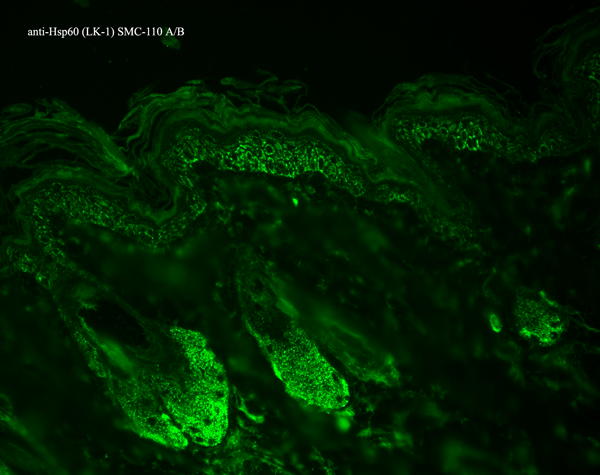

 ICC (Immunocytochemistry)
(Immunocytochemistry/Immunofluorescence analysis using Mouse Anti-Hsp60 Monoclonal Antibody, Clone LK1,. Tissue: skin Fibroblasts. Species: Human. Fixation: Cold 100% methanol for 30 minutes at -20 degree C . Primary Antibody: Mouse Anti-Hsp60 Monoclonal Antibody at 1:1000 for 1 hour at RT. Secondary Antibody: DAKO LSAB2 streptavidin-peroxidase system. Counterstain: Mayer Hematoxylin (purple/blue) nuclear stain. Left: control; Right: 24 hours after 7th passage of senescence. Courtesy of: Valentina di Felice, University of Palermo, Italy.)
ICC (Immunocytochemistry)
(Immunocytochemistry/Immunofluorescence analysis using Mouse Anti-Hsp60 Monoclonal Antibody, Clone LK1,. Tissue: skin Fibroblasts. Species: Human. Fixation: Cold 100% methanol for 30 minutes at -20 degree C . Primary Antibody: Mouse Anti-Hsp60 Monoclonal Antibody at 1:1000 for 1 hour at RT. Secondary Antibody: DAKO LSAB2 streptavidin-peroxidase system. Counterstain: Mayer Hematoxylin (purple/blue) nuclear stain. Left: control; Right: 24 hours after 7th passage of senescence. Courtesy of: Valentina di Felice, University of Palermo, Italy.)
HSP60, Monoclonal Antibody (Cat# AAA103397)

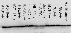
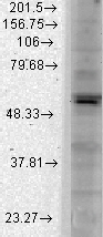
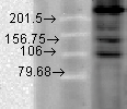
 WB (Western Blot)
(Western Blot analysis of Rat brain membrane lysate showing detection of GABA A Receptor protein using Mouse Anti-GABA A Receptor Monoclonal Antibody, Clone S87-25. Load: 15 ug. Block: 1.5% BSA for 30 minutes at RT. Primary Antibody: Mouse Anti-GABA A Receptor Monoclonal Antibody at 1:1000 for 2 hours at RT. Secondary Antibody: Sheep Anti-Mouse IgG: HRP for 1 hour at RT.)
WB (Western Blot)
(Western Blot analysis of Rat brain membrane lysate showing detection of GABA A Receptor protein using Mouse Anti-GABA A Receptor Monoclonal Antibody, Clone S87-25. Load: 15 ug. Block: 1.5% BSA for 30 minutes at RT. Primary Antibody: Mouse Anti-GABA A Receptor Monoclonal Antibody at 1:1000 for 2 hours at RT. Secondary Antibody: Sheep Anti-Mouse IgG: HRP for 1 hour at RT.)
GABA(A) Receptor Beta3, Monoclonal Antibody (Cat# AAA103407)
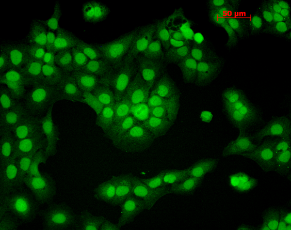
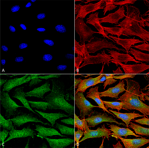
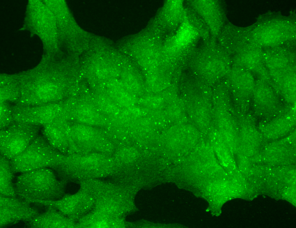
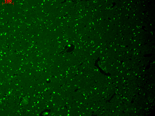
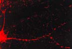 ICC (Immunocytochemistry)
(Immunocytochemistry/Immunofluorescence analysis using Mouse Anti-CaMKII Monoclonal Antibody, Clone 6G9. Tissue: dissociated hippocampal neurons. Species: Mouse. Fixation: Cold 4% paraformaldehyde/0.2% glutaraldehyde in 0.1M sodium phosphate buffer. Primary Antibody: Mouse Anti-CaMKII Monoclonal Antibody at 1:1000 for 12 hours at 4 degree C. Secondary Antibody: FITC Goat Anti-Mouse IgG (green) at 1:50 for 30 minutes at RT. Magnification: 10X. Courtesy of: Mary Kennedy, Caltech.)
ICC (Immunocytochemistry)
(Immunocytochemistry/Immunofluorescence analysis using Mouse Anti-CaMKII Monoclonal Antibody, Clone 6G9. Tissue: dissociated hippocampal neurons. Species: Mouse. Fixation: Cold 4% paraformaldehyde/0.2% glutaraldehyde in 0.1M sodium phosphate buffer. Primary Antibody: Mouse Anti-CaMKII Monoclonal Antibody at 1:1000 for 12 hours at 4 degree C. Secondary Antibody: FITC Goat Anti-Mouse IgG (green) at 1:50 for 30 minutes at RT. Magnification: 10X. Courtesy of: Mary Kennedy, Caltech.)
CaMKII (alpha-specific), Monoclonal Antibody (Cat# AAA103409)
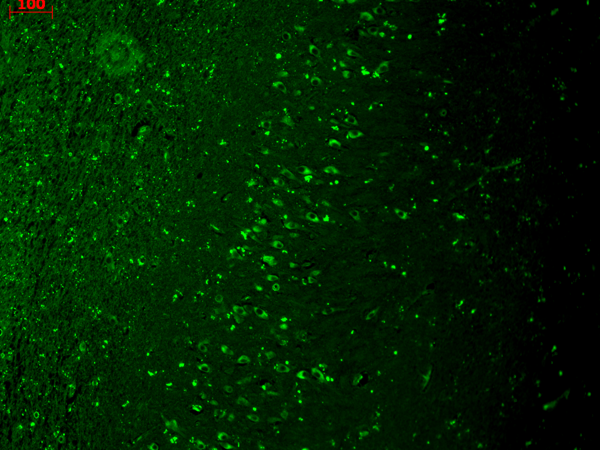
 IHC (Immunohistochemisry)
(Immunohistochemistry analysis using Mouse Anti-Kir2.1 Potassium Channel Monoclonal Antibody, Clone S112B-14. Tissue: hippocampus. Species: Human. Fixation: Bouin's Fixative and paraffin-embedded. Primary Antibody: Mouse Anti-Kir2.1 Potassium Channel Monoclonal Antibody at 1:1000 for 1 hour at RT. Secondary Antibody: FITC Goat Anti-Mouse (green) at 1:50 for 1 hour at RT.)
IHC (Immunohistochemisry)
(Immunohistochemistry analysis using Mouse Anti-Kir2.1 Potassium Channel Monoclonal Antibody, Clone S112B-14. Tissue: hippocampus. Species: Human. Fixation: Bouin's Fixative and paraffin-embedded. Primary Antibody: Mouse Anti-Kir2.1 Potassium Channel Monoclonal Antibody at 1:1000 for 1 hour at RT. Secondary Antibody: FITC Goat Anti-Mouse (green) at 1:50 for 1 hour at RT.)
Kir2.1, Monoclonal Antibody (Cat# AAA103425)
 WB (Western Blot)
(Western Blot analysis of Human Cell lysates showing detection of TrpM7 protein using Mouse Anti-TrpM7 Monoclonal Antibody, Clone S74-25. Load: 15 ug. Block: 1.5% BSA for 30 minutes at RT. Primary Antibody: Mouse Anti-TrpM7 Monoclonal Antibody at 1:1000 for 2 hours at RT. Secondary Antibody: Sheep Anti-Mouse IgG: HRP for 1 hour at RT.)
WB (Western Blot)
(Western Blot analysis of Human Cell lysates showing detection of TrpM7 protein using Mouse Anti-TrpM7 Monoclonal Antibody, Clone S74-25. Load: 15 ug. Block: 1.5% BSA for 30 minutes at RT. Primary Antibody: Mouse Anti-TrpM7 Monoclonal Antibody at 1:1000 for 2 hours at RT. Secondary Antibody: Sheep Anti-Mouse IgG: HRP for 1 hour at RT.)
TrpM7, Monoclonal Antibody (Cat# AAA103440)
 WB (Western Blot)
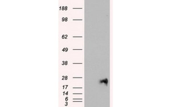
(Western Blot analysis of HEK293T cell lysates (5 ug) transfected with either recombinant NEUROG1 protein (Right) or empty vector (Left) detected with NEUROG1 antibody)
WB (Western Blot)
(Western Blot analysis of HEK293T cell lysates (5 ug) transfected with either recombinant NEUROG1 protein (Right) or empty vector (Left) detected with NEUROG1 antibody)
NEUROG1, Monoclonal Antibody (Cat# AAA108219)
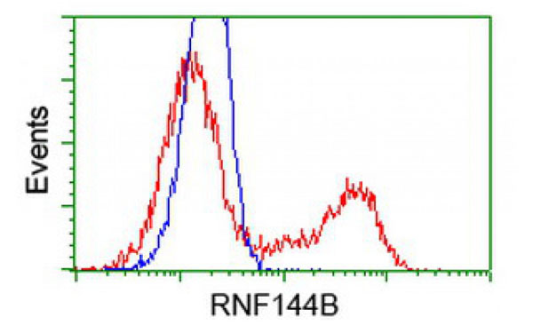
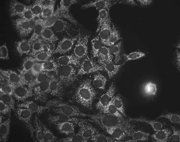
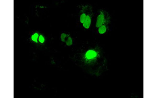
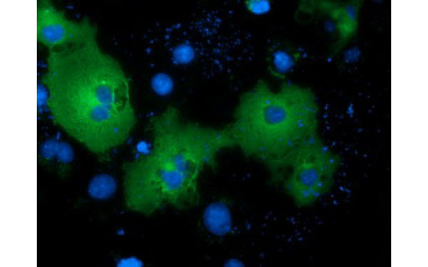
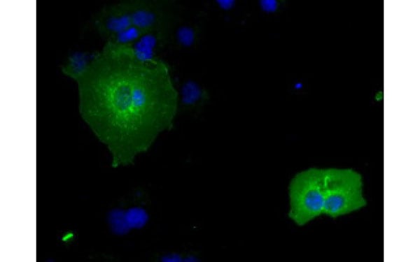 IF (Immunofluorescence)
(Immunofluorescent staining of COS7 cells transiently transfected with recombinant RNF144B protein using RNF144B antibody)
IF (Immunofluorescence)
(Immunofluorescent staining of COS7 cells transiently transfected with recombinant RNF144B protein using RNF144B antibody)
RNF144B, Monoclonal Antibody (Cat# AAA108238)
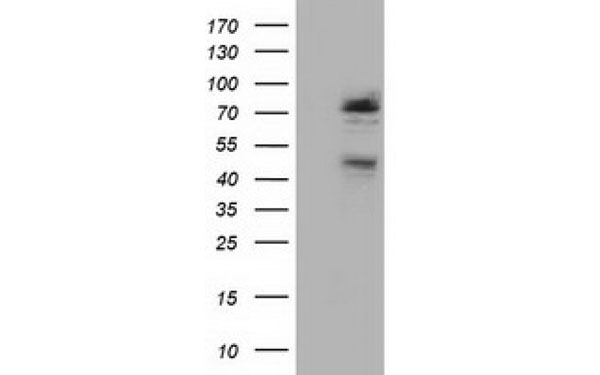
 WB (Western Blot)
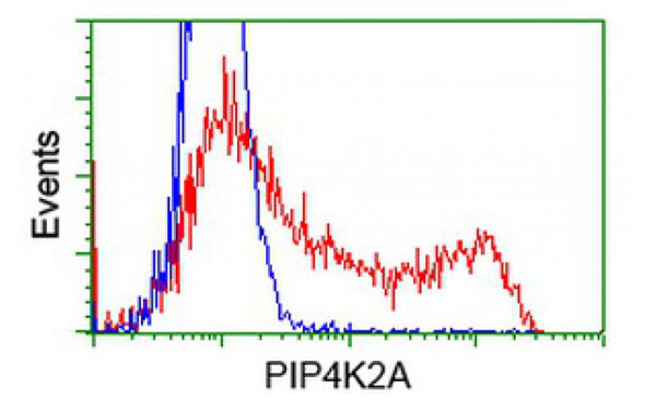
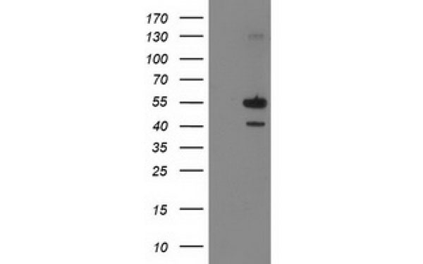
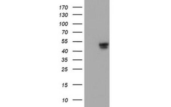
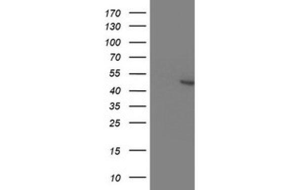
(Western Blot analysis of HEK293T cell lysates (5 ug) transfected with either recombinant PIP4K2A protein (Right) or empty vector (Left) detected with PIP4K2A antibody)
WB (Western Blot)
(Western Blot analysis of HEK293T cell lysates (5 ug) transfected with either recombinant PIP4K2A protein (Right) or empty vector (Left) detected with PIP4K2A antibody)
PIP4K2A, Monoclonal Antibody (Cat# AAA108248)
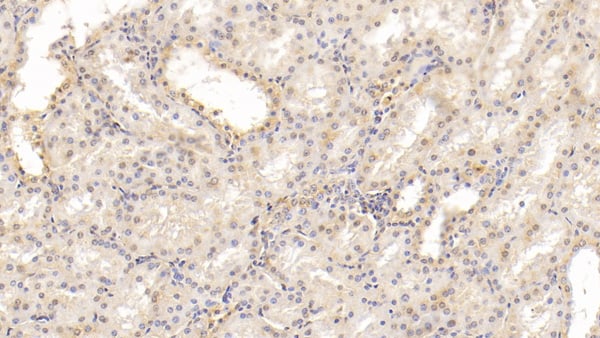
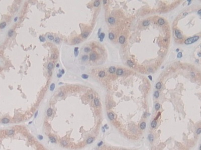
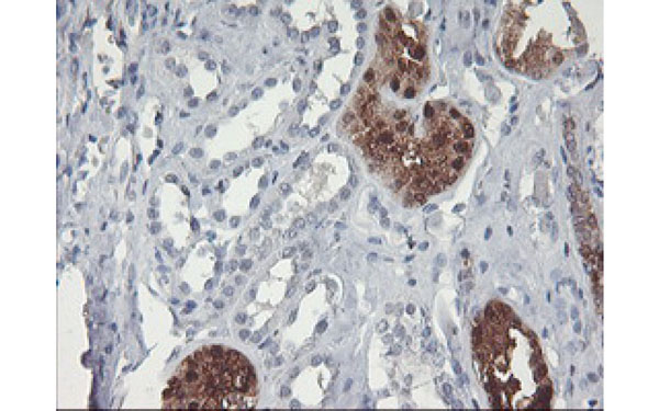
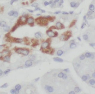
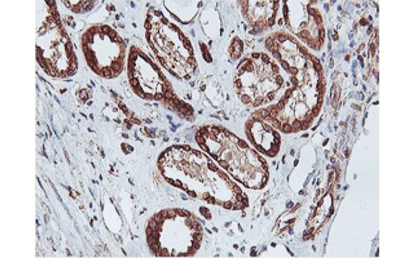 IHC (Immunohiostchemistry)
(Immunohistochemical analysis of C20orf3 protein in paraffin embedded Human Kidney tissue using C20orf3 antibody)
IHC (Immunohiostchemistry)
(Immunohistochemical analysis of C20orf3 protein in paraffin embedded Human Kidney tissue using C20orf3 antibody)
C20orf3, Monoclonal Antibody (Cat# AAA108268)
 WB (Western Blot)
(Western Blot analysis of HEK293T cell lysates (5 ug) transfected with either recombinant KCNAB1 protein (Right) or empty vector (Left) detected with KCNAB1 antibody)
WB (Western Blot)
(Western Blot analysis of HEK293T cell lysates (5 ug) transfected with either recombinant KCNAB1 protein (Right) or empty vector (Left) detected with KCNAB1 antibody)
KCNAB1, Monoclonal Antibody (Cat# AAA108273)
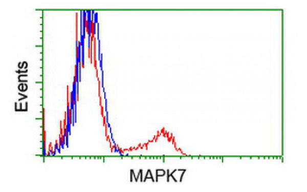
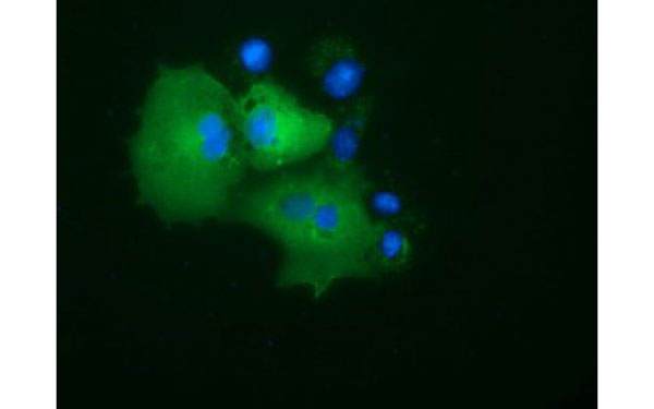
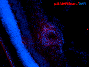
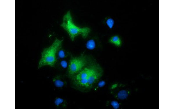 IF (Immunofluorescence)
(Immunofluorescent staining of COS7 cells transiently transfected with recombinant MAPK7 protein using MAPK7 antibody)
IF (Immunofluorescence)
(Immunofluorescent staining of COS7 cells transiently transfected with recombinant MAPK7 protein using MAPK7 antibody)
MAPK7, Monoclonal Antibody (Cat# AAA108289)
 IF (Immunofluorescence)
(Immunofluorescent staining of COS7 cells transiently transfected with recombinant PIM2 protein using PIM2 antibody)
IF (Immunofluorescence)
(Immunofluorescent staining of COS7 cells transiently transfected with recombinant PIM2 protein using PIM2 antibody)
PIM2, Monoclonal Antibody (Cat# AAA108100)
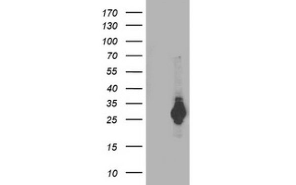 WB (Western Blot)
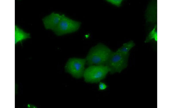
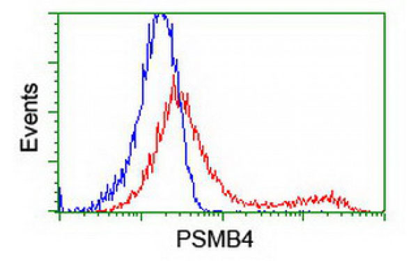
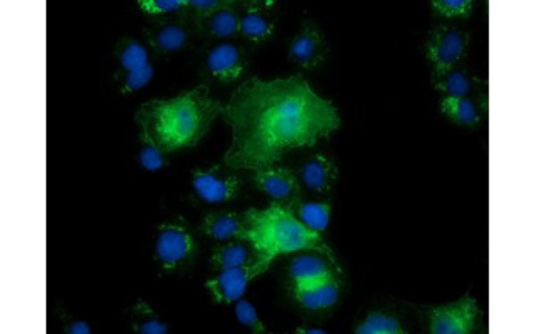
(Western Blot analysis of HEK293T cell lysates (5 ug) transfected with either recombinant PSMB4 protein (Right) or empty vector (Left) detected with PSMB4 antibody)
WB (Western Blot)
(Western Blot analysis of HEK293T cell lysates (5 ug) transfected with either recombinant PSMB4 protein (Right) or empty vector (Left) detected with PSMB4 antibody)
PSMB4, Monoclonal Antibody (Cat# AAA108107)
 WB (Western Blot)
(Western Blot analysis of HEK293T cell lysates (5 ug) transfected with either recombinant PYCRL protein (Right) or empty vector (Left) detected with PYCRL antibody)
WB (Western Blot)
(Western Blot analysis of HEK293T cell lysates (5 ug) transfected with either recombinant PYCRL protein (Right) or empty vector (Left) detected with PYCRL antibody)
PYCRL, Monoclonal Antibody (Cat# AAA108116)
Bt Cry1F, Monoclonal Antibody (Cat# AAA108149)
 IF (Immunofluorescence)
(Immunofluorescent staining of COS7 cells transiently transfected with recombinant PRKD2 protein using PRKD2 antibody)
IF (Immunofluorescence)
(Immunofluorescent staining of COS7 cells transiently transfected with recombinant PRKD2 protein using PRKD2 antibody)
PRKD2, Monoclonal Antibody (Cat# AAA107986)
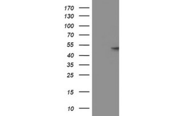
 WB (Western Blot)
(Western Blot analysis of HEK293T cell lysates (5 ug) transfected with either recombinant NEK11 protein (Right) or empty vector (Left) detected with NEK11 antibody)
WB (Western Blot)
(Western Blot analysis of HEK293T cell lysates (5 ug) transfected with either recombinant NEK11 protein (Right) or empty vector (Left) detected with NEK11 antibody)
NEK11, Monoclonal Antibody (Cat# AAA107991)
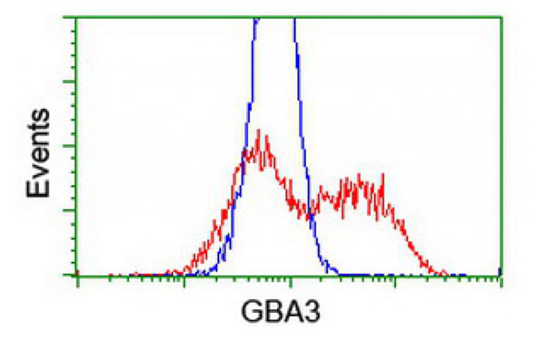
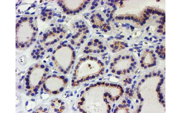
 IHC (Immunohistochemisry)
(Immunohistochemical analysis of GBA3 protein in paraffin embedded Human Kidney tissue using GBA3 antibody)
IHC (Immunohistochemisry)
(Immunohistochemical analysis of GBA3 protein in paraffin embedded Human Kidney tissue using GBA3 antibody)
GBA3, Monoclonal Antibody (Cat# AAA108002)
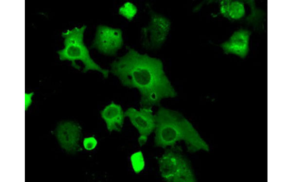
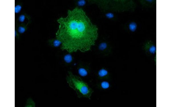 IF (Immunofluorescence)
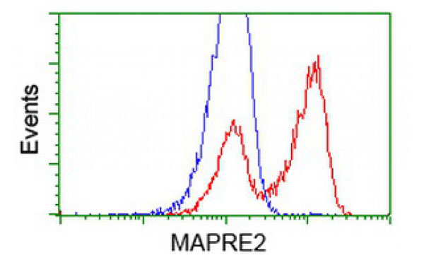
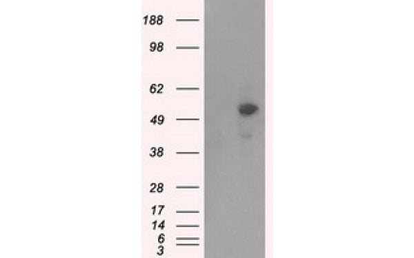
(Immunofluorescent staining of COS7 cells transiently transfected with recombinant MAPRE2 protein using MAPRE2 antibody)
IF (Immunofluorescence)
(Immunofluorescent staining of COS7 cells transiently transfected with recombinant MAPRE2 protein using MAPRE2 antibody)
MAPRE2, Monoclonal Antibody (Cat# AAA108004)
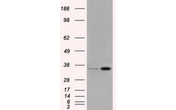
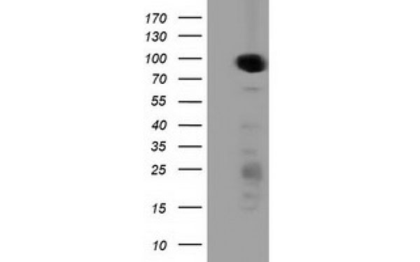
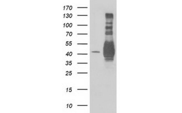
 WB (Western Blot)
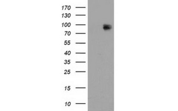
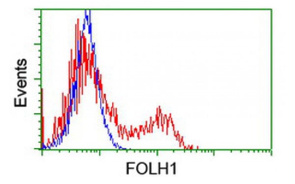
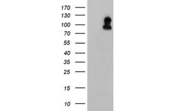
(Western Blot analysis of HEK293T cell lysates (5 ug) transfected with either recombinant FOLH1 protein (Right) or empty vector (Left) detected with FOLH1 antibody)
WB (Western Blot)
(Western Blot analysis of HEK293T cell lysates (5 ug) transfected with either recombinant FOLH1 protein (Right) or empty vector (Left) detected with FOLH1 antibody)
FOLH1, Monoclonal Antibody (Cat# AAA108021)
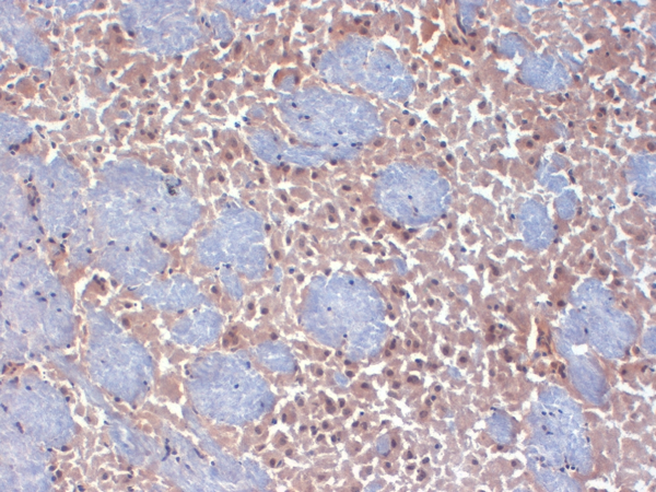
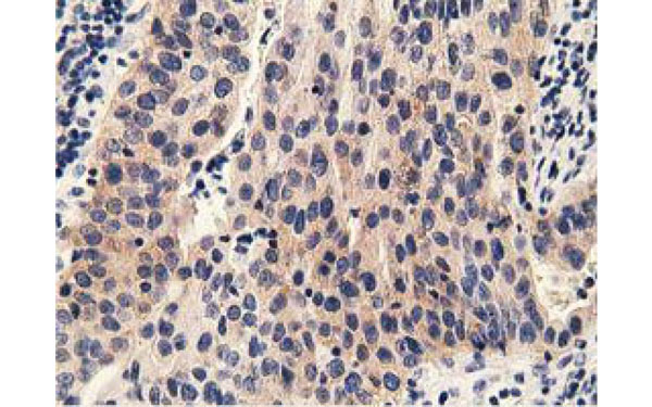
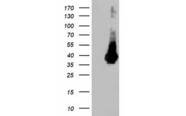
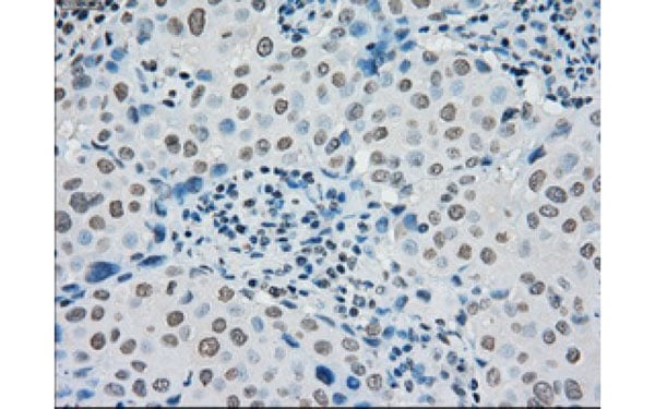
 IHC (Immunohistochemisry)
(Immunohistochemical analysis of PRKAR2A protein in paraffin embedded Carcinoma of Human prostate tissue using PRKAR2A antibody)
IHC (Immunohistochemisry)
(Immunohistochemical analysis of PRKAR2A protein in paraffin embedded Carcinoma of Human prostate tissue using PRKAR2A antibody)
PRKAR2A, Monoclonal Antibody (Cat# AAA108035)
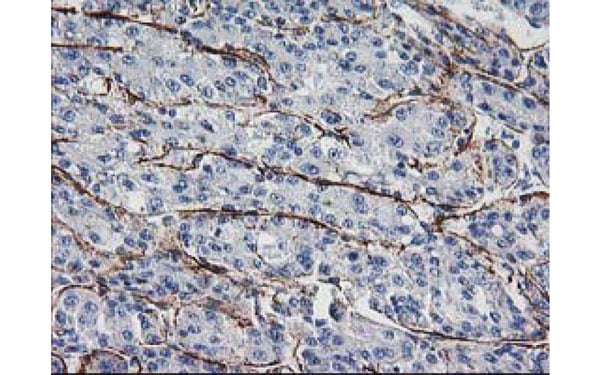 IHC (Immunohiostchemistry)
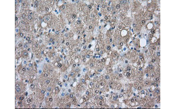
(Immunohistochemical analysis of LDLRAP1 protein in paraffin embedded Carcinoma of Human liver tissue using LDLRAP1 antibody)
IHC (Immunohiostchemistry)
(Immunohistochemical analysis of LDLRAP1 protein in paraffin embedded Carcinoma of Human liver tissue using LDLRAP1 antibody)
LDLRAP1, Monoclonal Antibody (Cat# AAA108076)
 IHC (Immunohiostchemistry)
(Immunohistochemical analysis of PMEL protein in paraffin embedded Adenocarcinoma of Human breast tissue using PMEL antibody)
IHC (Immunohiostchemistry)
(Immunohistochemical analysis of PMEL protein in paraffin embedded Adenocarcinoma of Human breast tissue using PMEL antibody)
PMEL, Monoclonal Antibody (Cat# AAA108077)
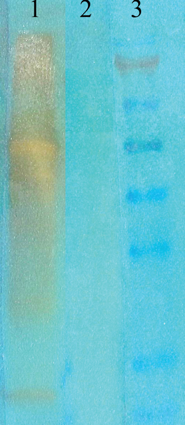 WB (Western Blot)
(Western Blot analysis of acetylated lysine showing detection of Acetylated Lysine protein using Mouse Anti-Acetylated Lysine Monoclonal Antibody, Clone 7F8. Primary Antibody: Mouse Anti-Acetylated Lysine Monoclonal Antibody at 1:1000. (1) acetylated BSA (75ng of protein), (2) non-acetylated BSA, and (3) marker.)
WB (Western Blot)
(Western Blot analysis of acetylated lysine showing detection of Acetylated Lysine protein using Mouse Anti-Acetylated Lysine Monoclonal Antibody, Clone 7F8. Primary Antibody: Mouse Anti-Acetylated Lysine Monoclonal Antibody at 1:1000. (1) acetylated BSA (75ng of protein), (2) non-acetylated BSA, and (3) marker.)
Acetylated Lysine, Monoclonal Antibody (Cat# AAA102914)
 WB (Western Blot)
(Western Blot analysis of Rat brain membrane lysate showing detection of GABA A Receptor protein using Mouse Anti-GABA A Receptor Monoclonal Antibody, Clone S87-25. Load: 15 ug. Block: 1.5% BSA for 30 minutes at RT. Primary Antibody: Mouse Anti-GABA A Receptor Monoclonal Antibody at 1:1000 for 2 hours at RT. Secondary Antibody: Sheep Anti-Mouse IgG: HRP for 1 hour at RT.)
WB (Western Blot)
(Western Blot analysis of Rat brain membrane lysate showing detection of GABA A Receptor protein using Mouse Anti-GABA A Receptor Monoclonal Antibody, Clone S87-25. Load: 15 ug. Block: 1.5% BSA for 30 minutes at RT. Primary Antibody: Mouse Anti-GABA A Receptor Monoclonal Antibody at 1:1000 for 2 hours at RT. Secondary Antibody: Sheep Anti-Mouse IgG: HRP for 1 hour at RT.)
GABA(A) Receptor Beta3, Monoclonal Antibody (Cat# AAA102952)
 WB (Western Blot)
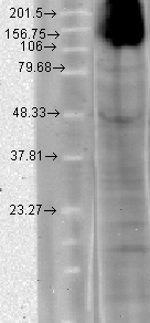
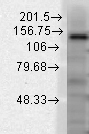
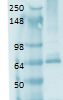
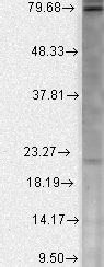
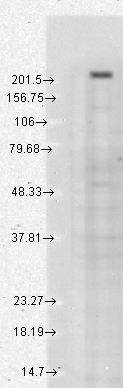
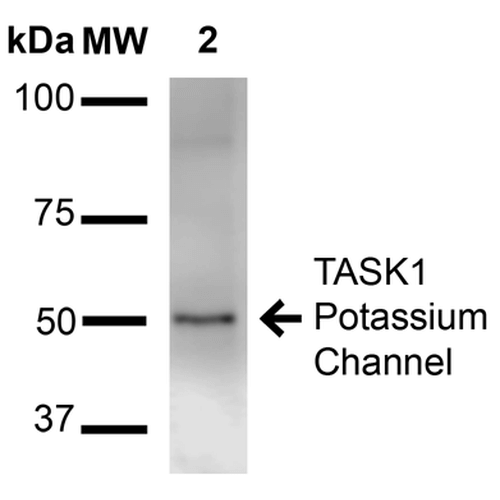
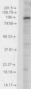
(Western Blot analysis of Rat Brain Membrane showing detection of ~50 kDa TASK1 Potassium Channel protein using Mouse Anti-TASK1 Potassium Channel Monoclonal Antibody, Clone S374-48 . Lane 1: Molecular Weight Ladder (MW). Lane 2: Rat brain membrane. Load: 15 ug. Block: 2% BSA and 2% Skim Milk in 1X TBST. Primary Antibody: Mouse Anti-TASK1 Potassium Channel Monoclonal Antibody at 1:1000 for 16 hours at 4 degree C. Secondary Antibody: Goat Anti-Mouse IgG: HRP at 1:2000 for 60 min at RT. Color Development: ECL solution for 6 min at RT. Predicted/Observed Size: ~50 kDa.)
WB (Western Blot)
(Western Blot analysis of Rat Brain Membrane showing detection of ~50 kDa TASK1 Potassium Channel protein using Mouse Anti-TASK1 Potassium Channel Monoclonal Antibody, Clone S374-48 . Lane 1: Molecular Weight Ladder (MW). Lane 2: Rat brain membrane. Load: 15 ug. Block: 2% BSA and 2% Skim Milk in 1X TBST. Primary Antibody: Mouse Anti-TASK1 Potassium Channel Monoclonal Antibody at 1:1000 for 16 hours at 4 degree C. Secondary Antibody: Goat Anti-Mouse IgG: HRP at 1:2000 for 60 min at RT. Color Development: ECL solution for 6 min at RT. Predicted/Observed Size: ~50 kDa.)
TASK1 Potassium Channel, Monoclonal Antibody (Cat# AAA102957)
 WB (Western Blot)
(Western Blot analysis of Human Cell line lysates showing detection of GABA A Receptor protein using Mouse Anti-GABA A Receptor Monoclonal Antibody, Clone S95-35. Load: 15 ug. Block: 1.5% BSA for 30 minutes at RT. Primary Antibody: Mouse Anti-GABA A Receptor Monoclonal Antibody at 1:1000 for 2 hours at RT. Secondary Antibody: Sheep Anti-Mouse IgG: HRP for 1 hour at RT.)
WB (Western Blot)
(Western Blot analysis of Human Cell line lysates showing detection of GABA A Receptor protein using Mouse Anti-GABA A Receptor Monoclonal Antibody, Clone S95-35. Load: 15 ug. Block: 1.5% BSA for 30 minutes at RT. Primary Antibody: Mouse Anti-GABA A Receptor Monoclonal Antibody at 1:1000 for 2 hours at RT. Secondary Antibody: Sheep Anti-Mouse IgG: HRP for 1 hour at RT.)
GABA(A) Receptor Alpha1, Monoclonal Antibody (Cat# AAA102990)
 ICC (Immunocytochemistry)
(Immunocytochemistry/Immunofluorescence analysis using Mouse Anti-TrpC7 Monoclonal Antibody, Clone S64A-36. Tissue: HaCaT cells. Species: Human. Fixation: Cold 100% methanol for 10 minutes at -20 degree C. Primary Antibody: Mouse Anti-TrpC7 Monoclonal Antibody at 1:100 for 1 hour at RT. Secondary Antibody: FITC Goat Anti-Mouse (green) at 1:50 for 1 hour at RT. Localization: Nuclear staining .)
ICC (Immunocytochemistry)
(Immunocytochemistry/Immunofluorescence analysis using Mouse Anti-TrpC7 Monoclonal Antibody, Clone S64A-36. Tissue: HaCaT cells. Species: Human. Fixation: Cold 100% methanol for 10 minutes at -20 degree C. Primary Antibody: Mouse Anti-TrpC7 Monoclonal Antibody at 1:100 for 1 hour at RT. Secondary Antibody: FITC Goat Anti-Mouse (green) at 1:50 for 1 hour at RT. Localization: Nuclear staining .)
TrpC7, Monoclonal Antibody (Cat# AAA102843)
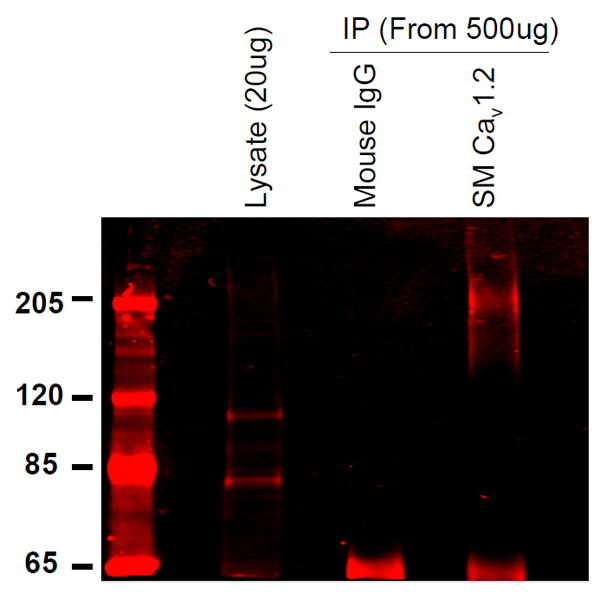
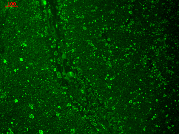
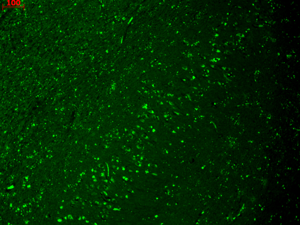
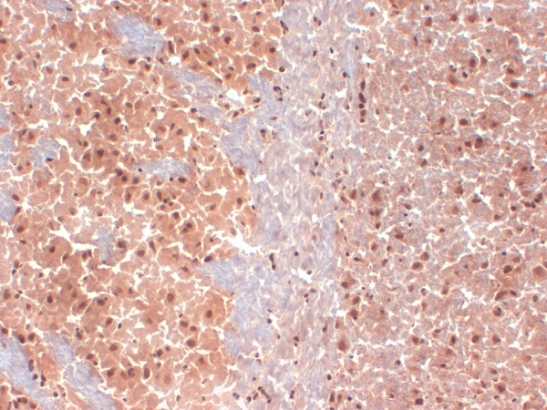
 IHC (Immunohistochemistry)
(Immunohistochemistry analysis using Mouse Anti-CaV1.2 Calcium channel Monoclonal Antibody, Clone S57-47. Tissue: Brain Tissue. Species: Mouse. Fixation: Formalin. Primary Antibody: Mouse Anti-CaV1.2 Calcium channel Monoclonal Antibody at 1:10000 for 12 hours at 4 degree C. Secondary Antibody: Biotin Goat Anti-Mouse at 1:2000 for 1 hour at RT. Counterstain: Mayer Hematoxylin (purple/blue) nuclear stain at 200 ul for 2 minutes at RT. Magnification: 40x.)
IHC (Immunohistochemistry)
(Immunohistochemistry analysis using Mouse Anti-CaV1.2 Calcium channel Monoclonal Antibody, Clone S57-47. Tissue: Brain Tissue. Species: Mouse. Fixation: Formalin. Primary Antibody: Mouse Anti-CaV1.2 Calcium channel Monoclonal Antibody at 1:10000 for 12 hours at 4 degree C. Secondary Antibody: Biotin Goat Anti-Mouse at 1:2000 for 1 hour at RT. Counterstain: Mayer Hematoxylin (purple/blue) nuclear stain at 200 ul for 2 minutes at RT. Magnification: 40x.)
Cav1.2, Monoclonal Antibody (Cat# AAA102861)
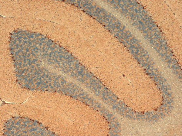
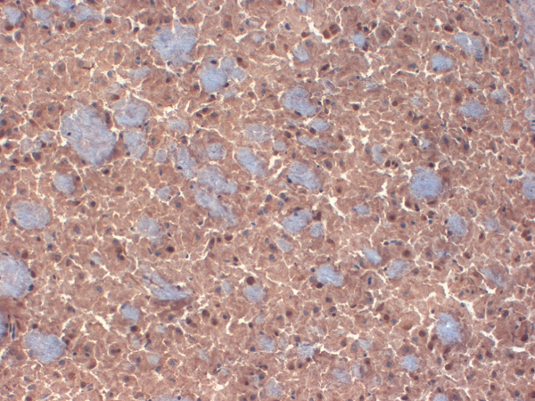
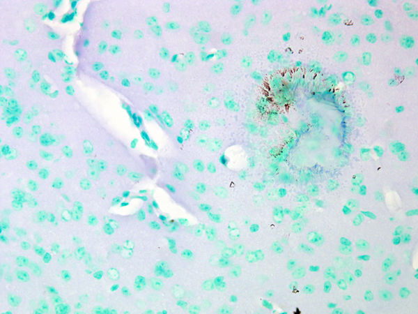
 IHC (Immunohistochemistry)
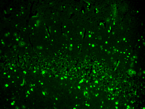
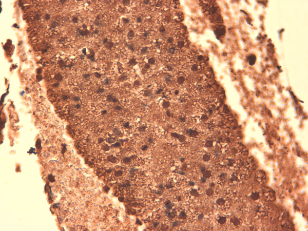
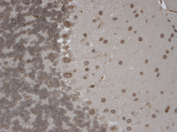
(Immunohistochemistry analysis using Mouse Anti-HCN1 Monoclonal Antibody, Clone S70-28. Tissue: Frozen brain section. Species: Mouse. Fixation: 10% Formalin Solution for 12-24 hours at RT. Primary Antibody: Mouse Anti-HCN1 Monoclonal Antibody at 1:1000 for 1 hour at RT. Secondary Antibody: HRP/DAB Detection System: Biotinylated Goat Anti-Mouse, Streptavidin Peroxidase, DAB Chromogen (brown) for 30 minutes at RT. Counterstain: Mayer Hematoxylin (purple/blue) nuclear stain at 250-500 ul for 5 minutes at RT.)
IHC (Immunohistochemistry)
(Immunohistochemistry analysis using Mouse Anti-HCN1 Monoclonal Antibody, Clone S70-28. Tissue: Frozen brain section. Species: Mouse. Fixation: 10% Formalin Solution for 12-24 hours at RT. Primary Antibody: Mouse Anti-HCN1 Monoclonal Antibody at 1:1000 for 1 hour at RT. Secondary Antibody: HRP/DAB Detection System: Biotinylated Goat Anti-Mouse, Streptavidin Peroxidase, DAB Chromogen (brown) for 30 minutes at RT. Counterstain: Mayer Hematoxylin (purple/blue) nuclear stain at 250-500 ul for 5 minutes at RT.)
HCN1, Monoclonal Antibody (Cat# AAA102885)
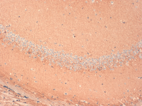
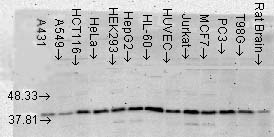 WB (Western Blot)
(Western Blot analysis of Human Cell lysates showing detection of p38 MAPK protein using Mouse Anti-p38 MAPK Monoclonal Antibody, Clone 9F12. Load: 15 ug. Block: 1.5% BSA for 30 minutes at RT. Primary Antibody: Mouse Anti-p38 MAPK Monoclonal Antibody at 1:1000 for 2 hours at RT. Secondary Antibody: Sheep Anti-Mouse IgG: HRP for 1 hour at RT.)
WB (Western Blot)
(Western Blot analysis of Human Cell lysates showing detection of p38 MAPK protein using Mouse Anti-p38 MAPK Monoclonal Antibody, Clone 9F12. Load: 15 ug. Block: 1.5% BSA for 30 minutes at RT. Primary Antibody: Mouse Anti-p38 MAPK Monoclonal Antibody at 1:1000 for 2 hours at RT. Secondary Antibody: Sheep Anti-Mouse IgG: HRP for 1 hour at RT.)
p38 alpha, Monoclonal Antibody (Cat# AAA103002)
EEA1, Monoclonal Antibody (Cat# AAA102783)
Protein A+G purification
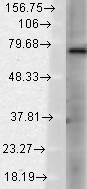
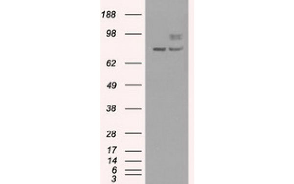
 WB (Western Blot)
(Jurkat cells were subjected to SDS PAGE followed by western blot with AAA102785 (EEF1D Antibody) at dilution of 1:1000)
WB (Western Blot)
(Jurkat cells were subjected to SDS PAGE followed by western blot with AAA102785 (EEF1D Antibody) at dilution of 1:1000)
EEF1D, Monoclonal Antibody (Cat# AAA102785)
Protein A+G purification
What are Monoclonal Antibodies?
Monoclonal antibodies are specialized laboratory-produced proteins developed for binding to specific biological antigens or other molecular targets. Since they come from a single cell (or clone), they are especially consistent and accurate in the data they are involved in producing.
This type of antibody material has been shown to be a powerful tool in finding and subsequently destroying harmful cells in an organism, such as those found in cancers or various autoimmune diseases. This makes them excellent aids in medical testing and research, which is why they are so widely used.
AAA Biotech offers a comprehensive range of high-quality monoclonal antibodies that perform effectively in various laboratory tests, including (amongst others) ELISA, western blotting, immunohistochemistry, and flow cytometry. All of the products in our catalog are thoroughly quality tested to make sure that they are reliable and will consistently perform well in your research.
What Are The Uses of Monoclonal Antibodies
Monoclonal antibodies are used in many lab tests, including (amongst others) ELISA, western blotting, immunohistochemistry, and flow cytometry.
ELISA is a test that helps detect a specific substance/analyte in a sample. It uses antibodies (often monoclonal) bound to a solid surface (such as the well of a microplate) to “capture” the substance/analyte in the sample and immobilize it so that the detection antibody component can then bind to it and produce a signal, which can then be measured.
Western blotting identifies specific proteins in a sample. The sample is first separated on a gel, and then antibodies are applied that will typically bind to the target, which will all be localized to a single band in a lane.
Immunohistochemistry helps locate specific proteins in cells or tissue samples using antibodies.
Flow cytometry looks at and sorts cells. It uses antibodies that are conjugated to reporter molecules called “fluorophores”, which, under special lights, emit light themselves, which can then be measured by a detector instrument.
How Monoclonal Antibodies Are Used as Medicine?
Please note that all of the products listed in AAA Biotech’s also known as AAA Bio or AAABio catalog are strictly for research-use only (RUO).
Monoclonal antibodies can also be used as therapeutic/medical treatments, particularly in the context of cancers. They are designed to find and bind to specific cells or proteins, helping the immune system recognize and attack the cancer. These treatments work in different ways, such as:
- Radioimmunotherapy attaches a small amount of radioactive molecule to the antibody, so it delivers the radiation directly to the cancer cells that the antibody is specifically binding to.
- Antibody-directed enzyme prodrug therapy uses antibodies that are specifically bound to special enzymes. These enzymes activate a harmless drug in the body and turn it into a cancer-killing drug only near the cancer cells—this helps avoid harming healthy cells.
- Immunoliposomes are tiny “bubbles” filled with medicine/drug and coated with antibodies. They carry the drug straight to the cancer cells.
Why Buy Monoclonal Antibodies From Us?
At AAA Biotech, we provide high-performance monoclonal antibodies designed to support a wide range of research needs.
1. Validated for Versatile Applications
The antibodies in our catalog are extensively validated and compatible with multiple techniques, including (but not limited to) ELISA, flow cytometry (FC), immunocytochemistry (ICC), immunofluorescence (IF), immunohistochemistry (IHC), immunoprecipitation (IP), and western blotting (WB).
2. Wide Selection & Specialized Options
We offer antibodies for common and rare species, that are available in various conjugated forms, and also in recombinant formats. Essentially, there is almost anything one might need to meet their experimental model’s requirements.
3. High-Quality Proteins
Our proteins meet high purity standards—90% or more as confirmed by SDS-PAGE. Many are available with tags like His, Flag, GST, or MBP, and we also supply native and biologically active proteins for functional studies.
Frequently Asked Questions
1. Are your monoclonal antibodies validated for specific applications?
Yes, our antibodies are tested and validated for use in methods such as ELISA, western blot, IHC, flow cytometry, and more. Refer to specific product pages or datasheets for individual product information.
2. How do I choose the right monoclonal antibody for my application?
Review the product details directly for application validation, species reactivity, and target information. You may also contact our support team at any time for help.
3. How quickly can I receive my order?
Most orders are processed and shipped within 1–3 business days, depending on product availability and your shipping location.