Filters
▼Clonality
▼Type
▼Reactivity
▼Gene Name
▼Isotype
▼Host
▼Application
▼Clone
▼Monoclonal Antibodies
Get accurate results in your research with our Monoclonal Antibodies, which are specially made to target exactly what you require for your research, and will produce consistent, reliable performance in lab tests.
Viewing 1350-1400 of 27597 product results
 WB (Western Blot)
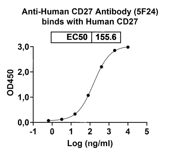
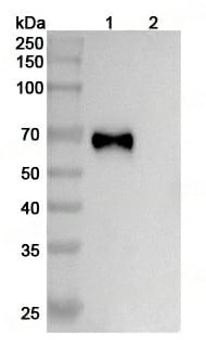
(Various lysates were subjected to SDS PAGE followed by western blot with CD27 antibody (FHD72220) at 1?ug/ml.Lane 1: CD27 transfected HEK293 cell lysateLane 2: Non-transfected HEK293 cell lysateSecond Ab: Goat Anti-Mouse IgG H&L Polyclonal antibody, HRP (PMB96431) at 0.1 ug/mL.Predict MW: 47 kDa)
WB (Western Blot)
(Various lysates were subjected to SDS PAGE followed by western blot with CD27 antibody (FHD72220) at 1?ug/ml.Lane 1: CD27 transfected HEK293 cell lysateLane 2: Non-transfected HEK293 cell lysateSecond Ab: Goat Anti-Mouse IgG H&L Polyclonal antibody, HRP (PMB96431) at 0.1 ug/mL.Predict MW: 47 kDa)
CD27, Monoclonal Antibody (Cat# AAA120673)
Protein A or G purified.
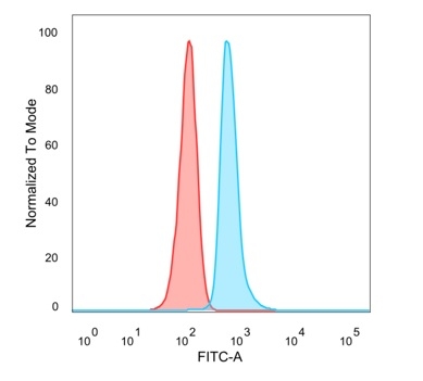
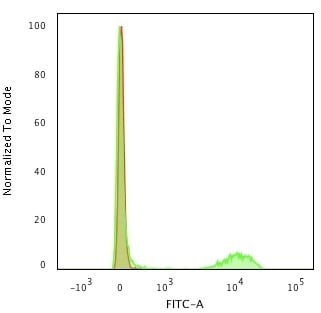
 FCM/FACS (Flow Cytometry)
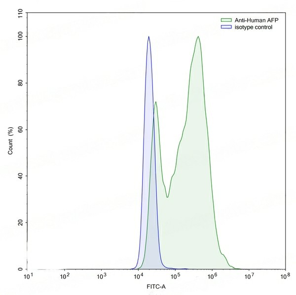
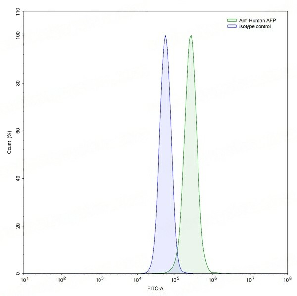
(Flow-cytometry using anti-human AFP antibody.HepG2 cells were stained with an irrelevant antibody (Blue Histogram) or an anti-human AFP antibody monoclonal antibody (Catalog # VHC01501 ,Green Histogram) at a concentration of 5 ?ug/ml for 30 mins at RT. After washing, bound antibody was detected using a FITC conjugated goat anti-human antibody (Catalog # PHB96441) and cells analysed on a NovoCyte Flow Cytometer.)
FCM/FACS (Flow Cytometry)
(Flow-cytometry using anti-human AFP antibody.HepG2 cells were stained with an irrelevant antibody (Blue Histogram) or an anti-human AFP antibody monoclonal antibody (Catalog # VHC01501 ,Green Histogram) at a concentration of 5 ?ug/ml for 30 mins at RT. After washing, bound antibody was detected using a FITC conjugated goat anti-human antibody (Catalog # PHB96441) and cells analysed on a NovoCyte Flow Cytometer.)
AFP/Alpha-fetoprotein, Monoclonal Antibody (Cat# AAA120677)
Protein A or G purified from cell culture supernatant.
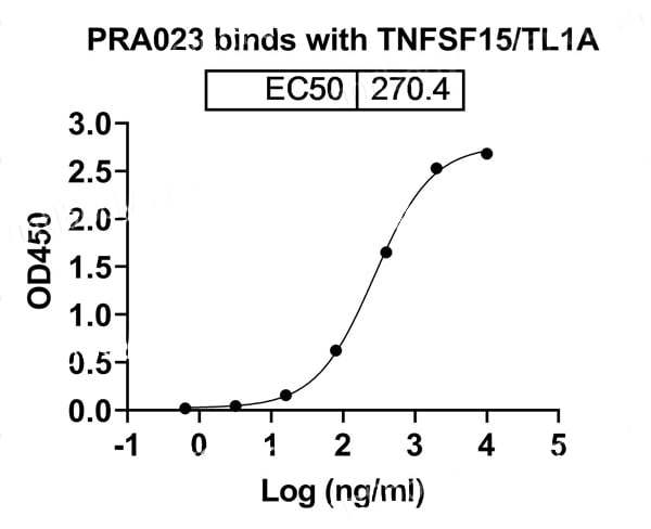
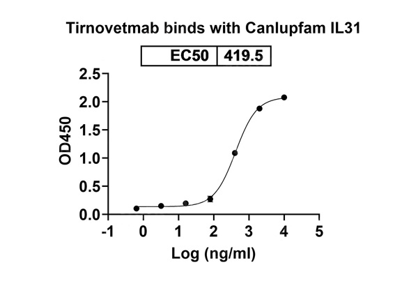 Bioactivity
(Detects IL31 in indirect ELISAs.)
Bioactivity
(Detects IL31 in indirect ELISAs.)
Tirnovetmab, Monoclonal Antibody (Cat# AAA120716)
Protein A or G purified from cell culture supernatant.
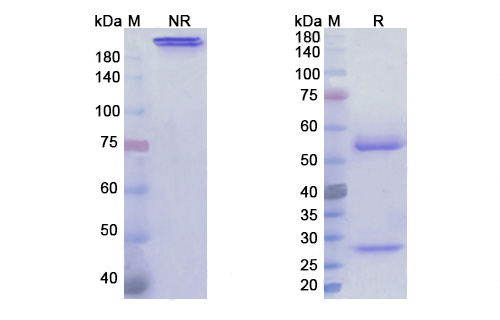
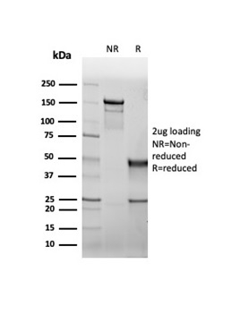
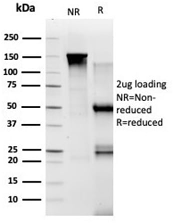
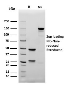
 SDS-PAGE
(SDS PAGE for Tulisokibart)
SDS-PAGE
(SDS PAGE for Tulisokibart)
Tulisokibart, Monoclonal Antibody (Cat# AAA120718)
Protein A or G purified from cell culture supernatant.
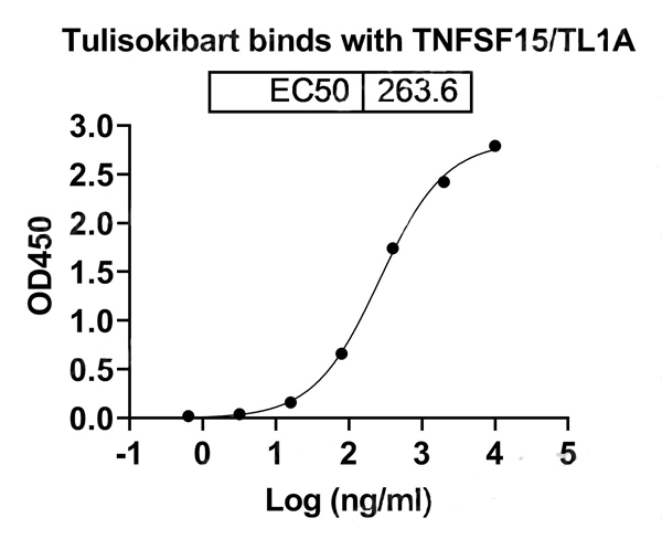
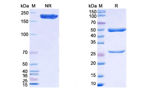
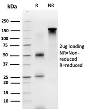
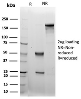
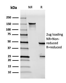
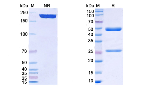 SDS-PAGE
(SDS PAGE for Human TNFSF15/TL1A antibody)
SDS-PAGE
(SDS PAGE for Human TNFSF15/TL1A antibody)
TNFSF15/TL1A, Monoclonal Recombinant Antibody (Cat# AAA120724)
Protein A/G purified from cell culture supernatant.
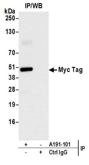
 WB (Western Blot)
(Detection of Myc-tagged protein by western blot of lysate from non-transfected human HEK293 cells, HEK293 transfected with Met-Myc-IDO1, HEK293 transfected with IDO1-Myc (C-terminal Tag), and HEK293 transfected with IDO1-Myc-HIS (Internal Tag). Antibody: Rabbit anti-Myc Tag recombinant monoclonal antibody (AAA210713 lot 1) used at 1:1000. Secondary: HRP-conjugated goat anti-rabbit IgG . Detection: Chemiluminescence with an exposure time of 3 seconds.)
WB (Western Blot)
(Detection of Myc-tagged protein by western blot of lysate from non-transfected human HEK293 cells, HEK293 transfected with Met-Myc-IDO1, HEK293 transfected with IDO1-Myc (C-terminal Tag), and HEK293 transfected with IDO1-Myc-HIS (Internal Tag). Antibody: Rabbit anti-Myc Tag recombinant monoclonal antibody (AAA210713 lot 1) used at 1:1000. Secondary: HRP-conjugated goat anti-rabbit IgG . Detection: Chemiluminescence with an exposure time of 3 seconds.)
Myc Tag, Monoclonal Recombinant Antibody (Cat# AAA210713)
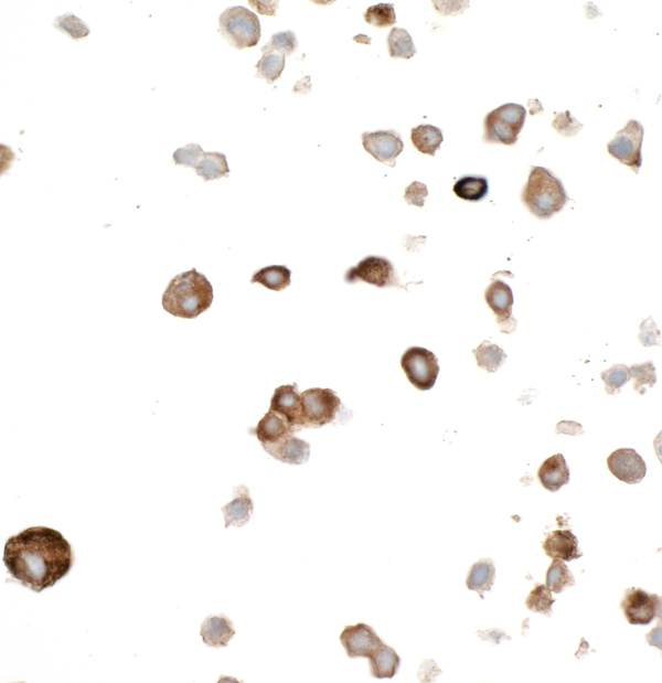
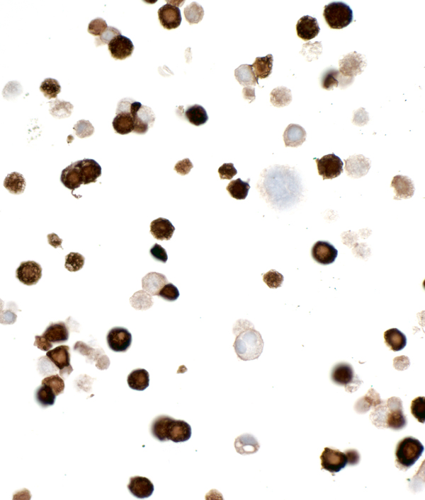
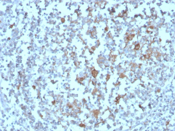
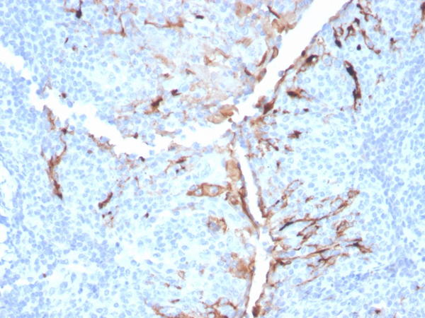
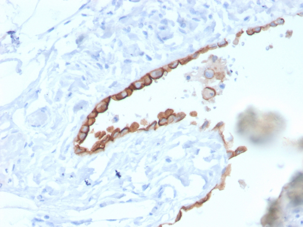
 IHC (Immunohiostchemistry)
(Formalin-fixed, paraffin-embedded human Lung SqCC stained with PD-L1 Recombinant Rabbit Monoclonal Antibody (PDL1/4451R).)
IHC (Immunohiostchemistry)
(Formalin-fixed, paraffin-embedded human Lung SqCC stained with PD-L1 Recombinant Rabbit Monoclonal Antibody (PDL1/4451R).)
PD-L1/PDCD1LG1/CD274/B7-H1, Monoclonal Antibody (Cat# AAA215501)
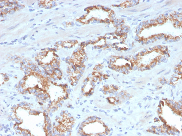
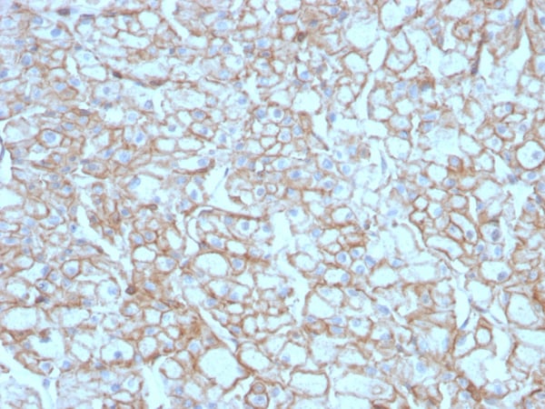
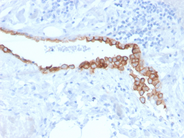
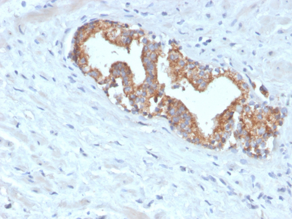 IHC (Immunohiostchemistry)
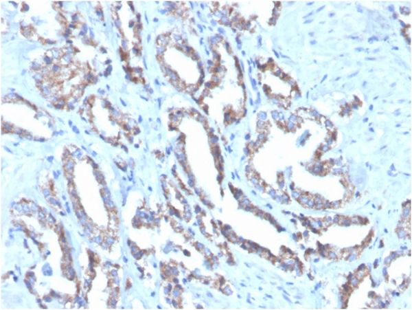
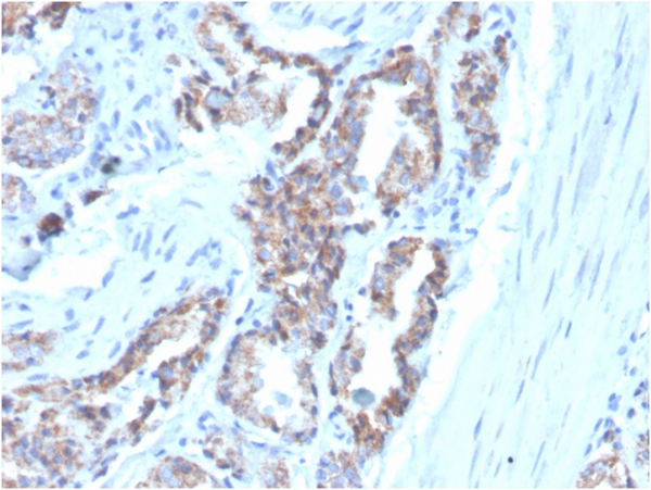
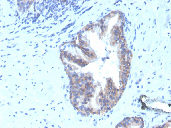
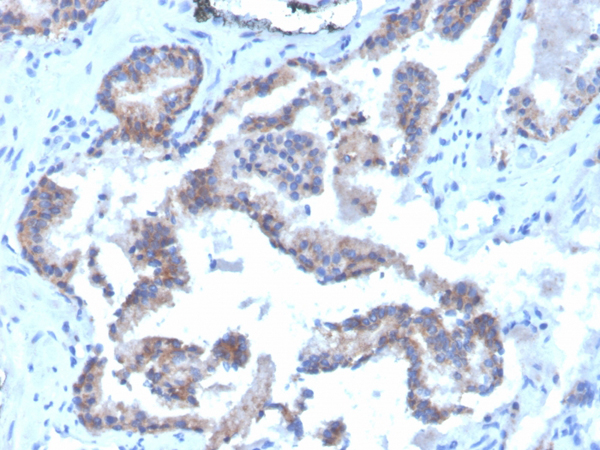
(Formalin-fixed, paraffin-embedded human prostate carcinoma stained with AMACR Recombinant Rabbit Monoclonal Antibody (AMACR/4572R).)
IHC (Immunohiostchemistry)
(Formalin-fixed, paraffin-embedded human prostate carcinoma stained with AMACR Recombinant Rabbit Monoclonal Antibody (AMACR/4572R).)
AMACR/P504s, Monoclonal Antibody (Cat# AAA215502)
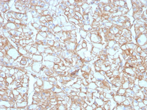
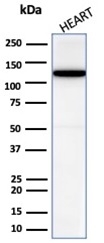 WB (Western Blot)
(Western blot analysis of human heart tissue lysate using N-Cadherin Recombinant Rabbit Monoclonal Antibody (CDH2/6857R).)
WB (Western Blot)
(Western blot analysis of human heart tissue lysate using N-Cadherin Recombinant Rabbit Monoclonal Antibody (CDH2/6857R).)
N-Cadherin/Cadherin-2/CD325 (NCAD), Monoclonal Antibody (Cat# AAA215504)
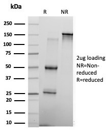
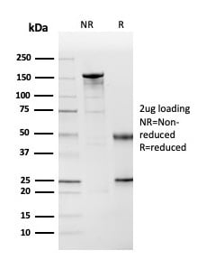
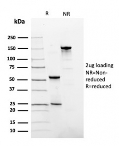
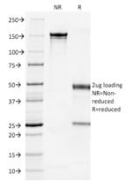
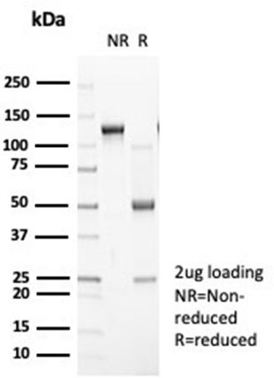 SDS-PAGE
(SDS-PAGE Analysis Purified N-Cadherin Recombinant Rabbit Monoclonal Antibody (CDH2/7070R). Confirmation of Purity and Integrity of Antibody.)
SDS-PAGE
(SDS-PAGE Analysis Purified N-Cadherin Recombinant Rabbit Monoclonal Antibody (CDH2/7070R). Confirmation of Purity and Integrity of Antibody.)
N-Cadherin/Cadherin-2/CD325 (NCAD), Monoclonal Antibody (Cat# AAA215505)
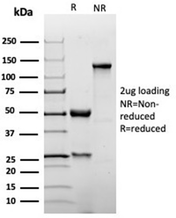 SDS-PAGE
(SDS-PAGE Analysis Purified p27 Recombinant Rabbit Monoclonal Antibody (KIP1/1355R). Confirmation of Purity and Integrity of Antibody.)
SDS-PAGE
(SDS-PAGE Analysis Purified p27 Recombinant Rabbit Monoclonal Antibody (KIP1/1355R). Confirmation of Purity and Integrity of Antibody.)
p27Kip1, Monoclonal Antibody (Cat# AAA215517)
 IHC (Immunohistochemistry)
(Formalin-fixed, paraffin-embedded human colon stained with P16INK4a Mouse Monoclonal Antibody (CDKN2A/3830).)
IHC (Immunohistochemistry)
(Formalin-fixed, paraffin-embedded human colon stained with P16INK4a Mouse Monoclonal Antibody (CDKN2A/3830).)
P16INK4a, Monoclonal Antibody (Cat# AAA215519)
 IHC (Immunohistochemistry)
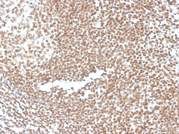
(Formalin-fixed, paraffin-embedded human cervix stained with P16INK4a Recombinant Rabbit Monoclonal Antibody (CDKN2A/4844R).)
IHC (Immunohistochemistry)
(Formalin-fixed, paraffin-embedded human cervix stained with P16INK4a Recombinant Rabbit Monoclonal Antibody (CDKN2A/4844R).)
P16INK4a, Monoclonal Antibody (Cat# AAA215522)
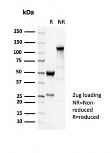 SDS-PAGE
(SDS-PAGE Analysis Purified TUBB3 Recombinant Rabbit Monoclonal Antibody (TUBB3/7089R). Confirmation of Purity and Integrity of Antibody.)
SDS-PAGE
(SDS-PAGE Analysis Purified TUBB3 Recombinant Rabbit Monoclonal Antibody (TUBB3/7089R). Confirmation of Purity and Integrity of Antibody.)
Tubulin beta 3/TUBB3, Monoclonal Antibody (Cat# AAA215525)
 SDS-PAGE
(SDS-PAGE Analysis Purified TUBB3 Recombinant Rabbit Monoclonal Antibody (TUBB3/7090R). Confirmation of Purity and Integrity of Antibody.)
SDS-PAGE
(SDS-PAGE Analysis Purified TUBB3 Recombinant Rabbit Monoclonal Antibody (TUBB3/7090R). Confirmation of Purity and Integrity of Antibody.)
Tubulin beta 3/TUBB3, Monoclonal Antibody (Cat# AAA215526)
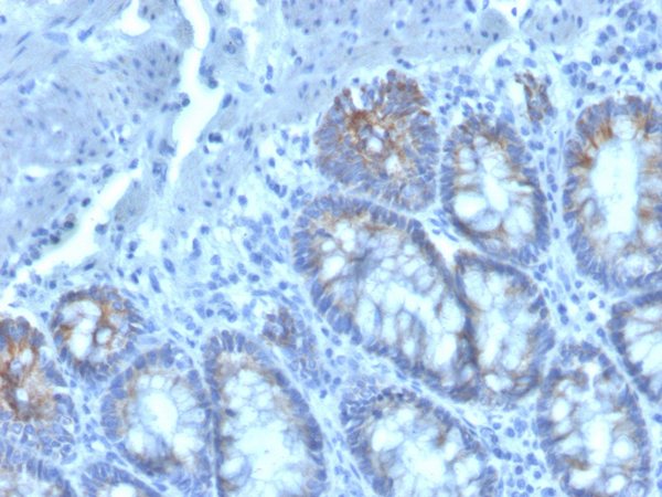
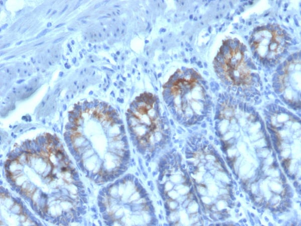
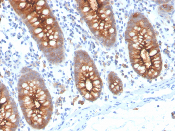 IHC (Immunohistochemistry)
(Formalin-fixed, paraffin-embedded human colon stained with CEA Recombinant Rabbit Monoclonal Antibody (C66/3707R).)
IHC (Immunohistochemistry)
(Formalin-fixed, paraffin-embedded human colon stained with CEA Recombinant Rabbit Monoclonal Antibody (C66/3707R).)
Carcinoembryonic Antigen (CEA)/CD66e, Monoclonal Antibody (Cat# AAA215529)
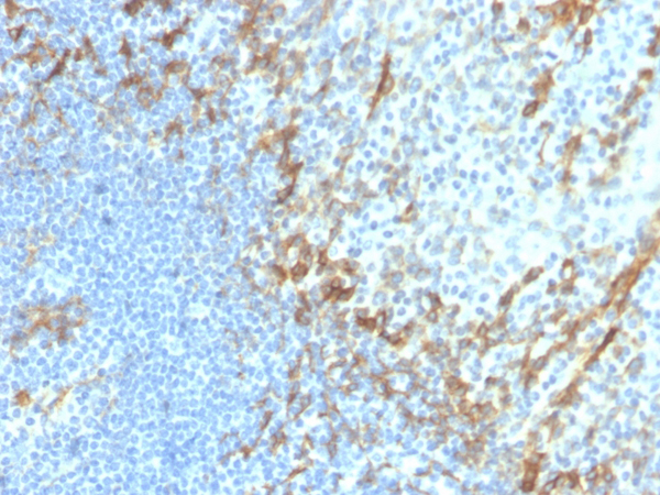 IHC (Immunohistochemistry)
(Formalin-fixed, paraffin-embedded human tonsil stained with Podoplanin Recombinant Rabbit Monoclonal Antibody (PDPN/4009R).)
IHC (Immunohistochemistry)
(Formalin-fixed, paraffin-embedded human tonsil stained with Podoplanin Recombinant Rabbit Monoclonal Antibody (PDPN/4009R).)
Podoplanin (PDPN), Monoclonal Antibody (Cat# AAA215531)
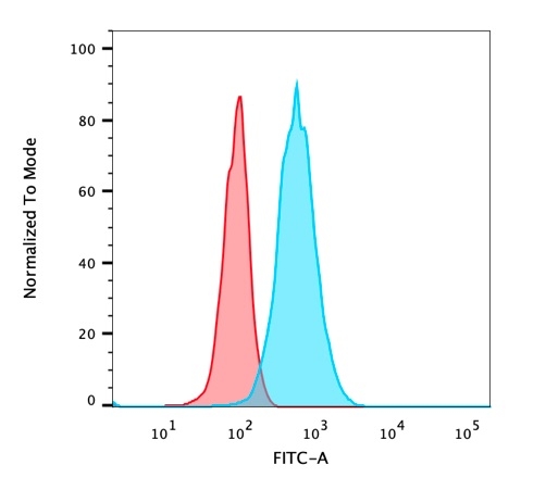
 FCM/FACS (Flow Cytometry)
(Flow Cytometric Analysis of PFA-fixed HeLa cells. DMRT2 Mouse Monoclonal Antibody (PCRP-DMRT2-1B11) followed by goat anti-mouse IgG-CF488 (blue); isotype control (red).)
FCM/FACS (Flow Cytometry)
(Flow Cytometric Analysis of PFA-fixed HeLa cells. DMRT2 Mouse Monoclonal Antibody (PCRP-DMRT2-1B11) followed by goat anti-mouse IgG-CF488 (blue); isotype control (red).)
DMRT2, Monoclonal Antibody (Cat# AAA215534)
 SDS-PAGE
(SDS-PAGE Analysis Purified CFTR Recombinant Mouse Monoclonal Antibody (rCFTR/6476). Confirmation of Purity and Integrity of Antibody.)
SDS-PAGE
(SDS-PAGE Analysis Purified CFTR Recombinant Mouse Monoclonal Antibody (rCFTR/6476). Confirmation of Purity and Integrity of Antibody.)
CFTR (Cystic Fibrosis Transmembrane Conductance Regulator), Monoclonal Antibody (Cat# AAA215536)
 SDS-PAGE
(SDS-PAGE Analysis Purified ECD Mouse Monoclonal Antibody (PCRP-ECD-1D10). Confirmation of Purity and Integrity of Antibody.)
SDS-PAGE
(SDS-PAGE Analysis Purified ECD Mouse Monoclonal Antibody (PCRP-ECD-1D10). Confirmation of Purity and Integrity of Antibody.)
ECD/SGT1, Monoclonal Antibody (Cat# AAA215546)
Predicted to react with Rabbit, Goat, Horse, Guinea pig, Dog, Pig, Rhesus monkey, Orangutan and Elephant (based on sequence homology).
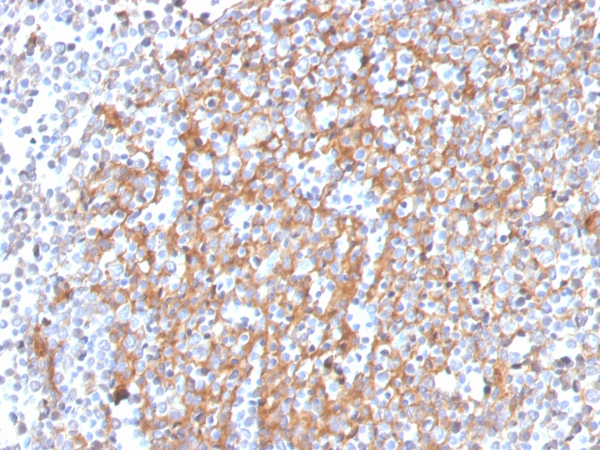
 IHC (Immunohistochemistry)
(Formalin-fixed, paraffin-embedded human tonsil stained with CD268/BAFFR Mouse Monoclonal Antibody (BAFFR/1558).)
IHC (Immunohistochemistry)
(Formalin-fixed, paraffin-embedded human tonsil stained with CD268/BAFFR Mouse Monoclonal Antibody (BAFFR/1558).)
CD268/BAFFR/TNFRSF13C, Monoclonal Antibody (Cat# AAA215547)
 SDS-PAGE
(SDS-PAGE Analysis Purified LRG1 Mouse Monoclonal Antibody (LRG1/4883). Confirmation of Purity and Integrity of Antibody.)
SDS-PAGE
(SDS-PAGE Analysis Purified LRG1 Mouse Monoclonal Antibody (LRG1/4883). Confirmation of Purity and Integrity of Antibody.)
LRG1/Leucine Rich alpha-2-glycoprotein 1, Monoclonal Antibody (Cat# AAA215550)
 SDS-PAGE
(SDS-PAGE Analysis Purified TADA1 Mouse Monoclonal Antibody (PCRP-TADA1-1C9). Confirmation of Purity and Integrity of Antibody.)
SDS-PAGE
(SDS-PAGE Analysis Purified TADA1 Mouse Monoclonal Antibody (PCRP-TADA1-1C9). Confirmation of Purity and Integrity of Antibody.)
TADA1, Monoclonal Antibody (Cat# AAA215551)
Predicted to react with Rabbit, Goat, Horse, Guinea pig, Dog, Pig, Rhesus monkey, Orangutan and Elephant (based on sequence homology).
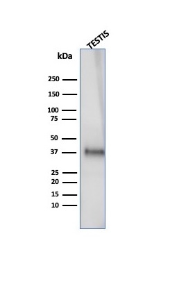 WB (Western Blot)
(Western Blot Analysis of human testis tissue lysate using Clusterin/APOJ Mouse Monoclonal Antibody (CLU/4727).)
WB (Western Blot)
(Western Blot Analysis of human testis tissue lysate using Clusterin/APOJ Mouse Monoclonal Antibody (CLU/4727).)
Clusterin/Apolipoprotein J (APO-J), Monoclonal Antibody (Cat# AAA215555)
 SDS-PAGE
(SDS-PAGE Analysis Purified KLF17 Mouse Monoclonal Antibody (PCRP-KLF17-1G2). Confirmation of Purity and Integrity of Antibody.)
SDS-PAGE
(SDS-PAGE Analysis Purified KLF17 Mouse Monoclonal Antibody (PCRP-KLF17-1G2). Confirmation of Purity and Integrity of Antibody.)
KLF17/ZNF393, Monoclonal Antibody (Cat# AAA215559)
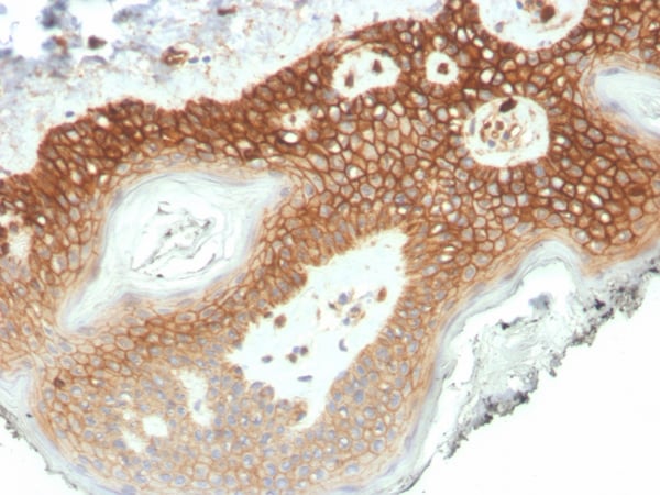
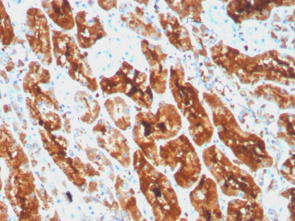
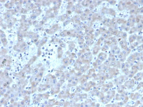
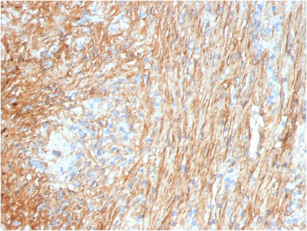
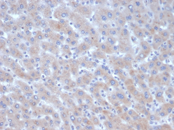 IHC (Immunohistochemistry)
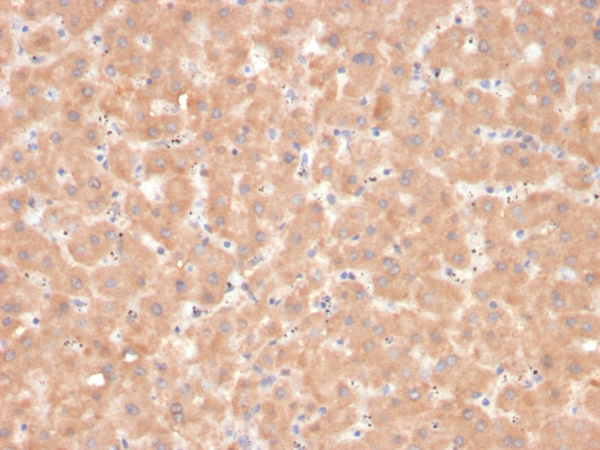
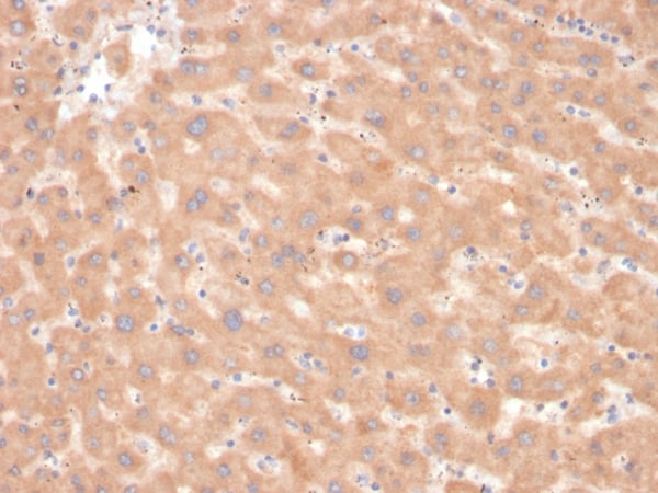
(Formalin-fixed, paraffin-embedded human liver stained with Alpha-1-Antichymotrypsin Mouse Monoclonal Antibody (SERPINA3/4185).)
IHC (Immunohistochemistry)
(Formalin-fixed, paraffin-embedded human liver stained with Alpha-1-Antichymotrypsin Mouse Monoclonal Antibody (SERPINA3/4185).)
Alpha-1-Antichymotrypsin (SERPINA3), Monoclonal Antibody (Cat# AAA215564)
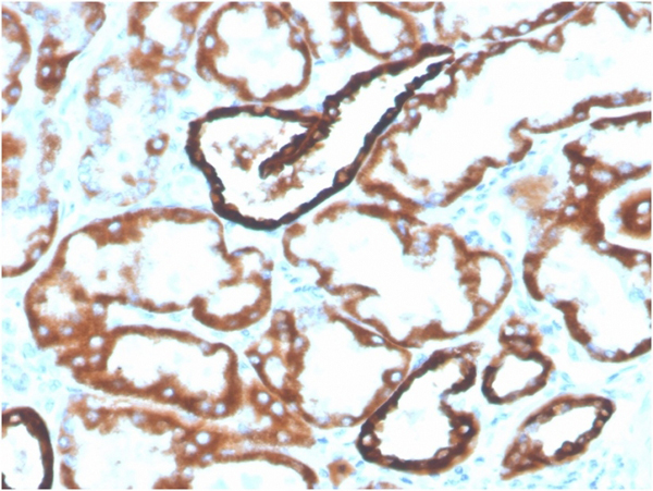
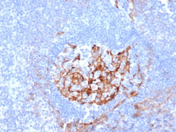 IHC (Immunohistochemistry)
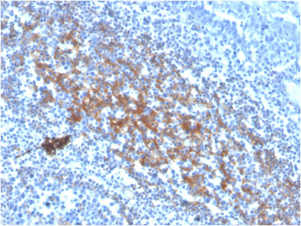
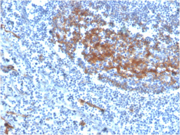
(Formalin-fixed, paraffin-embedded human tonsil stained with CD35 Mouse Monoclonal Antibody (CR1/6380) at 2ug/ml.)
IHC (Immunohistochemistry)
(Formalin-fixed, paraffin-embedded human tonsil stained with CD35 Mouse Monoclonal Antibody (CR1/6380) at 2ug/ml.)
CD35/CR1, Monoclonal Antibody (Cat# AAA215567)
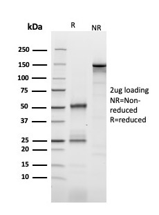
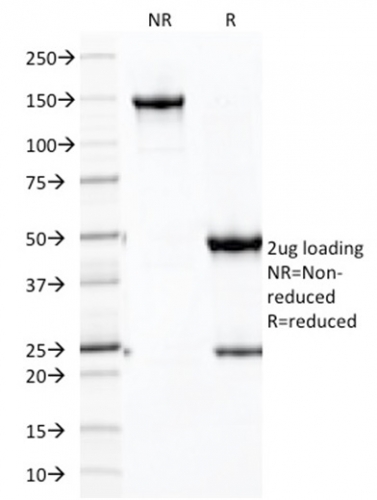
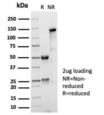 SDS-PAGE
(SDS-PAGE Analysis Purified CD35 Recombinant Rabbit Monoclonal Antibody (CD35/7016R). Confirmation of Purity and Integrity of Antibody.)
SDS-PAGE
(SDS-PAGE Analysis Purified CD35 Recombinant Rabbit Monoclonal Antibody (CD35/7016R). Confirmation of Purity and Integrity of Antibody.)
CD35/CR1, Monoclonal Antibody (Cat# AAA215569)
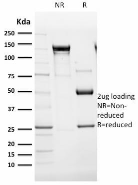
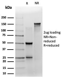 SDS-PAGE
(SDS-PAGE Analysis Purified ZNF358 Mouse Monoclonal Antibody (PCRP-ZNF358-1A6). Confirmation of Purity and Integrity of Antibody.)
SDS-PAGE
(SDS-PAGE Analysis Purified ZNF358 Mouse Monoclonal Antibody (PCRP-ZNF358-1A6). Confirmation of Purity and Integrity of Antibody.)
ZNF358 (Zinc Finger Protein 358), Monoclonal Antibody (Cat# AAA215573)
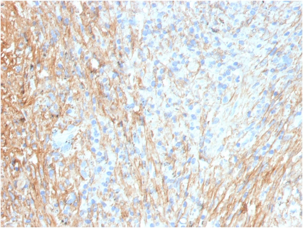
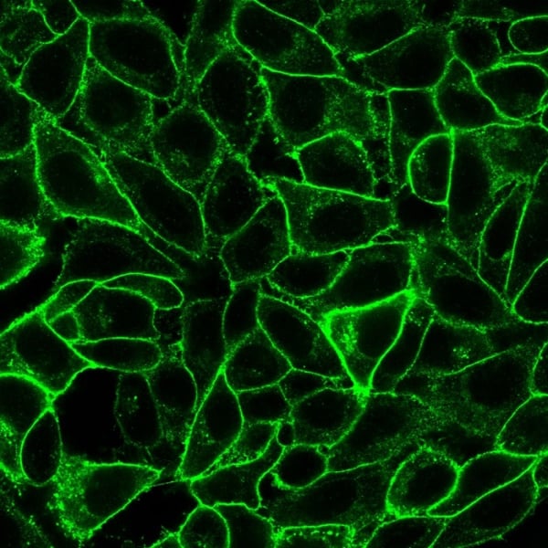
 IHC (Immunohistochemistry)
(IHC analysis of formalin-fixed, paraffin-embedded human colon. CTNND1/4501 at 2ug/ml in PBS for 30min RT. HIER: Tris/EDTA, pH9. 0, 45min. 2 degree : HRP-polymer, 30min. DAB, 5min.)
IHC (Immunohistochemistry)
(IHC analysis of formalin-fixed, paraffin-embedded human colon. CTNND1/4501 at 2ug/ml in PBS for 30min RT. HIER: Tris/EDTA, pH9. 0, 45min. 2 degree : HRP-polymer, 30min. DAB, 5min.)
p120/Catenin, delta-1 (CTNND1), Monoclonal Antibody (Cat# AAA215581)

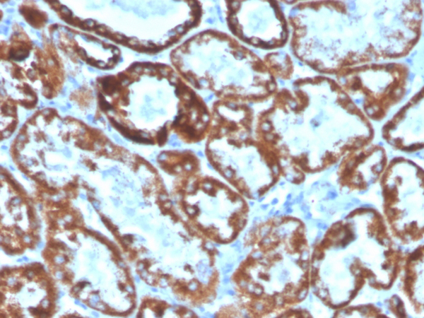
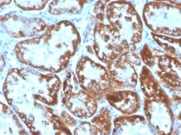
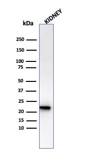 WB (Western Blot)
(Western Blot Analysis of human Kidney tissue lysate using Ferritin, Light Chain Mouse Monoclonal Antibody (FTL/1386).)
WB (Western Blot)
(Western Blot Analysis of human Kidney tissue lysate using Ferritin, Light Chain Mouse Monoclonal Antibody (FTL/1386).)
Ferritin, Monoclonal Antibody (Cat# AAA215425)
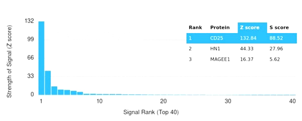 Application Data
(Analysis of Protein Array containing more than 19,000 full-length human proteins using CD25 / IL2RA Mouse Monoclonal Antibody (IL2RA/2393). Z- and S- Score: The Z-score represents the strength of a signal that a monoclonal antibody (MAb) (in combination with a fluorescently-tagged anti-IgG secondary antibody) produces when binding to a particular protein on the HuProtTM array. Z-scores are described in units of standard deviations (SD's) above the mean value of all signals generated on that array. If targets on HuProtTM are arranged in descending order of the Z-score, the S-score is the difference (also in units of SD's) between the Z-score. S-score therefore represents the relative target specificity of a MAb to its intended target. A MAb is considered to specific to its intended target, if the MAb has an S-score of at least 2.5. For example, if a MAb binds to protein X with a Z-score of 43 and to protein Y with a Z-score of 14, then the S-score for the binding of that MAb to protein X is equal to 29.)
Application Data
(Analysis of Protein Array containing more than 19,000 full-length human proteins using CD25 / IL2RA Mouse Monoclonal Antibody (IL2RA/2393). Z- and S- Score: The Z-score represents the strength of a signal that a monoclonal antibody (MAb) (in combination with a fluorescently-tagged anti-IgG secondary antibody) produces when binding to a particular protein on the HuProtTM array. Z-scores are described in units of standard deviations (SD's) above the mean value of all signals generated on that array. If targets on HuProtTM are arranged in descending order of the Z-score, the S-score is the difference (also in units of SD's) between the Z-score. S-score therefore represents the relative target specificity of a MAb to its intended target. A MAb is considered to specific to its intended target, if the MAb has an S-score of at least 2.5. For example, if a MAb binds to protein X with a Z-score of 43 and to protein Y with a Z-score of 14, then the S-score for the binding of that MAb to protein X is equal to 29.)
CD25/IL2RA, Monoclonal Antibody (Cat# AAA215426)
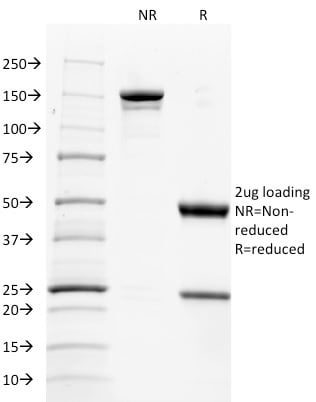
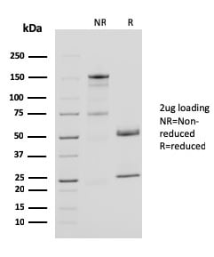
 SDS-PAGE
(SDS-PAGE Analysis Purified SOX9 Recombinant Rabbit Monoclonal Antibody (SOX9/3916R). Confirmation of Purity and Integrity of Antibody.)
SDS-PAGE
(SDS-PAGE Analysis Purified SOX9 Recombinant Rabbit Monoclonal Antibody (SOX9/3916R). Confirmation of Purity and Integrity of Antibody.)
SOX9/SRY-box 9, Monoclonal Antibody (Cat# AAA215428)
 FCM/FACS (Flow Cytometry)
(Flow Cytometric Analysis of PBMC cells using CD4 Recombinant Mouse Monoclonal Antibody (rC4/206) followed by goat anti-mouse IgG-CF488 (Green); Unstained cells (Red).)
FCM/FACS (Flow Cytometry)
(Flow Cytometric Analysis of PBMC cells using CD4 Recombinant Mouse Monoclonal Antibody (rC4/206) followed by goat anti-mouse IgG-CF488 (Green); Unstained cells (Red).)
CD4, Monoclonal Antibody (Cat# AAA215431)
 Application Data
(Analysis of Protein Array containing more than 19,000 full-length human proteins using Mesothelin Mouse Monoclonal Antibody (MSLN/3384). Z- and S- Score: The Z-score represents the strength of a signal that a monoclonal antibody (MAb) (in combination with a fluorescently-tagged anti-IgG secondary antibody) produces when binding to a particular protein on the HuProtTM array. Z-scores are described in units of standard deviations (SD's) above the mean value of all signals generated on that array. If targets on HuProtTM are arranged in descending order of the Z-score, the S-score is the difference (also in units of SD's) between the Z-score. S-score therefore represents the relative target specificity of a MAb to its intended target. A MAb is considered to specific to its intended target, if the MAb has an S-score of at least 2.5. For example, if a MAb binds to protein X with a Z-score of 43 and to protein Y with a Z-score of 14, then the S-score for the binding of that MAb to protein X is equal to 29.)
Application Data
(Analysis of Protein Array containing more than 19,000 full-length human proteins using Mesothelin Mouse Monoclonal Antibody (MSLN/3384). Z- and S- Score: The Z-score represents the strength of a signal that a monoclonal antibody (MAb) (in combination with a fluorescently-tagged anti-IgG secondary antibody) produces when binding to a particular protein on the HuProtTM array. Z-scores are described in units of standard deviations (SD's) above the mean value of all signals generated on that array. If targets on HuProtTM are arranged in descending order of the Z-score, the S-score is the difference (also in units of SD's) between the Z-score. S-score therefore represents the relative target specificity of a MAb to its intended target. A MAb is considered to specific to its intended target, if the MAb has an S-score of at least 2.5. For example, if a MAb binds to protein X with a Z-score of 43 and to protein Y with a Z-score of 14, then the S-score for the binding of that MAb to protein X is equal to 29.)
Mesothelin, Monoclonal Antibody (Cat# AAA215439)
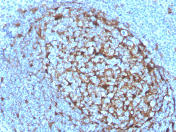
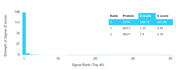 Application Data
(Analysis of Protein Array containing more than 19,000 full-length human proteins using Cystatin A Mouse Monoclonal Antibody (CSTA/3553). Z- and S- Score: The Z-score represents the strength of a signal that a monoclonal antibody (MAb) (in combination with a fluorescently-tagged anti-IgG secondary antibody) produces when binding to a particular protein on the HuProtTM array. Z-scores are described in units of standard deviations (SD's) above the mean value of all signals generated on that array. If targets on HuProtTM are arranged in descending order of the Z-score, the S-score is the difference (also in units of SD's) between the Z-score. S-score therefore represents the relative target specificity of a MAb to its intended target. A MAb is considered to specific to its intended target, if the MAb has an S-score of at least 2.5. For example, if a MAb binds to protein X with a Z-score of 43 and to protein Y with a Z-score of 14, then the S-score for the binding of that MAb to protein X is equal to 29.)
Application Data
(Analysis of Protein Array containing more than 19,000 full-length human proteins using Cystatin A Mouse Monoclonal Antibody (CSTA/3553). Z- and S- Score: The Z-score represents the strength of a signal that a monoclonal antibody (MAb) (in combination with a fluorescently-tagged anti-IgG secondary antibody) produces when binding to a particular protein on the HuProtTM array. Z-scores are described in units of standard deviations (SD's) above the mean value of all signals generated on that array. If targets on HuProtTM are arranged in descending order of the Z-score, the S-score is the difference (also in units of SD's) between the Z-score. S-score therefore represents the relative target specificity of a MAb to its intended target. A MAb is considered to specific to its intended target, if the MAb has an S-score of at least 2.5. For example, if a MAb binds to protein X with a Z-score of 43 and to protein Y with a Z-score of 14, then the S-score for the binding of that MAb to protein X is equal to 29.)
Cystatin A, Monoclonal Antibody (Cat# AAA215441)
 Application Data
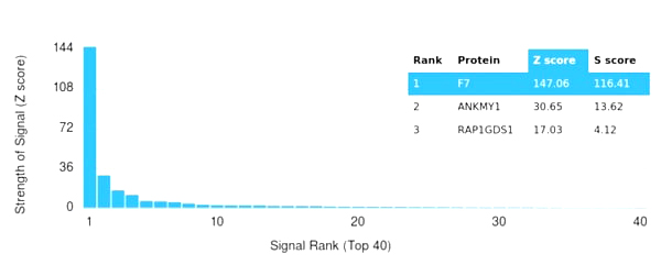
(Analysis of Protein Array containing more than 19,000 full-length human proteins using Coagulation Factor VII Mouse Monoclonal Antibody (F7/3511). Z- and S- Score: The Z-score represents the strength of a signal that a monoclonal antibody (MAb) (in combination with a fluorescently-tagged anti-IgG secondary antibody) produces when binding to a particular protein on the HuProtTM array. Z-scores are described in units of standard deviations (SD's) above the mean value of all signals generated on that array. If targets on HuProtTM are arranged in descending order of the Z-score, the S-score is the difference (also in units of SD's) between the Z-score. S-score therefore represents the relative target specificity of a MAb to its intended target. A MAb is considered to specific to its intended target, if the MAb has an S-score of at least 2.5. For example, if a MAb binds to protein X with a Z-score of 43 and to protein Y with a Z-score of 14, then the S-score for the binding of that MAb to protein X is equal to 29.)
Application Data
(Analysis of Protein Array containing more than 19,000 full-length human proteins using Coagulation Factor VII Mouse Monoclonal Antibody (F7/3511). Z- and S- Score: The Z-score represents the strength of a signal that a monoclonal antibody (MAb) (in combination with a fluorescently-tagged anti-IgG secondary antibody) produces when binding to a particular protein on the HuProtTM array. Z-scores are described in units of standard deviations (SD's) above the mean value of all signals generated on that array. If targets on HuProtTM are arranged in descending order of the Z-score, the S-score is the difference (also in units of SD's) between the Z-score. S-score therefore represents the relative target specificity of a MAb to its intended target. A MAb is considered to specific to its intended target, if the MAb has an S-score of at least 2.5. For example, if a MAb binds to protein X with a Z-score of 43 and to protein Y with a Z-score of 14, then the S-score for the binding of that MAb to protein X is equal to 29.)
VII/F7, Monoclonal Antibody (Cat# AAA215445)

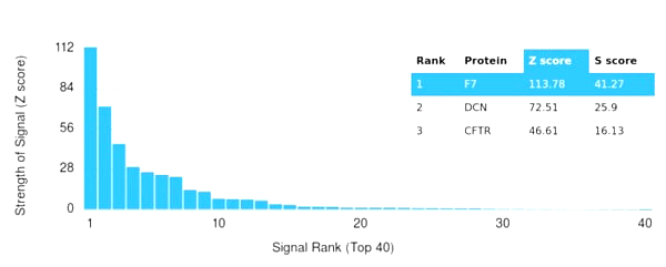 Application Data
(Analysis of Protein Array containing more than 19,000 full-length human proteins using Coagulation Factor VII Mouse Monoclonal Antibody (F7/3618). Z- and S- Score: The Z-score represents the strength of a signal that a monoclonal antibody (MAb) (in combination with a fluorescently-tagged anti-IgG secondary antibody) produces when binding to a particular protein on the HuProtTM array. Z-scores are described in units of standard deviations (SD's) above the mean value of all signals generated on that array. If targets on HuProtTM are arranged in descending order of the Z-score, the S-score is the difference (also in units of SD's) between the Z-score. S-score therefore represents the relative target specificity of a MAb to its intended target. A MAb is considered to specific to its intended target, if the MAb has an S-score of at least 2.5. For example, if a MAb binds to protein X with a Z-score of 43 and to protein Y with a Z-score of 14, then the S-score for the binding of that MAb to protein X is equal to 29.)
Application Data
(Analysis of Protein Array containing more than 19,000 full-length human proteins using Coagulation Factor VII Mouse Monoclonal Antibody (F7/3618). Z- and S- Score: The Z-score represents the strength of a signal that a monoclonal antibody (MAb) (in combination with a fluorescently-tagged anti-IgG secondary antibody) produces when binding to a particular protein on the HuProtTM array. Z-scores are described in units of standard deviations (SD's) above the mean value of all signals generated on that array. If targets on HuProtTM are arranged in descending order of the Z-score, the S-score is the difference (also in units of SD's) between the Z-score. S-score therefore represents the relative target specificity of a MAb to its intended target. A MAb is considered to specific to its intended target, if the MAb has an S-score of at least 2.5. For example, if a MAb binds to protein X with a Z-score of 43 and to protein Y with a Z-score of 14, then the S-score for the binding of that MAb to protein X is equal to 29.)
VII/F7, Monoclonal Antibody (Cat# AAA215448)
 Application Data
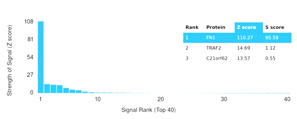
(Analysis of Protein Array containing more than 19,000 full-length human proteins using Fibronectin Mouse Monoclonal Antibody (FN1/3036). Z- and S- Score: The Z-score represents the strength of a signal that a monoclonal antibody (MAb) (in combination with a fluorescently-tagged anti-IgG secondary antibody) produces when binding to a particular protein on the HuProtTM array. Z-scores are described in units of standard deviations (SD's) above the mean value of all signals generated on that array. If targets on HuProtTM are arranged in descending order of the Z-score, the S-score is the difference (also in units of SD's) between the Z-score. S-score therefore represents the relative target specificity of a MAb to its intended target. A MAb is considered to specific to its intended target, if the MAb has an S-score of at least 2.5. For example, if a MAb binds to protein X with a Z-score of 43 and to protein Y with a Z-score of 14, then the S-score for the binding of that MAb to protein X is equal to 29.)
Application Data
(Analysis of Protein Array containing more than 19,000 full-length human proteins using Fibronectin Mouse Monoclonal Antibody (FN1/3036). Z- and S- Score: The Z-score represents the strength of a signal that a monoclonal antibody (MAb) (in combination with a fluorescently-tagged anti-IgG secondary antibody) produces when binding to a particular protein on the HuProtTM array. Z-scores are described in units of standard deviations (SD's) above the mean value of all signals generated on that array. If targets on HuProtTM are arranged in descending order of the Z-score, the S-score is the difference (also in units of SD's) between the Z-score. S-score therefore represents the relative target specificity of a MAb to its intended target. A MAb is considered to specific to its intended target, if the MAb has an S-score of at least 2.5. For example, if a MAb binds to protein X with a Z-score of 43 and to protein Y with a Z-score of 14, then the S-score for the binding of that MAb to protein X is equal to 29.)
Fibronectin, Monoclonal Antibody (Cat# AAA215450)
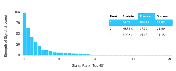 Application Data
(Analysis of Protein Array containing more than 19,000 full-length human proteins using DBC2 Mouse Monoclonal Antibody (DBC2/3362). Z- and S- Score: The Z-score represents the strength of a signal that a monoclonal antibody (MAb) (in combination with a fluorescently-tagged anti-IgG secondary antibody) produces when binding to a particular protein on the HuProtTM array. Z-scores are described in units of standard deviations (SD's) above the mean value of all signals generated on that array. If targets on HuProtTM are arranged in descending order of the Z-score, the S-score is the difference (also in units of SD's) between the Z-score. S-score therefore represents the relative target specificity of a MAb to its intended target. A MAb is considered to specific to its intended target, if the MAb has an S-score of at least 2.5. For example, if a MAb binds to protein X with a Z-score of 43 and to protein Y with a Z-score of 14, then the S-score for the binding of that MAb to protein X is equal to 29.)
Application Data
(Analysis of Protein Array containing more than 19,000 full-length human proteins using DBC2 Mouse Monoclonal Antibody (DBC2/3362). Z- and S- Score: The Z-score represents the strength of a signal that a monoclonal antibody (MAb) (in combination with a fluorescently-tagged anti-IgG secondary antibody) produces when binding to a particular protein on the HuProtTM array. Z-scores are described in units of standard deviations (SD's) above the mean value of all signals generated on that array. If targets on HuProtTM are arranged in descending order of the Z-score, the S-score is the difference (also in units of SD's) between the Z-score. S-score therefore represents the relative target specificity of a MAb to its intended target. A MAb is considered to specific to its intended target, if the MAb has an S-score of at least 2.5. For example, if a MAb binds to protein X with a Z-score of 43 and to protein Y with a Z-score of 14, then the S-score for the binding of that MAb to protein X is equal to 29.)
DBC2/RHOBTB2, Monoclonal Antibody (Cat# AAA215459)
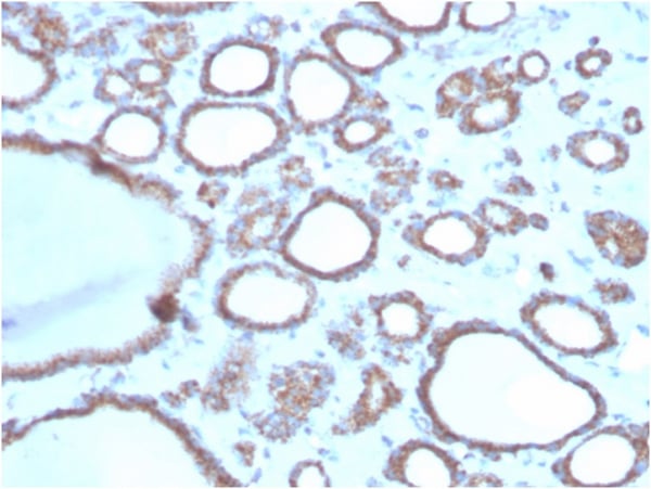
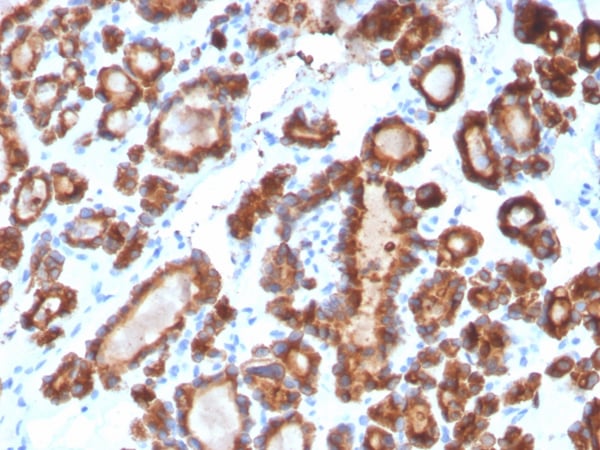
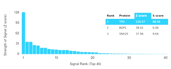
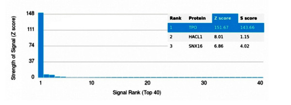 Application Data
(Analysis of Protein Array containing more than 19,000 full-length human proteins using Thyroid Peroxidase Mouse Monoclonal Antibody (TPO/3697). Z- and S- Score: The Z-score represents the strength of a signal that a monoclonal antibody (MAb) (in combination with a fluorescently-tagged anti-IgG secondary antibody) produces when binding to a particular protein on the HuProtTM array. Z-scores are described in units of standard deviations (SD's) above the mean value of all signals generated on that array. If targets on HuProtTM are arranged in descending order of the Z-score, the S-score is the difference (also in units of SD's) between the Z-score. S-score therefore represents the relative target specificity of a MAb to its intended target. A MAb is considered to specific to its intended target, if the MAb has an S-score of at least 2.5. For example, if a MAb binds to protein X with a Z-score of 43 and to protein Y with a Z-score of 14, then the S-score for the binding of that MAb to protein X is equal to 29.)
Application Data
(Analysis of Protein Array containing more than 19,000 full-length human proteins using Thyroid Peroxidase Mouse Monoclonal Antibody (TPO/3697). Z- and S- Score: The Z-score represents the strength of a signal that a monoclonal antibody (MAb) (in combination with a fluorescently-tagged anti-IgG secondary antibody) produces when binding to a particular protein on the HuProtTM array. Z-scores are described in units of standard deviations (SD's) above the mean value of all signals generated on that array. If targets on HuProtTM are arranged in descending order of the Z-score, the S-score is the difference (also in units of SD's) between the Z-score. S-score therefore represents the relative target specificity of a MAb to its intended target. A MAb is considered to specific to its intended target, if the MAb has an S-score of at least 2.5. For example, if a MAb binds to protein X with a Z-score of 43 and to protein Y with a Z-score of 14, then the S-score for the binding of that MAb to protein X is equal to 29.)
TPO (Thyroid Peroxidase), Monoclonal Antibody (Cat# AAA215468)
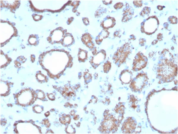
 Application Data
(Analysis of Protein Array containing more than 19,000 full-length human proteins using Thyroid Peroxidase Mouse Monoclonal Antibody (TPO/3698). Z- and S- Score: The Z-score represents the strength of a signal that a monoclonal antibody (MAb) (in combination with a fluorescently-tagged anti-IgG secondary antibody) produces when binding to a particular protein on the HuProtTM array. Z-scores are described in units of standard deviations (SD's) above the mean value of all signals generated on that array. If targets on HuProtTM are arranged in descending order of the Z-score, the S-score is the difference (also in units of SD's) between the Z-score. S-score therefore represents the relative target specificity of a MAb to its intended target. A MAb is considered to specific to its intended target, if the MAb has an S-score of at least 2.5. For example, if a MAb binds to protein X with a Z-score of 43 and to protein Y with a Z-score of 14, then the S-score for the binding of that MAb to protein X is equal to 29.)
Application Data
(Analysis of Protein Array containing more than 19,000 full-length human proteins using Thyroid Peroxidase Mouse Monoclonal Antibody (TPO/3698). Z- and S- Score: The Z-score represents the strength of a signal that a monoclonal antibody (MAb) (in combination with a fluorescently-tagged anti-IgG secondary antibody) produces when binding to a particular protein on the HuProtTM array. Z-scores are described in units of standard deviations (SD's) above the mean value of all signals generated on that array. If targets on HuProtTM are arranged in descending order of the Z-score, the S-score is the difference (also in units of SD's) between the Z-score. S-score therefore represents the relative target specificity of a MAb to its intended target. A MAb is considered to specific to its intended target, if the MAb has an S-score of at least 2.5. For example, if a MAb binds to protein X with a Z-score of 43 and to protein Y with a Z-score of 14, then the S-score for the binding of that MAb to protein X is equal to 29.)
TPO (Thyroid Peroxidase), Monoclonal Antibody (Cat# AAA215469)
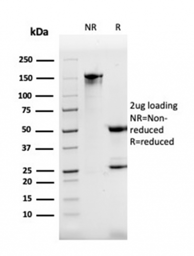
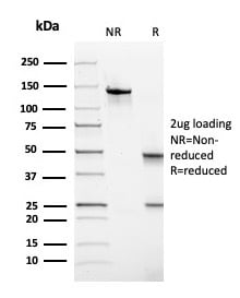
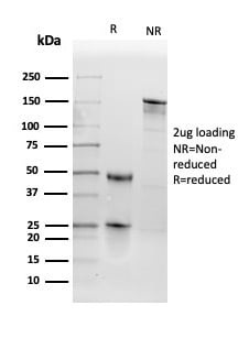
 SDS-PAGE
(SDS-PAGE Analysis Purified Endoglin / CD105 Mouse Monoclonal Antibody (ENG/1621).Confirmation of Integrity and Purity of Antibody.)
SDS-PAGE
(SDS-PAGE Analysis Purified Endoglin / CD105 Mouse Monoclonal Antibody (ENG/1621).Confirmation of Integrity and Purity of Antibody.)
Endoglin/CD105, Monoclonal Antibody (Cat# AAA215474)
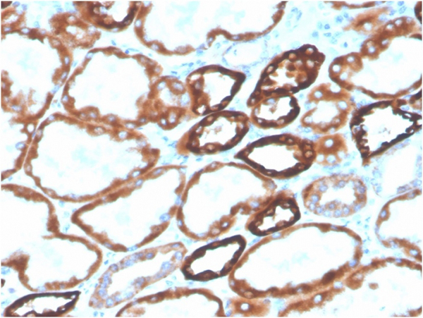
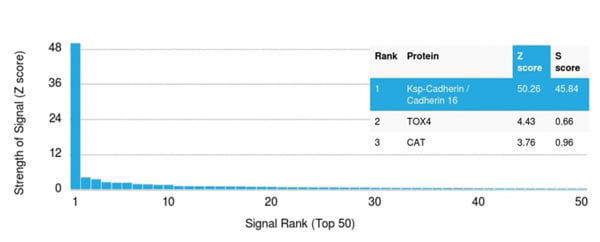 Application Data
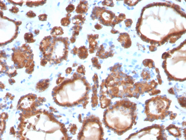
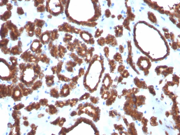
(Analysis of Protein Array containing more than 19,000 full-length human proteins using CDH16-Monospecific Mouse Monoclonal Antibody (CDH16/2125). Z- and S- Score: The Z-score represents the strength of a signal that a monoclonal antibody (MAb) (in combination with a fluorescently-tagged anti-IgG secondary antibody) produces when binding to a particular protein on the HuProtTM array. Z-scores are described in units of standard deviations (SD's) above the mean value of all signals generated on that array. If targets on HuProtTM are arranged in descending order of the Z-score, the S-score is the difference (also in units of SD's) between the Z-score. S-score therefore represents the relative target specificity of a MAb to its intended target. A MAb is considered to specific to its intended target, if the MAb has an S-score of at least 2.5. For example, if a MAb binds to protein X with a Z-score of 43 and to protein Y with a Z-score of 14, then the S-score for the binding of that MAb to protein X is equal to 29.)
Application Data
(Analysis of Protein Array containing more than 19,000 full-length human proteins using CDH16-Monospecific Mouse Monoclonal Antibody (CDH16/2125). Z- and S- Score: The Z-score represents the strength of a signal that a monoclonal antibody (MAb) (in combination with a fluorescently-tagged anti-IgG secondary antibody) produces when binding to a particular protein on the HuProtTM array. Z-scores are described in units of standard deviations (SD's) above the mean value of all signals generated on that array. If targets on HuProtTM are arranged in descending order of the Z-score, the S-score is the difference (also in units of SD's) between the Z-score. S-score therefore represents the relative target specificity of a MAb to its intended target. A MAb is considered to specific to its intended target, if the MAb has an S-score of at least 2.5. For example, if a MAb binds to protein X with a Z-score of 43 and to protein Y with a Z-score of 14, then the S-score for the binding of that MAb to protein X is equal to 29.)
Ksp-Cadherin/CDH16, Monoclonal Antibody (Cat# AAA215475)
 Application Data
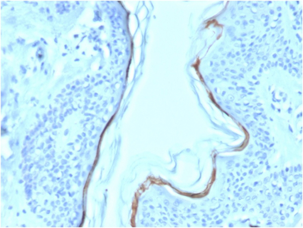
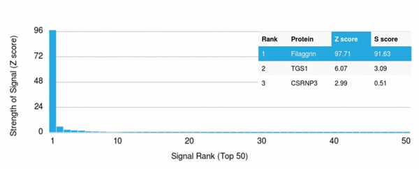
(Analysis of Protein Array containing more than 19,000 full-length human proteins using Filaggrin Mouse Monoclonal Antibody (FLG/1563). Z- and S- Score: The Z-score represents the strength of a signal that a monoclonal antibody (MAb) (in combination with a fluorescently-tagged anti-IgG secondary antibody) produces when binding to a particular protein on the HuProtTM array. Z-scores are described in units of standard deviations (SD's) above the mean value of all signals generated on that array. If targets on HuProtTM are arranged in descending order of the Z-score, the S-score is the difference (also in units of SD's) between the Z-score. S-score therefore represents the relative target specificity of a MAb to its intended target. A MAb is considered to specific to its intended target, if the MAb has an S-score of at least 2.5. For example, if a MAb binds to protein X with a Z-score of 43 and to protein Y with a Z-score of 14, then the S-score for the binding of that MAb to protein X is equal to 29.)
Application Data
(Analysis of Protein Array containing more than 19,000 full-length human proteins using Filaggrin Mouse Monoclonal Antibody (FLG/1563). Z- and S- Score: The Z-score represents the strength of a signal that a monoclonal antibody (MAb) (in combination with a fluorescently-tagged anti-IgG secondary antibody) produces when binding to a particular protein on the HuProtTM array. Z-scores are described in units of standard deviations (SD's) above the mean value of all signals generated on that array. If targets on HuProtTM are arranged in descending order of the Z-score, the S-score is the difference (also in units of SD's) between the Z-score. S-score therefore represents the relative target specificity of a MAb to its intended target. A MAb is considered to specific to its intended target, if the MAb has an S-score of at least 2.5. For example, if a MAb binds to protein X with a Z-score of 43 and to protein Y with a Z-score of 14, then the S-score for the binding of that MAb to protein X is equal to 29.)
Filaggrin, Monoclonal Antibody (Cat# AAA215480)
 SDS-PAGE
(SDS-PAGE Analysis Purified CK7 Mouse Monoclonal Antibody (OV-TL12/30) (unconjugated). Confirmation of Integrity and Purity of Antibody)
SDS-PAGE
(SDS-PAGE Analysis Purified CK7 Mouse Monoclonal Antibody (OV-TL12/30) (unconjugated). Confirmation of Integrity and Purity of Antibody)
Cytokeratin 7, Monoclonal Antibody (Cat# AAA215485)
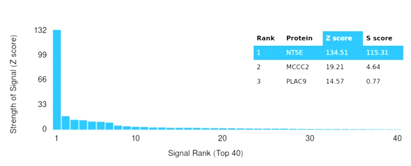 Application Data
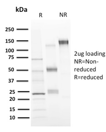
(Analysis of Protein Array containing more than 19,000 full-length human proteins using CD73 Mouse Monoclonal Antibody (CD73/2646) Z- and S- Score: The Z-score represents the strength of a signal that a monoclonal antibody (MAb) (in combination with a fluorescently-tagged anti-IgG secondary antibody) produces when binding to a particular protein on the HuProtTM array. Z-scores are described in units of standard deviations (SD's) above the mean value of all signals generated on that array. If targets on HuProtTM are arranged in descending order of the Z-score, the S-score is the difference (also in units of SD's) between the Z-score. S-score therefore represents the relative target specificity of a MAb to its intended target. A MAb is considered to specific to its intended target, if the MAb has an S-score of at least 2.5. For example, if a MAb binds to protein X with a Z-score of 43 and to protein Y with a Z-score of 14, then the S-score for the binding of that MAb to protein X is equal to 29.)
Application Data
(Analysis of Protein Array containing more than 19,000 full-length human proteins using CD73 Mouse Monoclonal Antibody (CD73/2646) Z- and S- Score: The Z-score represents the strength of a signal that a monoclonal antibody (MAb) (in combination with a fluorescently-tagged anti-IgG secondary antibody) produces when binding to a particular protein on the HuProtTM array. Z-scores are described in units of standard deviations (SD's) above the mean value of all signals generated on that array. If targets on HuProtTM are arranged in descending order of the Z-score, the S-score is the difference (also in units of SD's) between the Z-score. S-score therefore represents the relative target specificity of a MAb to its intended target. A MAb is considered to specific to its intended target, if the MAb has an S-score of at least 2.5. For example, if a MAb binds to protein X with a Z-score of 43 and to protein Y with a Z-score of 14, then the S-score for the binding of that MAb to protein X is equal to 29.)
CD73, Monoclonal Antibody (Cat# AAA215488)
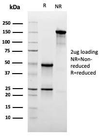
 SDS-PAGE
(SDS-PAGE Analysis Purified CD137L Mouse Monoclonal Antibody (CD137L/1547) (unconjugated). Confirmation of Purity and Integrity of Antibody.)
SDS-PAGE
(SDS-PAGE Analysis Purified CD137L Mouse Monoclonal Antibody (CD137L/1547) (unconjugated). Confirmation of Purity and Integrity of Antibody.)
CD137L/4-1BBL/TNFSF9, Monoclonal Antibody (Cat# AAA215491)
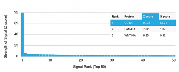 Application Data
(Analysis of Protein Array containing more than 19,000 full-length human proteins using CD40-Ligand Mouse Monoclonal Antibody (CD40LG/2761) Z- and S- Score: The Z-score represents the strength of a signal that a monoclonal antibody (MAb) (in combination with a fluorescently-tagged anti-IgG secondary antibody) produces when binding to a particular protein on the HuProtTM array. Z-scores are described in units of standard deviations (SD's) above the mean value of all signals generated on that array. If targets on HuProtTM are arranged in descending order of the Z-score, the S-score is the difference (also in units of SD's) between the Z-score. S-score therefore represents the relative target specificity of a MAb to its intended target. A MAb is considered to specific to its intended target, if the MAb has an S-score of at least 2.5. For example, if a MAb binds to protein X with a Z-score of 43 and to protein Y with a Z-score of 14, then the S-score for the binding of that MAb to protein X is equal to 29.)
Application Data
(Analysis of Protein Array containing more than 19,000 full-length human proteins using CD40-Ligand Mouse Monoclonal Antibody (CD40LG/2761) Z- and S- Score: The Z-score represents the strength of a signal that a monoclonal antibody (MAb) (in combination with a fluorescently-tagged anti-IgG secondary antibody) produces when binding to a particular protein on the HuProtTM array. Z-scores are described in units of standard deviations (SD's) above the mean value of all signals generated on that array. If targets on HuProtTM are arranged in descending order of the Z-score, the S-score is the difference (also in units of SD's) between the Z-score. S-score therefore represents the relative target specificity of a MAb to its intended target. A MAb is considered to specific to its intended target, if the MAb has an S-score of at least 2.5. For example, if a MAb binds to protein X with a Z-score of 43 and to protein Y with a Z-score of 14, then the S-score for the binding of that MAb to protein X is equal to 29.)
CD40 Ligand/CD154/TRAP1, Monoclonal Antibody (Cat# AAA215493)
 Application Data
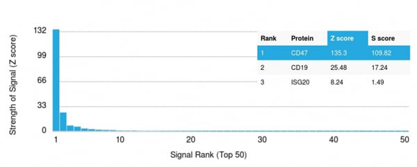
(Analysis of Protein Array containing more than 19,000 full-length human proteins using CD47 Mouse Monoclonal Antibody (CD47/3019) Z- and S- Score: The Z-score represents the strength of a signal that a monoclonal antibody (MAb) (in combination with a fluorescently-tagged anti-IgG secondary antibody) produces when binding to a particular protein on the HuProtTM array. Z-scores are described in units of standard deviations (SD's) above the mean value of all signals generated on that array. If targets on HuProtTM are arranged in descending order of the Z-score, the S-score is the difference (also in units of SD's) between the Z-score. S-score therefore represents the relative target specificity of a MAb to its intended target. A MAb is considered to specific to its intended target, if the MAb has an S-score of at least 2.5. For example, if a MAb binds to protein X with a Z-score of 43 and to protein Y with a Z-score of 14, then the S-score for the binding of that MAb to protein X is equal to 29.)
Application Data
(Analysis of Protein Array containing more than 19,000 full-length human proteins using CD47 Mouse Monoclonal Antibody (CD47/3019) Z- and S- Score: The Z-score represents the strength of a signal that a monoclonal antibody (MAb) (in combination with a fluorescently-tagged anti-IgG secondary antibody) produces when binding to a particular protein on the HuProtTM array. Z-scores are described in units of standard deviations (SD's) above the mean value of all signals generated on that array. If targets on HuProtTM are arranged in descending order of the Z-score, the S-score is the difference (also in units of SD's) between the Z-score. S-score therefore represents the relative target specificity of a MAb to its intended target. A MAb is considered to specific to its intended target, if the MAb has an S-score of at least 2.5. For example, if a MAb binds to protein X with a Z-score of 43 and to protein Y with a Z-score of 14, then the S-score for the binding of that MAb to protein X is equal to 29.)
CD47/IAP, Monoclonal Antibody (Cat# AAA215494)
What are Monoclonal Antibodies?
Monoclonal antibodies are specialized laboratory-produced proteins developed for binding to specific biological antigens or other molecular targets. Since they come from a single cell (or clone), they are especially consistent and accurate in the data they are involved in producing.
This type of antibody material has been shown to be a powerful tool in finding and subsequently destroying harmful cells in an organism, such as those found in cancers or various autoimmune diseases. This makes them excellent aids in medical testing and research, which is why they are so widely used.
AAA Biotech offers a comprehensive range of high-quality monoclonal antibodies that perform effectively in various laboratory tests, including (amongst others) ELISA, western blotting, immunohistochemistry, and flow cytometry. All of the products in our catalog are thoroughly quality tested to make sure that they are reliable and will consistently perform well in your research.
What Are The Uses of Monoclonal Antibodies
Monoclonal antibodies are used in many lab tests, including (amongst others) ELISA, western blotting, immunohistochemistry, and flow cytometry.
ELISA is a test that helps detect a specific substance/analyte in a sample. It uses antibodies (often monoclonal) bound to a solid surface (such as the well of a microplate) to “capture” the substance/analyte in the sample and immobilize it so that the detection antibody component can then bind to it and produce a signal, which can then be measured.
Western blotting identifies specific proteins in a sample. The sample is first separated on a gel, and then antibodies are applied that will typically bind to the target, which will all be localized to a single band in a lane.
Immunohistochemistry helps locate specific proteins in cells or tissue samples using antibodies.
Flow cytometry looks at and sorts cells. It uses antibodies that are conjugated to reporter molecules called “fluorophores”, which, under special lights, emit light themselves, which can then be measured by a detector instrument.
How Monoclonal Antibodies Are Used as Medicine?
Please note that all of the products listed in AAA Biotech’s also known as AAA Bio or AAABio catalog are strictly for research-use only (RUO).
Monoclonal antibodies can also be used as therapeutic/medical treatments, particularly in the context of cancers. They are designed to find and bind to specific cells or proteins, helping the immune system recognize and attack the cancer. These treatments work in different ways, such as:
- Radioimmunotherapy attaches a small amount of radioactive molecule to the antibody, so it delivers the radiation directly to the cancer cells that the antibody is specifically binding to.
- Antibody-directed enzyme prodrug therapy uses antibodies that are specifically bound to special enzymes. These enzymes activate a harmless drug in the body and turn it into a cancer-killing drug only near the cancer cells—this helps avoid harming healthy cells.
- Immunoliposomes are tiny “bubbles” filled with medicine/drug and coated with antibodies. They carry the drug straight to the cancer cells.
Why Buy Monoclonal Antibodies From Us?
At AAA Biotech, we provide high-performance monoclonal antibodies designed to support a wide range of research needs.
1. Validated for Versatile Applications
The antibodies in our catalog are extensively validated and compatible with multiple techniques, including (but not limited to) ELISA, flow cytometry (FC), immunocytochemistry (ICC), immunofluorescence (IF), immunohistochemistry (IHC), immunoprecipitation (IP), and western blotting (WB).
2. Wide Selection & Specialized Options
We offer antibodies for common and rare species, that are available in various conjugated forms, and also in recombinant formats. Essentially, there is almost anything one might need to meet their experimental model’s requirements.
3. High-Quality Proteins
Our proteins meet high purity standards—90% or more as confirmed by SDS-PAGE. Many are available with tags like His, Flag, GST, or MBP, and we also supply native and biologically active proteins for functional studies.
Frequently Asked Questions
1. Are your monoclonal antibodies validated for specific applications?
Yes, our antibodies are tested and validated for use in methods such as ELISA, western blot, IHC, flow cytometry, and more. Refer to specific product pages or datasheets for individual product information.
2. How do I choose the right monoclonal antibody for my application?
Review the product details directly for application validation, species reactivity, and target information. You may also contact our support team at any time for help.
3. How quickly can I receive my order?
Most orders are processed and shipped within 1–3 business days, depending on product availability and your shipping location.