Filters
▼Clonality
▼Type
▼Reactivity
▼Gene Name
▼Isotype
▼Host
▼Application
▼Clone
▼Monoclonal Antibodies
Get accurate results in your research with our Monoclonal Antibodies, which are specially made to target exactly what you require for your research, and will produce consistent, reliable performance in lab tests.
Viewing 1500-1550 of 27597 product results
 IHC (Immunohiostchemistry)
(DAB staining on IHCP;Samples: Human Cerebrum Tissue; Primary Ab: 30ug/ml Mouse AntiHuman SIGLEC8 AntibodySecond Ab: 2ug/mL HRPLinked Caprine AntiMouse IgG Polyclonal Antibody(Catalog: SAA544Mu19))
IHC (Immunohiostchemistry)
(DAB staining on IHCP;Samples: Human Cerebrum Tissue; Primary Ab: 30ug/ml Mouse AntiHuman SIGLEC8 AntibodySecond Ab: 2ug/mL HRPLinked Caprine AntiMouse IgG Polyclonal Antibody(Catalog: SAA544Mu19))
Sialic Acid Binding Ig Like Lectin 8 (SIGLEC8), Monoclonal Antibody (Cat# AAA151745)
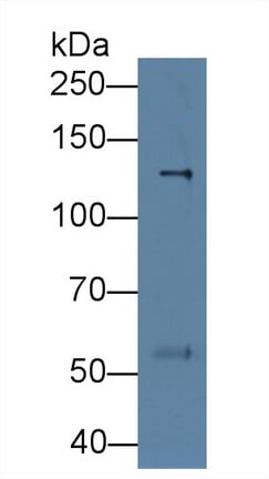
 WB (Western Blot)
(Western Blot; Sample: Rat Serum Primary Ab: 3ug/ml Mouse AntiRat DBP Antibody Second Ab: 0.2ug/mL HRPLinked Caprine AntiMouse IgG Polyclonal Antibody (Catalog: SAA544Mu19))
WB (Western Blot)
(Western Blot; Sample: Rat Serum Primary Ab: 3ug/ml Mouse AntiRat DBP Antibody Second Ab: 0.2ug/mL HRPLinked Caprine AntiMouse IgG Polyclonal Antibody (Catalog: SAA544Mu19))
Vitamin D Binding Protein (DBP), Monoclonal Antibody (Cat# AAA151752)
 WB (Western Blot)
(Western Blot; Sample: Rat Serum Primary Ab: 3ug/ml Mouse AntiRat DBP Antibody Second Ab: 0.2ug/mL HRPLinked Caprine AntiMouse IgG Polyclonal Antibody (Catalog: SAA544Mu19))
WB (Western Blot)
(Western Blot; Sample: Rat Serum Primary Ab: 3ug/ml Mouse AntiRat DBP Antibody Second Ab: 0.2ug/mL HRPLinked Caprine AntiMouse IgG Polyclonal Antibody (Catalog: SAA544Mu19))
Vitamin D Binding Protein (DBP), Monoclonal Antibody (Cat# AAA151753)
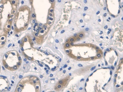
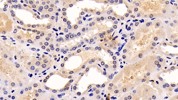
 IHC (Immunohistochemistry)
(DAB staining on IHCP;Sample: Human Kidney Tissue; Primary Ab: 30ug/ml Mouse AntiHuman VEGF121 AntibodySecond Ab: 2ug/mL HRPLinked Caprine AntiMouse IgG Polyclonal Antibody(Catalog: SAA544Mu19))
IHC (Immunohistochemistry)
(DAB staining on IHCP;Sample: Human Kidney Tissue; Primary Ab: 30ug/ml Mouse AntiHuman VEGF121 AntibodySecond Ab: 2ug/mL HRPLinked Caprine AntiMouse IgG Polyclonal Antibody(Catalog: SAA544Mu19))
Vascular Endothelial Growth Factor 121 (VEGF121), Monoclonal Antibody (Cat# AAA151759)
 WB (Western Blot)
(Western Blot; Sample: Human Urine lysate Primary Ab: 1ug/ml Mouse AntiHuman LOX1 Antibody Second Ab: 0.2ug/mL HRPLinked Caprine AntiMouse IgG Polyclonal Antibody (Catalog: SAA544Mu19))
WB (Western Blot)
(Western Blot; Sample: Human Urine lysate Primary Ab: 1ug/ml Mouse AntiHuman LOX1 Antibody Second Ab: 0.2ug/mL HRPLinked Caprine AntiMouse IgG Polyclonal Antibody (Catalog: SAA544Mu19))
Lectin Like Oxidized Low Density Lipoprotein Receptor 1 (LOX1), Monoclonal Antibody (Cat# AAA151760)
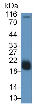
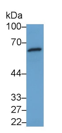
 WB (Western Blot)
(Western Blot; Sample: Human UrinePrimary Ab: 3ug/ml Mouse AntiHuman LRG1 AntibodySecond Ab: 0.2ug/mL HRPLinked Caprine AntiMouse IgG Polyclonal Antibody(Catalog: SAA544Mu19))
WB (Western Blot)
(Western Blot; Sample: Human UrinePrimary Ab: 3ug/ml Mouse AntiHuman LRG1 AntibodySecond Ab: 0.2ug/mL HRPLinked Caprine AntiMouse IgG Polyclonal Antibody(Catalog: SAA544Mu19))
Leucine Rich Alpha2Glycoprotein 1 (LRG1), Monoclonal Antibody (Cat# AAA151765)
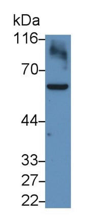
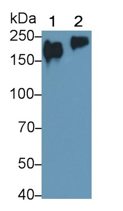
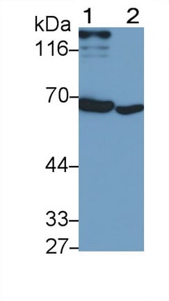
 WB (Western Blot)
(Western Blot; Sample: Lane1: Human Serum; Lane2: Human Urine Primary Ab: 0.2ug/ml Mouse AntiHuman LRG1 Antibody Second Ab: 0.2ug/mL HRPLinked Caprine AntiMouse IgG Polyclonal Antibody (Catalog: SAA544Mu19))
WB (Western Blot)
(Western Blot; Sample: Lane1: Human Serum; Lane2: Human Urine Primary Ab: 0.2ug/ml Mouse AntiHuman LRG1 Antibody Second Ab: 0.2ug/mL HRPLinked Caprine AntiMouse IgG Polyclonal Antibody (Catalog: SAA544Mu19))
Leucine Rich Alpha2Glycoprotein 1 (LRG1), Monoclonal Antibody (Cat# AAA151766)
 WB (Western Blot)
(Western Blot; Sample: Human Serum Primary Ab: 3ug/ml Mouse AntiHuman TGFb3 Antibody Second Ab: 0.2ug/mL HRPLinked Caprine AntiMouse IgG Polyclonal Antibody (Catalog: SAA544Mu19))
WB (Western Blot)
(Western Blot; Sample: Human Serum Primary Ab: 3ug/ml Mouse AntiHuman TGFb3 Antibody Second Ab: 0.2ug/mL HRPLinked Caprine AntiMouse IgG Polyclonal Antibody (Catalog: SAA544Mu19))
Transforming Growth Factor Beta 3 (TGFb3), Monoclonal Antibody (Cat# AAA151768)
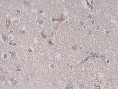
 IHC (Immunohiostchemistry)
(DAB staining on IHCP;Sample: Human Cerebrum Tissue; Primary Ab: 30ug/ml Mouse AntiHuman CD34 AntibodySecond Ab: 2ug/mL HRPLinked Caprine AntiMouse IgG Polyclonal Antibody(Catalog: SAA544Mu19))
IHC (Immunohiostchemistry)
(DAB staining on IHCP;Sample: Human Cerebrum Tissue; Primary Ab: 30ug/ml Mouse AntiHuman CD34 AntibodySecond Ab: 2ug/mL HRPLinked Caprine AntiMouse IgG Polyclonal Antibody(Catalog: SAA544Mu19))
Cluster Of Differentiation 34 (CD34), Monoclonal Antibody (Cat# AAA151769)
 IHC (Immunohistochemisry)
(DAB staining on IHCP;Sample: Human Kidney Tissue; Primary Ab: 20ug/ml Mouse AntiHuman TNC AntibodySecond Ab: 2ug/mL HRPLinked Caprine AntiMouse IgG Polyclonal Antibody(Catalog: SAA544Mu19))
IHC (Immunohistochemisry)
(DAB staining on IHCP;Sample: Human Kidney Tissue; Primary Ab: 20ug/ml Mouse AntiHuman TNC AntibodySecond Ab: 2ug/mL HRPLinked Caprine AntiMouse IgG Polyclonal Antibody(Catalog: SAA544Mu19))
Tenascin C (TNC), Monoclonal Antibody (Cat# AAA151773)
 WB (Western Blot)
(Western Blot; Sample: Human Urine Primary Ab: 1ug/ml Mouse AntiHuman RETN Antibody Second Ab: 0.2ug/mL HRPLinked Caprine AntiMouse IgG Polyclonal Antibody (Catalog: SAA544Mu19))
WB (Western Blot)
(Western Blot; Sample: Human Urine Primary Ab: 1ug/ml Mouse AntiHuman RETN Antibody Second Ab: 0.2ug/mL HRPLinked Caprine AntiMouse IgG Polyclonal Antibody (Catalog: SAA544Mu19))
Resistin (RETN), Monoclonal Antibody (Cat# AAA151642)
 IHC (Immunohistochemistry)
(DAB staining on IHCP;Sample: Mouse Uterus Tissue; Primary Ab: 40ug/ml Mouse AntiMouse RETN AntibodySecond Ab: 2ug/mL HRPLinked Caprine AntiMouse IgG Polyclonal Antibody(Catalog: SAA544Mu19))
IHC (Immunohistochemistry)
(DAB staining on IHCP;Sample: Mouse Uterus Tissue; Primary Ab: 40ug/ml Mouse AntiMouse RETN AntibodySecond Ab: 2ug/mL HRPLinked Caprine AntiMouse IgG Polyclonal Antibody(Catalog: SAA544Mu19))
Resistin (RETN), Monoclonal Antibody (Cat# AAA151643)
 WB (Western Blot)
(Western Blot; Sample: Jurkat cell lysate Primary Ab: 1.5ug/ml Mouse AntiHuman CASP8 Antibody Second Ab: 0.2ug/mL HRPLinked Caprine AntiMouse IgG Polyclonal Antibody (Catalog: SAA544Mu19))
WB (Western Blot)
(Western Blot; Sample: Jurkat cell lysate Primary Ab: 1.5ug/ml Mouse AntiHuman CASP8 Antibody Second Ab: 0.2ug/mL HRPLinked Caprine AntiMouse IgG Polyclonal Antibody (Catalog: SAA544Mu19))
Caspase 8 (CASP8), Monoclonal Antibody (Cat# AAA151646)
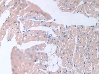
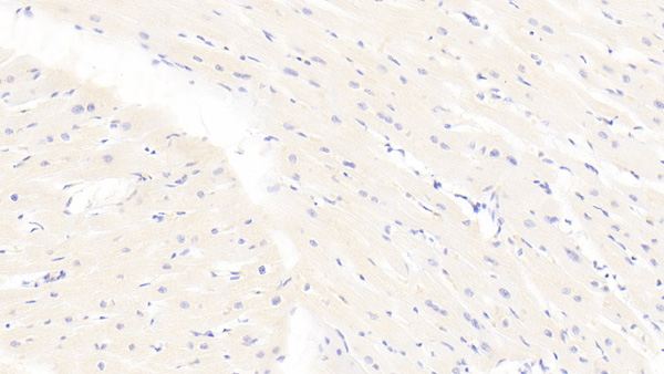
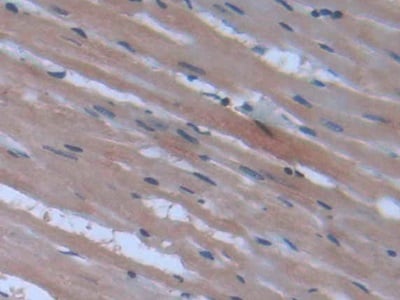 IHC (Immunohiostchemistry)
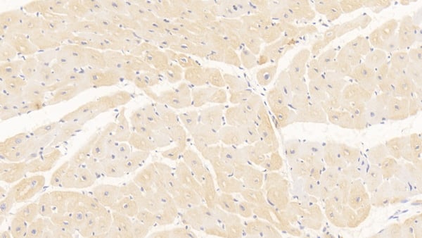
(DAB staining on IHCP;Sample: Rat Heart Tissue; Primary Ab: 40ug/ml Mouse AntiRat PPARg AntibodySecond Ab: 2ug/mL HRPLinked Caprine AntiMouse IgG Polyclonal Antibody(Catalog: SAA544Mu19))
IHC (Immunohiostchemistry)
(DAB staining on IHCP;Sample: Rat Heart Tissue; Primary Ab: 40ug/ml Mouse AntiRat PPARg AntibodySecond Ab: 2ug/mL HRPLinked Caprine AntiMouse IgG Polyclonal Antibody(Catalog: SAA544Mu19))
Peroxisome Proliferator Activated Receptor Gamma (PPARg), Monoclonal Antibody (Cat# AAA151649)
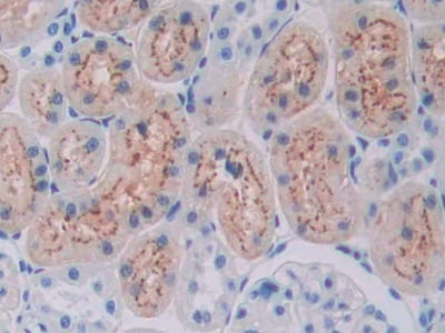
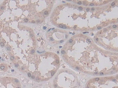
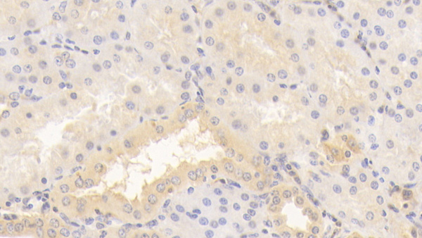
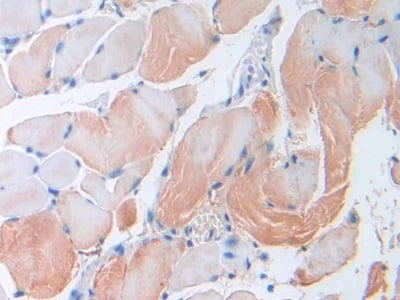
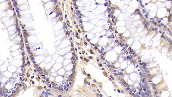
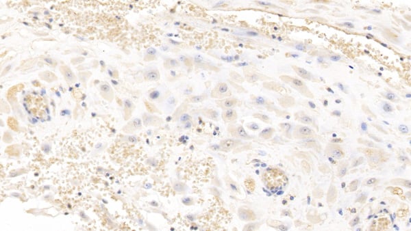
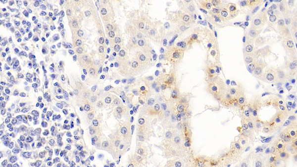
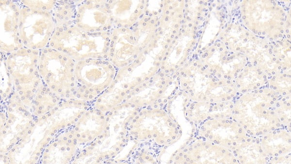
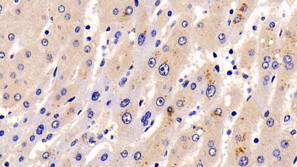
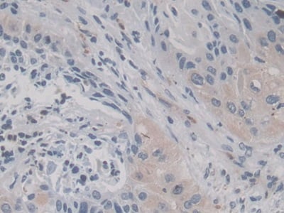
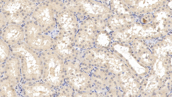 IHC (Immunohistochemistry)
(DAB staining on IHCP; Sample: Human Kidney Tissue; Primary Ab: 10ug/ml Mouse AntiHuman REN Antibody Second Ab: 2ug/mL HRPLinked Caprine AntiMouse IgG Polyclonal Antibody (Catalog: SAA544Mu19))
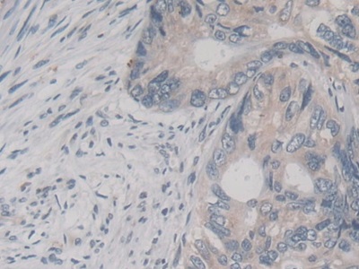
IHC (Immunohistochemistry)
(DAB staining on IHCP; Sample: Human Kidney Tissue; Primary Ab: 10ug/ml Mouse AntiHuman REN Antibody Second Ab: 2ug/mL HRPLinked Caprine AntiMouse IgG Polyclonal Antibody (Catalog: SAA544Mu19))
Renin (REN), Monoclonal Antibody (Cat# AAA151651)
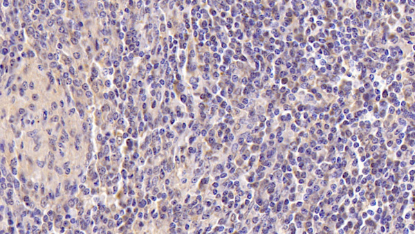
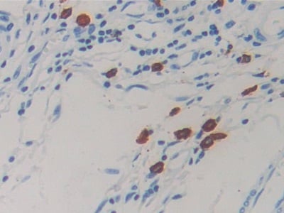
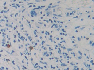
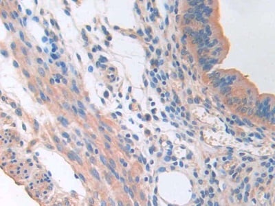
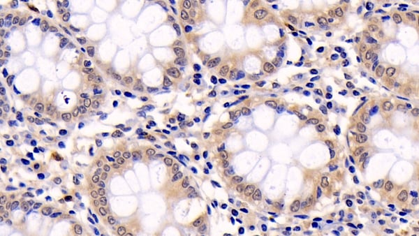
 IHC (Immunohiostchemistry)
(DAB staining on IHCP;Sample: Human Colon Tissue; Primary Ab: 20ug/ml Mouse AntiHuman NB1 AntibodySecond Ab: 2ug/mL HRPLinked Caprine AntiMouse IgG Polyclonal Antibody(Catalog: SAA544Mu19))
IHC (Immunohiostchemistry)
(DAB staining on IHCP;Sample: Human Colon Tissue; Primary Ab: 20ug/ml Mouse AntiHuman NB1 AntibodySecond Ab: 2ug/mL HRPLinked Caprine AntiMouse IgG Polyclonal Antibody(Catalog: SAA544Mu19))
Neutrophil Specific Antigen 1 (NB1), Monoclonal Antibody (Cat# AAA151659)
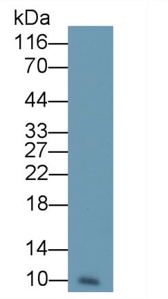
 WB (Western Blot)
(Western Blot;Sample: Rat Cerebrum lysatePrimary Ab: 5ug/ml Mouse AntiHuman PINP AntibodySecond Ab: 0.2ug/mL HRPLinked Caprine AntiMouse IgG Polyclonal Antibody)
WB (Western Blot)
(Western Blot;Sample: Rat Cerebrum lysatePrimary Ab: 5ug/ml Mouse AntiHuman PINP AntibodySecond Ab: 0.2ug/mL HRPLinked Caprine AntiMouse IgG Polyclonal Antibody)
Procollagen I NTerminal Propeptide (PINP), Monoclonal Antibody (Cat# AAA151663)
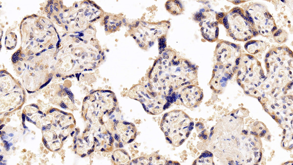
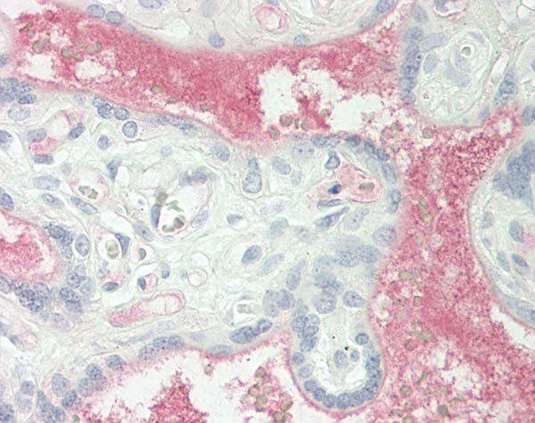
 IHC (Immunohiostchemistry)
(DAB staining on IHCP;Samples: Human Placenta Tissue; Primary Ab: 10ug/ml Mouse AntiHuman GPC3 AntibodySecond Ab: 2ug/mL HRPLinked Caprine AntiMouse IgG Polyclonal Antibody(Catalog: SAA544Mu19))
IHC (Immunohiostchemistry)
(DAB staining on IHCP;Samples: Human Placenta Tissue; Primary Ab: 10ug/ml Mouse AntiHuman GPC3 AntibodySecond Ab: 2ug/mL HRPLinked Caprine AntiMouse IgG Polyclonal Antibody(Catalog: SAA544Mu19))
Glypican 3 (GPC3), Monoclonal Antibody (Cat# AAA151664)
 WB (Western Blot)
(Western Blot; Sample: Lane1: Jurkat cell lysate; Lane2: THP1 cell lysate Primary Ab: 0.1ug/ml Mouse AntiHuman PTPRC Antibody Second Ab: 0.2ug/mL HRPLinked Caprine AntiMouse IgG Polyclonal Antibody (Catalog: SAA544Mu19))
WB (Western Blot)
(Western Blot; Sample: Lane1: Jurkat cell lysate; Lane2: THP1 cell lysate Primary Ab: 0.1ug/ml Mouse AntiHuman PTPRC Antibody Second Ab: 0.2ug/mL HRPLinked Caprine AntiMouse IgG Polyclonal Antibody (Catalog: SAA544Mu19))
Protein Tyrosine Phosphatase Receptor Type C (CD45), Monoclonal Antibody (Cat# AAA151670)


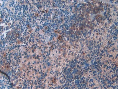
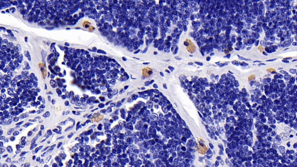
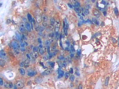
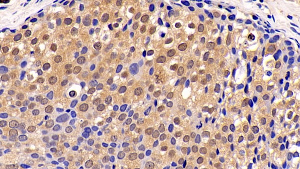
 IHC (Immunohistochemisry)
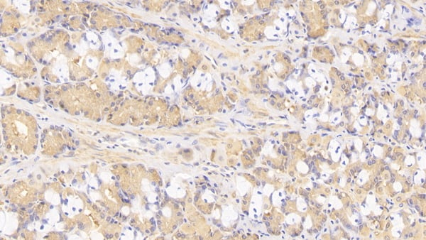
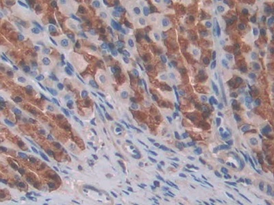
(DAB staining on IHCP;Sample: Human Stomach cancer Tissue; Primary Ab: 40ug/ml Mouse AntiHuman CCK8 AntibodySecond Ab: 2ug/mL HRPLinked Caprine AntiMouse IgG Polyclonal Antibody(Catalog: SAA544Mu19))
IHC (Immunohistochemisry)
(DAB staining on IHCP;Sample: Human Stomach cancer Tissue; Primary Ab: 40ug/ml Mouse AntiHuman CCK8 AntibodySecond Ab: 2ug/mL HRPLinked Caprine AntiMouse IgG Polyclonal Antibody(Catalog: SAA544Mu19))
Cholecystokinin 8 (CCK8), Monoclonal Antibody (Cat# AAA151673)

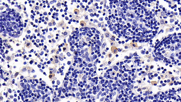
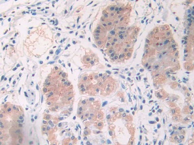
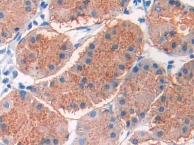
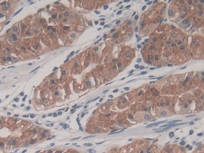
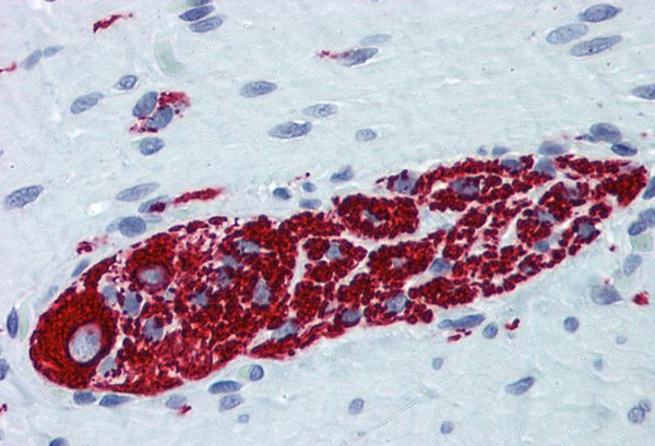
 IHC (Immunohistochemisry)
(DAB staining on IHCP;Sample: Human Pancreas Tissue; Primary Ab: 40ug/ml Mouse AntiHuman CCK8 AntibodySecond Ab: 2ug/mL HRPLinked Caprine AntiMouse IgG Polyclonal Antibody(Catalog: SAA544Mu19))
IHC (Immunohistochemisry)
(DAB staining on IHCP;Sample: Human Pancreas Tissue; Primary Ab: 40ug/ml Mouse AntiHuman CCK8 AntibodySecond Ab: 2ug/mL HRPLinked Caprine AntiMouse IgG Polyclonal Antibody(Catalog: SAA544Mu19))
Cholecystokinin 8 (CCK8), Monoclonal Antibody (Cat# AAA151677)
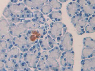
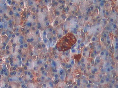
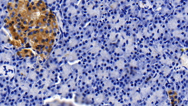
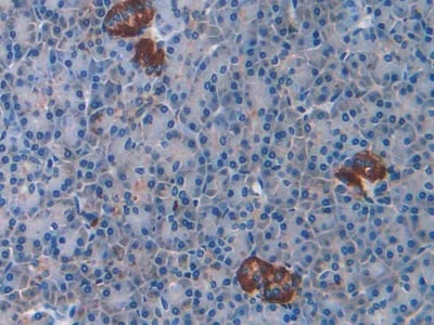 IHC (Immunohiostchemistry)
(DAB staining on IHCP;Samples: Human Pancreas Tissue;Primary Ab: 40ug/ml Mouse AntiMultispecies CCK8 AntibodySecond Ab: 2ug/mL HRPLinked Caprine AntiMouse IgG Polyclonal Antibody(Catalog: SAA544Mu19))
IHC (Immunohiostchemistry)
(DAB staining on IHCP;Samples: Human Pancreas Tissue;Primary Ab: 40ug/ml Mouse AntiMultispecies CCK8 AntibodySecond Ab: 2ug/mL HRPLinked Caprine AntiMouse IgG Polyclonal Antibody(Catalog: SAA544Mu19))
Cholecystokinin 8 (CCK8), Monoclonal Antibody (Cat# AAA151678)
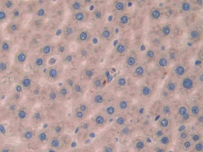
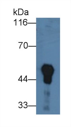
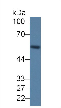
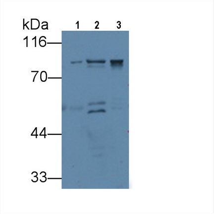
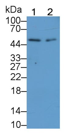
 WB (Western Blot)
(Western Blot Sample: Lane1: Human Liver lysate; Lane2: HepG2 cell lysate; Lane3: Porcine Liver lysate Primary Ab: 2ug/ml Mouse AntiHuman Arg Antibody Second Ab: 0.2ug/mL HRPLinked Caprine AntiMouse IgG Polyclonal Antibody (Catalog: SAA544Mu19))
WB (Western Blot)
(Western Blot Sample: Lane1: Human Liver lysate; Lane2: HepG2 cell lysate; Lane3: Porcine Liver lysate Primary Ab: 2ug/ml Mouse AntiHuman Arg Antibody Second Ab: 0.2ug/mL HRPLinked Caprine AntiMouse IgG Polyclonal Antibody (Catalog: SAA544Mu19))
Arginase (ARG), Monoclonal Antibody (Cat# AAA151681)

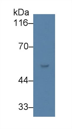
 WB (Western Blot)
(Western Blot; Sample: Lane1: Porcine Cerebrum lysate; Lane2: Rat Placenta lysate; Lane3: U87MG cell lysate Primary Ab: 3ug/ml Mouse AntiHuman VGF Antibody Second Ab: 0.2ug/mL HRPLinked Caprine AntiMouse IgG Polyclonal Antibody (Catalog: SAA544Mu19))
WB (Western Blot)
(Western Blot; Sample: Lane1: Porcine Cerebrum lysate; Lane2: Rat Placenta lysate; Lane3: U87MG cell lysate Primary Ab: 3ug/ml Mouse AntiHuman VGF Antibody Second Ab: 0.2ug/mL HRPLinked Caprine AntiMouse IgG Polyclonal Antibody (Catalog: SAA544Mu19))
VGF Nerve Growth Factor Inducible (VGF), Monoclonal Antibody (Cat# AAA151683)
 WB (Western Blot)
(Western Blot; Sample: Lane1: Porcine Serum; Lane2: Porcine Lung lysate; Lane3: Porcine Cerebrum lysate Primary Ab: 2ug/ml Mouse AntiPorcine CLU Antibody Second Ab: 0.2ug/mL HRPLinked Caprine AntiMouse IgG Polyclonal Antibody (Catalog: SAA544Mu19))
WB (Western Blot)
(Western Blot; Sample: Lane1: Porcine Serum; Lane2: Porcine Lung lysate; Lane3: Porcine Cerebrum lysate Primary Ab: 2ug/ml Mouse AntiPorcine CLU Antibody Second Ab: 0.2ug/mL HRPLinked Caprine AntiMouse IgG Polyclonal Antibody (Catalog: SAA544Mu19))
Clusterin (CLU), Monoclonal Antibody (Cat# AAA151690)
 WB (Western Blot)
(Western Blot; Sample: Mouse Cerebrum lysate Primary Ab: 5ug/ml Mouse AntiHuman BECN1 Antibody Second Ab: 0.2ug/mL HRPLinked Caprine AntiMouse IgG Polyclonal Antibody (Catalog: SAA544Mu19))
WB (Western Blot)
(Western Blot; Sample: Mouse Cerebrum lysate Primary Ab: 5ug/ml Mouse AntiHuman BECN1 Antibody Second Ab: 0.2ug/mL HRPLinked Caprine AntiMouse IgG Polyclonal Antibody (Catalog: SAA544Mu19))
Beclin 1 (BECN1), Monoclonal Antibody (Cat# AAA151895)
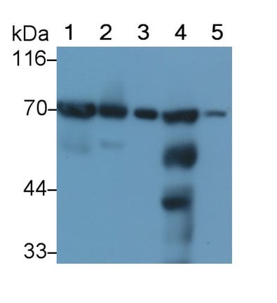 WB (Western Blot)
(Western Blot; Sample: Lane1: Mouse Heart lysate; Lane2: Mouse Liver lysate; Lane3: Mouse Cerebrum lysate; Lane4: Mouse Kidney lysate; Lane5: HepG2 cell lysate; Lane6: 293T cell lysate Primary Ab: 0.1ug/ml Mouse AntiMouse SDHA Antibody Second Ab: 0.2ug/mL HRPLinked Caprine AntiMouse IgG Polyclonal)
WB (Western Blot)
(Western Blot; Sample: Lane1: Mouse Heart lysate; Lane2: Mouse Liver lysate; Lane3: Mouse Cerebrum lysate; Lane4: Mouse Kidney lysate; Lane5: HepG2 cell lysate; Lane6: 293T cell lysate Primary Ab: 0.1ug/ml Mouse AntiMouse SDHA Antibody Second Ab: 0.2ug/mL HRPLinked Caprine AntiMouse IgG Polyclonal)
Succinate Dehydrogenase Complex Subunit A (SDHA), Monoclonal Antibody (Cat# AAA151897)
 WB (Western Blot)
(Western BlotSample: Lane1: Human Placenta lysateLane2: Porcine Lymph node lysateLane3: Raji cell lysatePrimary Ab: 2ug/ml Mouse Anti-Human CTLA4 AntibodySecond Ab: 0.2ug/mL HRP-Linked Caprine Anti-Mouse IgG Polyclonal Antibody)
WB (Western Blot)
(Western BlotSample: Lane1: Human Placenta lysateLane2: Porcine Lymph node lysateLane3: Raji cell lysatePrimary Ab: 2ug/ml Mouse Anti-Human CTLA4 AntibodySecond Ab: 0.2ug/mL HRP-Linked Caprine Anti-Mouse IgG Polyclonal Antibody)
Cytotoxic TLymphocyte Associated Antigen 4 (CTLA4), Monoclonal Antibody (Cat# AAA151694)
 WB (Western Blot)
(Western Blot; Sample: Lane1: Rat Lung lysate; Lane2: Rat Skin lysatePrimary Ab: 3ug/ml Mouse AntiHuman ELN AntibodySecond Ab: 0.2ug/mL HRPLinked Rabbit AntiMouse IgG Polyclonal Antibody(Catalog: SAA544Mu19))
WB (Western Blot)
(Western Blot; Sample: Lane1: Rat Lung lysate; Lane2: Rat Skin lysatePrimary Ab: 3ug/ml Mouse AntiHuman ELN AntibodySecond Ab: 0.2ug/mL HRPLinked Rabbit AntiMouse IgG Polyclonal Antibody(Catalog: SAA544Mu19))
Elastin (ELN), Monoclonal Antibody (Cat# AAA151701)
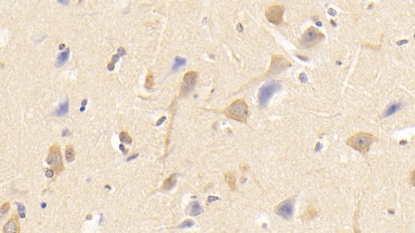
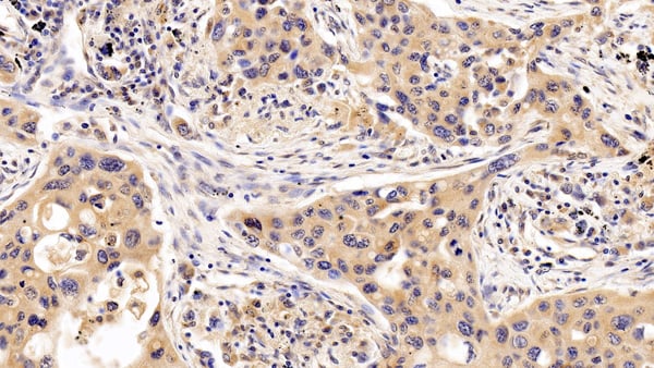
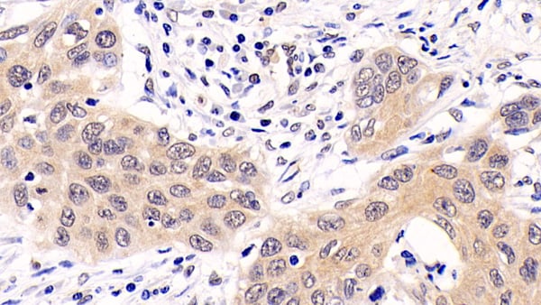 IHC (Immunohistochemistry)
(DAB staining on IHCP;Sample: Human Lung cancer Tissue; Primary Ab: 20ug/ml Mouse AntiHuman ERK1 AntibodySecond Ab: 2ug/mL HRPLinked Caprine AntiMouse IgG Polyclonal Antibody(Catalog: SAA544Mu19))
IHC (Immunohistochemistry)
(DAB staining on IHCP;Sample: Human Lung cancer Tissue; Primary Ab: 20ug/ml Mouse AntiHuman ERK1 AntibodySecond Ab: 2ug/mL HRPLinked Caprine AntiMouse IgG Polyclonal Antibody(Catalog: SAA544Mu19))
Extracellular Signal Regulated Kinase 1 (ERK1), Monoclonal Antibody (Cat# AAA151706)
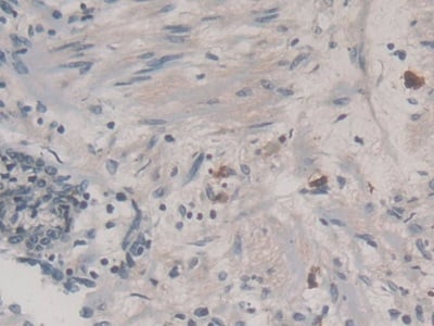
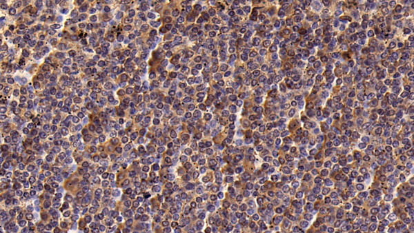
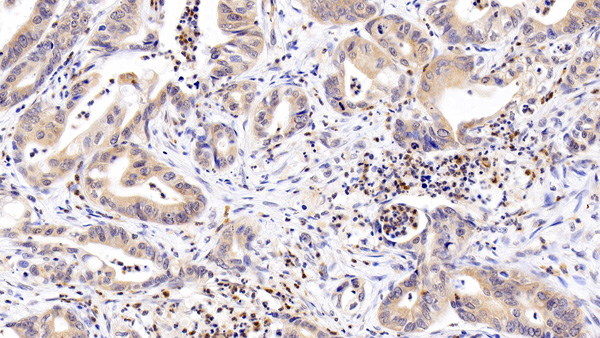
 IHC (Immunohistochemistry)
(DAB staining on IHCP;Sample: Human Colorectal cancer Tissue; Primary Ab: 20ug/ml Mouse AntiHuman LCN2 AntibodySecond Ab: 2ug/mL HRPLinked Caprine AntiMouse IgG Polyclonal Antibody(Catalog: SAA544Mu19))
IHC (Immunohistochemistry)
(DAB staining on IHCP;Sample: Human Colorectal cancer Tissue; Primary Ab: 20ug/ml Mouse AntiHuman LCN2 AntibodySecond Ab: 2ug/mL HRPLinked Caprine AntiMouse IgG Polyclonal Antibody(Catalog: SAA544Mu19))
Neutrophil gelatinaseassociated lipocalin (NGAL), Monoclonal Antibody (Cat# AAA151708)
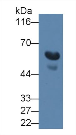
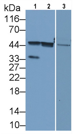
 WB (Western Blot)
(Western Blot; Sample: Lane1: Rat Cerebrum lysate; Lane2: Porcine Cerebrum lysatePrimary Ab: 0.6ug/ml Mouse AntiHuman IFNa/bR1 AntibodySecond Ab: 0.2ug/mL HRPLinked Caprine AntiMouse IgG Polyclonal Antibody(Catalog: SAA544Mu19))
WB (Western Blot)
(Western Blot; Sample: Lane1: Rat Cerebrum lysate; Lane2: Porcine Cerebrum lysatePrimary Ab: 0.6ug/ml Mouse AntiHuman IFNa/bR1 AntibodySecond Ab: 0.2ug/mL HRPLinked Caprine AntiMouse IgG Polyclonal Antibody(Catalog: SAA544Mu19))
Interferon Alpha/Beta Receptor 1 (IFNa/bR1), Monoclonal Antibody (Cat# AAA151710)
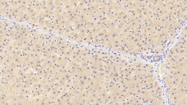
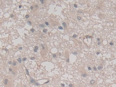
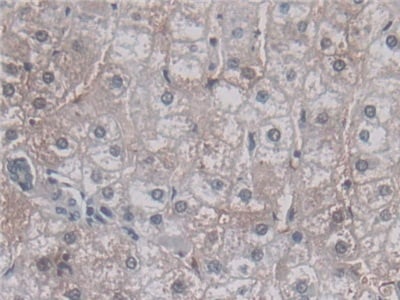
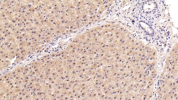
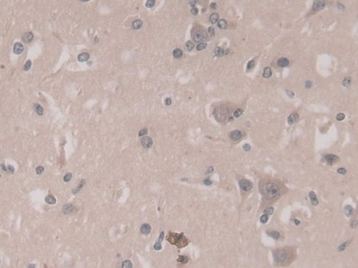
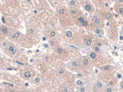
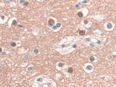
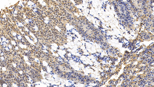
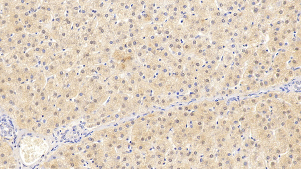
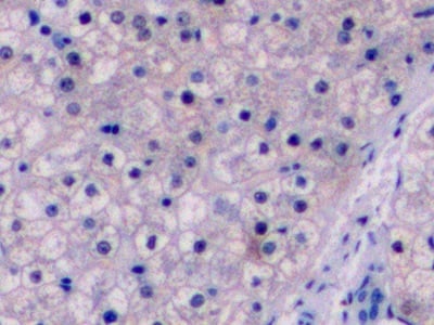
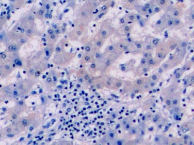
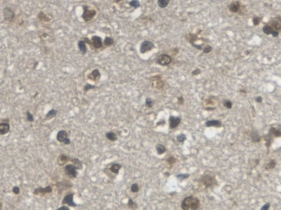
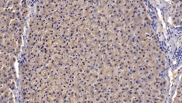
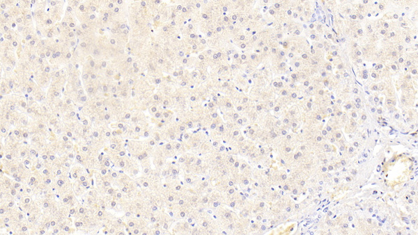
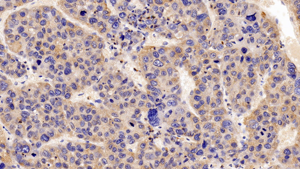 IHC (Immunohistochemistry)
(DAB staining on IHCP;Sample: Human Liver cancer Tissue; Primary Ab: 30ug/ml Mouse AntiHuman IFNa/bR1 AntibodySecond Ab: 2ug/mL HRPLinked Caprine AntiMouse IgG Polyclonal Antibody(Catalog: SAA544Mu19))
IHC (Immunohistochemistry)
(DAB staining on IHCP;Sample: Human Liver cancer Tissue; Primary Ab: 30ug/ml Mouse AntiHuman IFNa/bR1 AntibodySecond Ab: 2ug/mL HRPLinked Caprine AntiMouse IgG Polyclonal Antibody(Catalog: SAA544Mu19))
Interferon Alpha/Beta Receptor 1 (IFNa/bR1), Monoclonal Antibody (Cat# AAA151712)
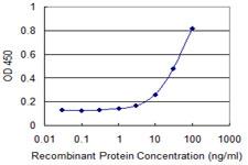
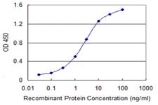 ELISA
(Detection limit for recombinant GST tagged VASH1 is 0.03 ng/ml as a capture antibody.)
ELISA
(Detection limit for recombinant GST tagged VASH1 is 0.03 ng/ml as a capture antibody.)
Vasohibin 1/VASH1, Monoclonal Antibody (Cat# AAA162271)


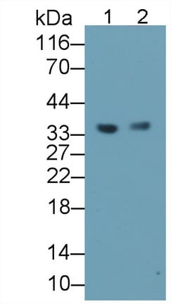
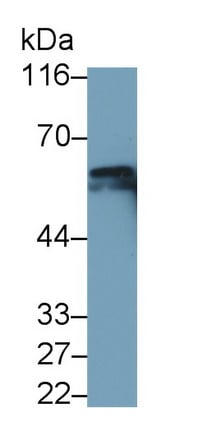
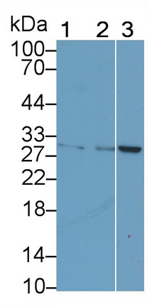
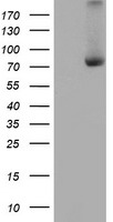
 WB (Western Blot)
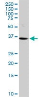
(Blots of crude HeLa cell extract stained with a panel of monoclonal antibodies to Galectin-3. Lane 12 was probed with revealing a band at the expected molecular weight of 30kDa.)
WB (Western Blot)
(Blots of crude HeLa cell extract stained with a panel of monoclonal antibodies to Galectin-3. Lane 12 was probed with revealing a band at the expected molecular weight of 30kDa.)
LGALS3/Galectin 3, Monoclonal Antibody (Cat# AAA162280)
Predicted: Mouse, Rat
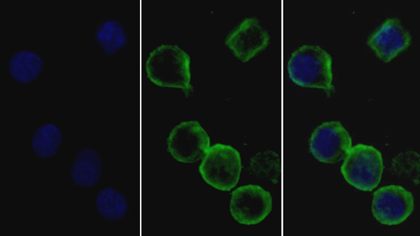
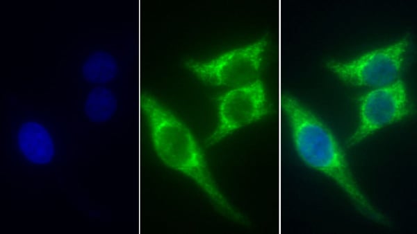
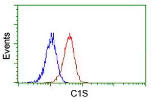 FCM/FACS (Flow Cytometry)
(Flow cytometry of Jurkat cells, using anti-C1S antibody (Red), compared to a nonspecific negative control antibody (Blue).)
FCM/FACS (Flow Cytometry)
(Flow cytometry of Jurkat cells, using anti-C1S antibody (Red), compared to a nonspecific negative control antibody (Blue).)
Complement C1s, Monoclonal Antibody (Cat# AAA162300)
Complement C3, Monoclonal Antibody (Cat# AAA162234)
 ELISA
(Detection limit for recombinant GST tagged PRPH is 1 ng/ml as a capture antibody.)
ELISA
(Detection limit for recombinant GST tagged PRPH is 1 ng/ml as a capture antibody.)
Peripherin, Monoclonal Antibody (Cat# AAA162236)
CD3, Monoclonal Antibody (Cat# AAA128263)
CD3, Monoclonal Antibody (Cat# AAA128264)
CD206, Monoclonal Antibody (Cat# AAA128272)
CD267, Monoclonal Antibody (Cat# AAA128279)
CD154, Monoclonal Antibody (Cat# AAA128290)
CD154, Monoclonal Antibody (Cat# AAA128292)
CD133, Monoclonal Antibody (Cat# AAA128297)
CD45, Monoclonal Antibody (Cat# AAA128306)
CD45, Monoclonal Antibody (Cat# AAA128309)
CD45, Monoclonal Antibody (Cat# AAA128310)
CD20, Monoclonal Antibody (Cat# AAA128314)
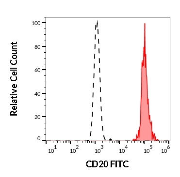
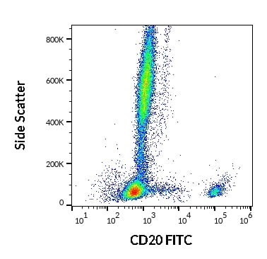 FCM/FACS (Flow Cytometry)
(Flow cytometry surface staining pattern of human peripheral whole blood stained using anti-human CD20 (2H7) FITC antibody (20 ul reagent / 100 ul of peripheral whole blood).)
FCM/FACS (Flow Cytometry)
(Flow cytometry surface staining pattern of human peripheral whole blood stained using anti-human CD20 (2H7) FITC antibody (20 ul reagent / 100 ul of peripheral whole blood).)
CD20, Monoclonal Antibody (Cat# AAA128315)
What are Monoclonal Antibodies?
Monoclonal antibodies are specialized laboratory-produced proteins developed for binding to specific biological antigens or other molecular targets. Since they come from a single cell (or clone), they are especially consistent and accurate in the data they are involved in producing.
This type of antibody material has been shown to be a powerful tool in finding and subsequently destroying harmful cells in an organism, such as those found in cancers or various autoimmune diseases. This makes them excellent aids in medical testing and research, which is why they are so widely used.
AAA Biotech offers a comprehensive range of high-quality monoclonal antibodies that perform effectively in various laboratory tests, including (amongst others) ELISA, western blotting, immunohistochemistry, and flow cytometry. All of the products in our catalog are thoroughly quality tested to make sure that they are reliable and will consistently perform well in your research.
What Are The Uses of Monoclonal Antibodies
Monoclonal antibodies are used in many lab tests, including (amongst others) ELISA, western blotting, immunohistochemistry, and flow cytometry.
ELISA is a test that helps detect a specific substance/analyte in a sample. It uses antibodies (often monoclonal) bound to a solid surface (such as the well of a microplate) to “capture” the substance/analyte in the sample and immobilize it so that the detection antibody component can then bind to it and produce a signal, which can then be measured.
Western blotting identifies specific proteins in a sample. The sample is first separated on a gel, and then antibodies are applied that will typically bind to the target, which will all be localized to a single band in a lane.
Immunohistochemistry helps locate specific proteins in cells or tissue samples using antibodies.
Flow cytometry looks at and sorts cells. It uses antibodies that are conjugated to reporter molecules called “fluorophores”, which, under special lights, emit light themselves, which can then be measured by a detector instrument.
How Monoclonal Antibodies Are Used as Medicine?
Please note that all of the products listed in AAA Biotech’s also known as AAA Bio or AAABio catalog are strictly for research-use only (RUO).
Monoclonal antibodies can also be used as therapeutic/medical treatments, particularly in the context of cancers. They are designed to find and bind to specific cells or proteins, helping the immune system recognize and attack the cancer. These treatments work in different ways, such as:
- Radioimmunotherapy attaches a small amount of radioactive molecule to the antibody, so it delivers the radiation directly to the cancer cells that the antibody is specifically binding to.
- Antibody-directed enzyme prodrug therapy uses antibodies that are specifically bound to special enzymes. These enzymes activate a harmless drug in the body and turn it into a cancer-killing drug only near the cancer cells—this helps avoid harming healthy cells.
- Immunoliposomes are tiny “bubbles” filled with medicine/drug and coated with antibodies. They carry the drug straight to the cancer cells.
Why Buy Monoclonal Antibodies From Us?
At AAA Biotech, we provide high-performance monoclonal antibodies designed to support a wide range of research needs.
1. Validated for Versatile Applications
The antibodies in our catalog are extensively validated and compatible with multiple techniques, including (but not limited to) ELISA, flow cytometry (FC), immunocytochemistry (ICC), immunofluorescence (IF), immunohistochemistry (IHC), immunoprecipitation (IP), and western blotting (WB).
2. Wide Selection & Specialized Options
We offer antibodies for common and rare species, that are available in various conjugated forms, and also in recombinant formats. Essentially, there is almost anything one might need to meet their experimental model’s requirements.
3. High-Quality Proteins
Our proteins meet high purity standards—90% or more as confirmed by SDS-PAGE. Many are available with tags like His, Flag, GST, or MBP, and we also supply native and biologically active proteins for functional studies.
Frequently Asked Questions
1. Are your monoclonal antibodies validated for specific applications?
Yes, our antibodies are tested and validated for use in methods such as ELISA, western blot, IHC, flow cytometry, and more. Refer to specific product pages or datasheets for individual product information.
2. How do I choose the right monoclonal antibody for my application?
Review the product details directly for application validation, species reactivity, and target information. You may also contact our support team at any time for help.
3. How quickly can I receive my order?
Most orders are processed and shipped within 1–3 business days, depending on product availability and your shipping location.