Filters
▼Clonality
▼Type
▼Reactivity
▼Gene Name
▼Isotype
▼Host
▼Application
▼Clone
▼Monoclonal Antibodies
Get accurate results in your research with our Monoclonal Antibodies, which are specially made to target exactly what you require for your research, and will produce consistent, reliable performance in lab tests.
Viewing 7650-7700 of 27597 product results
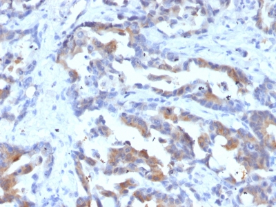
 IHC (Immunohistochemistry)
(Formalin-fixed, paraffin-embedded human Esophageal Carcinoma stained with HSV1 Recombinant Rabbit Monoclonal Antibody (HSV1/4055R).)
IHC (Immunohistochemistry)
(Formalin-fixed, paraffin-embedded human Esophageal Carcinoma stained with HSV1 Recombinant Rabbit Monoclonal Antibody (HSV1/4055R).)
HSV1 (Herpes Simplex Virus Type I), Monoclonal Antibody (Cat# AAA216097)
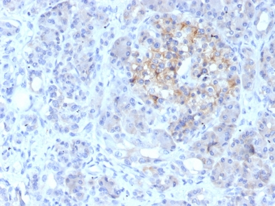
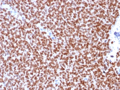
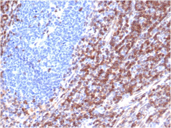 IHC (Immunohistochemistry)
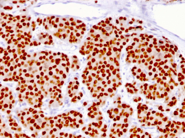
(Formalin-fixed, paraffin-embedded human tonsilstained with Bcl-2 Mouse Recombinant Monoclonal Antibody (rBCL2/6418).)
IHC (Immunohistochemistry)
(Formalin-fixed, paraffin-embedded human tonsilstained with Bcl-2 Mouse Recombinant Monoclonal Antibody (rBCL2/6418).)
Bcl-2, Monoclonal Antibody (Cat# AAA215889)
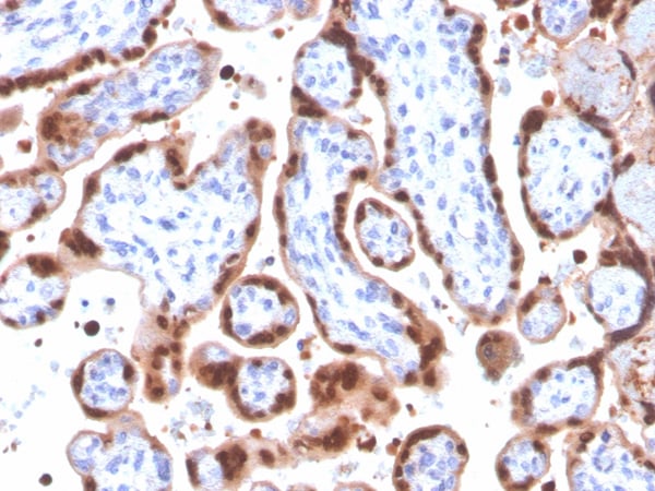
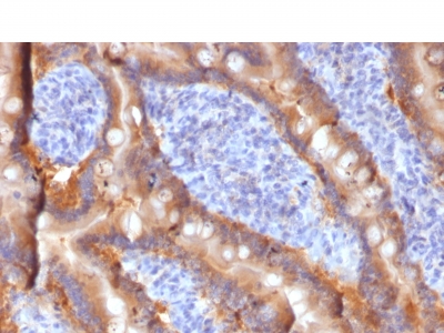
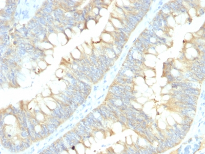
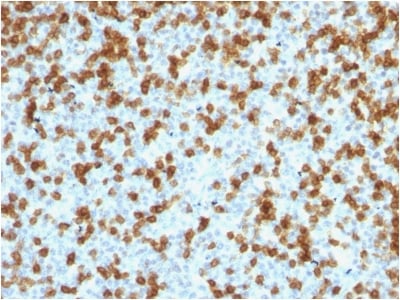
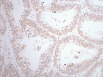
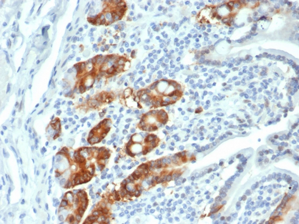 IHC (Immunohistochemistry)
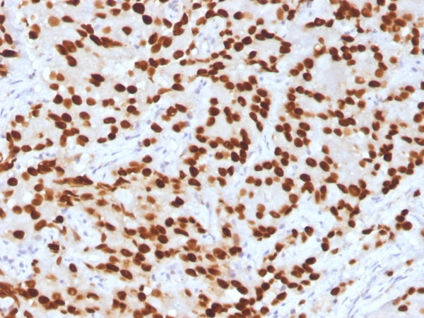
(Formalin-fixed, paraffin-embedded human small intestine stained with RRM1 Recombinant Rabbit Monoclonal (RRM1/4372R).)
IHC (Immunohistochemistry)
(Formalin-fixed, paraffin-embedded human small intestine stained with RRM1 Recombinant Rabbit Monoclonal (RRM1/4372R).)
Ribonucleotide Reductase M1/RRM1, Monoclonal Antibody (Cat# AAA215898)
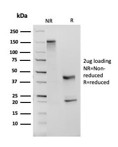
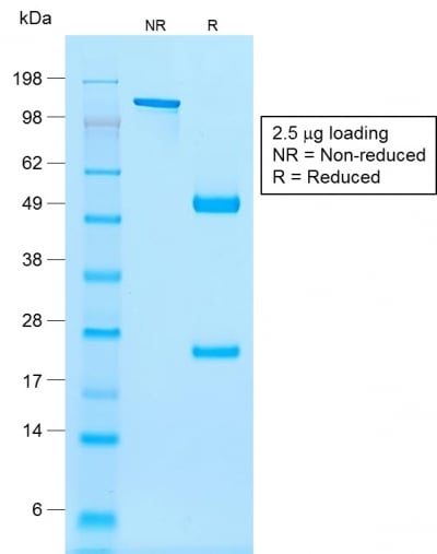
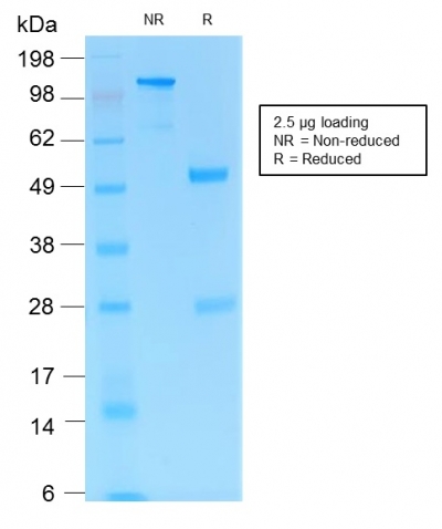
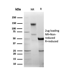 SDS-PAGE
(SDS-PAGE Analysis Purified DBC2 Mouse Monoclonal Antibody (DBC2/3361). Confirmation of Purity and Integrity of Antibody.)
SDS-PAGE
(SDS-PAGE Analysis Purified DBC2 Mouse Monoclonal Antibody (DBC2/3361). Confirmation of Purity and Integrity of Antibody.)
DBC2/RHOBTB2, Monoclonal Antibody (Cat# AAA215656)
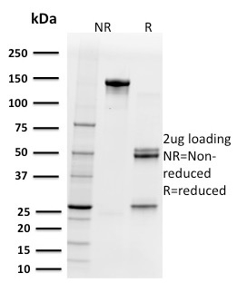
 SDS-PAGE
(SDS-PAGE Analysis Purified GNAQ Mouse Monoclonal Antibody (GNAQ/2434). Confirmation of Integrity and Purity of the Antibody.)
SDS-PAGE
(SDS-PAGE Analysis Purified GNAQ Mouse Monoclonal Antibody (GNAQ/2434). Confirmation of Integrity and Purity of the Antibody.)
Guanine nucleotide-binding protein alpha-q/GNAQ/G-ALPHA-q, Monoclonal Antibody (Cat# AAA214994)
 SDS-PAGE
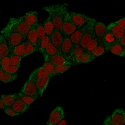
(SDS-PAGE Analysis Purified FOXA1 Mouse Monoclonal Antibody (FOXA1/1516). Confirmation of Purity and Integrity of Antibody.)
SDS-PAGE
(SDS-PAGE Analysis Purified FOXA1 Mouse Monoclonal Antibody (FOXA1/1516). Confirmation of Purity and Integrity of Antibody.)
FOXA1/HNF3A, Monoclonal Antibody (Cat# AAA215015)
 Application Data
Bcl-X, Monoclonal Antibody (Cat# AAA215184)
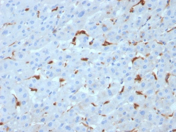
 Application Data
(Analysis of Protein Array containing more than 19,000 full-length human proteins using C1QA Mouse Monoclonal Antibody (C1QA/2953). Z- and S- Score: The Z-score represents the strength of a signal that a monoclonal antibody (MAb) (in combination with a fluorescently-tagged anti-IgG secondary antibody) produces when binding to a particular protein on the HuProtTM array. Z-scores are described in units of standard deviations (SD's) above the mean value of all signals generated on that array. If targets on HuProtTM are arranged in descending order of the Z-score, the S-score is the difference (also in units of SD's) between the Z-score. S-score therefore represents the relative target specificity of a MAb to its intended target. A MAb is considered to specific to its intended target, if the MAb has an S-score of at least 2.5. For example, if a MAb binds to protein X with a Z-score of 43 and to protein Y with a Z-score of 14, then the S-score for the binding of that MAb to protein X is equal to 29.)
Application Data
(Analysis of Protein Array containing more than 19,000 full-length human proteins using C1QA Mouse Monoclonal Antibody (C1QA/2953). Z- and S- Score: The Z-score represents the strength of a signal that a monoclonal antibody (MAb) (in combination with a fluorescently-tagged anti-IgG secondary antibody) produces when binding to a particular protein on the HuProtTM array. Z-scores are described in units of standard deviations (SD's) above the mean value of all signals generated on that array. If targets on HuProtTM are arranged in descending order of the Z-score, the S-score is the difference (also in units of SD's) between the Z-score. S-score therefore represents the relative target specificity of a MAb to its intended target. A MAb is considered to specific to its intended target, if the MAb has an S-score of at least 2.5. For example, if a MAb binds to protein X with a Z-score of 43 and to protein Y with a Z-score of 14, then the S-score for the binding of that MAb to protein X is equal to 29.)
C1QA/Complement C1q A-Chain, Monoclonal Antibody (Cat# AAA215221)
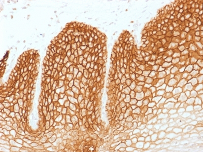
 IHC (Immunohistochemistry)
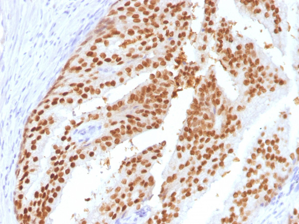
(Formalin-fixed, paraffin-embedded human colon stained with Cytokeratin 8 Recombinant Rabbit Monoclonal Antibody (KRT8/6472R).)
IHC (Immunohistochemistry)
(Formalin-fixed, paraffin-embedded human colon stained with Cytokeratin 8 Recombinant Rabbit Monoclonal Antibody (KRT8/6472R).)
Cytokeratin 8 (KRT8), Monoclonal Antibody (Cat# AAA215767)
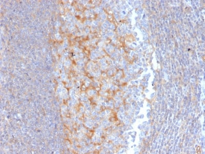
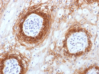 IHC (Immunohiostchemistry)
(Formalin-paraffin human Tongue Carcinoma stained with CD44v6 Monoclonal Antibody (CD44v6/1246))
IHC (Immunohiostchemistry)
(Formalin-paraffin human Tongue Carcinoma stained with CD44v6 Monoclonal Antibody (CD44v6/1246))
CD44v6, Monoclonal Antibody (Cat# AAA214607)
 Application Data
(Analysis of Protein Array containing more than 19,000 full-length human proteins using TYRP1-Monospecific Mouse Monoclonal Antibody (TYRP1/3282) Z- and S- Score: The Z-score represents the strength of a signal that a monoclonal antibody (Monoclonal Antibody) (in combination with a fluorescently-tagged anti-IgG secondary antibody) produces when binding to a particular protein on the HuProtTM array. Z-scores are described in units of standard deviations (SD’s) above the mean value of all signals generated on that array. If targets on HuProtTM are arranged in descending order of the Z-score, the S-score is the difference (also in units of SD’s) between the Z-score. S-score therefore represents the relative target specificity of a Monoclonal Antibody to its intended target. A Monoclonal Antibody is considered to specific to its intended target, if the Monoclonal Antibody has an S-score of at least 2.5. For example, if a Monoclonal Antibody binds to protein X with a Z-score of 43 and to protein Y with a Z-score of 14, then the S-score for the binding of that Monoclonal Antibody to protein X is equal to 29.)
Application Data
(Analysis of Protein Array containing more than 19,000 full-length human proteins using TYRP1-Monospecific Mouse Monoclonal Antibody (TYRP1/3282) Z- and S- Score: The Z-score represents the strength of a signal that a monoclonal antibody (Monoclonal Antibody) (in combination with a fluorescently-tagged anti-IgG secondary antibody) produces when binding to a particular protein on the HuProtTM array. Z-scores are described in units of standard deviations (SD’s) above the mean value of all signals generated on that array. If targets on HuProtTM are arranged in descending order of the Z-score, the S-score is the difference (also in units of SD’s) between the Z-score. S-score therefore represents the relative target specificity of a Monoclonal Antibody to its intended target. A Monoclonal Antibody is considered to specific to its intended target, if the Monoclonal Antibody has an S-score of at least 2.5. For example, if a Monoclonal Antibody binds to protein X with a Z-score of 43 and to protein Y with a Z-score of 14, then the S-score for the binding of that Monoclonal Antibody to protein X is equal to 29.)
Tyrosinase-Related Protein-1 (TYRP-1), Monoclonal Antibody (Cat# AAA215243)

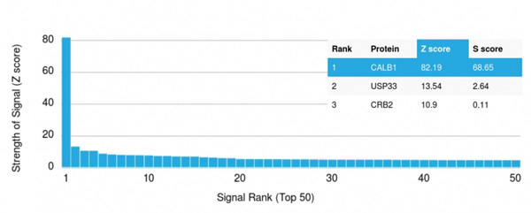 Application Data
(Analysis of Protein Array containing more than 19,000 full-length human proteins using Calbindin Mouse Monoclonal Antibody (CALB1/2364) Z- and S- Score: The Z-score represents the strength of a signal that a monoclonal antibody (Monoclonal Antibody) (in combination with a fluorescently-tagged anti-IgG secondary antibody) produces when binding to a particular protein on the HuProtTM array. Z-scores are described in units of standard deviations (SD's) above the mean value of all signals generated on that array. If targets on HuProtTM are arranged in descending order of the Z-score, the S-score is the difference (also in units of SD's) between the Z-score. S-score therefore represents the relative target specificity of a Monoclonal Antibody to its intended target. A Monoclonal Antibody is considered to specific to its intended target, if the Monoclonal Antibody has an S-score of at least 2.5. For example, if a Monoclonal Antibody binds to protein X with a Z-score of 43 and to protein Y with a Z-score of 14, then the S-score for the binding of that Monoclonal Antibody to protein X is equal to 29.)
Application Data
(Analysis of Protein Array containing more than 19,000 full-length human proteins using Calbindin Mouse Monoclonal Antibody (CALB1/2364) Z- and S- Score: The Z-score represents the strength of a signal that a monoclonal antibody (Monoclonal Antibody) (in combination with a fluorescently-tagged anti-IgG secondary antibody) produces when binding to a particular protein on the HuProtTM array. Z-scores are described in units of standard deviations (SD's) above the mean value of all signals generated on that array. If targets on HuProtTM are arranged in descending order of the Z-score, the S-score is the difference (also in units of SD's) between the Z-score. S-score therefore represents the relative target specificity of a Monoclonal Antibody to its intended target. A Monoclonal Antibody is considered to specific to its intended target, if the Monoclonal Antibody has an S-score of at least 2.5. For example, if a Monoclonal Antibody binds to protein X with a Z-score of 43 and to protein Y with a Z-score of 14, then the S-score for the binding of that Monoclonal Antibody to protein X is equal to 29.)
Calbindin 1 (CALB1), Monoclonal Antibody (Cat# AAA215260)
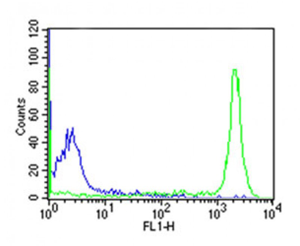
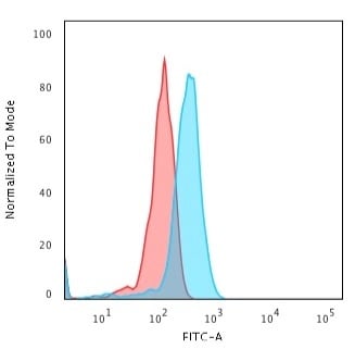 FCM/FACS (Flow Cytometry)
(Flow Cytometric Analysis of Jurkat cells. CD8 Mouse Monoclonal Antibody (UCHT4) followed by goat anti-Mouse IgG-CF488 (Blue); Isotype Control (Red).)
FCM/FACS (Flow Cytometry)
(Flow Cytometric Analysis of Jurkat cells. CD8 Mouse Monoclonal Antibody (UCHT4) followed by goat anti-Mouse IgG-CF488 (Blue); Isotype Control (Red).)
CD8A, Monoclonal Antibody (Cat# AAA215289)
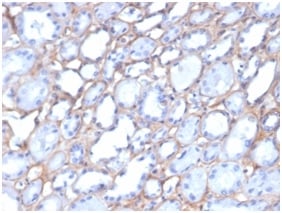
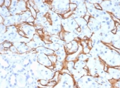
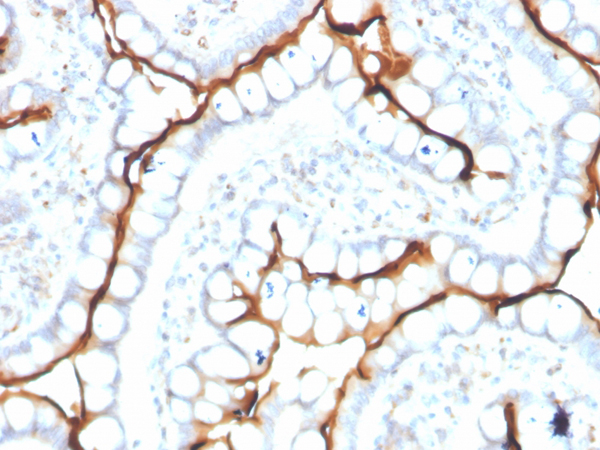
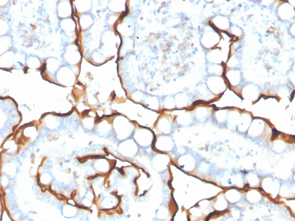
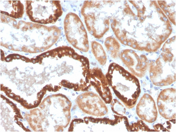
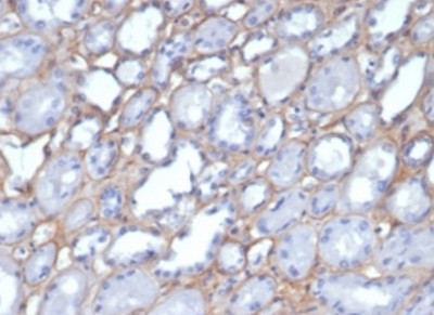 IHC (Immunohistochemisry)
(Formalin-fixed, paraffin-embedded human kidney stained with Collagen IVMouse Monoclonal Antibody (M3F7).)
IHC (Immunohistochemisry)
(Formalin-fixed, paraffin-embedded human kidney stained with Collagen IVMouse Monoclonal Antibody (M3F7).)
Collagen IV (COL4A1/COL4A2), Monoclonal Antibody (Cat# AAA215561)
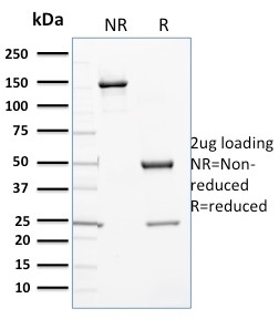 SDS-PAGE
(SDS-PAGE Analysis Purified Cytochrome P450 1A1/1A2 Mouse Monoclonal Antibody (P6). Confirmation of Purity and Integrity of Antibody.)
SDS-PAGE
(SDS-PAGE Analysis Purified Cytochrome P450 1A1/1A2 Mouse Monoclonal Antibody (P6). Confirmation of Purity and Integrity of Antibody.)
Cytochrome P450 3A1/CYP3A1, Monoclonal Antibody (Cat# AAA215327)
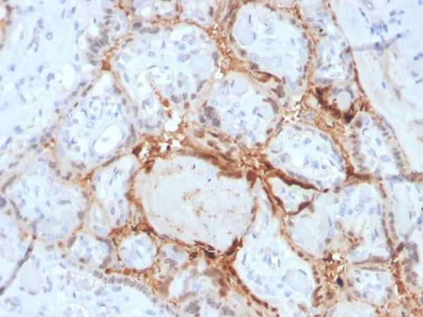
 IHC (Immunohistochemisry)
(Formalin-fixed, paraffin-embedded human placenta stained with HIF1 alpha Mouse Monoclonal Antibody (Ha111a).)
IHC (Immunohistochemisry)
(Formalin-fixed, paraffin-embedded human placenta stained with HIF1 alpha Mouse Monoclonal Antibody (Ha111a).)
HIF1 alpha (Hypoxia-Inducible Factor 1-alpha), Monoclonal Antibody (Cat# AAA215356)
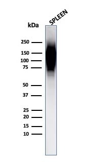
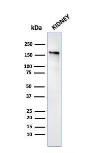 WB (Western Blot)
(Western Blot Analysis of human Kidney tissue lysate using ACE / CD143 Mouse Monoclonal Antibody (ACE/3763).)
WB (Western Blot)
(Western Blot Analysis of human Kidney tissue lysate using ACE / CD143 Mouse Monoclonal Antibody (ACE/3763).)
Angiotensin, Monoclonal Antibody (Cat# AAA215442)
 Application Data
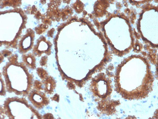
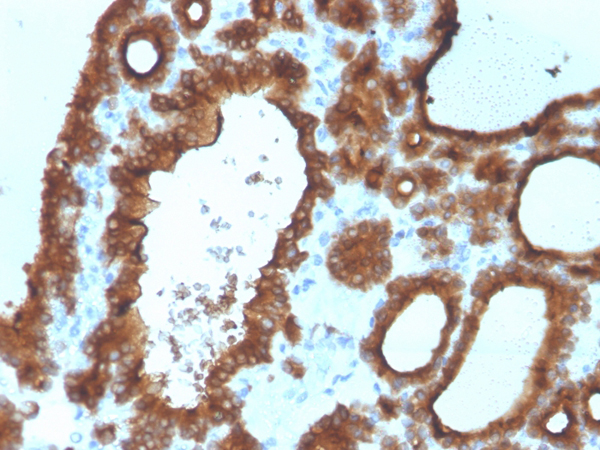
(Analysis of Protein Array containing more than 19,000 full-length human proteins using Thyroid Peroxidase Mouse Monoclonal Antibody (TPO/3700). Z- and S- Score: The Z-score represents the strength of a signal that a monoclonal antibody (MAb) (in combination with a fluorescently-tagged anti-IgG secondary antibody) produces when binding to a particular protein on the HuProtTM array. Z-scores are described in units of standard deviations (SD's) above the mean value of all signals generated on that array. If targets on HuProtTM are arranged in descending order of the Z-score, the S-score is the difference (also in units of SD's) between the Z-score. S-score therefore represents the relative target specificity of a MAb to its intended target. A MAb is considered to specific to its intended target, if the MAb has an S-score of at least 2.5. For example, if a MAb binds to protein X with a Z-score of 43 and to protein Y with a Z-score of 14, then the S-score for the binding of that MAb to protein X is equal to 29.)
Application Data
(Analysis of Protein Array containing more than 19,000 full-length human proteins using Thyroid Peroxidase Mouse Monoclonal Antibody (TPO/3700). Z- and S- Score: The Z-score represents the strength of a signal that a monoclonal antibody (MAb) (in combination with a fluorescently-tagged anti-IgG secondary antibody) produces when binding to a particular protein on the HuProtTM array. Z-scores are described in units of standard deviations (SD's) above the mean value of all signals generated on that array. If targets on HuProtTM are arranged in descending order of the Z-score, the S-score is the difference (also in units of SD's) between the Z-score. S-score therefore represents the relative target specificity of a MAb to its intended target. A MAb is considered to specific to its intended target, if the MAb has an S-score of at least 2.5. For example, if a MAb binds to protein X with a Z-score of 43 and to protein Y with a Z-score of 14, then the S-score for the binding of that MAb to protein X is equal to 29.)
TPO (Thyroid Peroxidase), Monoclonal Antibody (Cat# AAA215464)
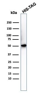 WB (Western Blot)
(Western Blot Analysis of recombinant His-Tag protein using Anti-Hexa-histidine Mouse Monoclonal 6HIS/3550).)
WB (Western Blot)
(Western Blot Analysis of recombinant His-Tag protein using Anti-Hexa-histidine Mouse Monoclonal 6HIS/3550).)
Hexa-histidine AT:AT, Monoclonal Antibody (Cat# AAA215473)
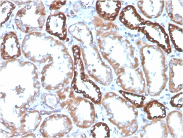
 WB (Western Blot)
(Western Blot Analysis of kidney tissue lysate using CD137 Recombinant Rabbit Monoclonal Antibody (4-1BB/4552R).)
WB (Western Blot)
(Western Blot Analysis of kidney tissue lysate using CD137 Recombinant Rabbit Monoclonal Antibody (4-1BB/4552R).)
CD137/4-1BB/TNFRSF9, Monoclonal Antibody (Cat# AAA215500)
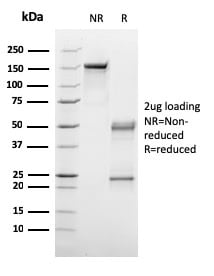
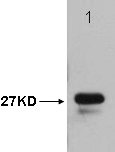
 SDS-PAGE
(SDS-PAGE Analysis Purified p27 Mouse Monoclonal Antibody (KIP1/1357). Confirmation of Purity and Integrity of Antibody.)
SDS-PAGE
(SDS-PAGE Analysis Purified p27 Mouse Monoclonal Antibody (KIP1/1357). Confirmation of Purity and Integrity of Antibody.)
p27Kip1, Monoclonal Antibody (Cat# AAA215516)
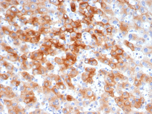
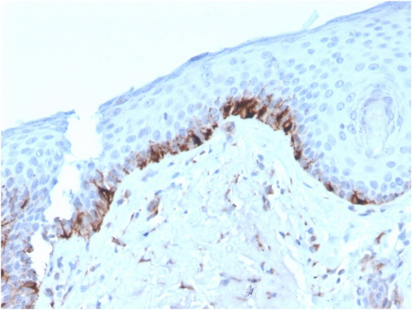
 IHC (Immunohistochemistry)
(Formalin-fixed, paraffin-embedded Human Colon Carcinoma stained with MRP1/ABCC1 Monoclonal Antibody (MRP1/1343).)
IHC (Immunohistochemistry)
(Formalin-fixed, paraffin-embedded Human Colon Carcinoma stained with MRP1/ABCC1 Monoclonal Antibody (MRP1/1343).)
MRP1/ABCC1 (Multidrug Resistance Related Protein 1), Monoclonal Antibody (Cat# AAA214451)
 SDS-PAGE
(SDS-PAGE Analysis Purified NKX2.2 Mouse Recombinant Monoclonal Antibody (rNX2/294).)
SDS-PAGE
(SDS-PAGE Analysis Purified NKX2.2 Mouse Recombinant Monoclonal Antibody (rNX2/294).)
NKX2.2, Monoclonal Antibody (Cat# AAA214468)
 SDS-PAGE
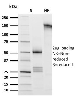
(SDS-PAGE Analysis Purified PD1 (CD279) Rabbit Monoclonal Antibody (PDCD1/1410R).)
SDS-PAGE
(SDS-PAGE Analysis Purified PD1 (CD279) Rabbit Monoclonal Antibody (PDCD1/1410R).)
PDCD1/PD1/CD279 (Programmed Cell Death 1), Monoclonal Antibody (Cat# AAA214473)
 Application Data
(Analysis of Protein Array containing more than 19,000 full-length human proteins using Glucose 6-Phosphate Isomerase Monoclonal Antibody (CPTC-GPI-1). Z- and S- Score: The Z-score represents the strength of a signal that a monoclonal antibody (MAb) (in combination with a fluorescently-tagged anti-IgG secondary antibody) produces when binding to a particular protein on the HuProtTM array. Z-scores are described in units of standard deviations (SD's) above the mean value of all signals generated on that array. If targets on HuProtTM are arranged in descending order of the Z-score, the S-score is the difference (also in units of SD's) between the Z-score. S-score therefore represents the relative target specificity of a MAb to its intended target. A MAb is considered to specific to its intended target, if the MAb has an S-score of at least 2.5. For example, if a MAb binds to protein X with a Z-score of 43 and to protein Y with a Z-score of 14, then the S-score for the binding of that MAb to protein X is equal to 29.)
Application Data
(Analysis of Protein Array containing more than 19,000 full-length human proteins using Glucose 6-Phosphate Isomerase Monoclonal Antibody (CPTC-GPI-1). Z- and S- Score: The Z-score represents the strength of a signal that a monoclonal antibody (MAb) (in combination with a fluorescently-tagged anti-IgG secondary antibody) produces when binding to a particular protein on the HuProtTM array. Z-scores are described in units of standard deviations (SD's) above the mean value of all signals generated on that array. If targets on HuProtTM are arranged in descending order of the Z-score, the S-score is the difference (also in units of SD's) between the Z-score. S-score therefore represents the relative target specificity of a MAb to its intended target. A MAb is considered to specific to its intended target, if the MAb has an S-score of at least 2.5. For example, if a MAb binds to protein X with a Z-score of 43 and to protein Y with a Z-score of 14, then the S-score for the binding of that MAb to protein X is equal to 29.)
Glucose 6-Phosphate Isomerase, Monoclonal Antibody (Cat# AAA214752)
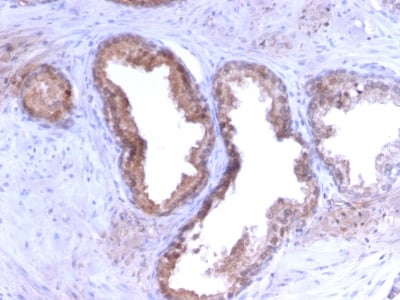
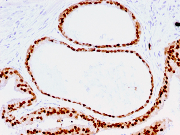
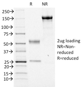
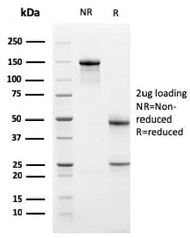
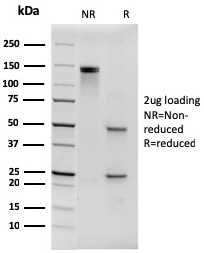
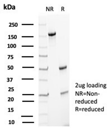
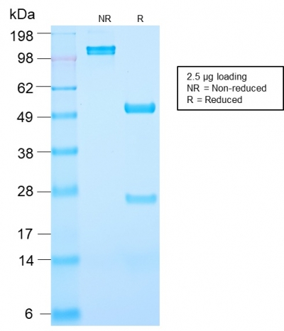 SDS-PAGE
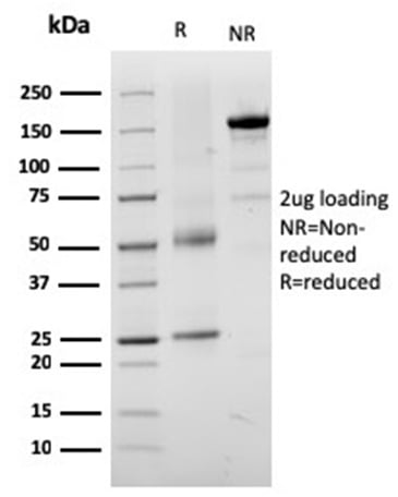
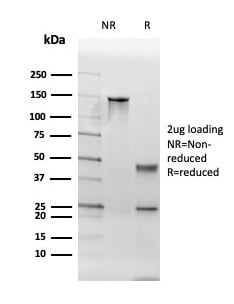
(SDS-PAGE Analysis Purified PSA Rabbit Recombinant Monoclonal Antibody (KLK3/2871R). Confirmation of Purity and Integrity of Antibody.)
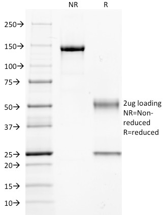
SDS-PAGE
(SDS-PAGE Analysis Purified PSA Rabbit Recombinant Monoclonal Antibody (KLK3/2871R). Confirmation of Purity and Integrity of Antibody.)
Prostate Specific Antigen (PSA), Monoclonal Antibody (Cat# AAA214764)
 SDS-PAGE
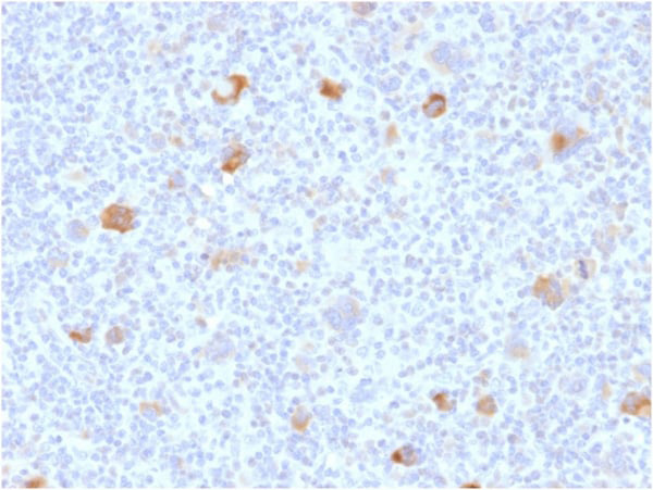
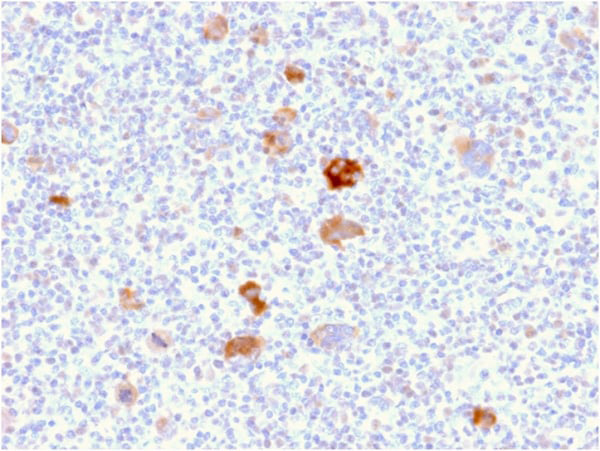
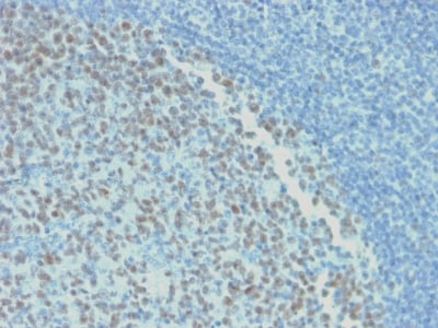
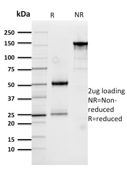
(SDS-PAGE Analysis Purified BCL-6 Mouse Recombinant Monoclonal Antibody (rBCL6/1718). Confirmation of Purity and Integrity of Antibody.)
SDS-PAGE
(SDS-PAGE Analysis Purified BCL-6 Mouse Recombinant Monoclonal Antibody (rBCL6/1718). Confirmation of Purity and Integrity of Antibody.)
Bcl-6, Monoclonal Antibody (Cat# AAA214809)
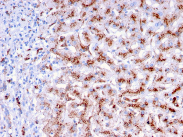
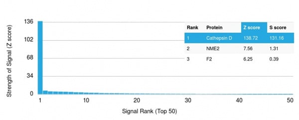 Application Data
(Analysis of Protein Array containing more than 19,000 full-length human proteins using Cathepsin D Mouse Monoclonal Antibody (CTSD/3276) Z- and S- Score: The Z-score represents the strength of a signal that a monoclonal antibody (MAb) (in combination with a fluorescently-tagged anti-IgG secondary antibody) produces when binding to a particular protein on the HuProtTM array. Z-scores are described in units of standard deviations (SD's) above the mean value of all signals generated on that array. If targets on HuProtTM are arranged in descending order of the Z-score, the S-score is the difference (also in units of SD's) between the Z-score. S-score therefore represents the relative target specificity of a MAb to its intended target. A MAb is considered to specific to its intended target, if the MAb has an S-score of at least 2.5. For example, if a MAb binds to protein X with a Z-score of 43 and to protein Y with a Z-score of 14, then the S-score for the binding of that MAb to protein X is equal to 29.)
Application Data
(Analysis of Protein Array containing more than 19,000 full-length human proteins using Cathepsin D Mouse Monoclonal Antibody (CTSD/3276) Z- and S- Score: The Z-score represents the strength of a signal that a monoclonal antibody (MAb) (in combination with a fluorescently-tagged anti-IgG secondary antibody) produces when binding to a particular protein on the HuProtTM array. Z-scores are described in units of standard deviations (SD's) above the mean value of all signals generated on that array. If targets on HuProtTM are arranged in descending order of the Z-score, the S-score is the difference (also in units of SD's) between the Z-score. S-score therefore represents the relative target specificity of a MAb to its intended target. A MAb is considered to specific to its intended target, if the MAb has an S-score of at least 2.5. For example, if a MAb binds to protein X with a Z-score of 43 and to protein Y with a Z-score of 14, then the S-score for the binding of that MAb to protein X is equal to 29.)
Cathepsin D, Monoclonal Antibody (Cat# AAA214913)
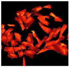 IF (Immunofluorescence)
(Immunofluorescent staining of SNAP25 in SH-SY-5Y cells using anti-SNAP-25.)
IF (Immunofluorescence)
(Immunofluorescent staining of SNAP25 in SH-SY-5Y cells using anti-SNAP-25.)
SNAP-25, Monoclonal Antibody (Cat# AAA311973)
 IHC (Immunohistochemisry)
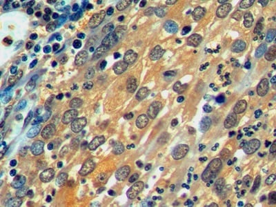
(DAB staining on IHC-P; Sample: Human Placenta Tissue; Primary Ab: 10ug/ml Mouse Anti-Human CA4 Antibody Second Ab: 2ug/mL HRP-Linked Caprine Anti-Mouse IgG Polyclonal Antibody ))
IHC (Immunohistochemisry)
(DAB staining on IHC-P; Sample: Human Placenta Tissue; Primary Ab: 10ug/ml Mouse Anti-Human CA4 Antibody Second Ab: 2ug/mL HRP-Linked Caprine Anti-Mouse IgG Polyclonal Antibody ))
Carbonic Anhydrase IV (CA4), Monoclonal Antibody (Cat# AAA130612)
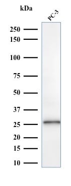
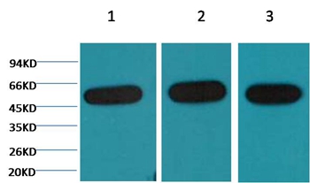
 Knockout Validation
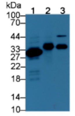
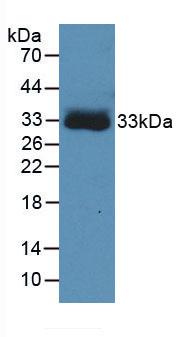
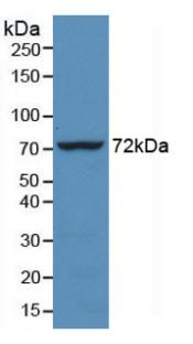
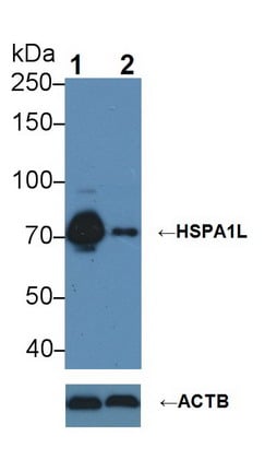
(Knockout Validation: Lane 1: Wild-type Hela cell lysate;;Lane 2: HSPA1L knockout Hela cell lysate;;Predicted MW: 70kDa ;Observed MW: 70kDa;Primary Ab: 3ug/ml Mouse Anti-Human HSPA1L Antibody;Second Ab: 0.2ug/mL HRP-Linked Caprine Anti-Mouse IgG Polyclonal Antibody;)
Knockout Validation
(Knockout Validation: Lane 1: Wild-type Hela cell lysate;;Lane 2: HSPA1L knockout Hela cell lysate;;Predicted MW: 70kDa ;Observed MW: 70kDa;Primary Ab: 3ug/ml Mouse Anti-Human HSPA1L Antibody;Second Ab: 0.2ug/mL HRP-Linked Caprine Anti-Mouse IgG Polyclonal Antibody;)
Heat Shock 70kDa Protein 1 Like Protein (HSPA1L), Monoclonal Antibody (Cat# AAA130637)
 IHC (Immunohiostchemistry)
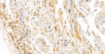
(DAB staining on IHC-P; Samples: Rat Heart Tissue)
IHC (Immunohiostchemistry)
(DAB staining on IHC-P; Samples: Rat Heart Tissue)
Von Willebrand Factor (vWF), Monoclonal Antibody (Cat# AAA130644)
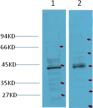
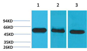
 WB (Western Blot)
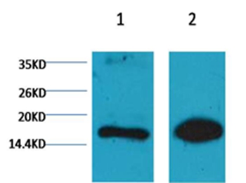
(Western blot analysis of Hela, using diluted at 1) 1:2,000 2) 1:5,000)
WB (Western Blot)
(Western blot analysis of Hela, using diluted at 1) 1:2,000 2) 1:5,000)
Histone H3, Monoclonal Antibody (Cat# AAA300705)
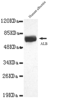
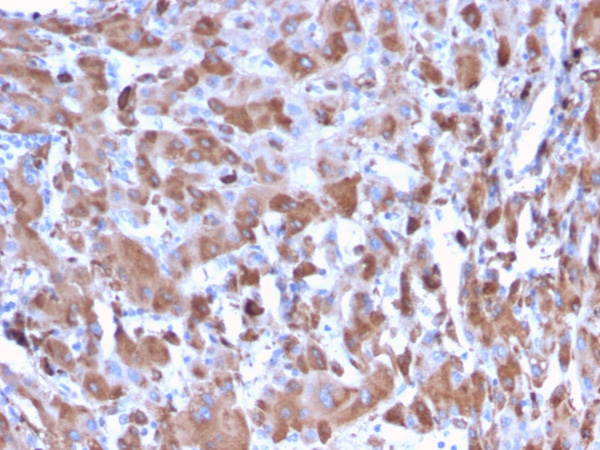
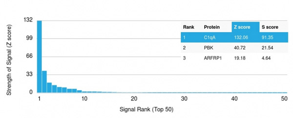
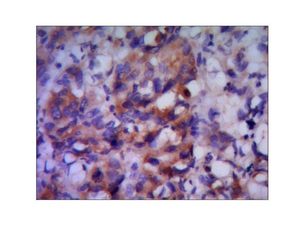 Application Data
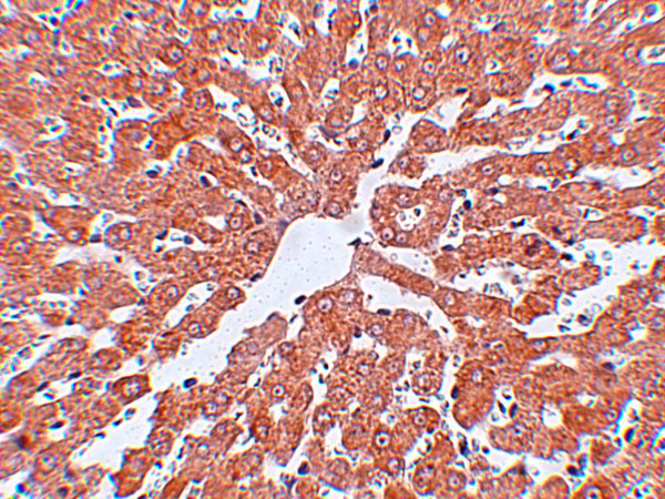
(Paraffin--embedded human liver cancer using anti- ALBdiluted 1:50-1:100)
Application Data
(Paraffin--embedded human liver cancer using anti- ALBdiluted 1:50-1:100)
Albumin, Monoclonal Antibody (Cat# AAA300709)
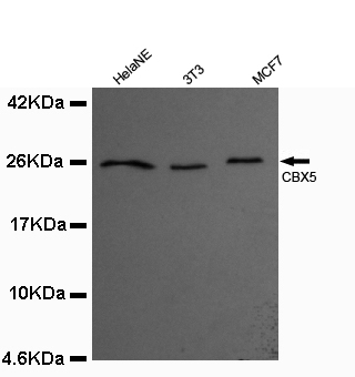
 Application Data
(1:100 dilution from a previous lot detected CBX5in Hela)
Application Data
(1:100 dilution from a previous lot detected CBX5in Hela)
CBX5, Monoclonal Antibody (Cat# AAA300710)
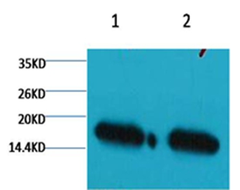
 WB (Western Blot)
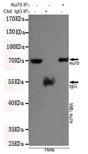
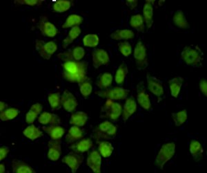
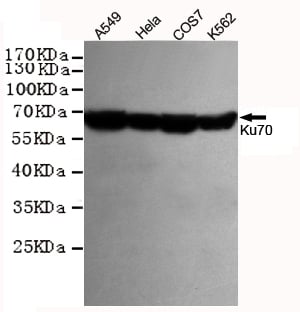
(Western blot detection of Ku70 in Hela,A549,COS7 and K562 cell lysates using Ku70 antibody (1:1000 diluted).Predicted band size:70KDa.Observed band size:67KDa.)
WB (Western Blot)
(Western blot detection of Ku70 in Hela,A549,COS7 and K562 cell lysates using Ku70 antibody (1:1000 diluted).Predicted band size:70KDa.Observed band size:67KDa.)
Ku70, Monoclonal Antibody (Cat# AAA300716)
 WB (Western Blot)
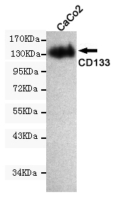
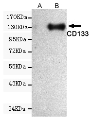
(Western blot detection of PROM1 expression in CHO-k1 cells non-transfected (A) or transfected (B) with PROM1 and using PROM1 antibody (1:1000 diluted).Predicted band size:97KDa.Observed band size:133KDa.)
WB (Western Blot)
(Western blot detection of PROM1 expression in CHO-k1 cells non-transfected (A) or transfected (B) with PROM1 and using PROM1 antibody (1:1000 diluted).Predicted band size:97KDa.Observed band size:133KDa.)
Prominin-1, Monoclonal Antibody (Cat# AAA300718)
 Application Data
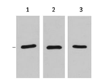
(1ug RFP fusion protein+ Primary antibody dilution at 11:3,000 21:5,000 31:10,000)
Application Data
(1ug RFP fusion protein+ Primary antibody dilution at 11:3,000 21:5,000 31:10,000)
RFP-Tag, Monoclonal Antibody (Cat# AAA300723)
 WB (Western Blot)
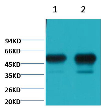
(Western blot detection of LRRFIP1 in Raji,C2C12,MCF7&Jurkat cell lysates using LRRFIP1antibody (1:1000 diluted)Predicted band size:89KDaObserved band size:160KDa)
WB (Western Blot)
(Western blot detection of LRRFIP1 in Raji,C2C12,MCF7&Jurkat cell lysates using LRRFIP1antibody (1:1000 diluted)Predicted band size:89KDaObserved band size:160KDa)
LRRFIP1, Monoclonal Antibody (Cat# AAA300725)
 Application Data
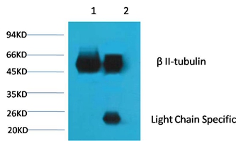
(1¡¢Input: Mouse Brain Tissue Lysate 2¡¢IP product: IP dilute 1:200 Western blot analysis: primary antibody : 1:10,000 Secondary antibody: Goat anti-Mouse IgG, Light chain specific, 1:5,000)
Application Data
(1¡¢Input: Mouse Brain Tissue Lysate 2¡¢IP product: IP dilute 1:200 Western blot analysis: primary antibody : 1:10,000 Secondary antibody: Goat anti-Mouse IgG, Light chain specific, 1:5,000)
beta II tubulin, Monoclonal Antibody (Cat# AAA300726)
 IHC (Immunohiostchemistry)
(Immunohistochemistry of ORAI1 in human spleen tissue with ORAI1 antibody at 2.5 ug/mL.)
IHC (Immunohiostchemistry)
(Immunohistochemistry of ORAI1 in human spleen tissue with ORAI1 antibody at 2.5 ug/mL.)
ORAI1, Monoclonal Antibody (Cat# AAA300732)
 IHC (Immunohiostchemistry)
(IHC staining of human rectal cancer tissue with CDX2 mouse mAb(14H6) diluted at 1:200.)
IHC (Immunohiostchemistry)
(IHC staining of human rectal cancer tissue with CDX2 mouse mAb(14H6) diluted at 1:200.)
CDX2, Monoclonal Antibody (Cat# AAA300735)
 IHC (Immunohiostchemistry)
(Immunohistochemistry of IRAK in rat liver tissue with IRAK antibody at 2.5 ug/mL.)
IHC (Immunohiostchemistry)
(Immunohistochemistry of IRAK in rat liver tissue with IRAK antibody at 2.5 ug/mL.)
IRAK, Monoclonal Antibody (Cat# AAA300736)
 Application Data
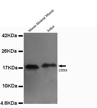
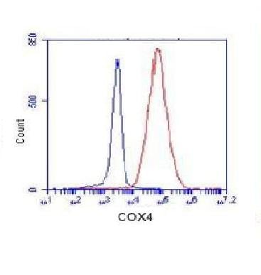
(K562 cells stained with COX4 (red, 1:100 dilution), followed by FITC-conjugated goat anti-mouse IgG. Blue line histogram represents the isotype control, normal mouse IgG)
Application Data
(K562 cells stained with COX4 (red, 1:100 dilution), followed by FITC-conjugated goat anti-mouse IgG. Blue line histogram represents the isotype control, normal mouse IgG)
COX4, Monoclonal Antibody (Cat# AAA300740)
 WB (Western Blot)
(Western blot analysis of GST-T7 fusion protein, using T7-tag mouse monoclonal antibody.)
WB (Western Blot)
(Western blot analysis of GST-T7 fusion protein, using T7-tag mouse monoclonal antibody.)
T7-Tag, Monoclonal Antibody (Cat# AAA300743)
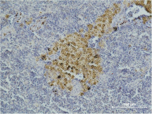 IHC (Immunohiostchemistry)
(Immunohistochemical analysis of paraffin-embedded Mouse Spleen Tissue using Caspase-8 Monoclonal Antibody.)
IHC (Immunohiostchemistry)
(Immunohistochemical analysis of paraffin-embedded Mouse Spleen Tissue using Caspase-8 Monoclonal Antibody.)
Caspase-8, Monoclonal Antibody (Cat# AAA300744)
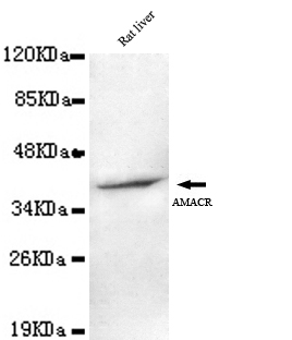
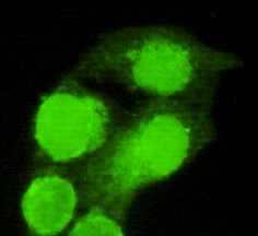 Application Data
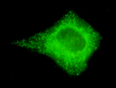
(HeLa cells using anti- AMACR (C-terminus) antibody diluted 1:150)
Application Data
(HeLa cells using anti- AMACR (C-terminus) antibody diluted 1:150)
AMACR, Monoclonal Antibody (Cat# AAA300748)
 IHC (Immunohiostchemistry)
(IHC staining of Human colon tissue with b-tubulin mouse mAb(5G3) diluted at 1:200.)
IHC (Immunohiostchemistry)
(IHC staining of Human colon tissue with b-tubulin mouse mAb(5G3) diluted at 1:200.)
beta-tubulin, Monoclonal Antibody (Cat# AAA300749)
 WB (Western Blot)
(Western blot analysis of 25 ng of recombinant H5 HA1 with H5 HA1 antibody at 2.5 ug/mL.)
WB (Western Blot)
(Western blot analysis of 25 ng of recombinant H5 HA1 with H5 HA1 antibody at 2.5 ug/mL.)
Hemagglutinin, Monoclonal Antibody (Cat# AAA300755)
 FCM/FACS (Flow Cytometry)
(Overlay histogram showing huuman peripheral blood lymphocytes stained with CD45 antibody(green line). The cells were icubated in 2% bovine serum albumin to block non-specific protein-protein interactions followed by the antibody (1:50 dilution) for 60min at 37 degree C. The secondary antibody used was Goat Anti-Mouse IgG, DyLight 488 Conjugated Highly Cross-Adsorbed(OJ192088) at 1/200 dilution for 40min at 37 degree C. Isotype control antibody (blue line) was mouse IgG2a (1mug/1x10^6 cells) used under the same conditions. Acquisition of >10, 000 events was performed.)
FCM/FACS (Flow Cytometry)
(Overlay histogram showing huuman peripheral blood lymphocytes stained with CD45 antibody(green line). The cells were icubated in 2% bovine serum albumin to block non-specific protein-protein interactions followed by the antibody (1:50 dilution) for 60min at 37 degree C. The secondary antibody used was Goat Anti-Mouse IgG, DyLight 488 Conjugated Highly Cross-Adsorbed(OJ192088) at 1/200 dilution for 40min at 37 degree C. Isotype control antibody (blue line) was mouse IgG2a (1mug/1x10^6 cells) used under the same conditions. Acquisition of >10, 000 events was performed.)
CD45, Monoclonal Antibody (Cat# AAA290899)
What are Monoclonal Antibodies?
Monoclonal antibodies are specialized laboratory-produced proteins developed for binding to specific biological antigens or other molecular targets. Since they come from a single cell (or clone), they are especially consistent and accurate in the data they are involved in producing.
This type of antibody material has been shown to be a powerful tool in finding and subsequently destroying harmful cells in an organism, such as those found in cancers or various autoimmune diseases. This makes them excellent aids in medical testing and research, which is why they are so widely used.
AAA Biotech offers a comprehensive range of high-quality monoclonal antibodies that perform effectively in various laboratory tests, including (amongst others) ELISA, western blotting, immunohistochemistry, and flow cytometry. All of the products in our catalog are thoroughly quality tested to make sure that they are reliable and will consistently perform well in your research.
What Are The Uses of Monoclonal Antibodies
Monoclonal antibodies are used in many lab tests, including (amongst others) ELISA, western blotting, immunohistochemistry, and flow cytometry.
ELISA is a test that helps detect a specific substance/analyte in a sample. It uses antibodies (often monoclonal) bound to a solid surface (such as the well of a microplate) to “capture” the substance/analyte in the sample and immobilize it so that the detection antibody component can then bind to it and produce a signal, which can then be measured.
Western blotting identifies specific proteins in a sample. The sample is first separated on a gel, and then antibodies are applied that will typically bind to the target, which will all be localized to a single band in a lane.
Immunohistochemistry helps locate specific proteins in cells or tissue samples using antibodies.
Flow cytometry looks at and sorts cells. It uses antibodies that are conjugated to reporter molecules called “fluorophores”, which, under special lights, emit light themselves, which can then be measured by a detector instrument.
How Monoclonal Antibodies Are Used as Medicine?
Please note that all of the products listed in AAA Biotech’s also known as AAA Bio or AAABio catalog are strictly for research-use only (RUO).
Monoclonal antibodies can also be used as therapeutic/medical treatments, particularly in the context of cancers. They are designed to find and bind to specific cells or proteins, helping the immune system recognize and attack the cancer. These treatments work in different ways, such as:
- Radioimmunotherapy attaches a small amount of radioactive molecule to the antibody, so it delivers the radiation directly to the cancer cells that the antibody is specifically binding to.
- Antibody-directed enzyme prodrug therapy uses antibodies that are specifically bound to special enzymes. These enzymes activate a harmless drug in the body and turn it into a cancer-killing drug only near the cancer cells—this helps avoid harming healthy cells.
- Immunoliposomes are tiny “bubbles” filled with medicine/drug and coated with antibodies. They carry the drug straight to the cancer cells.
Why Buy Monoclonal Antibodies From Us?
At AAA Biotech, we provide high-performance monoclonal antibodies designed to support a wide range of research needs.
1. Validated for Versatile Applications
The antibodies in our catalog are extensively validated and compatible with multiple techniques, including (but not limited to) ELISA, flow cytometry (FC), immunocytochemistry (ICC), immunofluorescence (IF), immunohistochemistry (IHC), immunoprecipitation (IP), and western blotting (WB).
2. Wide Selection & Specialized Options
We offer antibodies for common and rare species, that are available in various conjugated forms, and also in recombinant formats. Essentially, there is almost anything one might need to meet their experimental model’s requirements.
3. High-Quality Proteins
Our proteins meet high purity standards—90% or more as confirmed by SDS-PAGE. Many are available with tags like His, Flag, GST, or MBP, and we also supply native and biologically active proteins for functional studies.
Frequently Asked Questions
1. Are your monoclonal antibodies validated for specific applications?
Yes, our antibodies are tested and validated for use in methods such as ELISA, western blot, IHC, flow cytometry, and more. Refer to specific product pages or datasheets for individual product information.
2. How do I choose the right monoclonal antibody for my application?
Review the product details directly for application validation, species reactivity, and target information. You may also contact our support team at any time for help.
3. How quickly can I receive my order?
Most orders are processed and shipped within 1–3 business days, depending on product availability and your shipping location.