Filters
▼Clonality
▼Type
▼Reactivity
▼Gene Name
▼Isotype
▼Host
▼Application
▼Clone
▼Monoclonal Antibodies
Get accurate results in your research with our Monoclonal Antibodies, which are specially made to target exactly what you require for your research, and will produce consistent, reliable performance in lab tests.
Viewing 7800-7850 of 27597 product results
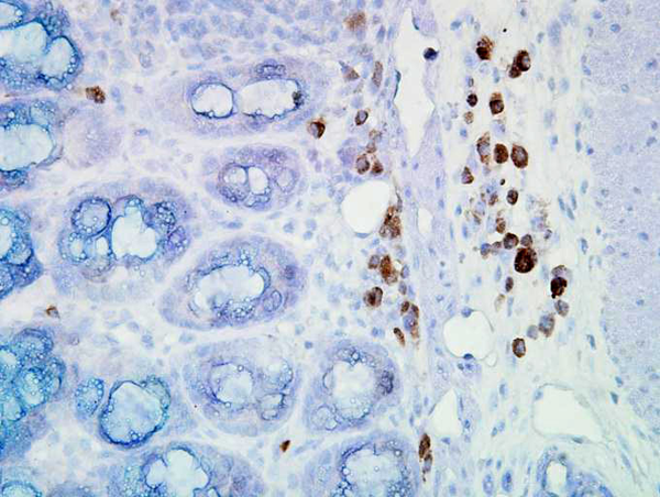
 IHC (Immunohiostchemistry)
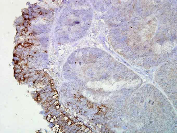
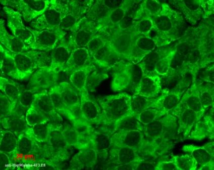
(Immunohistochemistry analysis using Mouse Anti-Hsp90 Monoclonal Antibody, Clone AC-16. Tissue: inflamed colon. Species: Mouse. Fixation: Formalin. Primary Antibody: Mouse Anti-Hsp90 Monoclonal Antibody at 1:2000 for 12 hours at 4 degree C. Secondary Antibody: Biotin Goat Anti-Mouse at 1:2000 for 1 hour at RT. Counterstain: Mayer Hematoxylin (purple/blue) nuclear stain at 200 ul for 2 minutes at RT. Localization: Inflammatory cells. Magnification: 40x. Mostly inflammatory cells, some mucosa.)
IHC (Immunohiostchemistry)
(Immunohistochemistry analysis using Mouse Anti-Hsp90 Monoclonal Antibody, Clone AC-16. Tissue: inflamed colon. Species: Mouse. Fixation: Formalin. Primary Antibody: Mouse Anti-Hsp90 Monoclonal Antibody at 1:2000 for 12 hours at 4 degree C. Secondary Antibody: Biotin Goat Anti-Mouse at 1:2000 for 1 hour at RT. Counterstain: Mayer Hematoxylin (purple/blue) nuclear stain at 200 ul for 2 minutes at RT. Localization: Inflammatory cells. Magnification: 40x. Mostly inflammatory cells, some mucosa.)
Hsp90, Monoclonal Antibody (Cat# AAA103474)
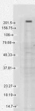
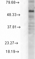
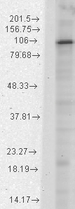
 WB (Western Blot)
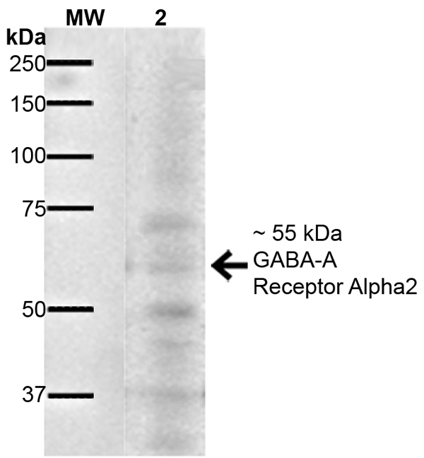
(Western Blot analysis of Rat Brain showing detection of ~55 kDa GABA A Receptor Alpha 2 protein using Mouse Anti-GABA A Receptor Alpha 2 Monoclonal Antibody, Clone S399-19. Lane 1: MW Ladder. Lane 2: Rat Brain. Load: 10 ug. Block: 5% Skim Milk for 1 hour at RT. Primary Antibody: Mouse Anti-GABA A Receptor Alpha 2 Monoclonal Antibody at 1:1000 for 1 hour at RT. Secondary Antibody: Goat Anti-Mouse IgG: HRP at 1:100 for 1 hour at RT. Color Development: ECL solution for 6 min at RT. Predicted/Observed Size: ~55 kDa.)
WB (Western Blot)
(Western Blot analysis of Rat Brain showing detection of ~55 kDa GABA A Receptor Alpha 2 protein using Mouse Anti-GABA A Receptor Alpha 2 Monoclonal Antibody, Clone S399-19. Lane 1: MW Ladder. Lane 2: Rat Brain. Load: 10 ug. Block: 5% Skim Milk for 1 hour at RT. Primary Antibody: Mouse Anti-GABA A Receptor Alpha 2 Monoclonal Antibody at 1:1000 for 1 hour at RT. Secondary Antibody: Goat Anti-Mouse IgG: HRP at 1:100 for 1 hour at RT. Color Development: ECL solution for 6 min at RT. Predicted/Observed Size: ~55 kDa.)
GABA-A Receptor Alpha2, Monoclonal Antibody (Cat# AAA103779)


 WB (Western Blot)
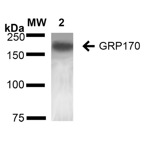
(Western Blot analysis of Rat Liver showing detection of ~170 kDa GRP170 protein using Mouse Anti-GRP170 Monoclonal Antibody, Clone 6E3-2C3 . Lane 1: Molecular Weight Ladder (MW). Lane 2: Rat Liver cell lysate. Load: 20 ug. Block: 2% BSA and 2% Skim Milk in 1X TBST. Primary Antibody: Mouse Anti-GRP170 Monoclonal Antibody at 1:1000 for 16 hours at 4 degree C. Secondary Antibody: Goat Anti-Mouse IgG: HRP at 1:100 for 60 min at RT. Color Development: ECL solution for 6 min in RT. Predicted/Observed Size: ~170 kDa.)
WB (Western Blot)
(Western Blot analysis of Rat Liver showing detection of ~170 kDa GRP170 protein using Mouse Anti-GRP170 Monoclonal Antibody, Clone 6E3-2C3 . Lane 1: Molecular Weight Ladder (MW). Lane 2: Rat Liver cell lysate. Load: 20 ug. Block: 2% BSA and 2% Skim Milk in 1X TBST. Primary Antibody: Mouse Anti-GRP170 Monoclonal Antibody at 1:1000 for 16 hours at 4 degree C. Secondary Antibody: Goat Anti-Mouse IgG: HRP at 1:100 for 60 min at RT. Color Development: ECL solution for 6 min in RT. Predicted/Observed Size: ~170 kDa.)
GRP170, Monoclonal Antibody (Cat# AAA103780)
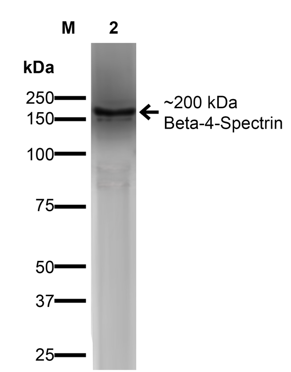 WB (Western Blot)
(Western Blot analysis of COS-Beta-4-Spectrin-His showing detection of ~ 200 kDa Beta-4-Spectrin protein using Mouse Anti-Beta-4-Spectrin Monoclonal Antibody, Clone S393-2. Lane 1: MW Ladder. Lane 2: COS-Beta-4-Spectrin-His. Load: 15 ug. Block: 2% GE Healthcare Blocker for 1 hour at RT. Primary Antibody: Mouse Anti-Beta-4-Spectrin Monoclonal Antibody at 1:1000 for 16 hours at 4 degree C. Secondary Antibody: Goat Anti-Mouse IgG: HRP at 1:200 for 1 hour at RT. Color Development: ECL solution for 6 min at RT. Predicted/Observed Size: ~ 200 kDa.)
WB (Western Blot)
(Western Blot analysis of COS-Beta-4-Spectrin-His showing detection of ~ 200 kDa Beta-4-Spectrin protein using Mouse Anti-Beta-4-Spectrin Monoclonal Antibody, Clone S393-2. Lane 1: MW Ladder. Lane 2: COS-Beta-4-Spectrin-His. Load: 15 ug. Block: 2% GE Healthcare Blocker for 1 hour at RT. Primary Antibody: Mouse Anti-Beta-4-Spectrin Monoclonal Antibody at 1:1000 for 16 hours at 4 degree C. Secondary Antibody: Goat Anti-Mouse IgG: HRP at 1:200 for 1 hour at RT. Color Development: ECL solution for 6 min at RT. Predicted/Observed Size: ~ 200 kDa.)
beta 4 Spectrin, Monoclonal Antibody (Cat# AAA103794)
 WB (Western Blot)
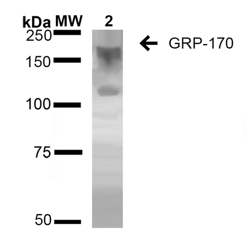
(Western Blot analysis of Rat Liver showing detection of ~170 kDa GRP170 protein using Mouse Anti-GRP170 Monoclonal Antibody, Clone 6E3-2C3 . Lane 1: Molecular Weight Ladder (MW). Lane 2: Rat Liver cell lysate. Load: 20 ug. Block: 2% BSA and 2% Skim Milk in 1X TBST. Primary Antibody: Mouse Anti-GRP170 Monoclonal Antibody at 1:1000 for 16 hours at 4 degree C. Secondary Antibody: Goat Anti-Mouse IgG: HRP at 1:100 for 60 min at RT. Color Development: ECL solution for 6 min in RT. Predicted/Observed Size: ~170 kDa.)
WB (Western Blot)
(Western Blot analysis of Rat Liver showing detection of ~170 kDa GRP170 protein using Mouse Anti-GRP170 Monoclonal Antibody, Clone 6E3-2C3 . Lane 1: Molecular Weight Ladder (MW). Lane 2: Rat Liver cell lysate. Load: 20 ug. Block: 2% BSA and 2% Skim Milk in 1X TBST. Primary Antibody: Mouse Anti-GRP170 Monoclonal Antibody at 1:1000 for 16 hours at 4 degree C. Secondary Antibody: Goat Anti-Mouse IgG: HRP at 1:100 for 60 min at RT. Color Development: ECL solution for 6 min in RT. Predicted/Observed Size: ~170 kDa.)
GRP170, Monoclonal Antibody (Cat# AAA103796)
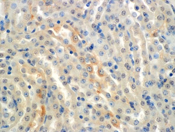
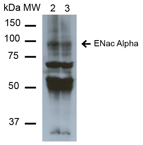 WB (Western Blot)
(Western Blot analysis of Mouse Whole kidney homogenates showing detection of ~85kDa ENaC alpha protein using Mouse Anti-ENaC alpha Monoclonal Antibody, Clone 2G4. Lane 1: Molecular Weight Ladder (MW). Lane 2: Low-salt diet. Lane 3: Normal-salt diet. Load: 20 ug. Primary Antibody: Mouse Anti-ENaC alpha Monoclonal Antibody at 1:1000. Predicted/Observed Size: ~85kDa.)
WB (Western Blot)
(Western Blot analysis of Mouse Whole kidney homogenates showing detection of ~85kDa ENaC alpha protein using Mouse Anti-ENaC alpha Monoclonal Antibody, Clone 2G4. Lane 1: Molecular Weight Ladder (MW). Lane 2: Low-salt diet. Lane 3: Normal-salt diet. Load: 20 ug. Primary Antibody: Mouse Anti-ENaC alpha Monoclonal Antibody at 1:1000. Predicted/Observed Size: ~85kDa.)
ENaC alpha, Monoclonal Antibody (Cat# AAA103889)
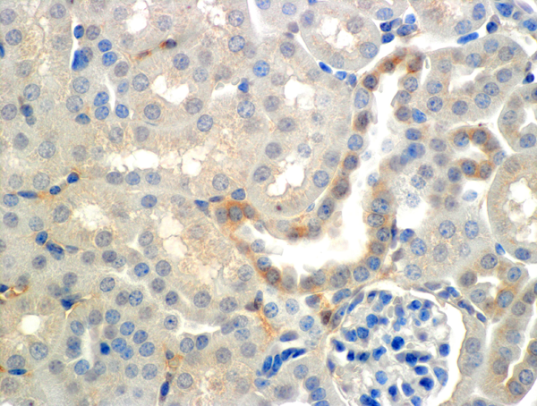
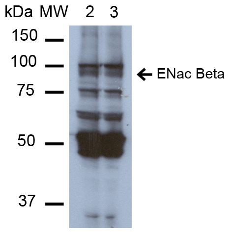 WB (Western Blot)
(Western Blot analysis of Mouse Whole kidney homogenates showing detection of ~87kDa ENaC beta protein using Mouse Anti-ENaC beta Monoclonal Antibody, Clone 7B8. Lane 1: Molecular Weight Ladder (MW). Lane 2: Low-salt diet. Lane 3: Normal-salt diet. Load: 20 ug. Primary Antibody: Mouse Anti-ENaC beta Monoclonal Antibody at 1:1000. Predicted/Observed Size: ~87kDa.)
WB (Western Blot)
(Western Blot analysis of Mouse Whole kidney homogenates showing detection of ~87kDa ENaC beta protein using Mouse Anti-ENaC beta Monoclonal Antibody, Clone 7B8. Lane 1: Molecular Weight Ladder (MW). Lane 2: Low-salt diet. Lane 3: Normal-salt diet. Load: 20 ug. Primary Antibody: Mouse Anti-ENaC beta Monoclonal Antibody at 1:1000. Predicted/Observed Size: ~87kDa.)
ENaC beta, Monoclonal Antibody (Cat# AAA103896)

 WB (Western Blot)
(Western Blot analysis of Mouse Whole kidney homogenates showing detection of ~87kDa ENaC beta protein using Mouse Anti-ENaC beta Monoclonal Antibody, Clone 7B8. Lane 1: Molecular Weight Ladder (MW). Lane 2: Low-salt diet. Lane 3: Normal-salt diet. Load: 20 ug. Primary Antibody: Mouse Anti-ENaC beta Monoclonal Antibody at 1:1000. Predicted/Observed Size: ~87kDa.)
WB (Western Blot)
(Western Blot analysis of Mouse Whole kidney homogenates showing detection of ~87kDa ENaC beta protein using Mouse Anti-ENaC beta Monoclonal Antibody, Clone 7B8. Lane 1: Molecular Weight Ladder (MW). Lane 2: Low-salt diet. Lane 3: Normal-salt diet. Load: 20 ug. Primary Antibody: Mouse Anti-ENaC beta Monoclonal Antibody at 1:1000. Predicted/Observed Size: ~87kDa.)
ENaC beta, Monoclonal Antibody (Cat# AAA103901)
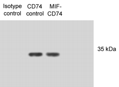
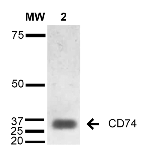 WB (Western Blot)
(Western Blot analysis of Human Lymphoblastoid cell line (Raji) showing detection of 33-35 kDa CD74 protein using Mouse Anti-CD74 Monoclonal Antibody, Clone 1B8 . Lane 1: Molecular Weight Ladder (MW). Lane 2: Raji cell lysate. Load: 15 ug. Block: 5% Skim Milk in TBST. Primary Antibody: Mouse Anti-CD74 Monoclonal Antibody at 1:1000 for 2 hours at RT. Secondary Antibody: Goat Anti-Mouse IgG: HRP at 1:1000 for 60 min at RT. Color Development: ECL solution for 5 min in RT. Predicted/Observed Size: 33-35 kDa.)
WB (Western Blot)
(Western Blot analysis of Human Lymphoblastoid cell line (Raji) showing detection of 33-35 kDa CD74 protein using Mouse Anti-CD74 Monoclonal Antibody, Clone 1B8 . Lane 1: Molecular Weight Ladder (MW). Lane 2: Raji cell lysate. Load: 15 ug. Block: 5% Skim Milk in TBST. Primary Antibody: Mouse Anti-CD74 Monoclonal Antibody at 1:1000 for 2 hours at RT. Secondary Antibody: Goat Anti-Mouse IgG: HRP at 1:1000 for 60 min at RT. Color Development: ECL solution for 5 min in RT. Predicted/Observed Size: 33-35 kDa.)
CD74, Monoclonal Antibody (Cat# AAA103935)
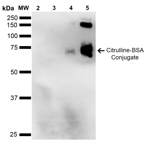
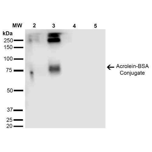
 WB (Western Blot)
(Western Blot analysis of Citrulline-BSA Conjugate showing detection of 67 kDa Citrulline protein using Mouse Anti-Citrulline Monoclonal Antibody, Clone 2D3-1B9. Lane 1: Molecular Weight Ladder (MW). Lane 2: BSA (0.5 ug). Lane 3: BSA (2.0 ug). Lane 4: Citrulline-BSA (0.5 ug). Lane 5: Citrulline-BSA (2.0 ug). Block: 5% Skim Milk in TBST. Primary Antibody: Mouse Anti-Citrulline Monoclonal Antibody at 1:1000 for 2 hours at RT. Secondary Antibody: Goat Anti-Mouse IgG: HRP at 1:2000 for 60 min at RT. Color Development: ECL solution for 5 min in RT. Predicted/Observed Size: 67 kDa.)
WB (Western Blot)
(Western Blot analysis of Citrulline-BSA Conjugate showing detection of 67 kDa Citrulline protein using Mouse Anti-Citrulline Monoclonal Antibody, Clone 2D3-1B9. Lane 1: Molecular Weight Ladder (MW). Lane 2: BSA (0.5 ug). Lane 3: BSA (2.0 ug). Lane 4: Citrulline-BSA (0.5 ug). Lane 5: Citrulline-BSA (2.0 ug). Block: 5% Skim Milk in TBST. Primary Antibody: Mouse Anti-Citrulline Monoclonal Antibody at 1:1000 for 2 hours at RT. Secondary Antibody: Goat Anti-Mouse IgG: HRP at 1:2000 for 60 min at RT. Color Development: ECL solution for 5 min in RT. Predicted/Observed Size: 67 kDa.)
Citrulline, Monoclonal Antibody (Cat# AAA103962)

 WB (Western Blot)
(Western Blot analysis of Citrulline-BSA Conjugate showing detection of 67 kDa Citrulline protein using Mouse Anti-Citrulline Monoclonal Antibody, Clone 2D3-1B9. Lane 1: Molecular Weight Ladder (MW). Lane 2: BSA (0.5 ug). Lane 3: BSA (2.0 ug). Lane 4: Citrulline-BSA (0.5 ug). Lane 5: Citrulline-BSA (2.0 ug). Block: 5% Skim Milk in TBST. Primary Antibody: Mouse Anti-Citrulline Monoclonal Antibody at 1:1000 for 2 hours at RT. Secondary Antibody: Goat Anti-Mouse IgG: HRP at 1:2000 for 60 min at RT. Color Development: ECL solution for 5 min in RT. Predicted/Observed Size: 67 kDa.)
WB (Western Blot)
(Western Blot analysis of Citrulline-BSA Conjugate showing detection of 67 kDa Citrulline protein using Mouse Anti-Citrulline Monoclonal Antibody, Clone 2D3-1B9. Lane 1: Molecular Weight Ladder (MW). Lane 2: BSA (0.5 ug). Lane 3: BSA (2.0 ug). Lane 4: Citrulline-BSA (0.5 ug). Lane 5: Citrulline-BSA (2.0 ug). Block: 5% Skim Milk in TBST. Primary Antibody: Mouse Anti-Citrulline Monoclonal Antibody at 1:1000 for 2 hours at RT. Secondary Antibody: Goat Anti-Mouse IgG: HRP at 1:2000 for 60 min at RT. Color Development: ECL solution for 5 min in RT. Predicted/Observed Size: 67 kDa.)
Citrulline, Monoclonal Antibody (Cat# AAA103965)
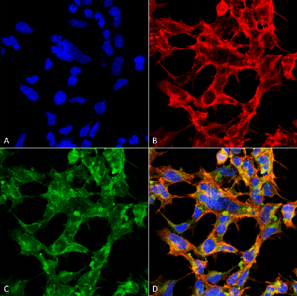
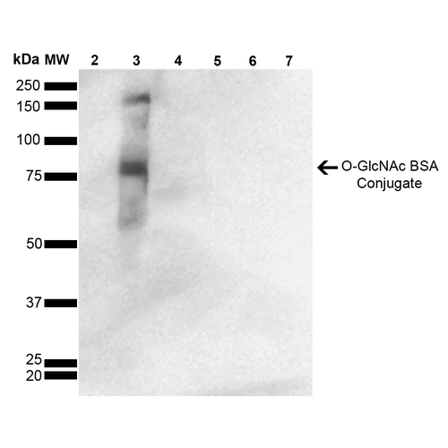 WB (Western Blot)
(Western Blot analysis of GlcNAc-BSA Conjugate showing detection of 67 kDa O-GlcNAc protein using Mouse Anti-O-GlcNAc Monoclonal Antibody, Clone 9H6. Lane 1: Molecular Weight Ladder (MW). Lane 2: BSA. Lane 3: GlcNAc-BSA. Lane 4: GalNAc-BSA. Lane 5: Galactose-BSA. Lane 6: Glucose-BSA. Lane 7: Citrulline-BSA. Load: 2.0 ug. Block: 5% Skim Milk in TBST. Primary Antibody: Mouse Anti-O-GlcNAc Monoclonal Antibody at 1:1000 for 2 hours at RT. Secondary Antibody: Goat Anti-Mouse IgG: HRP at 1:2000 for 60 min at RT. Color Development: ECL solution for 5 min in RT. Predicted/Observed Size: 67 kDa.)
WB (Western Blot)
(Western Blot analysis of GlcNAc-BSA Conjugate showing detection of 67 kDa O-GlcNAc protein using Mouse Anti-O-GlcNAc Monoclonal Antibody, Clone 9H6. Lane 1: Molecular Weight Ladder (MW). Lane 2: BSA. Lane 3: GlcNAc-BSA. Lane 4: GalNAc-BSA. Lane 5: Galactose-BSA. Lane 6: Glucose-BSA. Lane 7: Citrulline-BSA. Load: 2.0 ug. Block: 5% Skim Milk in TBST. Primary Antibody: Mouse Anti-O-GlcNAc Monoclonal Antibody at 1:1000 for 2 hours at RT. Secondary Antibody: Goat Anti-Mouse IgG: HRP at 1:2000 for 60 min at RT. Color Development: ECL solution for 5 min in RT. Predicted/Observed Size: 67 kDa.)
O-GlcNAc, Monoclonal Antibody (Cat# AAA103980)

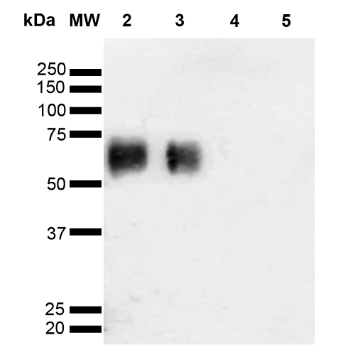 WB (Western Blot)
(Western Blot analysis of Glycoconjugates showing detection of 67 kDa O-GalNAC protein using Mouse Anti-O-GalNAC Monoclonal Antibody, Clone 9B9. Lane 1: Molecular Weight Ladder (MW). Lane 2: GlcNAc-BSA. Lane 3: GalNAc-BSA. Lane 4: Galactose-BSA. Lane 5: Glucose-BSA. Load: 2.0 ug. Block: 5% Skim Milk in TBST. Primary Antibody: Mouse Anti-O-GalNAC Monoclonal Antibody at 1:1000 for 2 hours at RT. Secondary Antibody: Goat Anti-Mouse IgG: HRP at 1:2000 for 60 min at RT. Color Development: ECL solution for 5 min in RT. Predicted/Observed Size: 67 kDa.)
WB (Western Blot)
(Western Blot analysis of Glycoconjugates showing detection of 67 kDa O-GalNAC protein using Mouse Anti-O-GalNAC Monoclonal Antibody, Clone 9B9. Lane 1: Molecular Weight Ladder (MW). Lane 2: GlcNAc-BSA. Lane 3: GalNAc-BSA. Lane 4: Galactose-BSA. Lane 5: Glucose-BSA. Load: 2.0 ug. Block: 5% Skim Milk in TBST. Primary Antibody: Mouse Anti-O-GalNAC Monoclonal Antibody at 1:1000 for 2 hours at RT. Secondary Antibody: Goat Anti-Mouse IgG: HRP at 1:2000 for 60 min at RT. Color Development: ECL solution for 5 min in RT. Predicted/Observed Size: 67 kDa.)
O-GalNAc, Monoclonal Antibody (Cat# AAA103988)
 WB (Western Blot)
(Western Blot analysis of Glycoconjugates showing detection of 67 kDa O-GalNAC protein using Mouse Anti-O-GalNAC Monoclonal Antibody, Clone 9B9. Lane 1: Molecular Weight Ladder (MW). Lane 2: GlcNAc-BSA. Lane 3: GalNAc-BSA. Lane 4: Galactose-BSA. Lane 5: Glucose-BSA. Load: 2.0 ug. Block: 5% Skim Milk in TBST. Primary Antibody: Mouse Anti-O-GalNAC Monoclonal Antibody at 1:1000 for 2 hours at RT. Secondary Antibody: Goat Anti-Mouse IgG: HRP at 1:2000 for 60 min at RT. Color Development: ECL solution for 5 min in RT. Predicted/Observed Size: 67 kDa.)
WB (Western Blot)
(Western Blot analysis of Glycoconjugates showing detection of 67 kDa O-GalNAC protein using Mouse Anti-O-GalNAC Monoclonal Antibody, Clone 9B9. Lane 1: Molecular Weight Ladder (MW). Lane 2: GlcNAc-BSA. Lane 3: GalNAc-BSA. Lane 4: Galactose-BSA. Lane 5: Glucose-BSA. Load: 2.0 ug. Block: 5% Skim Milk in TBST. Primary Antibody: Mouse Anti-O-GalNAC Monoclonal Antibody at 1:1000 for 2 hours at RT. Secondary Antibody: Goat Anti-Mouse IgG: HRP at 1:2000 for 60 min at RT. Color Development: ECL solution for 5 min in RT. Predicted/Observed Size: 67 kDa.)
O-GalNAc, Monoclonal Antibody (Cat# AAA103994)
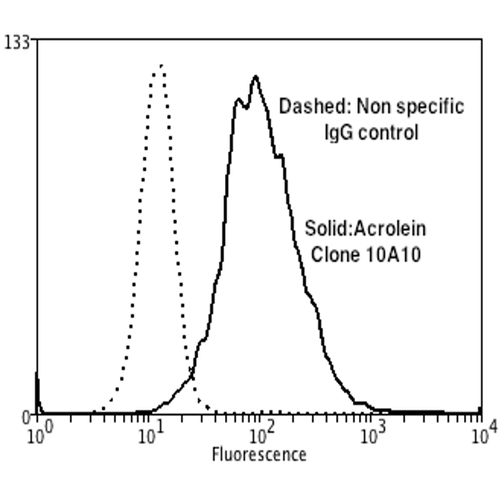
 FCM/FACS (Flow Cytometry)
(Flow Cytometry analysis using Mouse Anti-Acrolein Monoclonal Antibody, Clone 2H2. Tissue: Neuroblastoma cells (SH-SY5Y). Species: Human. Fixation: 90% Methanol. Primary Antibody: Mouse Anti-Acrolein Monoclonal Antibody at 1:50 for 30 min on ice. Secondary Antibody: Goat Anti-Mouse: PE at 1:100 for 20 min at RT. Isotype Control: Non Specific IgG. Cells were subject to oxidative stress by treating with 250 uM H2O2 for 24 hours.)
FCM/FACS (Flow Cytometry)
(Flow Cytometry analysis using Mouse Anti-Acrolein Monoclonal Antibody, Clone 2H2. Tissue: Neuroblastoma cells (SH-SY5Y). Species: Human. Fixation: 90% Methanol. Primary Antibody: Mouse Anti-Acrolein Monoclonal Antibody at 1:50 for 30 min on ice. Secondary Antibody: Goat Anti-Mouse: PE at 1:100 for 20 min at RT. Isotype Control: Non Specific IgG. Cells were subject to oxidative stress by treating with 250 uM H2O2 for 24 hours.)
Acrolein, Monoclonal Antibody (Cat# AAA104000)
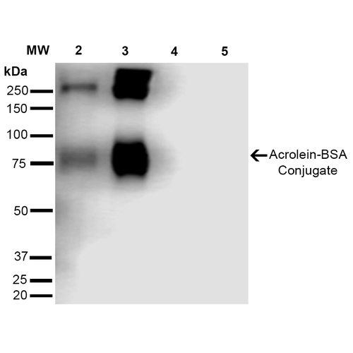
 WB (Western Blot)
(Western Blot analysis of Human Cervical cancer cell line (HeLa) lysate showing detection of Acrolein protein using Mouse Anti-Acrolein Monoclonal Antibody, Clone 10A10. Lane 1: Molecular Weight Ladder (MW). Lane 2: HeLa cell lysate. Lane 3: H2O2 treated HeLa cell lysate. Load: 12 ug. Block: 5% Skim Milk in TBST. Primary Antibody: Mouse Anti-Acrolein Monoclonal Antibody at 1:1000 for 2 hours at RT. Secondary Antibody: Goat Anti-Mouse IgG: HRP at 1:2000 for 60 min at RT. Color Development: ECL solution for 5 min in RT.)
WB (Western Blot)
(Western Blot analysis of Human Cervical cancer cell line (HeLa) lysate showing detection of Acrolein protein using Mouse Anti-Acrolein Monoclonal Antibody, Clone 10A10. Lane 1: Molecular Weight Ladder (MW). Lane 2: HeLa cell lysate. Lane 3: H2O2 treated HeLa cell lysate. Load: 12 ug. Block: 5% Skim Milk in TBST. Primary Antibody: Mouse Anti-Acrolein Monoclonal Antibody at 1:1000 for 2 hours at RT. Secondary Antibody: Goat Anti-Mouse IgG: HRP at 1:2000 for 60 min at RT. Color Development: ECL solution for 5 min in RT.)
Acrolein, Monoclonal Antibody (Cat# AAA104010)
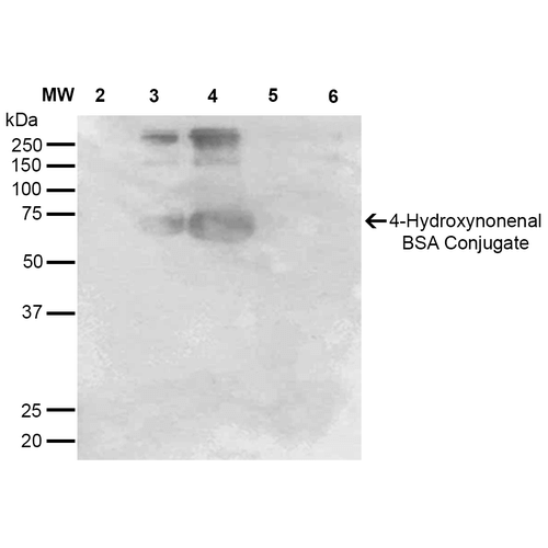 WB (Western Blot)
(Western Blot analysis of 4-hydroxy-nonenal-BSA Conjugate showing detection of 67 kDa 4-hydroxy-nonenal protein using Mouse Anti-4-hydroxy-nonenal Monoclonal Antibody, Clone 12F7. Lane 1: Molecular Weight Ladder (MW). Lane 2: BSA (0.5 ug). Lane 3: 4-hydroxyl nonenal-BSA (0.5 ug). Lane 4: 4-hydroxy nonenal-BSA (2.0 ug). Lane 5: 4-hydroxy-2-hexenal (0.5 ug). Lane 6: 4-hydroxy-2-hexenal (2.0 ug). Block: 5% Skim Milk in TBST. Primary Antibody: Mouse Anti-4-hydroxy-nonenal Monoclonal Antibody at 1:1000 for 2 hours at RT. Secondary Antibody: Goat Anti-Mouse IgG: HRP at 1:2000 for 60 min at RT. Color Development: ECL solution for 5 min in RT. Predicted/Observed Size: 67 kDa.)
WB (Western Blot)
(Western Blot analysis of 4-hydroxy-nonenal-BSA Conjugate showing detection of 67 kDa 4-hydroxy-nonenal protein using Mouse Anti-4-hydroxy-nonenal Monoclonal Antibody, Clone 12F7. Lane 1: Molecular Weight Ladder (MW). Lane 2: BSA (0.5 ug). Lane 3: 4-hydroxyl nonenal-BSA (0.5 ug). Lane 4: 4-hydroxy nonenal-BSA (2.0 ug). Lane 5: 4-hydroxy-2-hexenal (0.5 ug). Lane 6: 4-hydroxy-2-hexenal (2.0 ug). Block: 5% Skim Milk in TBST. Primary Antibody: Mouse Anti-4-hydroxy-nonenal Monoclonal Antibody at 1:1000 for 2 hours at RT. Secondary Antibody: Goat Anti-Mouse IgG: HRP at 1:2000 for 60 min at RT. Color Development: ECL solution for 5 min in RT. Predicted/Observed Size: 67 kDa.)
4-Hydroxynonenal, Monoclonal Antibody (Cat# AAA104043)
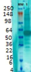
 WB (Western Blot)
(Western Blot analysis of Human Cervical cancer cell line (HeLa) lysate showing detection of FKBP51 protein using Mouse Anti-FKBP51 Monoclonal Antibody, Clone Hi51B. Load: 15 ug. Block: 1.5% BSA for 30 minutes at RT. Primary Antibody: Mouse Anti-FKBP51 Monoclonal Antibody at 1:1000 for 2 hours at RT. Secondary Antibody: Sheep Anti-Mouse IgG: HRP for 1 hour at RT.)
WB (Western Blot)
(Western Blot analysis of Human Cervical cancer cell line (HeLa) lysate showing detection of FKBP51 protein using Mouse Anti-FKBP51 Monoclonal Antibody, Clone Hi51B. Load: 15 ug. Block: 1.5% BSA for 30 minutes at RT. Primary Antibody: Mouse Anti-FKBP51 Monoclonal Antibody at 1:1000 for 2 hours at RT. Secondary Antibody: Sheep Anti-Mouse IgG: HRP for 1 hour at RT.)
FKBP51, Monoclonal Antibody (Cat# AAA103464)
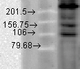
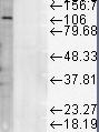
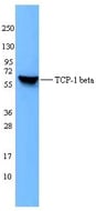 WB (Western Blot)
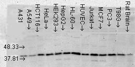
(HepG2 cell extracts were resolved by electrophoresis, transferred to nitrocellulose, and probed with purified monoclonal TCP-1beta antibody (clone F39P7F11). Proteins were visualized using a goat anti-mouse-IgG secondary conjugated to HRP and chemiluminescence detection.)
WB (Western Blot)
(HepG2 cell extracts were resolved by electrophoresis, transferred to nitrocellulose, and probed with purified monoclonal TCP-1beta antibody (clone F39P7F11). Proteins were visualized using a goat anti-mouse-IgG secondary conjugated to HRP and chemiluminescence detection.)
CCT2 / CCT Beta, Monoclonal Antibody (Cat# AAA51781)
Predicted Reactivity: Chicken (at least 90% immunogen sequence identity)
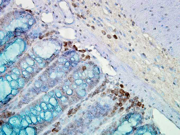
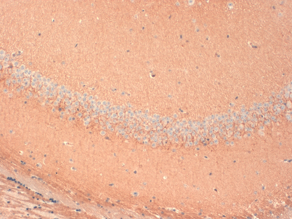
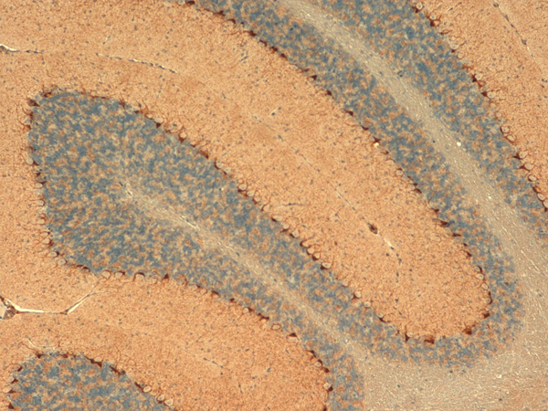
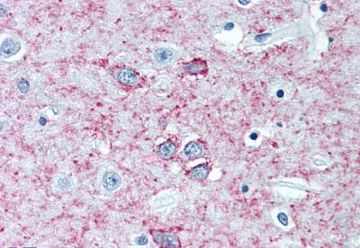 IHC (Immunohistochemistry)
(Anti-ADORA2A antibody IHC of human brain, putamen. Immunohistochemistry of formalin-fixed, paraffin-embedded tissue after heat-induced antigen retrieval. Antibody concentration 5 ug/ml.)
IHC (Immunohistochemistry)
(Anti-ADORA2A antibody IHC of human brain, putamen. Immunohistochemistry of formalin-fixed, paraffin-embedded tissue after heat-induced antigen retrieval. Antibody concentration 5 ug/ml.)
ADORA2A/Adenosine A2A Receptor, Monoclonal Antibody (Cat# AAA51801)
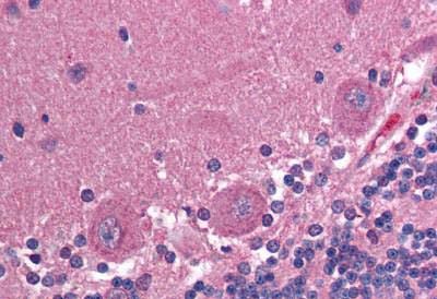
 IHC (Immunohistochemistry)
Nestin, Monoclonal Antibody (Cat# AAA59525)

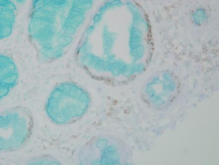
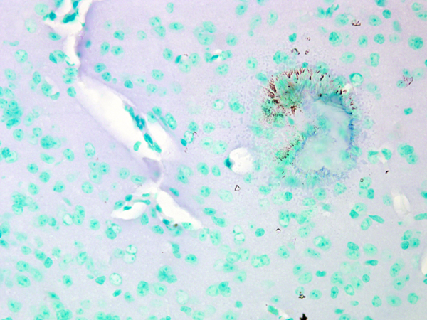
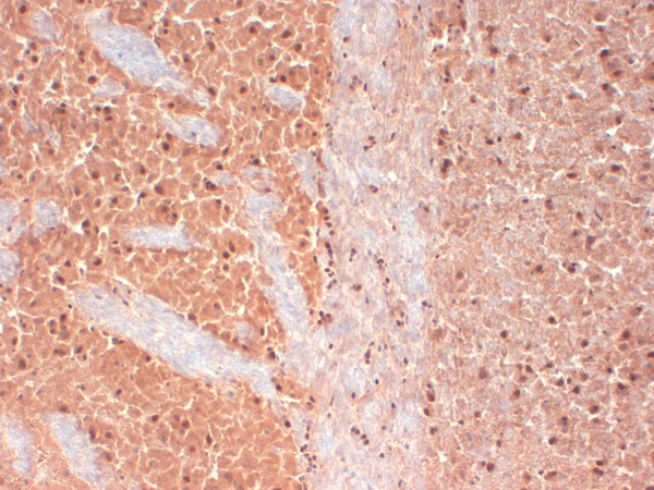
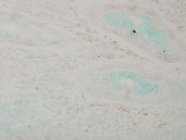
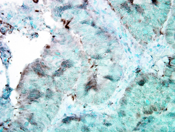
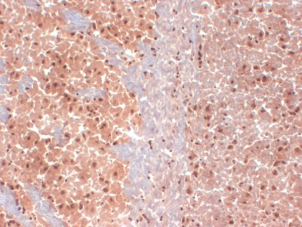
 IHC (Immunohistochemisry)
(Immunohistochemistry analysis using Mouse Anti-DNMT1 Monoclonal Antibody, Clone 60B1220.1. Tissue: medullar kidney tissue. Species: Mouse. Primary Antibody: Mouse Anti-DNMT1 Monoclonal Antibody at 1:1000. Secondary Antibody: HRP/DAB Detection System: Biotinylated Goat Anti-Mouse, Streptavidin Peroxidase, DAB Chromogen (brown). Counterstain: Mayer Hematoxylin (purple/blue) nuclear stain.)
IHC (Immunohistochemisry)
(Immunohistochemistry analysis using Mouse Anti-DNMT1 Monoclonal Antibody, Clone 60B1220.1. Tissue: medullar kidney tissue. Species: Mouse. Primary Antibody: Mouse Anti-DNMT1 Monoclonal Antibody at 1:1000. Secondary Antibody: HRP/DAB Detection System: Biotinylated Goat Anti-Mouse, Streptavidin Peroxidase, DAB Chromogen (brown). Counterstain: Mayer Hematoxylin (purple/blue) nuclear stain.)
DNMT1, Monoclonal Antibody (Cat# AAA103333)
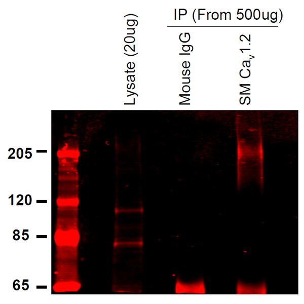
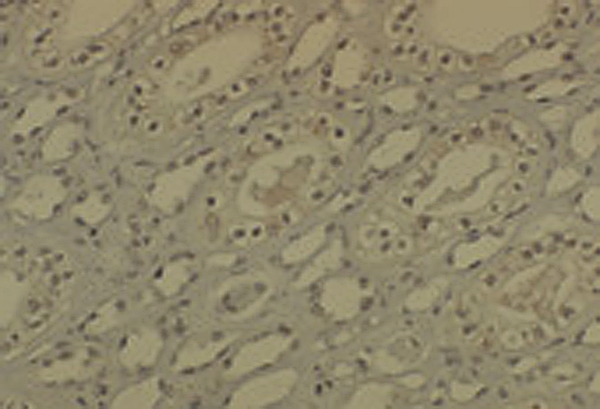
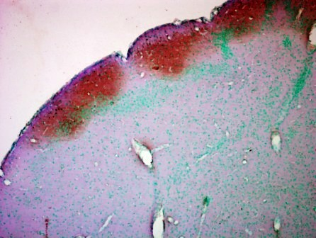
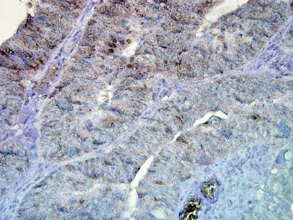
 IHC (Immunohistochemistry)
(Immunohistochemistry analysis using Mouse Anti-CaV1.2 Calcium channel Monoclonal Antibody, Clone S57-47. Tissue: Brain Tissue. Species: Mouse. Fixation: Formalin. Primary Antibody: Mouse Anti-CaV1.2 Calcium channel Monoclonal Antibody at 1:10000 for 12 hours at 4 degree C. Secondary Antibody: Biotin Goat Anti-Mouse at 1:2000 for 1 hour at RT. Counterstain: Mayer Hematoxylin (purple/blue) nuclear stain at 200 ul for 2 minutes at RT. Magnification: 40x.)
IHC (Immunohistochemistry)
(Immunohistochemistry analysis using Mouse Anti-CaV1.2 Calcium channel Monoclonal Antibody, Clone S57-47. Tissue: Brain Tissue. Species: Mouse. Fixation: Formalin. Primary Antibody: Mouse Anti-CaV1.2 Calcium channel Monoclonal Antibody at 1:10000 for 12 hours at 4 degree C. Secondary Antibody: Biotin Goat Anti-Mouse at 1:2000 for 1 hour at RT. Counterstain: Mayer Hematoxylin (purple/blue) nuclear stain at 200 ul for 2 minutes at RT. Magnification: 40x.)
Cav1.2, Monoclonal Antibody (Cat# AAA103347)
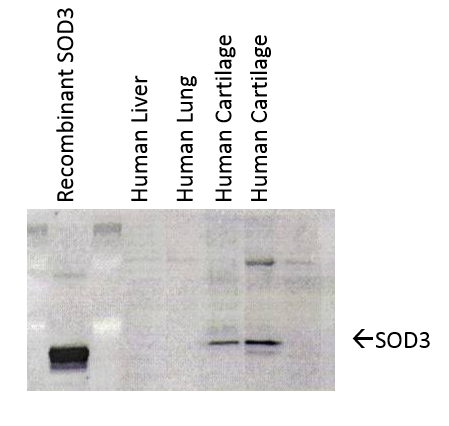 WB (Western Blot)
(Western Blot analysis of Human cartilage lysates showing detection of SOD3 protein using Mouse Anti-SOD3 Monoclonal Antibody, Clone 4GG11G6. Primary Antibody: Mouse Anti-SOD3 Monoclonal Antibody at 1:1000. Left: Control, Middle: Young cartilage, Right: Cartilage sample with osteoarthritis-arthritis.)
WB (Western Blot)
(Western Blot analysis of Human cartilage lysates showing detection of SOD3 protein using Mouse Anti-SOD3 Monoclonal Antibody, Clone 4GG11G6. Primary Antibody: Mouse Anti-SOD3 Monoclonal Antibody at 1:1000. Left: Control, Middle: Young cartilage, Right: Cartilage sample with osteoarthritis-arthritis.)
SOD (EC), Monoclonal Antibody (Cat# AAA103354)
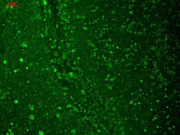
 IHC (Immunohiostchemistry)
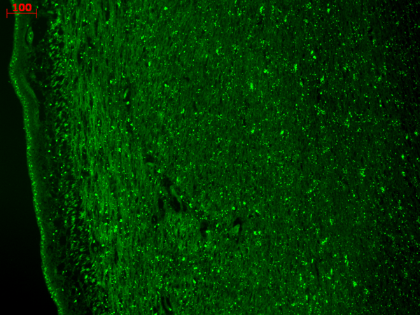
(Immunohistochemistry analysis using Mouse Anti-CaV3.2 Calcium channel Monoclonal Antibody, Clone S55-10. Tissue: frozen brain section. Species: human. Fixation: 10% Formalin Solution for 12-24 hours at RT. Primary Antibody: Mouse Anti-CaV3.2 Calcium channel Monoclonal Antibody at 1:1000 for 1 hour at RT. Secondary Antibody: HRP/DAB Detection System: Biotinylated Goat Anti-Mouse, Streptavidin Peroxidase, DAB Chromogen (brown) for 30 minutes at RT. Counterstain: Mayer Hematoxylin (purple/blue) nuclear stain at 250-500 ul for 5 minutes at RT.)
IHC (Immunohiostchemistry)
(Immunohistochemistry analysis using Mouse Anti-CaV3.2 Calcium channel Monoclonal Antibody, Clone S55-10. Tissue: frozen brain section. Species: human. Fixation: 10% Formalin Solution for 12-24 hours at RT. Primary Antibody: Mouse Anti-CaV3.2 Calcium channel Monoclonal Antibody at 1:1000 for 1 hour at RT. Secondary Antibody: HRP/DAB Detection System: Biotinylated Goat Anti-Mouse, Streptavidin Peroxidase, DAB Chromogen (brown) for 30 minutes at RT. Counterstain: Mayer Hematoxylin (purple/blue) nuclear stain at 250-500 ul for 5 minutes at RT.)
Cav3.2, Monoclonal Antibody (Cat# AAA103370)
 WB (Western Blot)
(Western Blot analysis of Human Cell lysates showing detection of TrpM7 protein using Mouse Anti-TrpM7 Monoclonal Antibody, Clone S74-25. Load: 15 ug. Block: 1.5% BSA for 30 minutes at RT. Primary Antibody: Mouse Anti-TrpM7 Monoclonal Antibody at 1:1000 for 2 hours at RT. Secondary Antibody: Sheep Anti-Mouse IgG: HRP for 1 hour at RT.)
WB (Western Blot)
(Western Blot analysis of Human Cell lysates showing detection of TrpM7 protein using Mouse Anti-TrpM7 Monoclonal Antibody, Clone S74-25. Load: 15 ug. Block: 1.5% BSA for 30 minutes at RT. Primary Antibody: Mouse Anti-TrpM7 Monoclonal Antibody at 1:1000 for 2 hours at RT. Secondary Antibody: Sheep Anti-Mouse IgG: HRP for 1 hour at RT.)
TrpM7, Monoclonal Antibody (Cat# AAA103374)
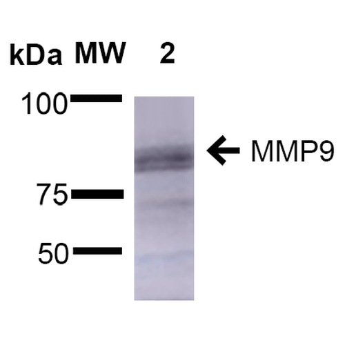 WB (Western Blot)
(Western Blot analysis of Rat Brain showing detection of ~92 kDa and ~82 kDa (pro and active) MMP9 protein using Mouse Anti-MMP9 Monoclonal Antibody, Clone S51-82 . Lane 1: Molecular Weight Ladder (MW). Lane 2: Rat Brain. Load: 15 ug. Block: 5% Skim Milk in 1X TBST. Primary Antibody: Mouse Anti-MMP9 Monoclonal Antibody at 1:1000 for 2 hours at RT. Secondary Antibody: Goat Anti-Mouse IgG: HRP at 1:2000 for 60 min at RT. Color Development: ECL solution for 5 min at RT. Predicted/Observed Size: ~92 kDa and ~82 kDa (pro and active).)
WB (Western Blot)
(Western Blot analysis of Rat Brain showing detection of ~92 kDa and ~82 kDa (pro and active) MMP9 protein using Mouse Anti-MMP9 Monoclonal Antibody, Clone S51-82 . Lane 1: Molecular Weight Ladder (MW). Lane 2: Rat Brain. Load: 15 ug. Block: 5% Skim Milk in 1X TBST. Primary Antibody: Mouse Anti-MMP9 Monoclonal Antibody at 1:1000 for 2 hours at RT. Secondary Antibody: Goat Anti-Mouse IgG: HRP at 1:2000 for 60 min at RT. Color Development: ECL solution for 5 min at RT. Predicted/Observed Size: ~92 kDa and ~82 kDa (pro and active).)
MMP9, Monoclonal Antibody (Cat# AAA103377)


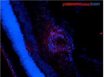
 WB (Western Blot)
(Western Blot analysis of Human Cell lysates showing detection of p38 MAPK protein using Mouse Anti-p38 MAPK Monoclonal Antibody, Clone 9F12. Load: 15 ug. Block: 1.5% BSA for 30 minutes at RT. Primary Antibody: Mouse Anti-p38 MAPK Monoclonal Antibody at 1:1000 for 2 hours at RT. Secondary Antibody: Sheep Anti-Mouse IgG: HRP for 1 hour at RT.)
WB (Western Blot)
(Western Blot analysis of Human Cell lysates showing detection of p38 MAPK protein using Mouse Anti-p38 MAPK Monoclonal Antibody, Clone 9F12. Load: 15 ug. Block: 1.5% BSA for 30 minutes at RT. Primary Antibody: Mouse Anti-p38 MAPK Monoclonal Antibody at 1:1000 for 2 hours at RT. Secondary Antibody: Sheep Anti-Mouse IgG: HRP for 1 hour at RT.)
p38 alpha, Monoclonal Antibody (Cat# AAA103379)
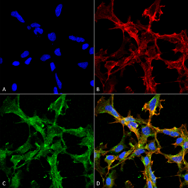
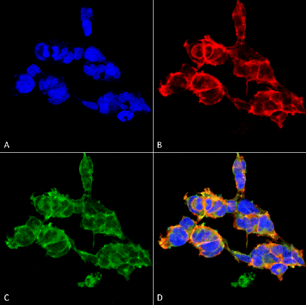
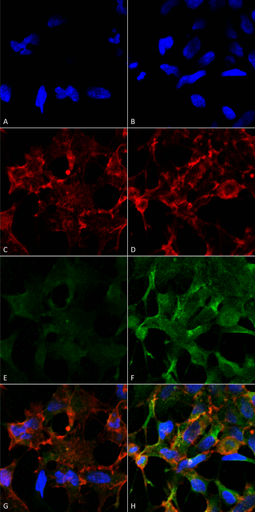
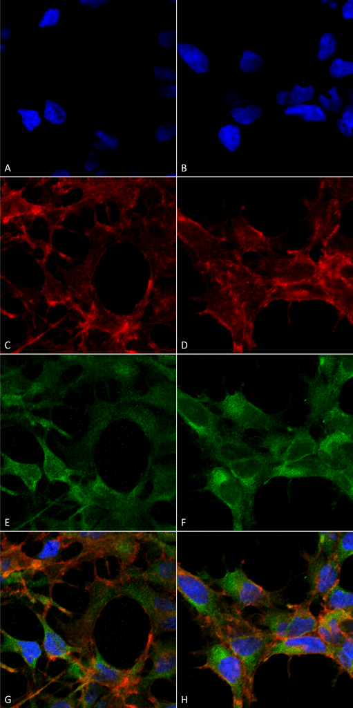
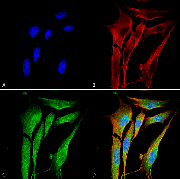
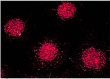
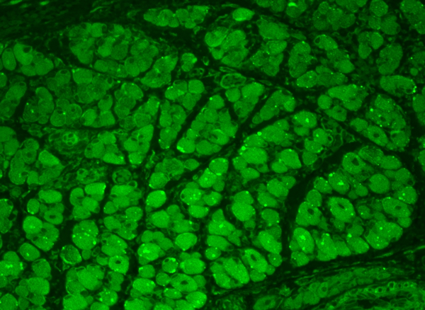
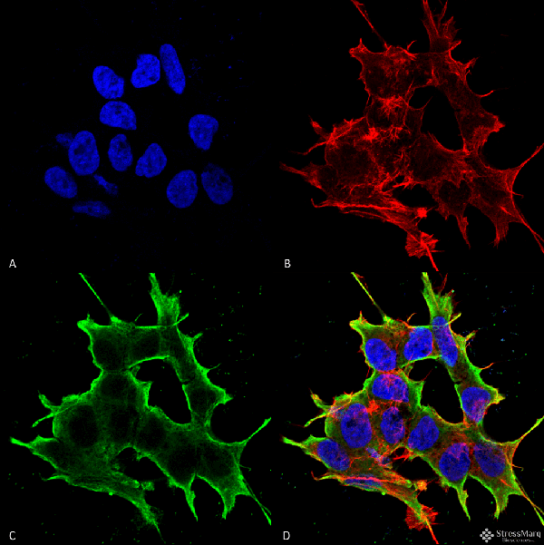
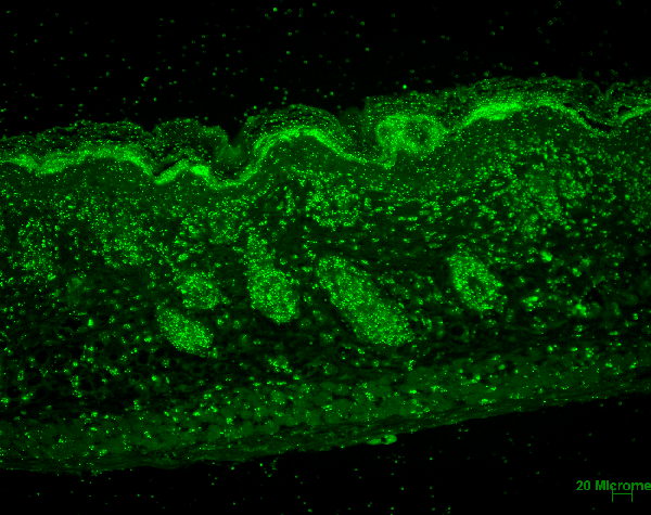
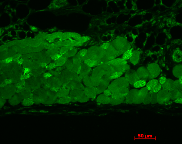
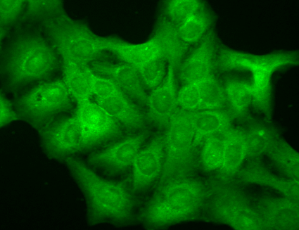
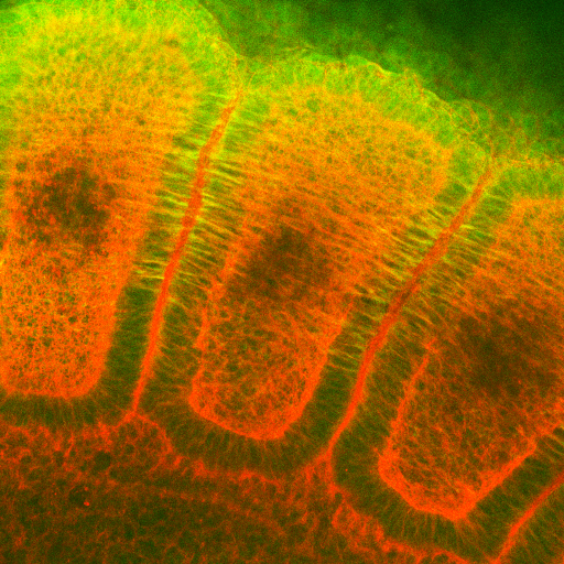
 ICC (Immunocytochemistry)
(Immunocytochemistry/Immunofluorescence analysis using Mouse Anti-Cav Beta2 Calcium Channel Monoclonal Antibody, Clone S8b-1. Tissue: HaCaT cells. Species: Human. Fixation: Cold 100% methanol for 10 minutes at -20 degree C. Primary Antibody: Mouse Anti-Cav Beta2 Calcium Channel Monoclonal Antibody at 1:100 for 1 hour at RT. Secondary Antibody: FITC Goat Anti-Mouse (green) at 1:50 for 1 hour at RT. Localization: All cells positive. Bright dottiness located throughout cytoplasm and in nuclei.)
ICC (Immunocytochemistry)
(Immunocytochemistry/Immunofluorescence analysis using Mouse Anti-Cav Beta2 Calcium Channel Monoclonal Antibody, Clone S8b-1. Tissue: HaCaT cells. Species: Human. Fixation: Cold 100% methanol for 10 minutes at -20 degree C. Primary Antibody: Mouse Anti-Cav Beta2 Calcium Channel Monoclonal Antibody at 1:100 for 1 hour at RT. Secondary Antibody: FITC Goat Anti-Mouse (green) at 1:50 for 1 hour at RT. Localization: All cells positive. Bright dottiness located throughout cytoplasm and in nuclei.)
Cavbeta2, Monoclonal Antibody (Cat# AAA103389)
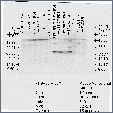
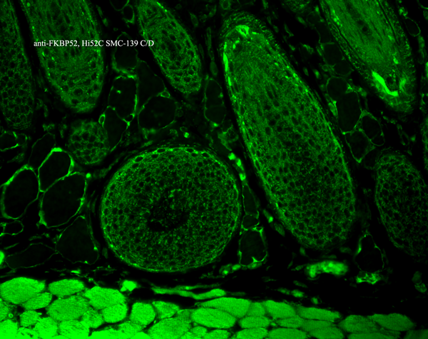
 WB (Western Blot)
(Western Blot analysis of Human Cell lysates showing detection of FKBP52 protein using Mouse Anti-FKBP52 Monoclonal Antibody, Clone Hi52C. Load: 15 ug. Block: 1.5% BSA for 30 minutes at RT. Primary Antibody: Mouse Anti-FKBP52 Monoclonal Antibody at 1.5 ug/mL for 2 hours at RT. Secondary Antibody: Sheep Anti-Mouse IgG: HRP for 1 hour at RT.)
WB (Western Blot)
(Western Blot analysis of Human Cell lysates showing detection of FKBP52 protein using Mouse Anti-FKBP52 Monoclonal Antibody, Clone Hi52C. Load: 15 ug. Block: 1.5% BSA for 30 minutes at RT. Primary Antibody: Mouse Anti-FKBP52 Monoclonal Antibody at 1.5 ug/mL for 2 hours at RT. Secondary Antibody: Sheep Anti-Mouse IgG: HRP for 1 hour at RT.)
FKBP52, Monoclonal Antibody (Cat# AAA103403)
 WB (Western Blot)
(Western Blot analysis of Mouse Ventricle lysates showing detection of CaMKII protein using Mouse Anti-CaMKII Monoclonal Antibody, Clone 22B1. Primary Antibody: Mouse Anti-CaMKII Monoclonal Antibody at 1:1000. Analysis of CaMKII and NFAT phosphorylation in ventricles of 14 day old mice over-expressing CaMK.)
WB (Western Blot)
(Western Blot analysis of Mouse Ventricle lysates showing detection of CaMKII protein using Mouse Anti-CaMKII Monoclonal Antibody, Clone 22B1. Primary Antibody: Mouse Anti-CaMKII Monoclonal Antibody at 1:1000. Analysis of CaMKII and NFAT phosphorylation in ventricles of 14 day old mice over-expressing CaMK.)
CaMKII, Monoclonal Antibody (Cat# AAA103036)

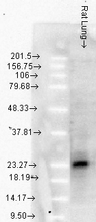
 WB (Western Blot)
(Western Blot analysis of Rat brain membrane lysate showing detection of VGLUT1 protein using Mouse Anti-VGLUT1 Monoclonal Antibody, Clone S28-9. Primary Antibody: Mouse Anti-VGLUT1 Monoclonal Antibody at 1:1000.)
WB (Western Blot)
(Western Blot analysis of Rat brain membrane lysate showing detection of VGLUT1 protein using Mouse Anti-VGLUT1 Monoclonal Antibody, Clone S28-9. Primary Antibody: Mouse Anti-VGLUT1 Monoclonal Antibody at 1:1000.)
VGLUT1, Monoclonal Antibody (Cat# AAA103038)
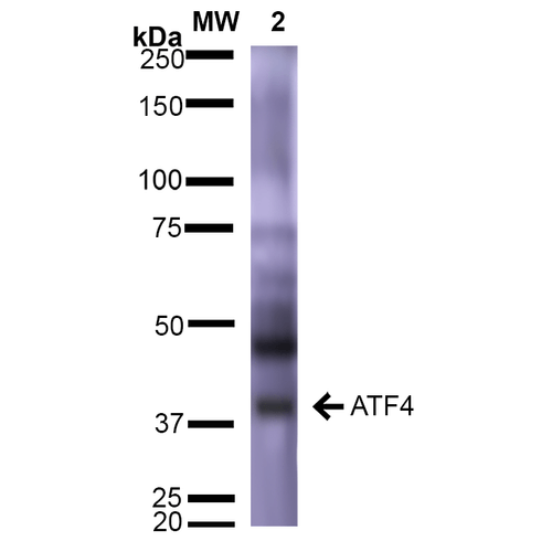 WB (Western Blot)
(Western Blot analysis of Rat Brain showing detection of ~39 kDa (isoform 2) ATF4 protein using Mouse Anti-ATF4 Monoclonal Antibody, Clone S360A-24 . Lane 1: Molecular Weight Ladder (MW). Lane 2: Rat Brain. Load: 15 ug. Block: 5% Skim Milk in 1X TBST. Primary Antibody: Mouse Anti-ATF4 Monoclonal Antibody at 1:1000 for 2 hours at RT. Secondary Antibody: Goat Anti-Mouse IgG: HRP at 1:2000 for 60 min at RT. Color Development: ECL solution for 5 min at RT. Predicted/Observed Size: ~39 kDa (isoform 2).)
WB (Western Blot)
(Western Blot analysis of Rat Brain showing detection of ~39 kDa (isoform 2) ATF4 protein using Mouse Anti-ATF4 Monoclonal Antibody, Clone S360A-24 . Lane 1: Molecular Weight Ladder (MW). Lane 2: Rat Brain. Load: 15 ug. Block: 5% Skim Milk in 1X TBST. Primary Antibody: Mouse Anti-ATF4 Monoclonal Antibody at 1:1000 for 2 hours at RT. Secondary Antibody: Goat Anti-Mouse IgG: HRP at 1:2000 for 60 min at RT. Color Development: ECL solution for 5 min at RT. Predicted/Observed Size: ~39 kDa (isoform 2).)
ATF4, Monoclonal Antibody (Cat# AAA103039)
 WB (Western Blot)
(Western Blot analysis of Human Cell lysates showing detection of Hsp27 protein using Mouse Anti-Hsp27 Monoclonal Antibody, Clone 5D12-A3. Load: 15 ug. Block: 1.5% BSA for 30 minutes at RT. Primary Antibody: Mouse Anti-Hsp27 Monoclonal Antibody at 1:1000 for 2 hours at RT. Secondary Antibody: Sheep Anti-Mouse IgG: HRP for 1 hour at RT.)
WB (Western Blot)
(Western Blot analysis of Human Cell lysates showing detection of Hsp27 protein using Mouse Anti-Hsp27 Monoclonal Antibody, Clone 5D12-A3. Load: 15 ug. Block: 1.5% BSA for 30 minutes at RT. Primary Antibody: Mouse Anti-Hsp27 Monoclonal Antibody at 1:1000 for 2 hours at RT. Secondary Antibody: Sheep Anti-Mouse IgG: HRP for 1 hour at RT.)
Hsp27, Monoclonal Antibody (Cat# AAA103041)
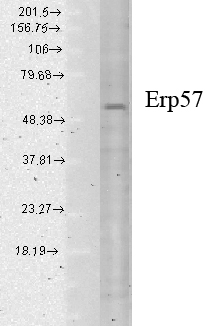
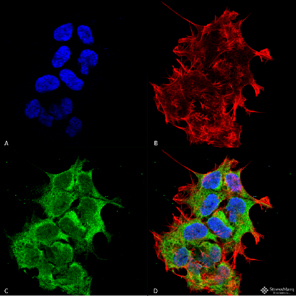
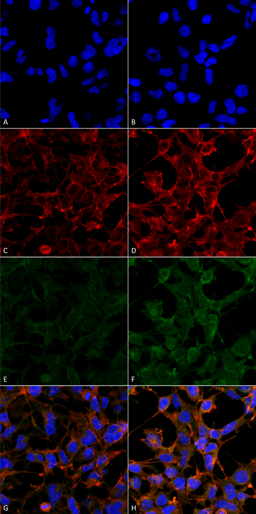
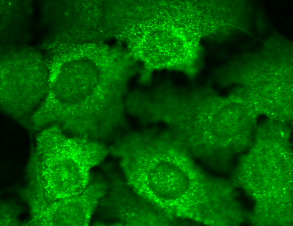
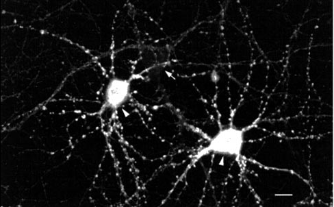
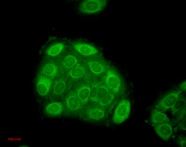
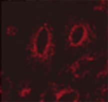
 ICC (Immunocytochemistry)
(Immunocytochemistry/Immunofluorescence analysis using Mouse Anti-Erp57 Monoclonal Antibody, Clone Map.ERP57. Tissue: HaCaT cells. Species: Human. Fixation: Cold 100% methanol for 10 minutes at -20 degree C. Primary Antibody: Mouse Anti-Erp57 Monoclonal Antibody at 1:100 for 1 hour at RT. Secondary Antibody: FITC Goat Anti-Mouse (green) at 1:50 for 1 hour at RT. Localization: Cytoplasmic and perinuclear staining.)
ICC (Immunocytochemistry)
(Immunocytochemistry/Immunofluorescence analysis using Mouse Anti-Erp57 Monoclonal Antibody, Clone Map.ERP57. Tissue: HaCaT cells. Species: Human. Fixation: Cold 100% methanol for 10 minutes at -20 degree C. Primary Antibody: Mouse Anti-Erp57 Monoclonal Antibody at 1:100 for 1 hour at RT. Secondary Antibody: FITC Goat Anti-Mouse (green) at 1:50 for 1 hour at RT. Localization: Cytoplasmic and perinuclear staining.)
Erp57 (Grp58), Monoclonal Antibody (Cat# AAA103051)
 WB (Western Blot)
(Western Blot analysis of Human Cell lysates showing detection of p38 MAPK protein using Mouse Anti-p38 MAPK Monoclonal Antibody, Clone 9F12. Load: 15 ug. Block: 1.5% BSA for 30 minutes at RT. Primary Antibody: Mouse Anti-p38 MAPK Monoclonal Antibody at 1:1000 for 2 hours at RT. Secondary Antibody: Sheep Anti-Mouse IgG: HRP for 1 hour at RT.)
WB (Western Blot)
(Western Blot analysis of Human Cell lysates showing detection of p38 MAPK protein using Mouse Anti-p38 MAPK Monoclonal Antibody, Clone 9F12. Load: 15 ug. Block: 1.5% BSA for 30 minutes at RT. Primary Antibody: Mouse Anti-p38 MAPK Monoclonal Antibody at 1:1000 for 2 hours at RT. Secondary Antibody: Sheep Anti-Mouse IgG: HRP for 1 hour at RT.)
p38 alpha, Monoclonal Antibody (Cat# AAA103059)
 WB (Western Blot)
(Western Blot analysis of Rat Brain Membrane showing detection of 36-38 kDa QKI (pan) protein using Mouse Anti-QKI (pan) Monoclonal Antibody, Clone S147-6. Lane 1: Molecular Weight Ladder. Lane 2: Rat Brain Membrane. Load: 15 ug. Block: 2% BSA and 2% Skim Milk in 1X TBST. Primary Antibody: Mouse Anti-QKI (pan) Monoclonal Antibody at 1:200 for 16 hours at 4 degree C. Secondary Antibody: Goat Anti-Mouse IgG: HRP at 1:1000 for 1 hour RT. Color Development: ECL solution for 6 min in RT. Predicted/Observed Size: 36-38 kDa.)
WB (Western Blot)
(Western Blot analysis of Rat Brain Membrane showing detection of 36-38 kDa QKI (pan) protein using Mouse Anti-QKI (pan) Monoclonal Antibody, Clone S147-6. Lane 1: Molecular Weight Ladder. Lane 2: Rat Brain Membrane. Load: 15 ug. Block: 2% BSA and 2% Skim Milk in 1X TBST. Primary Antibody: Mouse Anti-QKI (pan) Monoclonal Antibody at 1:200 for 16 hours at 4 degree C. Secondary Antibody: Goat Anti-Mouse IgG: HRP at 1:1000 for 1 hour RT. Color Development: ECL solution for 6 min in RT. Predicted/Observed Size: 36-38 kDa.)
Pan-QKI, Monoclonal Antibody (Cat# AAA103060)

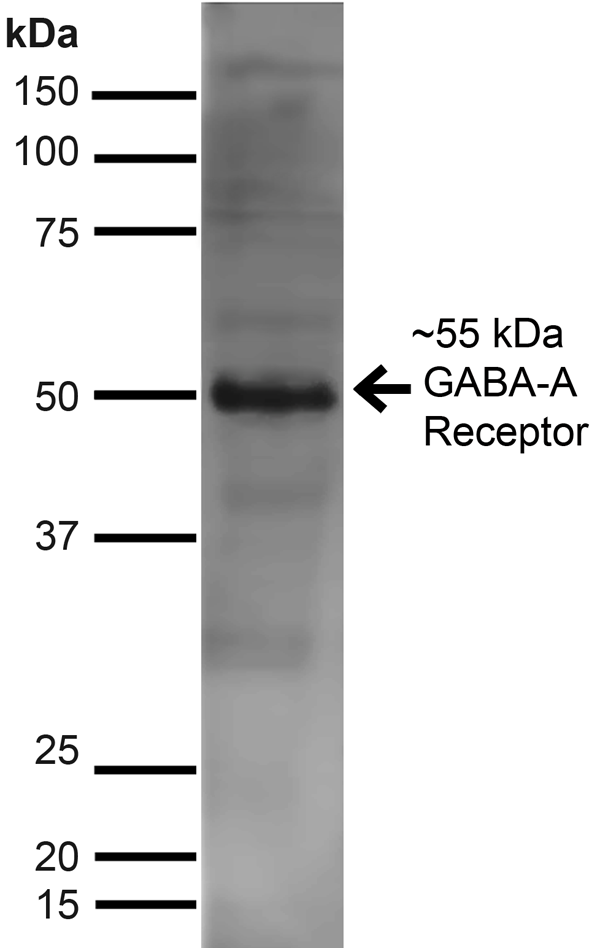
 WB (Western Blot)
(Western Blot analysis of Rat Cell line lysates showing detection of GABA A Receptor protein using Mouse Anti-GABA A Receptor Monoclonal Antibody, Clone S151-3. Load: 15 ug. Block: 1.5% BSA for 30 minutes at RT. Primary Antibody: Mouse Anti-GABA A Receptor Monoclonal Antibody at 1:1000 for 2 hours at RT. Secondary Antibody: Sheep Anti-Mouse IgG: HRP for 1 hour at RT.)
WB (Western Blot)
(Western Blot analysis of Rat Cell line lysates showing detection of GABA A Receptor protein using Mouse Anti-GABA A Receptor Monoclonal Antibody, Clone S151-3. Load: 15 ug. Block: 1.5% BSA for 30 minutes at RT. Primary Antibody: Mouse Anti-GABA A Receptor Monoclonal Antibody at 1:1000 for 2 hours at RT. Secondary Antibody: Sheep Anti-Mouse IgG: HRP for 1 hour at RT.)
GABA(A) Receptor Delta, Monoclonal Antibody (Cat# AAA103068)
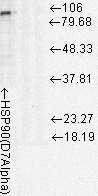 WB (Western Blot)
(Western Blot analysis of Rat cell lysates showing detection of Hsp90 protein using Mouse Anti-Hsp90 Monoclonal Antibody, Clone D7Alpha. Load: 15 ug. Block: 1.5% BSA for 30 minutes at RT. Primary Antibody: Mouse Anti-Hsp90 Monoclonal Antibody at 1:1000 for 2 hours at RT. Secondary Antibody: Sheep Anti-Mouse IgG: HRP for 1 hour at RT.)
WB (Western Blot)
(Western Blot analysis of Rat cell lysates showing detection of Hsp90 protein using Mouse Anti-Hsp90 Monoclonal Antibody, Clone D7Alpha. Load: 15 ug. Block: 1.5% BSA for 30 minutes at RT. Primary Antibody: Mouse Anti-Hsp90 Monoclonal Antibody at 1:1000 for 2 hours at RT. Secondary Antibody: Sheep Anti-Mouse IgG: HRP for 1 hour at RT.)
Hsp90, Monoclonal Antibody (Cat# AAA103087)
 Application Data
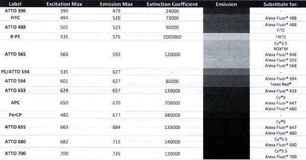
(HRP, Alkaline Phosphatase, Streptavidin and the following fluorescent conjugates are available, please inquire.)
Application Data
(HRP, Alkaline Phosphatase, Streptavidin and the following fluorescent conjugates are available, please inquire.)
GFAP, Monoclonal Antibody (Cat# AAA103262)
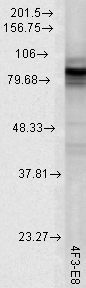
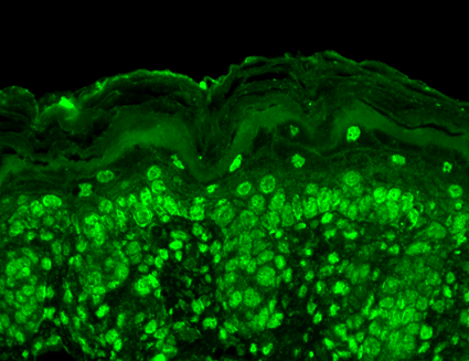
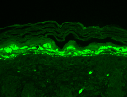
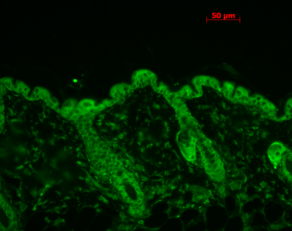 IHC (Immunohistochemistry)
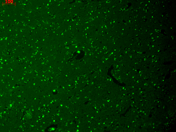
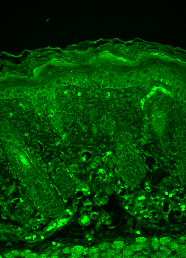
(Immunohistochemistry analysis using Mouse Anti-Hsp90 Monoclonal Antibody, Clone 4F3.E8. Tissue: backskin. Species: Mouse. Fixation: Bouin's Fixative and paraffin-embedded. Primary Antibody: Mouse Anti-Hsp90 Monoclonal Antibody at 1:100 for 1 hour at RT. Secondary Antibody: FITC Goat Anti-Mouse (green) at 1:50 for 1 hour at RT.)
IHC (Immunohistochemistry)
(Immunohistochemistry analysis using Mouse Anti-Hsp90 Monoclonal Antibody, Clone 4F3.E8. Tissue: backskin. Species: Mouse. Fixation: Bouin's Fixative and paraffin-embedded. Primary Antibody: Mouse Anti-Hsp90 Monoclonal Antibody at 1:100 for 1 hour at RT. Secondary Antibody: FITC Goat Anti-Mouse (green) at 1:50 for 1 hour at RT.)
Hsp90, Monoclonal Antibody (Cat# AAA103266)
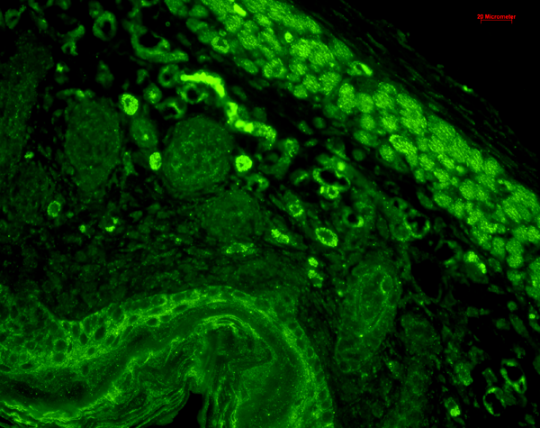
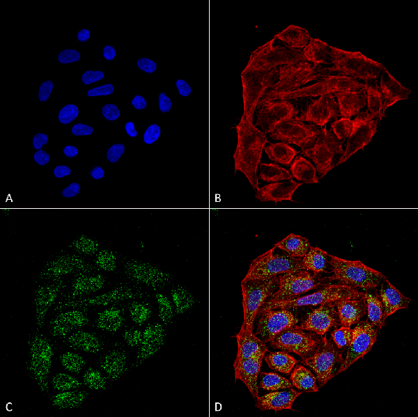
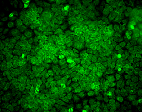
 ICC (Immunocytochemistry)
(Immunocytochemistry/Immunofluorescence analysis using Mouse Anti-CD74 Monoclonal Antibody, Clone PIN 1.1. Tissue: HaCaT cells. Species: Human. Fixation: Cold 100% methanol for 10 minutes at -20 degree C. Primary Antibody: Mouse Anti-CD74 Monoclonal Antibody at 1:100 for 1 hour at RT. Secondary Antibody: FITC Goat Anti-Mouse (green) at 1:50 for 1 hour at RT. Localization: Cytoplasmic Staining.)
ICC (Immunocytochemistry)
(Immunocytochemistry/Immunofluorescence analysis using Mouse Anti-CD74 Monoclonal Antibody, Clone PIN 1.1. Tissue: HaCaT cells. Species: Human. Fixation: Cold 100% methanol for 10 minutes at -20 degree C. Primary Antibody: Mouse Anti-CD74 Monoclonal Antibody at 1:100 for 1 hour at RT. Secondary Antibody: FITC Goat Anti-Mouse (green) at 1:50 for 1 hour at RT. Localization: Cytoplasmic Staining.)
CD74, Monoclonal Antibody (Cat# AAA103267)
 WB (Western Blot)
(Western Blot analysis of Rat brain membrane lysate showing detection of SHANK1 protein using Mouse Anti-SHANK1 Monoclonal Antibody, Clone S22-21. Load: 15 ug. Block: 1.5% BSA for 30 minutes at RT. Primary Antibody: Mouse Anti-SHANK1 Monoclonal Antibody at 1:1000 for 2 hours at RT. Secondary Antibody: Sheep Anti-Mouse IgG: HRP for 1 hour at RT.)
WB (Western Blot)
(Western Blot analysis of Rat brain membrane lysate showing detection of SHANK1 protein using Mouse Anti-SHANK1 Monoclonal Antibody, Clone S22-21. Load: 15 ug. Block: 1.5% BSA for 30 minutes at RT. Primary Antibody: Mouse Anti-SHANK1 Monoclonal Antibody at 1:1000 for 2 hours at RT. Secondary Antibody: Sheep Anti-Mouse IgG: HRP for 1 hour at RT.)
Shank1, Monoclonal Antibody (Cat# AAA103278)
 WB (Western Blot)
(Western Blot analysis of Rat Cell line lysates showing detection of GABA A Receptor protein using Mouse Anti-GABA A Receptor Monoclonal Antibody, Clone S151-3. Load: 15 ug. Block: 1.5% BSA for 30 minutes at RT. Primary Antibody: Mouse Anti-GABA A Receptor Monoclonal Antibody at 1:1000 for 2 hours at RT. Secondary Antibody: Sheep Anti-Mouse IgG: HRP for 1 hour at RT.)
WB (Western Blot)
(Western Blot analysis of Rat Cell line lysates showing detection of GABA A Receptor protein using Mouse Anti-GABA A Receptor Monoclonal Antibody, Clone S151-3. Load: 15 ug. Block: 1.5% BSA for 30 minutes at RT. Primary Antibody: Mouse Anti-GABA A Receptor Monoclonal Antibody at 1:1000 for 2 hours at RT. Secondary Antibody: Sheep Anti-Mouse IgG: HRP for 1 hour at RT.)
GABA(A) Receptor Delta, Monoclonal Antibody (Cat# AAA103291)
 WB (Western Blot)
(Western Blot analysis of Rat liver microsome lysate showing detection of LAMP1 protein using Mouse Anti-LAMP1 Monoclonal Antibody, Clone Ly1C6. Load: 15 ug. Block: 1.5% BSA for 30 minutes at RT. Primary Antibody: Mouse Anti-LAMP1 Monoclonal Antibody at 1:1000 for 2 hours at RT. Secondary Antibody: Sheep Anti-Mouse IgG: HRP for 1 hour at RT.)
WB (Western Blot)
(Western Blot analysis of Rat liver microsome lysate showing detection of LAMP1 protein using Mouse Anti-LAMP1 Monoclonal Antibody, Clone Ly1C6. Load: 15 ug. Block: 1.5% BSA for 30 minutes at RT. Primary Antibody: Mouse Anti-LAMP1 Monoclonal Antibody at 1:1000 for 2 hours at RT. Secondary Antibody: Sheep Anti-Mouse IgG: HRP for 1 hour at RT.)
LAMP1, Monoclonal Antibody (Cat# AAA103297)
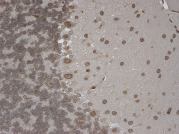
 IHC (Immunohistochemistry)
(Immunohistochemistry analysis using Mouse Anti-HCN1 Monoclonal Antibody, Clone S70-28. Tissue: Frozen brain section. Species: Mouse. Fixation: 10% Formalin Solution for 12-24 hours at RT. Primary Antibody: Mouse Anti-HCN1 Monoclonal Antibody at 1:1000 for 1 hour at RT. Secondary Antibody: HRP/DAB Detection System: Biotinylated Goat Anti-Mouse, Streptavidin Peroxidase, DAB Chromogen (brown) for 30 minutes at RT. Counterstain: Mayer Hematoxylin (purple/blue) nuclear stain at 250-500 ul for 5 minutes at RT.)
IHC (Immunohistochemistry)
(Immunohistochemistry analysis using Mouse Anti-HCN1 Monoclonal Antibody, Clone S70-28. Tissue: Frozen brain section. Species: Mouse. Fixation: 10% Formalin Solution for 12-24 hours at RT. Primary Antibody: Mouse Anti-HCN1 Monoclonal Antibody at 1:1000 for 1 hour at RT. Secondary Antibody: HRP/DAB Detection System: Biotinylated Goat Anti-Mouse, Streptavidin Peroxidase, DAB Chromogen (brown) for 30 minutes at RT. Counterstain: Mayer Hematoxylin (purple/blue) nuclear stain at 250-500 ul for 5 minutes at RT.)
HCN1, Monoclonal Antibody (Cat# AAA103316)
 WB (Western Blot)
(Western Blot analysis of Human Cell lysates showing detection of Rhodopsin protein using Mouse Anti-Rhodopsin Monoclonal Antibody, Clone 1D4. Load: 15 ug. Block: 1.5% BSA for 30 minutes at RT. Primary Antibody: Mouse Anti-Rhodopsin Monoclonal Antibody at 1:1000 for 2 hours at RT. Secondary Antibody: Sheep Anti-Mouse IgG: HRP for 1 hour at RT.)
WB (Western Blot)
(Western Blot analysis of Human Cell lysates showing detection of Rhodopsin protein using Mouse Anti-Rhodopsin Monoclonal Antibody, Clone 1D4. Load: 15 ug. Block: 1.5% BSA for 30 minutes at RT. Primary Antibody: Mouse Anti-Rhodopsin Monoclonal Antibody at 1:1000 for 2 hours at RT. Secondary Antibody: Sheep Anti-Mouse IgG: HRP for 1 hour at RT.)
Rhodopsin, Monoclonal Antibody (Cat# AAA103091)

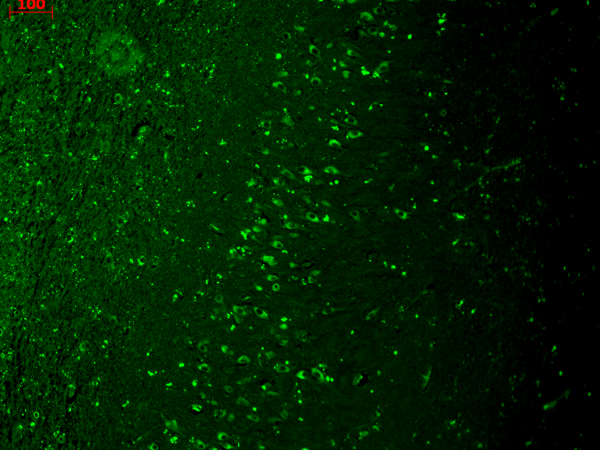
 IHC (Immunohiostchemistry)
(Immunohistochemistry analysis using Mouse Anti-CaV3.2 Calcium channel Monoclonal Antibody, Clone S55-10. Tissue: frozen brain section. Species: human. Fixation: 10% Formalin Solution for 12-24 hours at RT. Primary Antibody: Mouse Anti-CaV3.2 Calcium channel Monoclonal Antibody at 1:1000 for 1 hour at RT. Secondary Antibody: HRP/DAB Detection System: Biotinylated Goat Anti-Mouse, Streptavidin Peroxidase, DAB Chromogen (brown) for 30 minutes at RT. Counterstain: Mayer Hematoxylin (purple/blue) nuclear stain at 250-500 ul for 5 minutes at RT.)
IHC (Immunohiostchemistry)
(Immunohistochemistry analysis using Mouse Anti-CaV3.2 Calcium channel Monoclonal Antibody, Clone S55-10. Tissue: frozen brain section. Species: human. Fixation: 10% Formalin Solution for 12-24 hours at RT. Primary Antibody: Mouse Anti-CaV3.2 Calcium channel Monoclonal Antibody at 1:1000 for 1 hour at RT. Secondary Antibody: HRP/DAB Detection System: Biotinylated Goat Anti-Mouse, Streptavidin Peroxidase, DAB Chromogen (brown) for 30 minutes at RT. Counterstain: Mayer Hematoxylin (purple/blue) nuclear stain at 250-500 ul for 5 minutes at RT.)
Cav3.2, Monoclonal Antibody (Cat# AAA103099)

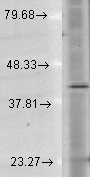
 WB (Western Blot)
(Western Blot analysis of Rat brain membrane lysate showing detection of GluN2B/NR2B protein using Mouse Anti-GluN2B/NR2B Monoclonal Antibody, Clone S59-36. Load: 15 ug. Block: 1.5% BSA for 30 minutes at RT. Primary Antibody: Mouse Anti-GluN2B/NR2B Monoclonal Antibody at 1:1000 for 2 hours at RT. Secondary Antibody: Sheep Anti-Mouse IgG: HRP for 1 hour at RT.)
WB (Western Blot)
(Western Blot analysis of Rat brain membrane lysate showing detection of GluN2B/NR2B protein using Mouse Anti-GluN2B/NR2B Monoclonal Antibody, Clone S59-36. Load: 15 ug. Block: 1.5% BSA for 30 minutes at RT. Primary Antibody: Mouse Anti-GluN2B/NR2B Monoclonal Antibody at 1:1000 for 2 hours at RT. Secondary Antibody: Sheep Anti-Mouse IgG: HRP for 1 hour at RT.)
NR2B, Monoclonal Antibody (Cat# AAA103100)
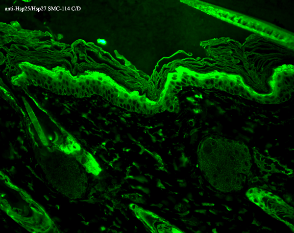

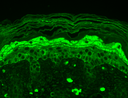
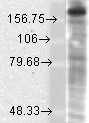
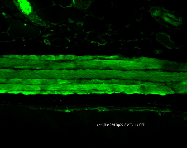 IHC (Immunohistochemistry)
(Immunohistochemistry analysis using Mouse Anti-Hsp27 Monoclonal Antibody, Clone 8A7. Tissue: backskin. Species: Mouse. Fixation: Bouin's Fixative and paraffin-embedded. Primary Antibody: Mouse Anti-Hsp27 Monoclonal Antibody at 1:100 for 1 hour at RT. Secondary Antibody: FITC Goat Anti-Mouse (green) at 1:50 for 1 hour at RT. Localization: Epidermis.)
IHC (Immunohistochemistry)
(Immunohistochemistry analysis using Mouse Anti-Hsp27 Monoclonal Antibody, Clone 8A7. Tissue: backskin. Species: Mouse. Fixation: Bouin's Fixative and paraffin-embedded. Primary Antibody: Mouse Anti-Hsp27 Monoclonal Antibody at 1:100 for 1 hour at RT. Secondary Antibody: FITC Goat Anti-Mouse (green) at 1:50 for 1 hour at RT. Localization: Epidermis.)
Hsp25/Hsp27, Monoclonal Antibody (Cat# AAA103122)
What are Monoclonal Antibodies?
Monoclonal antibodies are specialized laboratory-produced proteins developed for binding to specific biological antigens or other molecular targets. Since they come from a single cell (or clone), they are especially consistent and accurate in the data they are involved in producing.
This type of antibody material has been shown to be a powerful tool in finding and subsequently destroying harmful cells in an organism, such as those found in cancers or various autoimmune diseases. This makes them excellent aids in medical testing and research, which is why they are so widely used.
AAA Biotech offers a comprehensive range of high-quality monoclonal antibodies that perform effectively in various laboratory tests, including (amongst others) ELISA, western blotting, immunohistochemistry, and flow cytometry. All of the products in our catalog are thoroughly quality tested to make sure that they are reliable and will consistently perform well in your research.
What Are The Uses of Monoclonal Antibodies
Monoclonal antibodies are used in many lab tests, including (amongst others) ELISA, western blotting, immunohistochemistry, and flow cytometry.
ELISA is a test that helps detect a specific substance/analyte in a sample. It uses antibodies (often monoclonal) bound to a solid surface (such as the well of a microplate) to “capture” the substance/analyte in the sample and immobilize it so that the detection antibody component can then bind to it and produce a signal, which can then be measured.
Western blotting identifies specific proteins in a sample. The sample is first separated on a gel, and then antibodies are applied that will typically bind to the target, which will all be localized to a single band in a lane.
Immunohistochemistry helps locate specific proteins in cells or tissue samples using antibodies.
Flow cytometry looks at and sorts cells. It uses antibodies that are conjugated to reporter molecules called “fluorophores”, which, under special lights, emit light themselves, which can then be measured by a detector instrument.
How Monoclonal Antibodies Are Used as Medicine?
Please note that all of the products listed in AAA Biotech’s also known as AAA Bio or AAABio catalog are strictly for research-use only (RUO).
Monoclonal antibodies can also be used as therapeutic/medical treatments, particularly in the context of cancers. They are designed to find and bind to specific cells or proteins, helping the immune system recognize and attack the cancer. These treatments work in different ways, such as:
- Radioimmunotherapy attaches a small amount of radioactive molecule to the antibody, so it delivers the radiation directly to the cancer cells that the antibody is specifically binding to.
- Antibody-directed enzyme prodrug therapy uses antibodies that are specifically bound to special enzymes. These enzymes activate a harmless drug in the body and turn it into a cancer-killing drug only near the cancer cells—this helps avoid harming healthy cells.
- Immunoliposomes are tiny “bubbles” filled with medicine/drug and coated with antibodies. They carry the drug straight to the cancer cells.
Why Buy Monoclonal Antibodies From Us?
At AAA Biotech, we provide high-performance monoclonal antibodies designed to support a wide range of research needs.
1. Validated for Versatile Applications
The antibodies in our catalog are extensively validated and compatible with multiple techniques, including (but not limited to) ELISA, flow cytometry (FC), immunocytochemistry (ICC), immunofluorescence (IF), immunohistochemistry (IHC), immunoprecipitation (IP), and western blotting (WB).
2. Wide Selection & Specialized Options
We offer antibodies for common and rare species, that are available in various conjugated forms, and also in recombinant formats. Essentially, there is almost anything one might need to meet their experimental model’s requirements.
3. High-Quality Proteins
Our proteins meet high purity standards—90% or more as confirmed by SDS-PAGE. Many are available with tags like His, Flag, GST, or MBP, and we also supply native and biologically active proteins for functional studies.
Frequently Asked Questions
1. Are your monoclonal antibodies validated for specific applications?
Yes, our antibodies are tested and validated for use in methods such as ELISA, western blot, IHC, flow cytometry, and more. Refer to specific product pages or datasheets for individual product information.
2. How do I choose the right monoclonal antibody for my application?
Review the product details directly for application validation, species reactivity, and target information. You may also contact our support team at any time for help.
3. How quickly can I receive my order?
Most orders are processed and shipped within 1–3 business days, depending on product availability and your shipping location.