Filters
▼Clonality
▼Type
▼Reactivity
▼Gene Name
▼Isotype
▼Host
▼Application
▼Clone
▼Monoclonal Antibodies
Get accurate results in your research with our Monoclonal Antibodies, which are specially made to target exactly what you require for your research, and will produce consistent, reliable performance in lab tests.
Viewing 7600-7650 of 27597 product results
 WB (Western Blot)
(COLO 320 cells were subjected to SDS PAGE followed by western blot with AAA248093 (ST6GALNAC6 Antibody) at dilution of 1:500)
WB (Western Blot)
(COLO 320 cells were subjected to SDS PAGE followed by western blot with AAA248093 (ST6GALNAC6 Antibody) at dilution of 1:500)
ST6GALNAC6, Monoclonal Antibody (Cat# AAA248093)
Protein A+G purification
 WB (Western Blot)
(HEK-293 cells were subjected to SDS PAGE followed by western blot with AAA248094 (STC2 antibody) at dilution of 1:1000)
WB (Western Blot)
(HEK-293 cells were subjected to SDS PAGE followed by western blot with AAA248094 (STC2 antibody) at dilution of 1:1000)
Stanniocalcin 2, Monoclonal Antibody (Cat# AAA248094)
Protein A+G purification
 WB (Western Blot)
(SH-SY5Y cells were subjected to SDS PAGE followed by western blot with AAA248126 (TTL antibody) at dilution of 1:500)
WB (Western Blot)
(SH-SY5Y cells were subjected to SDS PAGE followed by western blot with AAA248126 (TTL antibody) at dilution of 1:500)
TTL, Monoclonal Antibody (Cat# AAA248126)
Protein A+G purification
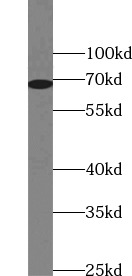
 WB (Western Blot)
(Jurkat cells were subjected to SDS PAGE followed by western blot with AAA247956 (IL2 Antibody) at dilution of 1:1000)
WB (Western Blot)
(Jurkat cells were subjected to SDS PAGE followed by western blot with AAA247956 (IL2 Antibody) at dilution of 1:1000)
IL2, Monoclonal Antibody (Cat# AAA247956)
Protein A+G purification

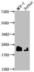
 WB (Western Blot)
(IL27 fusion protein were subjected to SDS PAGE followed by western blot with AAA247959 (IL27 Antibody) at dilution of 1:5000)
WB (Western Blot)
(IL27 fusion protein were subjected to SDS PAGE followed by western blot with AAA247959 (IL27 Antibody) at dilution of 1:5000)
IL-27, Monoclonal Antibody (Cat# AAA247959)
Protein A+G purification
 WB (Western Blot)
(human blood tissue were subjected to SDS PAGE followed by western blot with AAA247978 (LGALS3BP Antibody) at dilution of 1:2000)
WB (Western Blot)
(human blood tissue were subjected to SDS PAGE followed by western blot with AAA247978 (LGALS3BP Antibody) at dilution of 1:2000)
LGALS3BP, Monoclonal Antibody (Cat# AAA247978)
Protein A+G purification
 WB (Western Blot)
(pig brain tissue were subjected to SDS PAGE followed by western blot with AAA248013 (NF200 Antibody) at dilution of 1:4000)
WB (Western Blot)
(pig brain tissue were subjected to SDS PAGE followed by western blot with AAA248013 (NF200 Antibody) at dilution of 1:4000)
NF200, Monoclonal Antibody (Cat# AAA248013)
Protein A+G purification
 WB (Western Blot)
(HeLa cells were subjected to SDS PAGE followed by western blot with AAA247938 (HSP70 Antibody) at dilution of 1:10000)
WB (Western Blot)
(HeLa cells were subjected to SDS PAGE followed by western blot with AAA247938 (HSP70 Antibody) at dilution of 1:10000)
HSP70, Monoclonal Antibody (Cat# AAA247938)
Protein A+G purification
 WB (Western Blot)
(NCCIT cell were subjected to SDS PAGE followed by western blot with AAA247947 (IFT88 Antibody) at dilution of 1:4000)
WB (Western Blot)
(NCCIT cell were subjected to SDS PAGE followed by western blot with AAA247947 (IFT88 Antibody) at dilution of 1:4000)
IFT88, Monoclonal Antibody (Cat# AAA247947)
Protein A+G purification
 WB (Western Blot)
(fetal human brain tissue were subjected to SDS PAGE followed by western blot with AAA248130 (UCHL1 Antibody) at dilution of 1:20000)
WB (Western Blot)
(fetal human brain tissue were subjected to SDS PAGE followed by western blot with AAA248130 (UCHL1 Antibody) at dilution of 1:20000)
UCHL1, Monoclonal Antibody (Cat# AAA248130)
Protein A+G purification
 WB (Western Blot)
(HepG2 cells were subjected to SDS PAGE followed by western blot with AAA248131 (USP1 Antibody) at dilution of 1:2000)
WB (Western Blot)
(HepG2 cells were subjected to SDS PAGE followed by western blot with AAA248131 (USP1 Antibody) at dilution of 1:2000)
USP1, Monoclonal Antibody (Cat# AAA248131)
Protein A+G purification
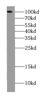
 WB (Western Blot)
(Jurkat cells were subjected to SDS PAGE followed by western blot with AAA248138 (ZAP70 antibody) at dilution of 1:3000)
WB (Western Blot)
(Jurkat cells were subjected to SDS PAGE followed by western blot with AAA248138 (ZAP70 antibody) at dilution of 1:3000)
ZAP70, Monoclonal Antibody (Cat# AAA248138)
Protein A+G purification
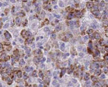
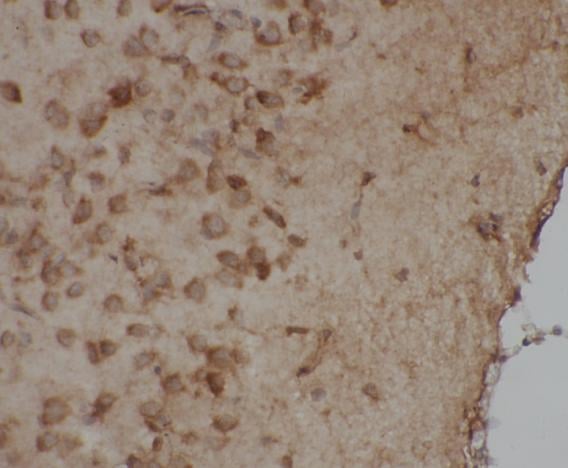
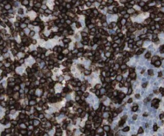
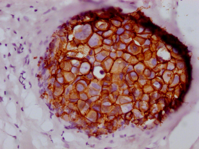
 IHC (Immunohiostchemistry)
(IHC image diluted at 1:100 and staining in paraffin-embedded human tonsil tissue performed on a Leica BondTM system. After dewaxing and hydration, antigen retrieval was mediated by high pressure in a citrate buffer (pH 6.0). Section was blocked with 10% normal goat serum 30min at RT. Then primary antibody (1% BSA) was incubated at 4 degree C overnight. The primary is detected by a Goat anti-rabbit IgG polymer labeled by HRP and visualized using 0.05% DAB.)
IHC (Immunohiostchemistry)
(IHC image diluted at 1:100 and staining in paraffin-embedded human tonsil tissue performed on a Leica BondTM system. After dewaxing and hydration, antigen retrieval was mediated by high pressure in a citrate buffer (pH 6.0). Section was blocked with 10% normal goat serum 30min at RT. Then primary antibody (1% BSA) was incubated at 4 degree C overnight. The primary is detected by a Goat anti-rabbit IgG polymer labeled by HRP and visualized using 0.05% DAB.)
MGMT, Monoclonal Recombinant Antibody (Cat# AAA244000)
 FCM/FACS (Flow Cytometry)
(Overlay histogram showing 293 cells stained with (red line) at 1?50. The cells were fixed with 70% Ethylalcohol (18h) and then incubated in 10% normal goat serum to block non-specific protein-protein interactions followedby the antibody (1ug/1*106cells) for 1 h at 4?.The secondary antibody used was FITC-conjugated goat anti-rabbit IgG (H+L) at 1/200 dilution for 30min at 4?. Control antibody (green line) was Rabbit IgG (1ug/1*106cells) used under the same conditions. Acquisition of >10,000 events was performed.)
FCM/FACS (Flow Cytometry)
(Overlay histogram showing 293 cells stained with (red line) at 1?50. The cells were fixed with 70% Ethylalcohol (18h) and then incubated in 10% normal goat serum to block non-specific protein-protein interactions followedby the antibody (1ug/1*106cells) for 1 h at 4?.The secondary antibody used was FITC-conjugated goat anti-rabbit IgG (H+L) at 1/200 dilution for 30min at 4?. Control antibody (green line) was Rabbit IgG (1ug/1*106cells) used under the same conditions. Acquisition of >10,000 events was performed.)
SDHB, Monoclonal Recombinant Antibody (Cat# AAA244014)
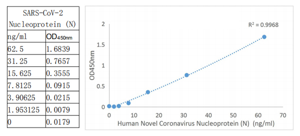 ELISA
(It is a solid phase sandwich Enzyme Linked-Immuno-Sorbent Assay (Sandwich ELISA). An antibody specific for SARS-CoV-2 Nucleoprotein (N) has been pre-coated onto the microwells. The SARS-CoV-2 Nucleoprotein (N) protein in samples is captured by the coated antibody after incubation. Following extensive washing, another antibody Biotin conjugated specific for SARS-CoV-2 Nucleoprotein (N) is added to detect the captured SARS-CoV-2 Nucleoprotein (N) protein. Followed by Tetramethyl-benzidine (TMB) reagent. Solution containing sulfuric acid is used to stop color development and the color intensity which is proportional to the quantity of bound protein is measurable at 450nm.)
ELISA
(It is a solid phase sandwich Enzyme Linked-Immuno-Sorbent Assay (Sandwich ELISA). An antibody specific for SARS-CoV-2 Nucleoprotein (N) has been pre-coated onto the microwells. The SARS-CoV-2 Nucleoprotein (N) protein in samples is captured by the coated antibody after incubation. Following extensive washing, another antibody Biotin conjugated specific for SARS-CoV-2 Nucleoprotein (N) is added to detect the captured SARS-CoV-2 Nucleoprotein (N) protein. Followed by Tetramethyl-benzidine (TMB) reagent. Solution containing sulfuric acid is used to stop color development and the color intensity which is proportional to the quantity of bound protein is measurable at 450nm.)
COVID 19 Nucleocapsid (NP) Coronavirus, Monoclonal Antibody Pair Kit (Cat# AAA244026)
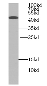
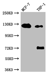
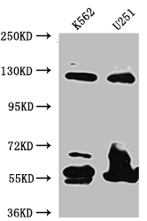
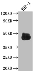 WB (Western Blot)
(Western BlotPositive WB detected in: THP-1 whole cell lysateAll lanes: CD32 antibody at 1:1000SecondaryGoat polyclonal to rabbit IgG at 1/50000 dilutionPredicted band size: 35, 36 kDaObserved band size: 45 kDa)
WB (Western Blot)
(Western BlotPositive WB detected in: THP-1 whole cell lysateAll lanes: CD32 antibody at 1:1000SecondaryGoat polyclonal to rabbit IgG at 1/50000 dilutionPredicted band size: 35, 36 kDaObserved band size: 45 kDa)
FCGR2A, Monoclonal Recombinant Antibody (Cat# AAA243816)


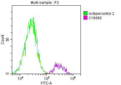
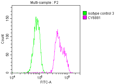
 FCM/FACS (Flow Cytometry)
(Overlay histogram showing Hela cells stained with (red line) at 1?50. The cells were fixed with 70% Ethylalcohol (18h) and then incubated in 10% normal goat serum to block non-specific protein-protein interactions followedby the antibody (1ug/1*106cells) for 1 h at 4?.The secondary antibody used was FITC-conjugated goat anti-rabbit IgG (H+L) at 1/200 dilution for 30min at 4?. Control antibody (green line) was Rabbit IgG (1ug/1*106cells) used under the same conditions. Acquisition of >10,000 events was performed.)
FCM/FACS (Flow Cytometry)
(Overlay histogram showing Hela cells stained with (red line) at 1?50. The cells were fixed with 70% Ethylalcohol (18h) and then incubated in 10% normal goat serum to block non-specific protein-protein interactions followedby the antibody (1ug/1*106cells) for 1 h at 4?.The secondary antibody used was FITC-conjugated goat anti-rabbit IgG (H+L) at 1/200 dilution for 30min at 4?. Control antibody (green line) was Rabbit IgG (1ug/1*106cells) used under the same conditions. Acquisition of >10,000 events was performed.)
DNM2, Monoclonal Recombinant Antibody (Cat# AAA243968)
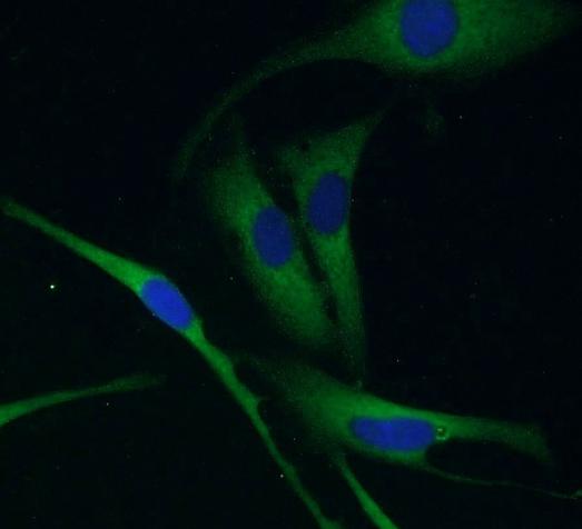
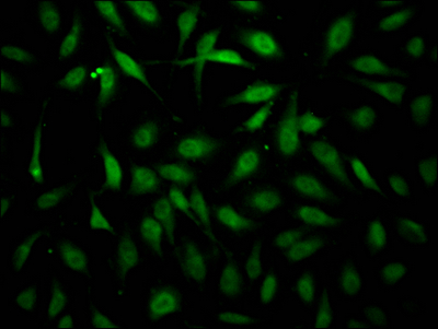
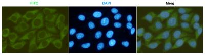
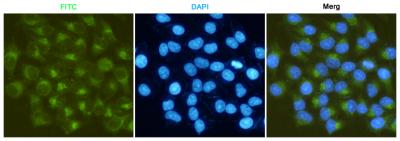
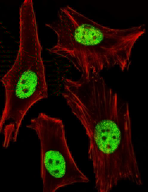
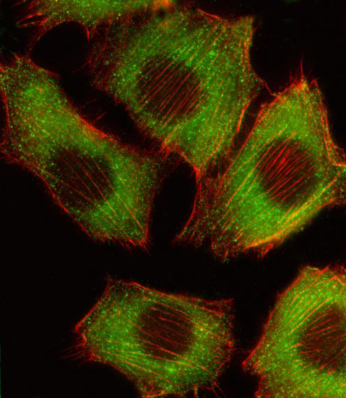
 IF (Immunofluorescence)
(Immunofluorescence staining of Hela Cells at 1?50, counter-stained with DAPI. The cells were fixed in 4% formaldehyde, permeated by 0.2% TritonX-100, and blocked in 10% normal Goat Serum. The cells were then incubated with the antibody overnight at 4 degree C. Nuclear DNA was labeled in blue with DAPI. The secondary antibody was FITC-conjugated AffiniPure Goat Anti-Rabbit IgG ?H+L?.)
IF (Immunofluorescence)
(Immunofluorescence staining of Hela Cells at 1?50, counter-stained with DAPI. The cells were fixed in 4% formaldehyde, permeated by 0.2% TritonX-100, and blocked in 10% normal Goat Serum. The cells were then incubated with the antibody overnight at 4 degree C. Nuclear DNA was labeled in blue with DAPI. The secondary antibody was FITC-conjugated AffiniPure Goat Anti-Rabbit IgG ?H+L?.)
EIF4A1, Monoclonal Recombinant Antibody (Cat# AAA243829)
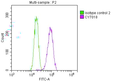
 FCM/FACS (Flow Cytometry)
(Overlay histogram showing Jurkat cells stained with (red line) at 1?50. The cells were fixed with 70% Ethylalcohol (18h) and then incubated in 10% normal goat serum to block non-specific protein-protein interactions followedby the antibody (1ug/1*106cells) for 1 h at 4?.The secondary antibody used was FITC-conjugated goat anti-rabbit IgG (H+L) at 1/200 dilution for 30min at 4?. Control antibody (green line) was Rabbit IgG (1ug/1*106cells) used under the same conditions. Acquisition of >10,000 events was performed.)
FCM/FACS (Flow Cytometry)
(Overlay histogram showing Jurkat cells stained with (red line) at 1?50. The cells were fixed with 70% Ethylalcohol (18h) and then incubated in 10% normal goat serum to block non-specific protein-protein interactions followedby the antibody (1ug/1*106cells) for 1 h at 4?.The secondary antibody used was FITC-conjugated goat anti-rabbit IgG (H+L) at 1/200 dilution for 30min at 4?. Control antibody (green line) was Rabbit IgG (1ug/1*106cells) used under the same conditions. Acquisition of >10,000 events was performed.)
ELANE, Monoclonal Recombinant Antibody (Cat# AAA243835)
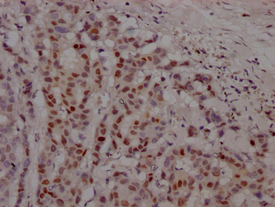
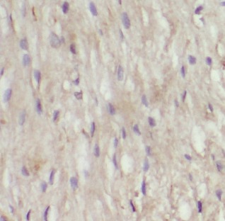
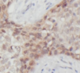
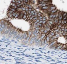
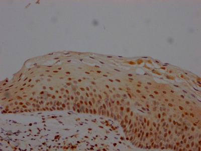
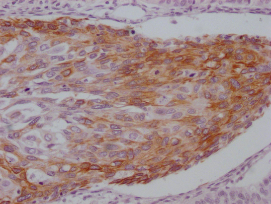
 IHC (Immunohistochemisry)
(IHC image diluted at 1:100 and staining in paraffin-embedded human prostate cancer performed on a Leica BondTM system. After dewaxing and hydration, antigen retrieval was mediated by high pressure in a citrate buffer (pH 6.0). Section was blocked with 10% normal goat serum 30min at RT. Then primary antibody (1% BSA) was incubated at 4 degree C overnight. The primary is detected by a Goat anti-rabbit IgG polymer labeled by HRP and visualized using 0.05% DAB.)
IHC (Immunohistochemisry)
(IHC image diluted at 1:100 and staining in paraffin-embedded human prostate cancer performed on a Leica BondTM system. After dewaxing and hydration, antigen retrieval was mediated by high pressure in a citrate buffer (pH 6.0). Section was blocked with 10% normal goat serum 30min at RT. Then primary antibody (1% BSA) was incubated at 4 degree C overnight. The primary is detected by a Goat anti-rabbit IgG polymer labeled by HRP and visualized using 0.05% DAB.)
AR, Monoclonal Recombinant Antibody (Cat# AAA243842)
 IF (Immunofluorescence)
(Immunofluorescence staining of Hela Cells at 1?50, counter-stained with DAPI. The cells were fixed in 4% formaldehyde, permeated by 0.2% TritonX-100, and blocked in 10% normal Goat Serum. The cells were then incubated with the antibody overnight at 4 degree C. Nuclear DNA was labeled in blue with DAPI. The secondary antibody was FITC-conjugated AffiniPure Goat Anti-Rabbit IgG ?H+L?.)
IF (Immunofluorescence)
(Immunofluorescence staining of Hela Cells at 1?50, counter-stained with DAPI. The cells were fixed in 4% formaldehyde, permeated by 0.2% TritonX-100, and blocked in 10% normal Goat Serum. The cells were then incubated with the antibody overnight at 4 degree C. Nuclear DNA was labeled in blue with DAPI. The secondary antibody was FITC-conjugated AffiniPure Goat Anti-Rabbit IgG ?H+L?.)
ERBB2, Monoclonal Recombinant Antibody (Cat# AAA243876)
 FCM/FACS (Flow Cytometry)
(Overlay histogram showing Hela cells stained with (red line) at 1?50. The cells were fixed with 70% Ethylalcohol (18h) and then incubated in 10% normal goat serum to block non-specific protein-protein interactions followedby the antibody (1ug/1*106cells) for 1 h at 4?.The secondary antibody used was FITC-conjugated goat anti-rabbit IgG (H+L) at 1/200 dilution for 30min at 4?. Control antibody (green line) was Rabbit IgG (1ug/1*106cells) used under the same conditions. Acquisition of >10,000 events was performed.)
FCM/FACS (Flow Cytometry)
(Overlay histogram showing Hela cells stained with (red line) at 1?50. The cells were fixed with 70% Ethylalcohol (18h) and then incubated in 10% normal goat serum to block non-specific protein-protein interactions followedby the antibody (1ug/1*106cells) for 1 h at 4?.The secondary antibody used was FITC-conjugated goat anti-rabbit IgG (H+L) at 1/200 dilution for 30min at 4?. Control antibody (green line) was Rabbit IgG (1ug/1*106cells) used under the same conditions. Acquisition of >10,000 events was performed.)
BUB1B, Monoclonal Recombinant Antibody (Cat# AAA243880)
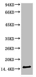 WB (Western Blot)
(Western blot analysis of Human Serum using TTR Mouse mAb diluted at 1:2000.)
WB (Western Blot)
(Western blot analysis of Human Serum using TTR Mouse mAb diluted at 1:2000.)
TTR, Monoclonal Antibody (Cat# AAA243669)
 WB (Western Blot)
(Western blot analysis of Human Serum using TTR Mouse mAb diluted at 1:2000)
WB (Western Blot)
(Western blot analysis of Human Serum using TTR Mouse mAb diluted at 1:2000)
TTR, Monoclonal Antibody (Cat# AAA243673)

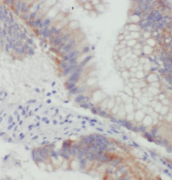
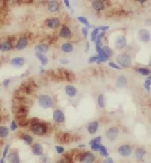
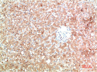
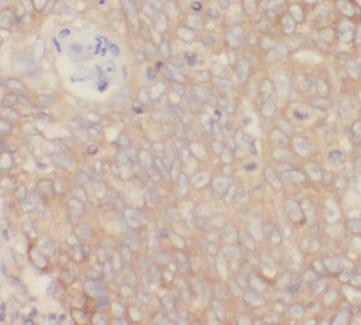
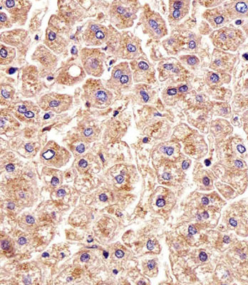
 IHC (Immunohiostchemistry)
(IHC image of AAA243699 diluted at 1:100 and staining in paraffin-embedded human liver cancer performed on a Leica BondTM system. After dewaxing and hydration, antigen retrieval was mediated by high pressure in a citrate buffer (pH 6.0). Section was blocked with 10% normal goat serum 30min at RT. Then primary antibody (1% BSA) was incubated at 4 degree C overnight. The primary is detected by a Goat anti-mouse IgG polymer labeled by HRP and visualized using 0.05% DAB.)
IHC (Immunohiostchemistry)
(IHC image of AAA243699 diluted at 1:100 and staining in paraffin-embedded human liver cancer performed on a Leica BondTM system. After dewaxing and hydration, antigen retrieval was mediated by high pressure in a citrate buffer (pH 6.0). Section was blocked with 10% normal goat serum 30min at RT. Then primary antibody (1% BSA) was incubated at 4 degree C overnight. The primary is detected by a Goat anti-mouse IgG polymer labeled by HRP and visualized using 0.05% DAB.)
ARG1, Monoclonal Antibody (Cat# AAA243699)
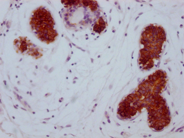
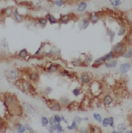
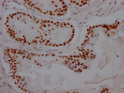
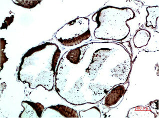
 IHC (Immunohiostchemistry)
(IHC image of AAA243706 diluted at 1:100 and staining in paraffin-embedded human cervical cancer performed on a Leica BondTM system. After dewaxing and hydration, antigen retrieval was mediated by high pressure in a citrate buffer (pH 6.0). Section was blocked with 10% normal goat serum 30min at RT. Then primary antibody (1% BSA) was incubated at 4 degree C overnight. The primary is detected by a Goat anti-mouse IgG polymer labeled by HRP and visualized using 0.05% DAB.)
IHC (Immunohiostchemistry)
(IHC image of AAA243706 diluted at 1:100 and staining in paraffin-embedded human cervical cancer performed on a Leica BondTM system. After dewaxing and hydration, antigen retrieval was mediated by high pressure in a citrate buffer (pH 6.0). Section was blocked with 10% normal goat serum 30min at RT. Then primary antibody (1% BSA) was incubated at 4 degree C overnight. The primary is detected by a Goat anti-mouse IgG polymer labeled by HRP and visualized using 0.05% DAB.)
KRT15, Monoclonal Antibody (Cat# AAA243706)
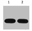 WB (Western Blot)
(Western blot analysis of Avi-Recombinant protein, diluted at 1) 1:5000 2) 1:10000)
WB (Western Blot)
(Western blot analysis of Avi-Recombinant protein, diluted at 1) 1:5000 2) 1:10000)
Avi-Tag, Monoclonal Antibody (Cat# AAA243717)

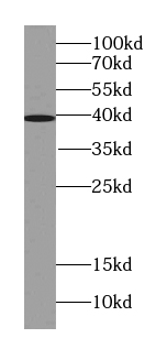
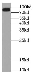
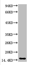
 WB (Western Blot)
(mouse brain tissue were subjected to SDS PAGE followed by western blot with AAA249045 (SNCA antibody) at dilution of 1:1000)
WB (Western Blot)
(mouse brain tissue were subjected to SDS PAGE followed by western blot with AAA249045 (SNCA antibody) at dilution of 1:1000)
alpha-Synuclein, Monoclonal Antibody (Cat# AAA249045)
Protein A+G Purified
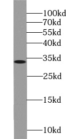
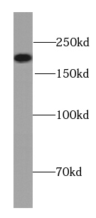
 WB (Western Blot)
(human spleen tissue were subjected to SDS PAGE followed by western blot with AAA249056 (VCAM-1 Antibody) at dilution of 1:800)
WB (Western Blot)
(human spleen tissue were subjected to SDS PAGE followed by western blot with AAA249056 (VCAM-1 Antibody) at dilution of 1:800)
VCAM-1, Monoclonal Antibody (Cat# AAA249056)
Protein A+G purified
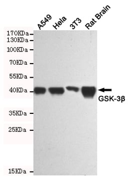
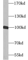
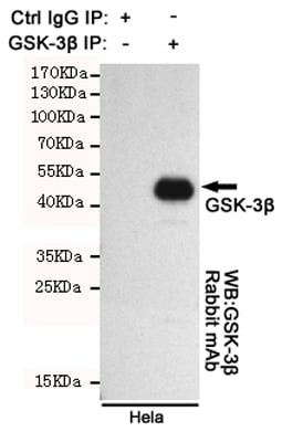 IP (Immunoprecipitation)
(Immunoprecipitation analysis of Hela cell lysates using GSK-3beta.GSK-3beta Rabbit mAb was used for the western blot analysis (1:1000 diluted).)
IP (Immunoprecipitation)
(Immunoprecipitation analysis of Hela cell lysates using GSK-3beta.GSK-3beta Rabbit mAb was used for the western blot analysis (1:1000 diluted).)
GSK3 beta, Monoclonal Antibody (Cat# AAA290858)
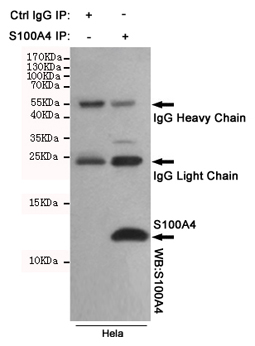 IP (Immunoprecipitation)
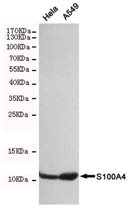
(Immunoprecipitation analysis of Hela cell lysates using S100A4 mouse mAb.)
IP (Immunoprecipitation)
(Immunoprecipitation analysis of Hela cell lysates using S100A4 mouse mAb.)
S100A4, Monoclonal Antibody (Cat# AAA290861)
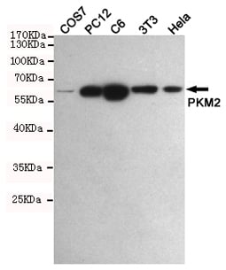
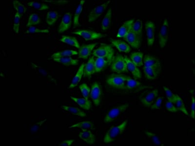
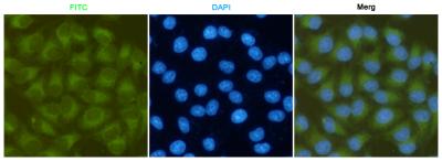
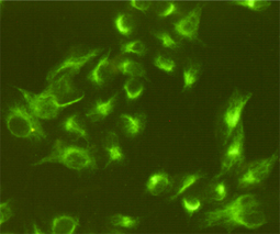
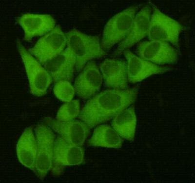
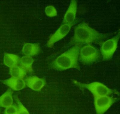 ICC (Immunocytochemistry)
(Immunocytochemistry staining of Hela cells fixed with 4% Paraformaldehyde and using anti-PKM2 mouse mAb (dilution 1:400).)
ICC (Immunocytochemistry)
(Immunocytochemistry staining of Hela cells fixed with 4% Paraformaldehyde and using anti-PKM2 mouse mAb (dilution 1:400).)
PKM2, Monoclonal Antibody (Cat# AAA290864)
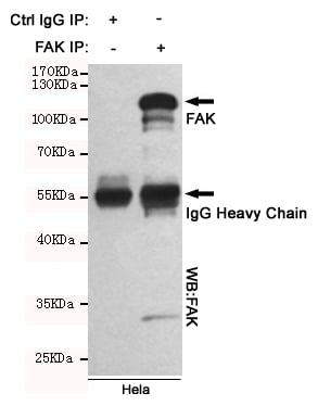 IP (Immunoprecipitation)
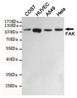
(Immunoprecipitation analysis of Hela cell lysates using FAK mouse mAb.)
IP (Immunoprecipitation)
(Immunoprecipitation analysis of Hela cell lysates using FAK mouse mAb.)
FAK, Monoclonal Antibody (Cat# AAA290866)
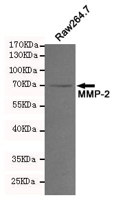
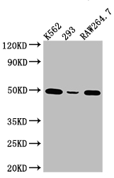
 WB (Western Blot)
(Western blot analysis of extracts from Raw264.7 cell lysates using MMP-2 mouse mAb (1:200 diluted). Predicted band size:64,72KDa. Observed band size:72KDa.)
WB (Western Blot)
(Western blot analysis of extracts from Raw264.7 cell lysates using MMP-2 mouse mAb (1:200 diluted). Predicted band size:64,72KDa. Observed band size:72KDa.)
MMP-2, Monoclonal Antibody (Cat# AAA290868)
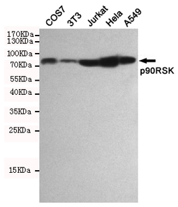
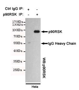 IP (Immunoprecipitation)
(Immunoprecipitation analysis of Hela cell lysates using p90RSK mouse mAb.)
IP (Immunoprecipitation)
(Immunoprecipitation analysis of Hela cell lysates using p90RSK mouse mAb.)
p90RSK, Monoclonal Antibody (Cat# AAA290870)
 IP (Immunoprecipitation)
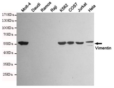
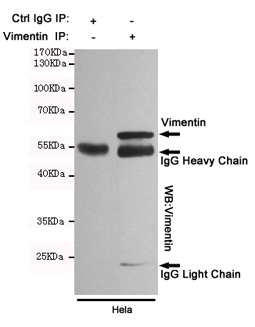
(Immunoprecipitation analysis of Hela cell lysates using Vimentin mouse mAb.)
IP (Immunoprecipitation)
(Immunoprecipitation analysis of Hela cell lysates using Vimentin mouse mAb.)
Vimentin, Monoclonal Antibody (Cat# AAA290872)
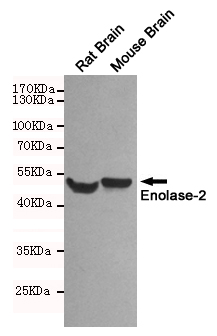

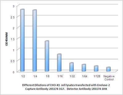 ELISA
(Observed Enolase-2 levels in CHO-K1 cell lysates transfected with Enolase-2 at different dilution.)
ELISA
(Observed Enolase-2 levels in CHO-K1 cell lysates transfected with Enolase-2 at different dilution.)
Enolase-2, Monoclonal Antibody (Cat# AAA290875)
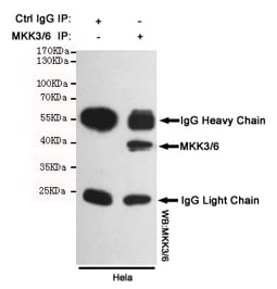 IP (Immunoprecipitation)
(Immunoprecipitation analysis of Hela cell lysates using MKK3/6 mouse mAb.)
IP (Immunoprecipitation)
(Immunoprecipitation analysis of Hela cell lysates using MKK3/6 mouse mAb.)
MKK3/6, Monoclonal Antibody (Cat# AAA290876)
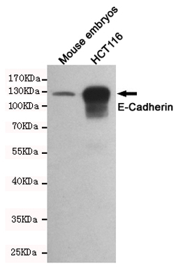
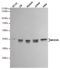
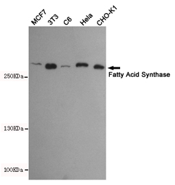
 WB (Western Blot)
(Western blot detection of E-Cadherin in Mouse embryos and HCT116 cell lysates using E-Cadherin mouse mAb (dilution 1:1000).Predicted band size:135kDa.Observed band size:135kDa.)
WB (Western Blot)
(Western blot detection of E-Cadherin in Mouse embryos and HCT116 cell lysates using E-Cadherin mouse mAb (dilution 1:1000).Predicted band size:135kDa.Observed band size:135kDa.)
E-Cadherin, Monoclonal Antibody (Cat# AAA290878)
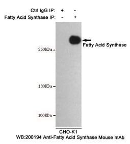 IP (Immunoprecipitation)
(Immunoprecipitation analysis of CHO-K1 cell lysates using Fatty Acid Synthase mouse mAb.)
IP (Immunoprecipitation)
(Immunoprecipitation analysis of CHO-K1 cell lysates using Fatty Acid Synthase mouse mAb.)
Fatty Acid Synthase, Monoclonal Antibody (Cat# AAA290879)
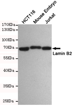
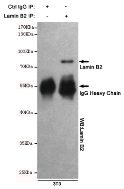 IP (Immunoprecipitation)
(Immunoprecipitation analysis of Hela cell lysates using Lamin B2 mouse mAb.)
IP (Immunoprecipitation)
(Immunoprecipitation analysis of Hela cell lysates using Lamin B2 mouse mAb.)
Lamin B2, Monoclonal Antibody (Cat# AAA290880)

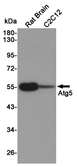 WB (Western Blot)
(Western blot detection of Atg5 in Rat Brain and C2C12 cell lysates using Atg5 mouse mAb (1:1000 diluted).Predicted band size:55KDa.Observed band size:55KDa.)
WB (Western Blot)
(Western blot detection of Atg5 in Rat Brain and C2C12 cell lysates using Atg5 mouse mAb (1:1000 diluted).Predicted band size:55KDa.Observed band size:55KDa.)
Atg5, Monoclonal Antibody (Cat# AAA290884)
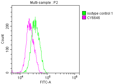
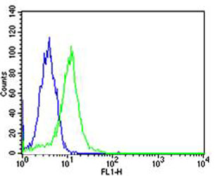
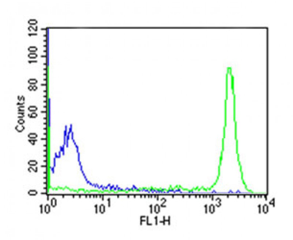 FCM/FACS (Flow Cytometry)
(Overlay histogram showing huuman peripheral blood lymphocytes stained with CD45 antibody(green line). The cells were icubated in 2% bovine serum albumin to block non-specific protein-protein interactions followed by the antibody (1:50 dilution) for 60min at 37 degree C. The secondary antibody used was Goat Anti-Mouse IgG, DyLight 488 Conjugated Highly Cross-Adsorbed(OJ192088) at 1/200 dilution for 40min at 37 degree C. Isotype control antibody (blue line) was mouse IgG2a (1mug/1x10^6 cells) used under the same conditions. Acquisition of >10, 000 events was performed.)
FCM/FACS (Flow Cytometry)
(Overlay histogram showing huuman peripheral blood lymphocytes stained with CD45 antibody(green line). The cells were icubated in 2% bovine serum albumin to block non-specific protein-protein interactions followed by the antibody (1:50 dilution) for 60min at 37 degree C. The secondary antibody used was Goat Anti-Mouse IgG, DyLight 488 Conjugated Highly Cross-Adsorbed(OJ192088) at 1/200 dilution for 40min at 37 degree C. Isotype control antibody (blue line) was mouse IgG2a (1mug/1x10^6 cells) used under the same conditions. Acquisition of >10, 000 events was performed.)
CD45, Monoclonal Antibody (Cat# AAA290899)
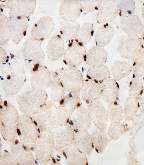
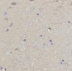
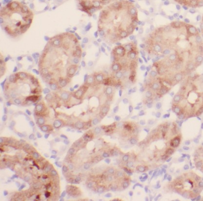
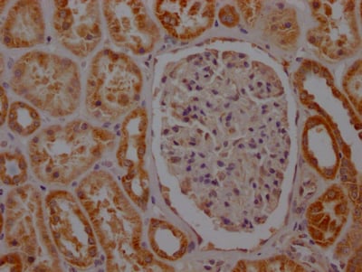
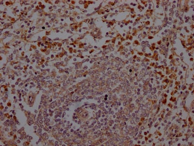
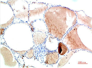
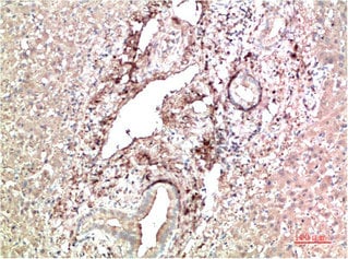
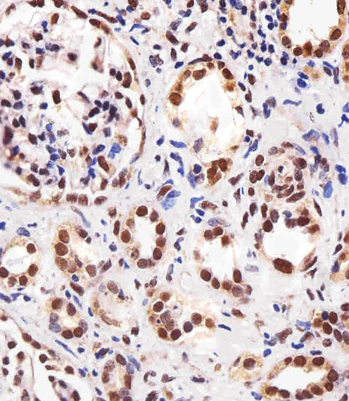 IHC (Immunohistochemistry)
(Immunohistochemical analysis of paraffin-embedded H. kidney section using PPARA Antibody . It was diluted at 1:25 dilution. A peroxidase-conjugated goat anti-mouse IgG at 1:400 dilution was used as the secondary antibody, followed by DAB staining.)
IHC (Immunohistochemistry)
(Immunohistochemical analysis of paraffin-embedded H. kidney section using PPARA Antibody . It was diluted at 1:25 dilution. A peroxidase-conjugated goat anti-mouse IgG at 1:400 dilution was used as the secondary antibody, followed by DAB staining.)
PPARA, Monoclonal Antibody (Cat# AAA290936)
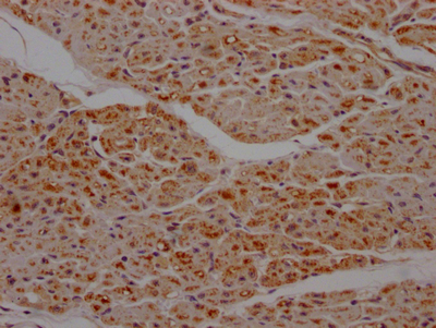
 IHC (Immunohiostchemistry)
(Immunohistochemical analysis of paraffin-embedded H. liver section using CTSD Antibody(AAA290942). AAA290942 was diluted at 1:25 dilution. A undiluted biotinylated goat polyvalent antibody was used as the secondary, followed by DAB staining.)
IHC (Immunohiostchemistry)
(Immunohistochemical analysis of paraffin-embedded H. liver section using CTSD Antibody(AAA290942). AAA290942 was diluted at 1:25 dilution. A undiluted biotinylated goat polyvalent antibody was used as the secondary, followed by DAB staining.)
CTSD, Monoclonal Antibody (Cat# AAA290942)
Predicted: Mouse, Bovine
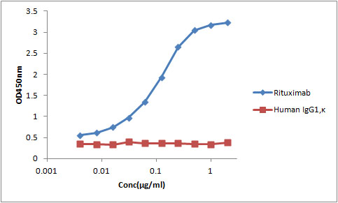 ELISA
(Plate was coated with Mabthera and Human IgG1,kappa at 1.25 ug/ml in PBS, and then incubated with anti-Mabthera monoclonal antibody (1F3C10) from 0.004 ug/ml to 2 ug/ml. The secondary antibody, HRP conjugated goat anti-mouse antibody,were used at 1:10000 dilution.)
ELISA
(Plate was coated with Mabthera and Human IgG1,kappa at 1.25 ug/ml in PBS, and then incubated with anti-Mabthera monoclonal antibody (1F3C10) from 0.004 ug/ml to 2 ug/ml. The secondary antibody, HRP conjugated goat anti-mouse antibody,were used at 1:10000 dilution.)
Rituximab, Monoclonal Antibody (Cat# AAA290958)
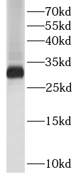
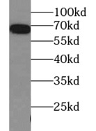
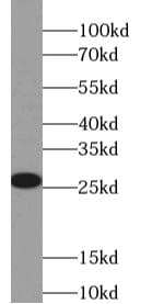
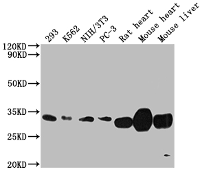
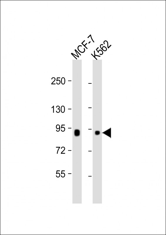
 WB (Western Blot)
(WB - RAB27B Antibody AM8508b detail Anti-RAB27B Antibody at 1:2000 dilution + mouse stomach lysate Lysates/proteins at 20 ug per lane. Secondary Goat Anti-mouse IgG, (H+L), Peroxidase conjugated at 1/10000 dilution. Predicted band size : 25 kDa Blocking/Dilution buffer: 5% NFDM/TBST.)
WB (Western Blot)
(WB - RAB27B Antibody AM8508b detail Anti-RAB27B Antibody at 1:2000 dilution + mouse stomach lysate Lysates/proteins at 20 ug per lane. Secondary Goat Anti-mouse IgG, (H+L), Peroxidase conjugated at 1/10000 dilution. Predicted band size : 25 kDa Blocking/Dilution buffer: 5% NFDM/TBST.)
RAB27B, Monoclonal Antibody (Cat# AAA290610)
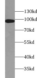
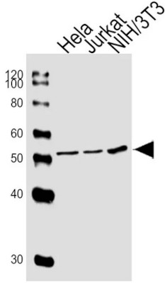
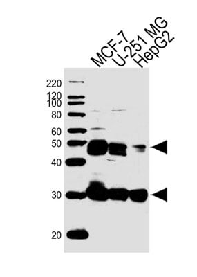
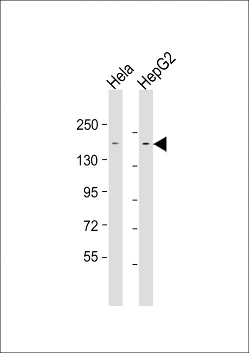 WB (Western Blot)
(All lanes : Anti-LAP2 Antibody at 1:1000-1:2000 dilutionLane 1: Hela whole cell lysateLane 2: HepG2 whole cell lysateLysates/proteins at 20 ug per lane.SecondaryGoat Anti-mouse IgG, (H+L), Peroxidase conjugated at 1/10000 dilution.Predicted band size : 158 kDaBlocking/Dilution buffer: 5% NFDM/TBST.)
WB (Western Blot)
(All lanes : Anti-LAP2 Antibody at 1:1000-1:2000 dilutionLane 1: Hela whole cell lysateLane 2: HepG2 whole cell lysateLysates/proteins at 20 ug per lane.SecondaryGoat Anti-mouse IgG, (H+L), Peroxidase conjugated at 1/10000 dilution.Predicted band size : 158 kDaBlocking/Dilution buffer: 5% NFDM/TBST.)
LAP2, Monoclonal Antibody (Cat# AAA290618)
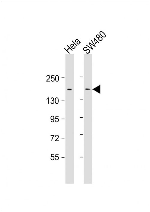
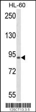 WB (Western Blot)
(Western blot analysis of anti-BRAF Antibody in HL-60 cell line lysates (35mug/lane). BRAF (arrow) was detected using the purified Mab.)
WB (Western Blot)
(Western blot analysis of anti-BRAF Antibody in HL-60 cell line lysates (35mug/lane). BRAF (arrow) was detected using the purified Mab.)
BRAF, Monoclonal Antibody (Cat# AAA290642)
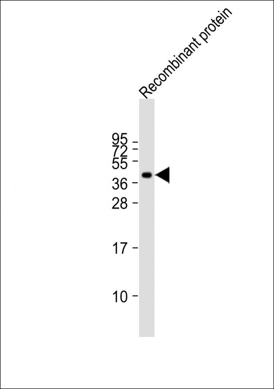
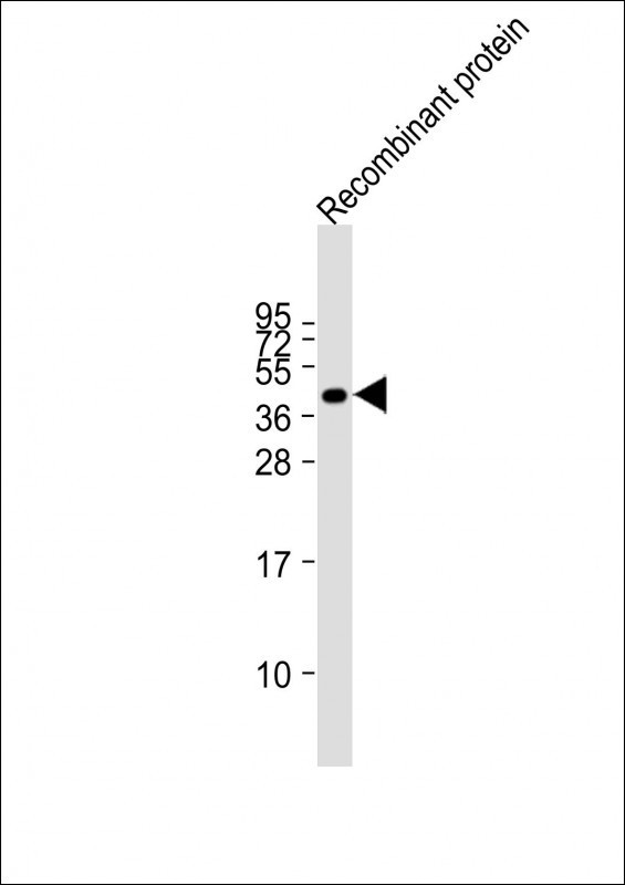
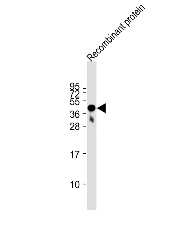 WB (Western Blot)
(Anti-FAT4 Antibody at 1:2000 dilution + Recombinant proteinLysates/proteins at 20 ug per lane.SecondaryGoat Anti-mouse IgG, (H+L), Peroxidase conjugated at 1/10000 dilution.Predicted band size : 543 kDaBlocking/Dilution buffer: 5% NFDM/TBST.)
WB (Western Blot)
(Anti-FAT4 Antibody at 1:2000 dilution + Recombinant proteinLysates/proteins at 20 ug per lane.SecondaryGoat Anti-mouse IgG, (H+L), Peroxidase conjugated at 1/10000 dilution.Predicted band size : 543 kDaBlocking/Dilution buffer: 5% NFDM/TBST.)
FAT4, Monoclonal Antibody (Cat# AAA290645)
What are Monoclonal Antibodies?
Monoclonal antibodies are specialized laboratory-produced proteins developed for binding to specific biological antigens or other molecular targets. Since they come from a single cell (or clone), they are especially consistent and accurate in the data they are involved in producing.
This type of antibody material has been shown to be a powerful tool in finding and subsequently destroying harmful cells in an organism, such as those found in cancers or various autoimmune diseases. This makes them excellent aids in medical testing and research, which is why they are so widely used.
AAA Biotech offers a comprehensive range of high-quality monoclonal antibodies that perform effectively in various laboratory tests, including (amongst others) ELISA, western blotting, immunohistochemistry, and flow cytometry. All of the products in our catalog are thoroughly quality tested to make sure that they are reliable and will consistently perform well in your research.
What Are The Uses of Monoclonal Antibodies
Monoclonal antibodies are used in many lab tests, including (amongst others) ELISA, western blotting, immunohistochemistry, and flow cytometry.
ELISA is a test that helps detect a specific substance/analyte in a sample. It uses antibodies (often monoclonal) bound to a solid surface (such as the well of a microplate) to “capture” the substance/analyte in the sample and immobilize it so that the detection antibody component can then bind to it and produce a signal, which can then be measured.
Western blotting identifies specific proteins in a sample. The sample is first separated on a gel, and then antibodies are applied that will typically bind to the target, which will all be localized to a single band in a lane.
Immunohistochemistry helps locate specific proteins in cells or tissue samples using antibodies.
Flow cytometry looks at and sorts cells. It uses antibodies that are conjugated to reporter molecules called “fluorophores”, which, under special lights, emit light themselves, which can then be measured by a detector instrument.
How Monoclonal Antibodies Are Used as Medicine?
Please note that all of the products listed in AAA Biotech’s also known as AAA Bio or AAABio catalog are strictly for research-use only (RUO).
Monoclonal antibodies can also be used as therapeutic/medical treatments, particularly in the context of cancers. They are designed to find and bind to specific cells or proteins, helping the immune system recognize and attack the cancer. These treatments work in different ways, such as:
- Radioimmunotherapy attaches a small amount of radioactive molecule to the antibody, so it delivers the radiation directly to the cancer cells that the antibody is specifically binding to.
- Antibody-directed enzyme prodrug therapy uses antibodies that are specifically bound to special enzymes. These enzymes activate a harmless drug in the body and turn it into a cancer-killing drug only near the cancer cells—this helps avoid harming healthy cells.
- Immunoliposomes are tiny “bubbles” filled with medicine/drug and coated with antibodies. They carry the drug straight to the cancer cells.
Why Buy Monoclonal Antibodies From Us?
At AAA Biotech, we provide high-performance monoclonal antibodies designed to support a wide range of research needs.
1. Validated for Versatile Applications
The antibodies in our catalog are extensively validated and compatible with multiple techniques, including (but not limited to) ELISA, flow cytometry (FC), immunocytochemistry (ICC), immunofluorescence (IF), immunohistochemistry (IHC), immunoprecipitation (IP), and western blotting (WB).
2. Wide Selection & Specialized Options
We offer antibodies for common and rare species, that are available in various conjugated forms, and also in recombinant formats. Essentially, there is almost anything one might need to meet their experimental model’s requirements.
3. High-Quality Proteins
Our proteins meet high purity standards—90% or more as confirmed by SDS-PAGE. Many are available with tags like His, Flag, GST, or MBP, and we also supply native and biologically active proteins for functional studies.
Frequently Asked Questions
1. Are your monoclonal antibodies validated for specific applications?
Yes, our antibodies are tested and validated for use in methods such as ELISA, western blot, IHC, flow cytometry, and more. Refer to specific product pages or datasheets for individual product information.
2. How do I choose the right monoclonal antibody for my application?
Review the product details directly for application validation, species reactivity, and target information. You may also contact our support team at any time for help.
3. How quickly can I receive my order?
Most orders are processed and shipped within 1–3 business days, depending on product availability and your shipping location.