Filters
▼Clonality
▼Type
▼Reactivity
▼Gene Name
▼Isotype
▼Host
▼Application
▼Clone
▼Monoclonal Antibodies
Get accurate results in your research with our Monoclonal Antibodies, which are specially made to target exactly what you require for your research, and will produce consistent, reliable performance in lab tests.
Viewing 7400-7450 of 27597 product results
 IHC (Immunohistochemisry)
(DAB staining on IHCP;Samples: Rat Ovary Tissue;Primary Ab: 20ug/ml Mouse AntiRat Trx AntibodySecond Ab: 2ug/mL HRPLinked Caprine AntiMouse IgG Polyclonal Antibody(Catalog: SAA544Mu19))
IHC (Immunohistochemisry)
(DAB staining on IHCP;Samples: Rat Ovary Tissue;Primary Ab: 20ug/ml Mouse AntiRat Trx AntibodySecond Ab: 2ug/mL HRPLinked Caprine AntiMouse IgG Polyclonal Antibody(Catalog: SAA544Mu19))
Thioredoxin (Trx), Monoclonal Antibody (Cat# AAA151606)
 WB (Western Blot)
(Western Blot; Sample: Rat Serum Primary Ab: 3ug/ml Mouse AntiRat APOE Antibody Second Ab: 0.2ug/mL HRPLinked Caprine AntiMouse IgG Polyclonal Antibody (Catalog: SAA544Mu19))
WB (Western Blot)
(Western Blot; Sample: Rat Serum Primary Ab: 3ug/ml Mouse AntiRat APOE Antibody Second Ab: 0.2ug/mL HRPLinked Caprine AntiMouse IgG Polyclonal Antibody (Catalog: SAA544Mu19))
Apolipoprotein E (APOE), Monoclonal Antibody (Cat# AAA151607)
 WB (Western Blot)
(Western Blot; Sample: Rat Serum Primary Ab: 3ug/ml Mouse AntiRat APOE Antibody Second Ab: 0.2ug/mL HRPLinked Caprine AntiMouse IgG Polyclonal Antibody (Catalog: SAA544Mu19))
WB (Western Blot)
(Western Blot; Sample: Rat Serum Primary Ab: 3ug/ml Mouse AntiRat APOE Antibody Second Ab: 0.2ug/mL HRPLinked Caprine AntiMouse IgG Polyclonal Antibody (Catalog: SAA544Mu19))
Apolipoprotein E (APOE), Monoclonal Antibody (Cat# AAA151608)
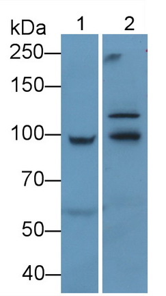
 WB (Western Blot)
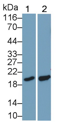
(Western Blot; Sample: Lane1: Hela cell lysate; Lane2: Porcine Liver lysate Primary Ab: 0.2ug/ml Mouse AntiHuman CYPB Antibody Second Ab: 0.2ug/mL HRPLinked Caprine AntiMouse IgG Polyclonal Antibody (Catalog: SAA544Mu19))
WB (Western Blot)
(Western Blot; Sample: Lane1: Hela cell lysate; Lane2: Porcine Liver lysate Primary Ab: 0.2ug/ml Mouse AntiHuman CYPB Antibody Second Ab: 0.2ug/mL HRPLinked Caprine AntiMouse IgG Polyclonal Antibody (Catalog: SAA544Mu19))
Cyclophilin B (CYPB), Monoclonal Antibody (Cat# AAA151510)
 IHC (Immunohistochemisry)
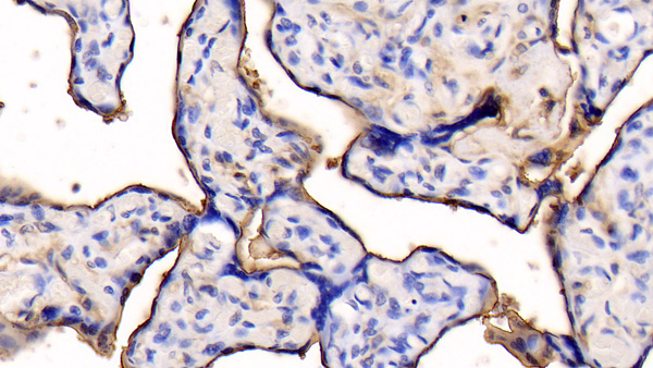
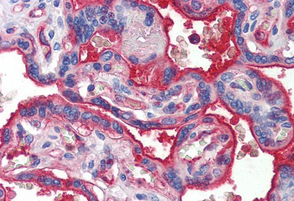
(DAB staining on IHCP;Samples: Human Placenta Tissue;Primary Ab: 20ug/ml Mouse AntiHuman ANXA5 AntibodySecond Ab: 2ug/mL HRPLinked Caprine AntiMouse IgG Polyclonal Antibody(Catalog: SAA544Mu19))
IHC (Immunohistochemisry)
(DAB staining on IHCP;Samples: Human Placenta Tissue;Primary Ab: 20ug/ml Mouse AntiHuman ANXA5 AntibodySecond Ab: 2ug/mL HRPLinked Caprine AntiMouse IgG Polyclonal Antibody(Catalog: SAA544Mu19))
Annexin V (ANXA5), Monoclonal Antibody (Cat# AAA151516)
 WB (Western Blot)
(Western Blot; Sample: Lane1: Mouse Cerebrum lysate; Lane2: Rat Cerebrum lysate Primary Ab: 2ug/mL Mouse AntiHuman NRGN Antibody Second Ab: 0.2ug/mL HRPLinked Caprine AntiMouse IgG Polyclonal Antibody (Catalog: SAA544Mu19))
WB (Western Blot)
(Western Blot; Sample: Lane1: Mouse Cerebrum lysate; Lane2: Rat Cerebrum lysate Primary Ab: 2ug/mL Mouse AntiHuman NRGN Antibody Second Ab: 0.2ug/mL HRPLinked Caprine AntiMouse IgG Polyclonal Antibody (Catalog: SAA544Mu19))
Neurogranin (NRGN), Monoclonal Antibody (Cat# AAA151532)
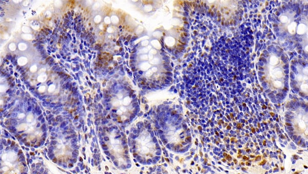
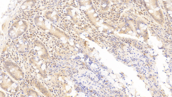
 IHC (Immunohiostchemistry)
(DAB staining on IHCP;Sample: Porcine Colon Tissue; Primary Ab: 10ug/ml Mouse AntiHuman PKCe AntibodySecond Ab: 2ug/mL HRPLinked Caprine AntiMouse IgG Polyclonal Antibody(Catalog: SAA544Mu19))
IHC (Immunohiostchemistry)
(DAB staining on IHCP;Sample: Porcine Colon Tissue; Primary Ab: 10ug/ml Mouse AntiHuman PKCe AntibodySecond Ab: 2ug/mL HRPLinked Caprine AntiMouse IgG Polyclonal Antibody(Catalog: SAA544Mu19))
Protein Kinase C Epsilon (PKCe), Monoclonal Antibody (Cat# AAA151538)
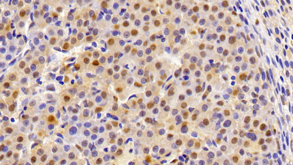
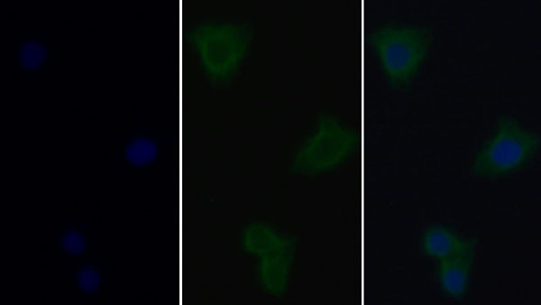
 IHC (Immunohistochemisry)
(DAB staining on IHCP;Sample: Porcine Pancreas Tissue; Primary Ab: 10ug/ml Mouse AntiHuman PKCe AntibodySecond Ab: 2ug/mL HRPLinked Caprine AntiMouse IgG Polyclonal Antibody(Catalog: SAA544Mu19))
IHC (Immunohistochemisry)
(DAB staining on IHCP;Sample: Porcine Pancreas Tissue; Primary Ab: 10ug/ml Mouse AntiHuman PKCe AntibodySecond Ab: 2ug/mL HRPLinked Caprine AntiMouse IgG Polyclonal Antibody(Catalog: SAA544Mu19))
Protein Kinase C Epsilon (PKCe), Monoclonal Antibody (Cat# AAA151539)
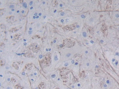
 IHC (Immunohiostchemistry)
(DAB staining on IHCP;Sample: Human Glioma Tissue; Primary Ab: 30ug/ml Mouse AntiHuman NES AntibodySecond Ab: 2ug/mL HRPLinked Caprine AntiMouse IgG Polyclonal Antibody(Catalog: SAA544Mu19))
IHC (Immunohiostchemistry)
(DAB staining on IHCP;Sample: Human Glioma Tissue; Primary Ab: 30ug/ml Mouse AntiHuman NES AntibodySecond Ab: 2ug/mL HRPLinked Caprine AntiMouse IgG Polyclonal Antibody(Catalog: SAA544Mu19))
Nestin (NES), Monoclonal Antibody (Cat# AAA151548)
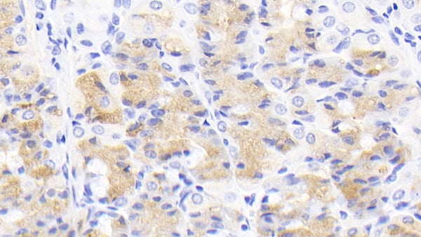
 IHC (Immunohiostchemistry)
(DAB staining on IHCP;Sample: Rat Stomach Tissue; Primary Ab: 20ug/ml Mouse AntiRat EGF AntibodySecond Ab: 2ug/mL HRPLinked Caprine AntiMouse IgG Polyclonal Antibody(Catalog: SAA544Mu19))
IHC (Immunohiostchemistry)
(DAB staining on IHCP;Sample: Rat Stomach Tissue; Primary Ab: 20ug/ml Mouse AntiRat EGF AntibodySecond Ab: 2ug/mL HRPLinked Caprine AntiMouse IgG Polyclonal Antibody(Catalog: SAA544Mu19))
Epidermal Growth Factor (EGF), Monoclonal Antibody (Cat# AAA151562)


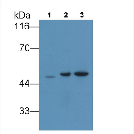
 WB (Western Blot)
(Western Blot; Sample: Lane1: K562 cell lysate; Lane2: Hela cell lysate; Lane3: HepG2 cell lysate; Primary Ab: 3ug/ml Mouse AntiRat ANXA1 Antibody Second Ab: 0.2ug/mL HRPLinked Caprine AntiMouse IgG Polyclonal Antibody (Catalog: SAA544Mu19))
WB (Western Blot)
(Western Blot; Sample: Lane1: K562 cell lysate; Lane2: Hela cell lysate; Lane3: HepG2 cell lysate; Primary Ab: 3ug/ml Mouse AntiRat ANXA1 Antibody Second Ab: 0.2ug/mL HRPLinked Caprine AntiMouse IgG Polyclonal Antibody (Catalog: SAA544Mu19))
Annexin A1 (ANXA1), Monoclonal Antibody (Cat# AAA151864)
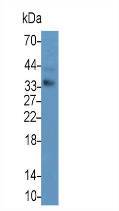
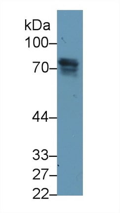
 WB (Western Blot)
(Western Blot; Sample: Porcine Skeletal muscle lysate Primary Ab: 2ug/ml Mouse AntiHuman ACVR2A Antibody Second Ab: 0.2ug/mL HRPLinked Caprine AntiMouse IgG Polyclonal Antibody (Catalog: SAA544Mu19))
WB (Western Blot)
(Western Blot; Sample: Porcine Skeletal muscle lysate Primary Ab: 2ug/ml Mouse AntiHuman ACVR2A Antibody Second Ab: 0.2ug/mL HRPLinked Caprine AntiMouse IgG Polyclonal Antibody (Catalog: SAA544Mu19))
Activin A Receptor Type II A (ACVR2A), Monoclonal Antibody (Cat# AAA151791)
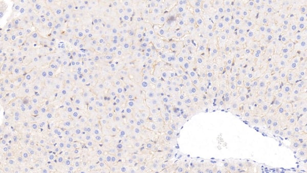
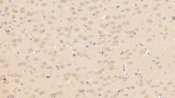
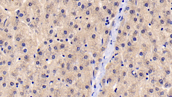
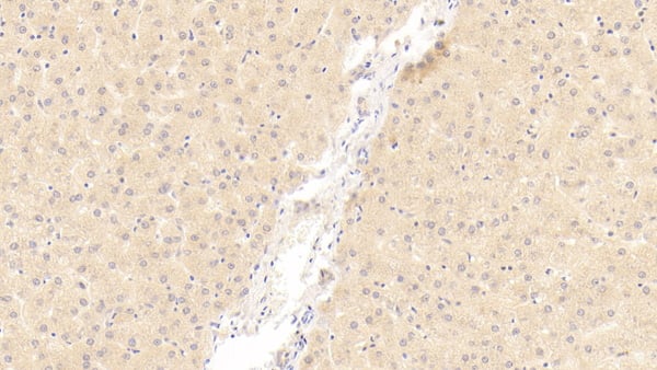
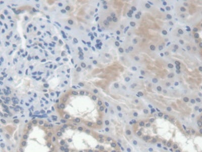
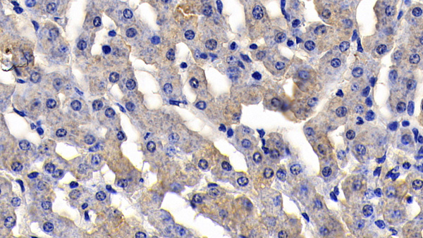
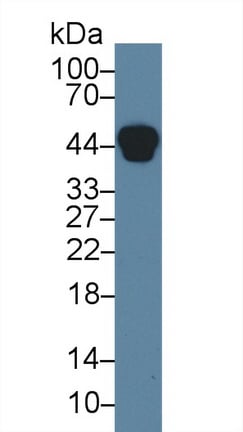
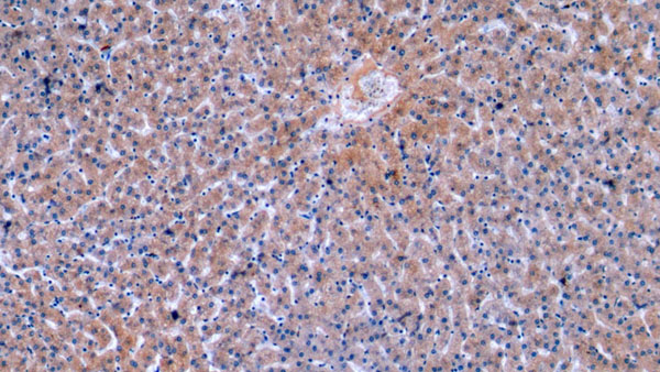
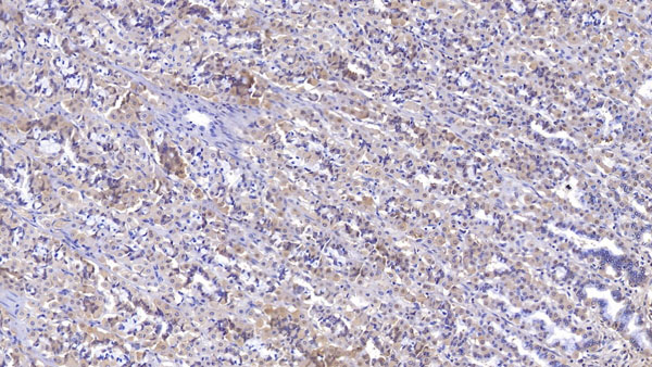
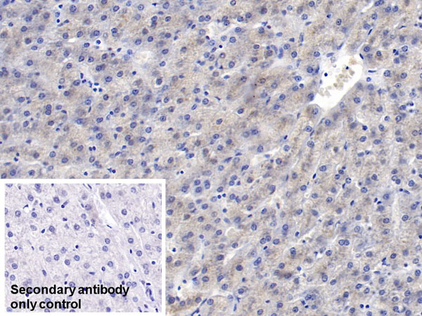
 IHC (Immunohiostchemistry)
(DAB staining on IHCP; Samples: Rat Liver Tissue; Primary Ab: 40ug/ml Mouse AntiRat CRAT Antibody Second Ab: 2ug/mL HRPLinked Caprine AntiMouse IgG Polyclonal Antibody (Catalog: SAA544Mu19))
IHC (Immunohiostchemistry)
(DAB staining on IHCP; Samples: Rat Liver Tissue; Primary Ab: 40ug/ml Mouse AntiRat CRAT Antibody Second Ab: 2ug/mL HRPLinked Caprine AntiMouse IgG Polyclonal Antibody (Catalog: SAA544Mu19))
Carnitine Acetyltransferase (CRAT), Monoclonal Antibody (Cat# AAA151806)
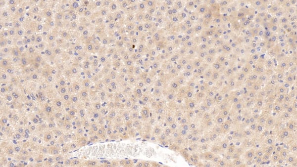
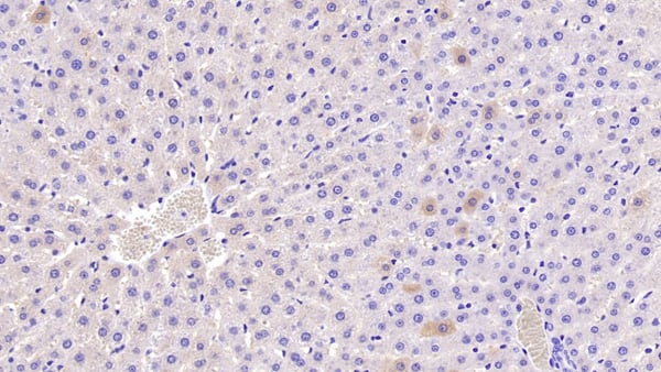
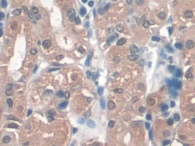
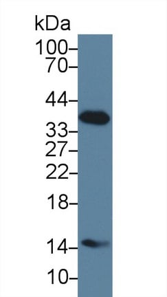
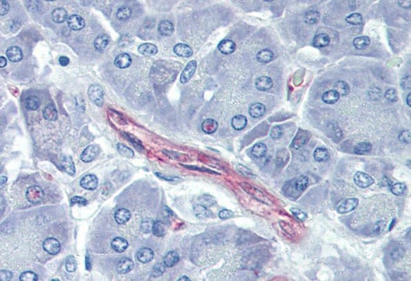
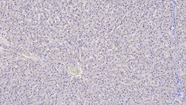
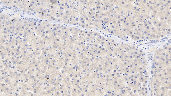
 IHC (Immunohiostchemistry)
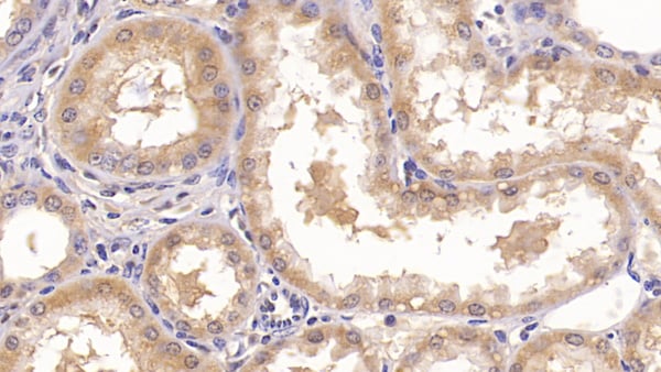
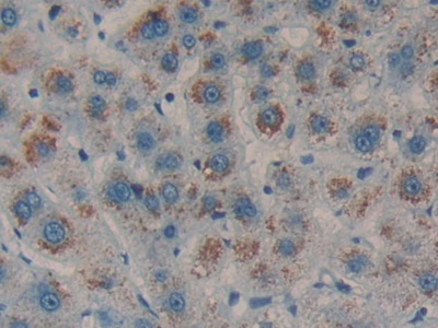
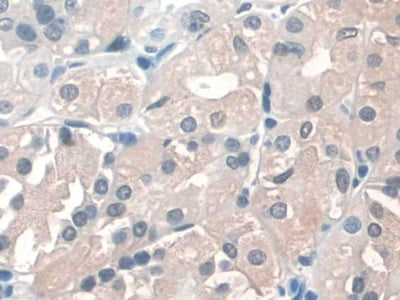
(DAB staining on IHCP;Sample: Human Liver Tissue; Primary Ab: 30ug/ml Mouse AntiHuman OAS1 AntibodySecond Ab: 2ug/mL HRPLinked Caprine AntiMouse IgG Polyclonal Antibody(Catalog: SAA544Mu19))
IHC (Immunohiostchemistry)
(DAB staining on IHCP;Sample: Human Liver Tissue; Primary Ab: 30ug/ml Mouse AntiHuman OAS1 AntibodySecond Ab: 2ug/mL HRPLinked Caprine AntiMouse IgG Polyclonal Antibody(Catalog: SAA544Mu19))
2',5'Oligoadenylate Synthetase 1 (OAS1), Monoclonal Antibody (Cat# AAA151813)
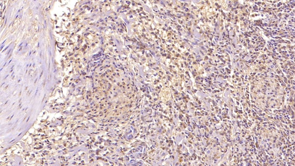
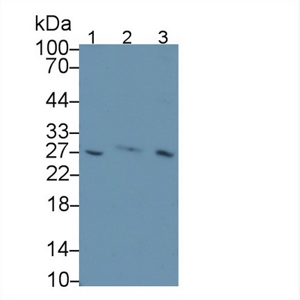
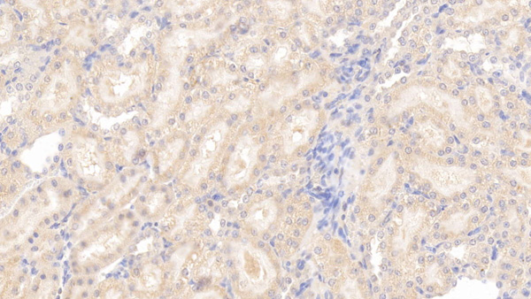
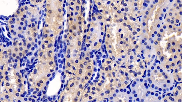
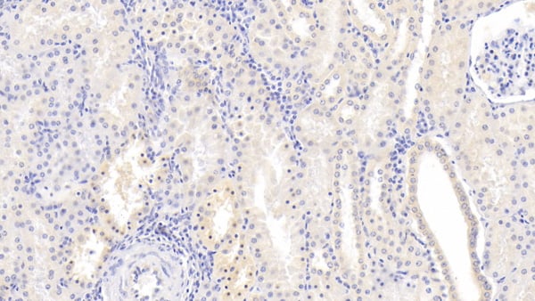
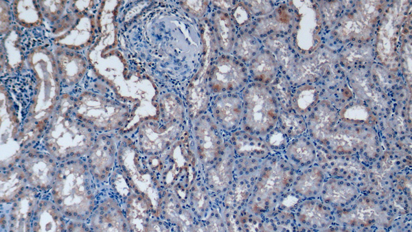
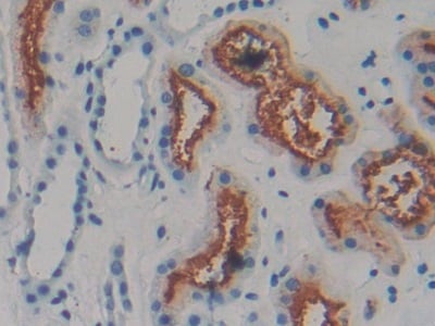
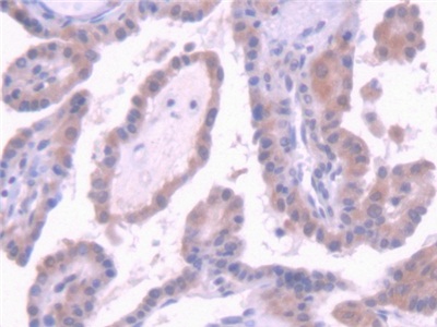
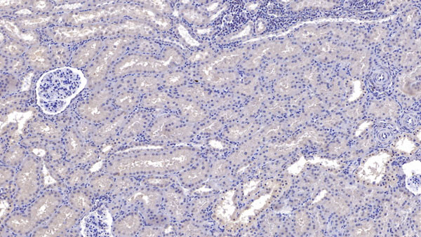
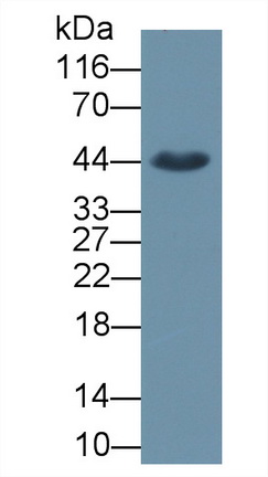
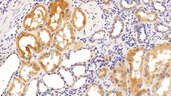
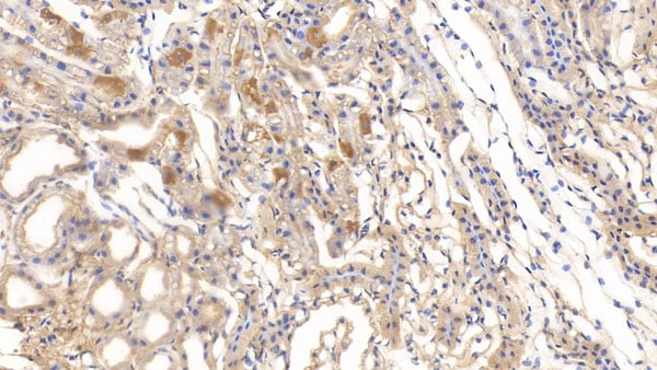
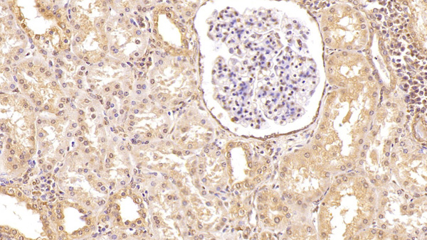 IHC (Immunohistochemisry)
(DAB staining on IHC-P; Samples: Human Kidney Tissue; Primary Ab: 40ug/ml Mouse Anti-Human CFD Antibody Second Ab: 2ug/mL HRP-Linked Caprine Anti-Mouse IgG Polyclonal Antibody)
IHC (Immunohistochemisry)
(DAB staining on IHC-P; Samples: Human Kidney Tissue; Primary Ab: 40ug/ml Mouse Anti-Human CFD Antibody Second Ab: 2ug/mL HRP-Linked Caprine Anti-Mouse IgG Polyclonal Antibody)
Complement Factor D (CFD), Monoclonal Antibody (Cat# AAA151126)
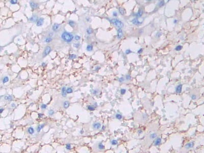
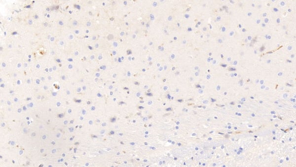
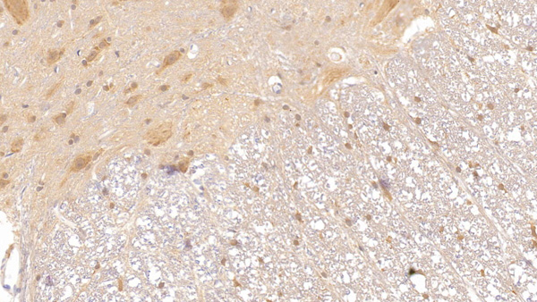
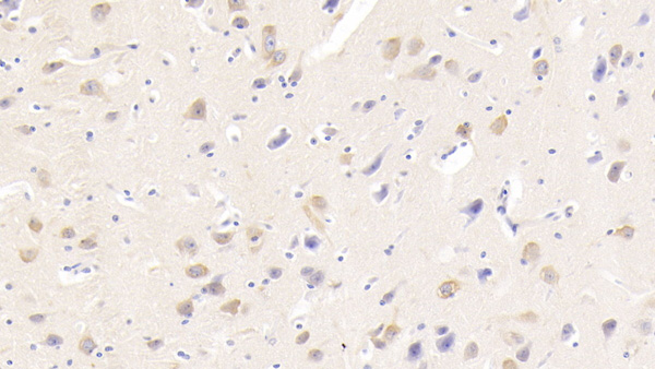
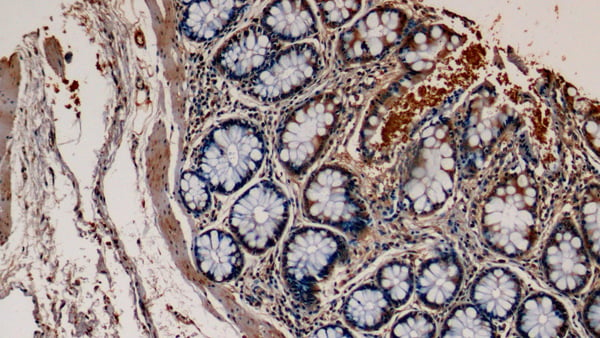
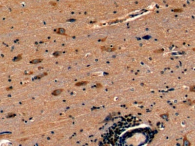
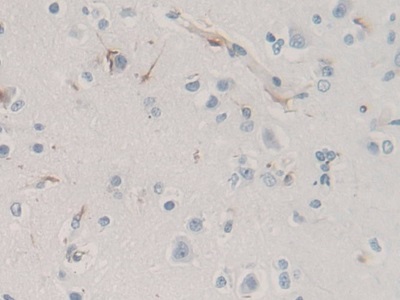
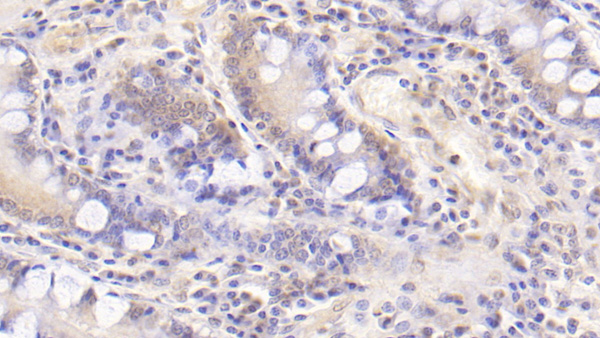
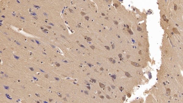
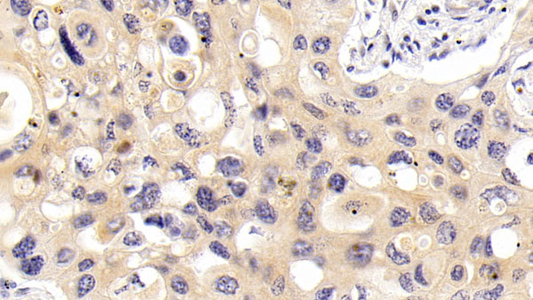
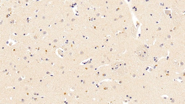
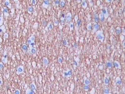 IHC (Immunohistochemistry)
(DAB staining on IHCP;Sample: Human Brain Tissue; Primary Ab: 30ug/ml Mouse AntiHuman AREG AntibodySecond Ab: 2ug/mL HRPLinked Caprine AntiMouse IgG Polyclonal Antibody(Catalog: SAA544Mu19))
IHC (Immunohistochemistry)
(DAB staining on IHCP;Sample: Human Brain Tissue; Primary Ab: 30ug/ml Mouse AntiHuman AREG AntibodySecond Ab: 2ug/mL HRPLinked Caprine AntiMouse IgG Polyclonal Antibody(Catalog: SAA544Mu19))
Amphiregulin (AREG), Monoclonal Antibody (Cat# AAA151429)
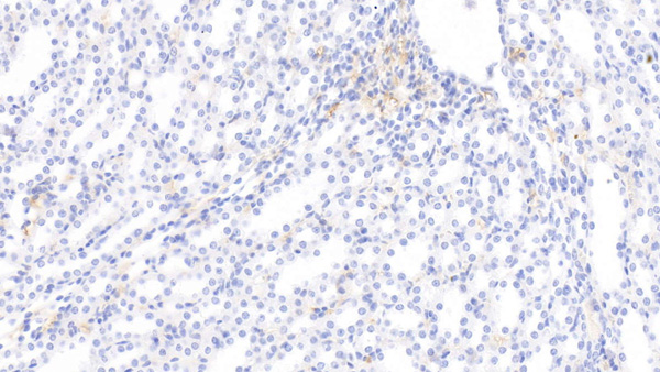
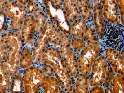
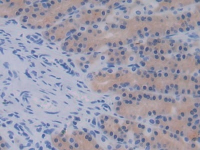
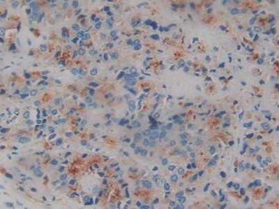
 IHC (Immunohistochemisry)
(DAB staining on IHCP;Sample: Human Stomach Tissue; Primary Ab: 30ug/ml Mouse AntiHuman EPO AntibodySecond Ab: 2ug/mL HRPLinked Caprine AntiMouse IgG Polyclonal Antibody(Catalog: SAA544Mu19))
IHC (Immunohistochemisry)
(DAB staining on IHCP;Sample: Human Stomach Tissue; Primary Ab: 30ug/ml Mouse AntiHuman EPO AntibodySecond Ab: 2ug/mL HRPLinked Caprine AntiMouse IgG Polyclonal Antibody(Catalog: SAA544Mu19))
Erythropoietin (EPO), Monoclonal Antibody (Cat# AAA151435)
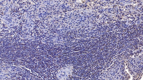
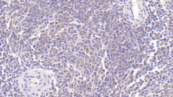 IHC (Immunohiostchemistry)
(DAB staining on IHCP;Sample: Porcine Spleen Tissue; Primary Ab: 30ug/ml Mouse AntiPorcine IL10 AntibodySecond Ab: 2ug/mL HRPLinked Caprine AntiMouse IgG Polyclonal Antibody(Catalog: SAA544Mu19))
IHC (Immunohiostchemistry)
(DAB staining on IHCP;Sample: Porcine Spleen Tissue; Primary Ab: 30ug/ml Mouse AntiPorcine IL10 AntibodySecond Ab: 2ug/mL HRPLinked Caprine AntiMouse IgG Polyclonal Antibody(Catalog: SAA544Mu19))
Interleukin 10 (IL10), Monoclonal Antibody (Cat# AAA151446)
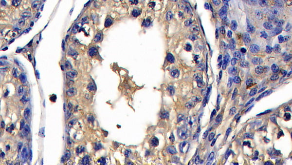 IHC (Immunohiostchemistry)
(DAB staining on IHCP;Sample: Bovine Testis Tissue; Primary Ab: 20ug/ml Mouse AntiBovine TNFa AntibodySecond Ab: 2ug/mL HRPLinked Caprine AntiMouse IgG Polyclonal Antibody(Catalog: SAA544Mu19))
IHC (Immunohiostchemistry)
(DAB staining on IHCP;Sample: Bovine Testis Tissue; Primary Ab: 20ug/ml Mouse AntiBovine TNFa AntibodySecond Ab: 2ug/mL HRPLinked Caprine AntiMouse IgG Polyclonal Antibody(Catalog: SAA544Mu19))
Tumor Necrosis Factor Alpha (TNFa), Monoclonal Antibody (Cat# AAA151491)
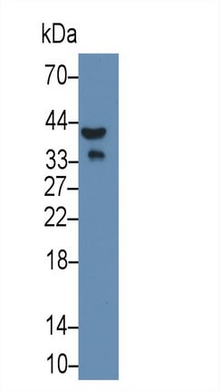
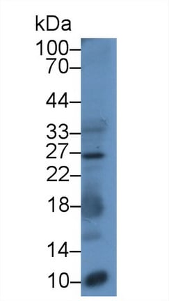
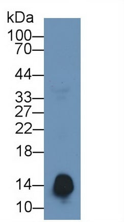
 WB (Western Blot)
(Western Blot; Sample: Human Urine Primary Ab: 2ug/ml Mouse AntiHuman PGA Antibody Second Ab: 0.2ug/mL HRPLinked Caprine AntiMouse IgG Polyclonal Antibody (Catalog: SAA544Mu19))
WB (Western Blot)
(Western Blot; Sample: Human Urine Primary Ab: 2ug/ml Mouse AntiHuman PGA Antibody Second Ab: 0.2ug/mL HRPLinked Caprine AntiMouse IgG Polyclonal Antibody (Catalog: SAA544Mu19))
Pepsinogen A (PGA), Monoclonal Antibody (Cat# AAA151502)
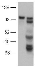
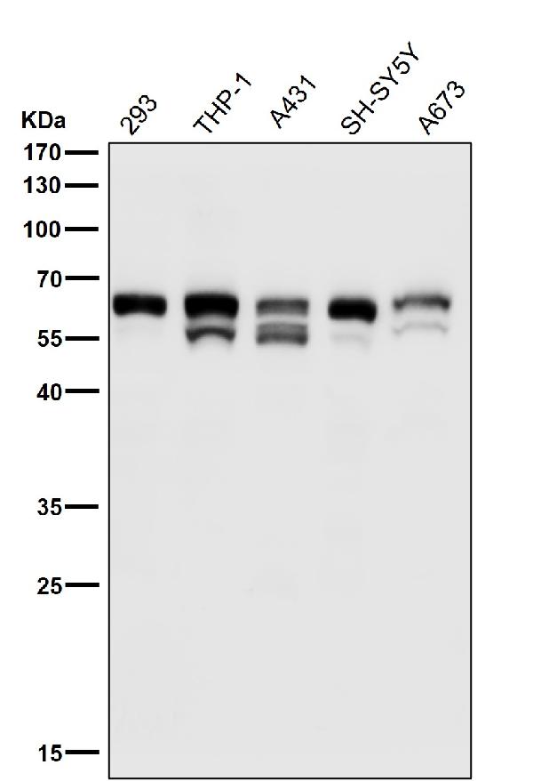
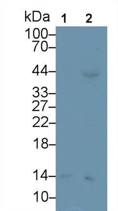
 WB (Western Blot)
(Western Blot; Sample: Lane1: Human Skeletal muscle lysate; Lane2: Human MCF7 cell lysate; Primary Ab)
WB (Western Blot)
(Western Blot; Sample: Lane1: Human Skeletal muscle lysate; Lane2: Human MCF7 cell lysate; Primary Ab)
Insulin Like Growth Factor 1 Receptor (IGF1R), Monoclonal Antibody (Cat# AAA150815)
 WB (Western Blot)
(Western Blot; Sample: Rat Liver lysate; Primary Ab: 3ug/mL Mouse Anti-Multi-species Ub AntibodySecond Ab: 0.2ug/mL HRP-Linked Caprine Anti-Mouse IgG Polyclonal Antibody)
WB (Western Blot)
(Western Blot; Sample: Rat Liver lysate; Primary Ab: 3ug/mL Mouse Anti-Multi-species Ub AntibodySecond Ab: 0.2ug/mL HRP-Linked Caprine Anti-Mouse IgG Polyclonal Antibody)
Ubiquitin (Ub), Monoclonal Antibody (Cat# AAA150819)
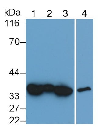
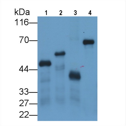 WB (Western Blot)
(Western Blot;Sample: Four differentRecombinant proteins with GST Tag (Lane 1-4).)
WB (Western Blot)
(Western Blot;Sample: Four differentRecombinant proteins with GST Tag (Lane 1-4).)
Glutathione S Transferase (GST), Monoclonal Antibody (Cat# AAA150822)
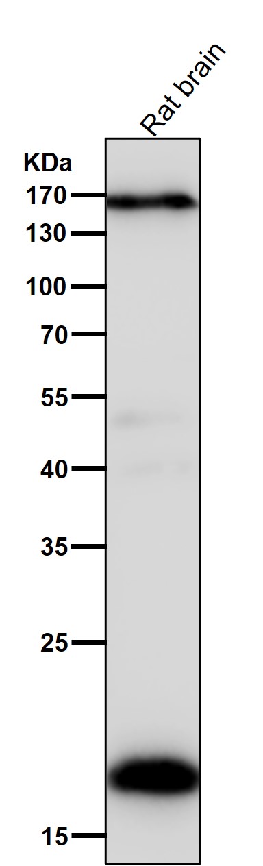
 WB (Western Blot)
(Western Blot; Sample: Lane1: Rat Liver lysate; Lane2: Rat CerebuM lysate; Lane3: Hela cell lysate; Lane4: HepG2 cell lysate; Lane5: HL60 cell lysate Primary Ab: 0.1ug/ml Mouse Anti-Rat IGF1 Antibody Second Ab: 0.2ug/mL HRP-Linked Caprine Anti-Mouse IgG Polyclonal Antibody)
WB (Western Blot)
(Western Blot; Sample: Lane1: Rat Liver lysate; Lane2: Rat CerebuM lysate; Lane3: Hela cell lysate; Lane4: HepG2 cell lysate; Lane5: HL60 cell lysate Primary Ab: 0.1ug/ml Mouse Anti-Rat IGF1 Antibody Second Ab: 0.2ug/mL HRP-Linked Caprine Anti-Mouse IgG Polyclonal Antibody)
Calretiulin (CALR), Monoclonal Antibody (Cat# AAA152804)
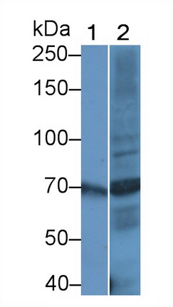
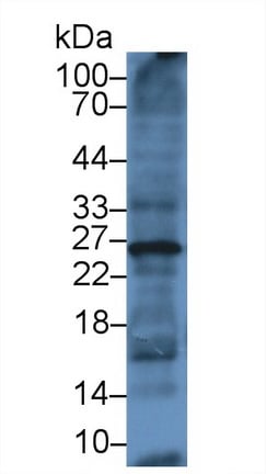
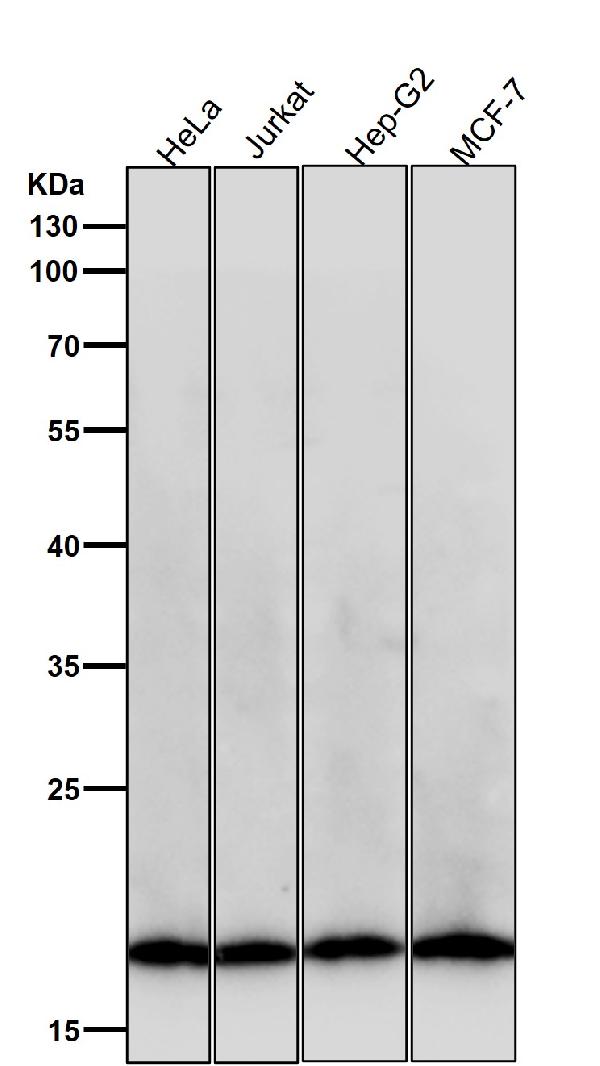
 WB (Western Blot)
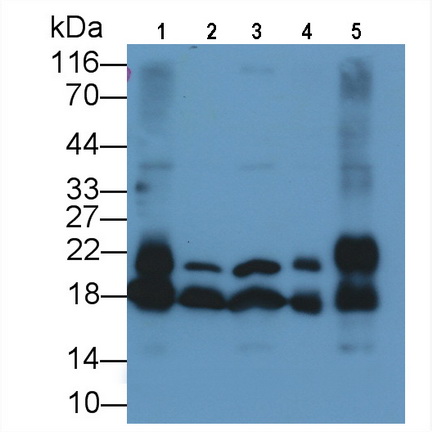
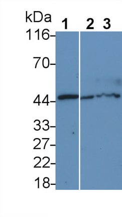
(Western Blot; Sample: Lane1: Rat Liver lysate; Lane2: Rat CerebuM lysate; Lane3: Rat Thymus lysate; Lane4: Rat Testis lysate; Lane5: Rat Ovary lysate Primary Ab: 0.2ug/ml Mouse Anti-Rat CYPA Antibody Second Ab: 0.2ug/mL HRP-Linked Caprine Anti-Mouse IgG Polyclonal Antibody)
WB (Western Blot)
(Western Blot; Sample: Lane1: Rat Liver lysate; Lane2: Rat CerebuM lysate; Lane3: Rat Thymus lysate; Lane4: Rat Testis lysate; Lane5: Rat Ovary lysate Primary Ab: 0.2ug/ml Mouse Anti-Rat CYPA Antibody Second Ab: 0.2ug/mL HRP-Linked Caprine Anti-Mouse IgG Polyclonal Antibody)
Cyclophilin A (CYPA), Monoclonal Antibody (Cat# AAA152810)
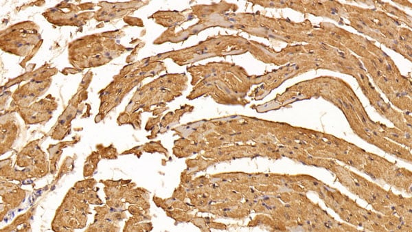
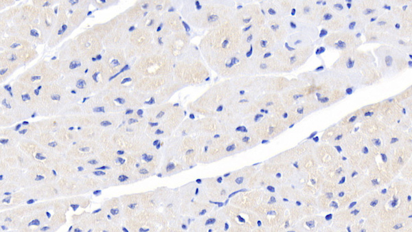
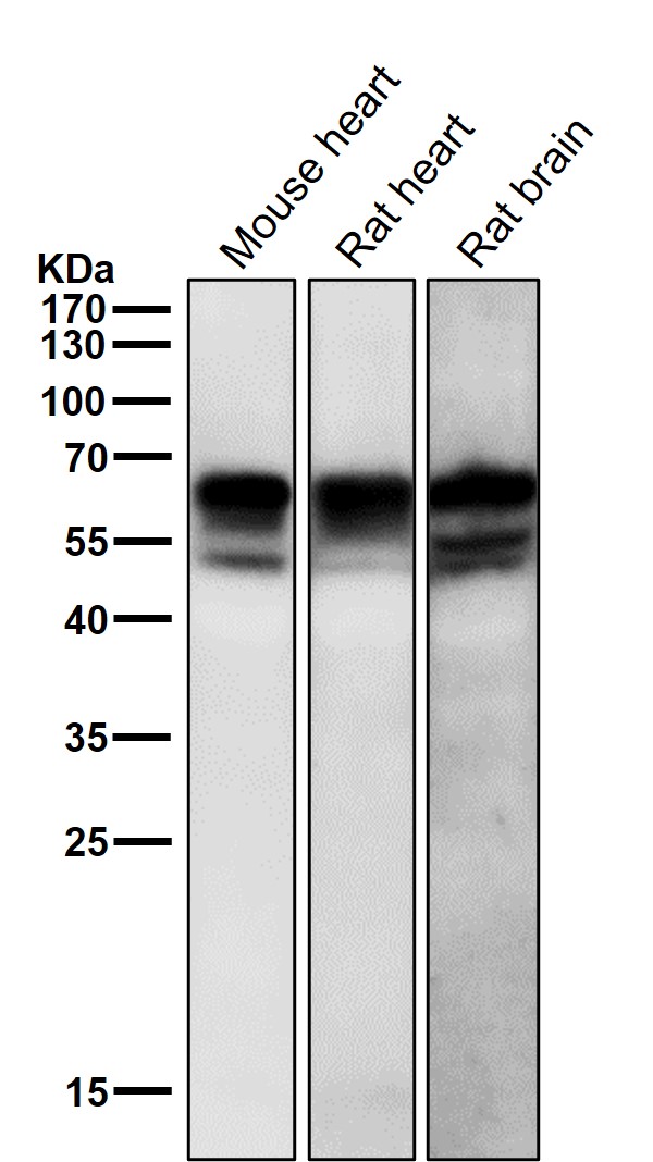
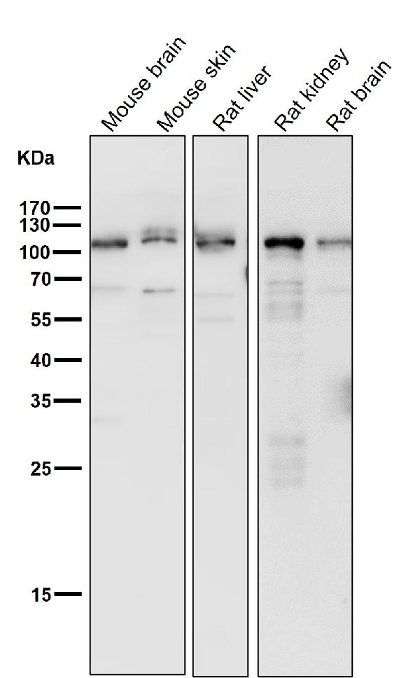
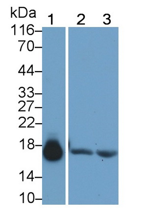 WB (Western Blot)
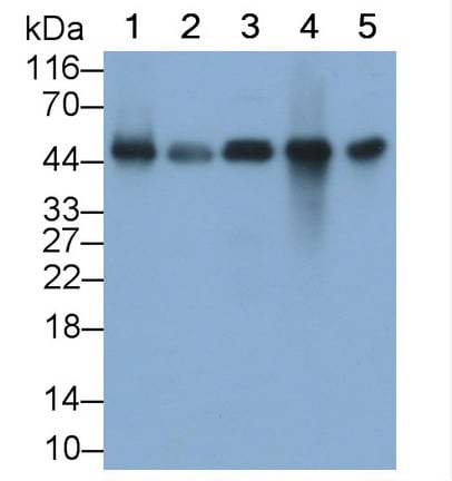
(Western Blot; Sample: Lane1: Porcine Heart lysate; Lane2: Rat Heart lysate; Lane3: Mouse Heart lysate Primary Ab: 0.4ug/ml Mouse Anti-human MYO Antibody Second Ab: 0.2ug/mL HRP-Linked Caprine Anti-Mouse IgG Polyclonal Antibody)
WB (Western Blot)
(Western Blot; Sample: Lane1: Porcine Heart lysate; Lane2: Rat Heart lysate; Lane3: Mouse Heart lysate Primary Ab: 0.4ug/ml Mouse Anti-human MYO Antibody Second Ab: 0.2ug/mL HRP-Linked Caprine Anti-Mouse IgG Polyclonal Antibody)
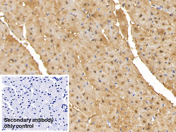
Myoglobin (MYO), Monoclonal Antibody (Cat# AAA152832)
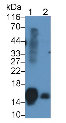
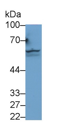
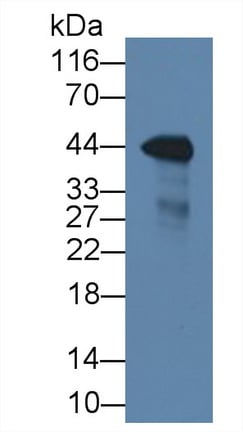
 WB (Western Blot)
(Western Blot; Sample: human Liver lysatePrimary Ab: 2ug/ml Mouse Anti-human Hpt AntibodySecond Ab: 0.2ug/mL HRP-Linked Caprine Anti-Mouse IgG Polyclonal Antibody)
WB (Western Blot)
(Western Blot; Sample: human Liver lysatePrimary Ab: 2ug/ml Mouse Anti-human Hpt AntibodySecond Ab: 0.2ug/mL HRP-Linked Caprine Anti-Mouse IgG Polyclonal Antibody)
Haptoglobin (Hpt), Monoclonal Antibody (Cat# AAA152916)
 WB (Western Blot)
(EDTA-passaged (left) and Trypsin-passaged (right) Human Umbilical Vein Endothelial Cells (HUVEC) were lysed, and lysates were loaded at 1x10⁵ cells/lane, probed with 2 ug/mL purified 16B1 and revealed with HRP anti-rat IgG.)
WB (Western Blot)
(EDTA-passaged (left) and Trypsin-passaged (right) Human Umbilical Vein Endothelial Cells (HUVEC) were lysed, and lysates were loaded at 1x10⁵ cells/lane, probed with 2 ug/mL purified 16B1 and revealed with HRP anti-rat IgG.)
CD144/CDH5/VE Cadherin, Monoclonal Antibody (Cat# AAA162233)
 WB (Western Blot)
(All lanes use the Antibody at 1:3K dilution for 1 hour at room temperature.)
WB (Western Blot)
(All lanes use the Antibody at 1:3K dilution for 1 hour at room temperature.)
Src, Monoclonal Antibody (Cat# AAA128108)
 WB (Western Blot)
(All lanes use the Antibody at 1:5K dilution for 1 hour at room temperature.)
WB (Western Blot)
(All lanes use the Antibody at 1:5K dilution for 1 hour at room temperature.)
Pleiotrophin, Monoclonal Antibody (Cat# AAA128133)
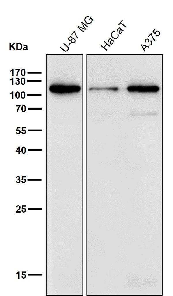
 WB (Western Blot)
(Western blot analysis of Desmocollin 1 expression in A375 cell lysate.)
WB (Western Blot)
(Western blot analysis of Desmocollin 1 expression in A375 cell lysate.)
Desmocollin 1, Monoclonal Antibody (Cat# AAA128157)
Clara Cell Protein 16 (CC16), Monoclonal Antibody (Cat# AAA151404)
 WB (Western Blot)
(Western Blot; Sample: Lane1: Porcine CerebeluM lysate; Lane2: Rat CerebuM lysate; Lane3: Rat CerebeluM lysatePrimary Ab: 1ug/ml Mouse Anti-human PTEN AntibodySecond Ab: 0.2ug/mL HRP-Linked Caprine Anti-Mouse IgG Polyclonal Antibody)
WB (Western Blot)
(Western Blot; Sample: Lane1: Porcine CerebeluM lysate; Lane2: Rat CerebuM lysate; Lane3: Rat CerebeluM lysatePrimary Ab: 1ug/ml Mouse Anti-human PTEN AntibodySecond Ab: 0.2ug/mL HRP-Linked Caprine Anti-Mouse IgG Polyclonal Antibody)
Phosphatase And Tensin Homolog (PTEN), Monoclonal Antibody (Cat# AAA152612)
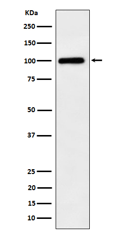
 WB (Western Blot)
(Western Blot; Sample: Rat Lung lysate; Primary Ab: 2ug/ml Mouse Anti-human POSTN Antibody Second Ab: 0.2ug/mL HRP-Linked Caprine Anti-Mouse IgG Polyclonal Antibody)
WB (Western Blot)
(Western Blot; Sample: Rat Lung lysate; Primary Ab: 2ug/ml Mouse Anti-human POSTN Antibody Second Ab: 0.2ug/mL HRP-Linked Caprine Anti-Mouse IgG Polyclonal Antibody)
Periostin (POSTN), Monoclonal Antibody (Cat# AAA152645)
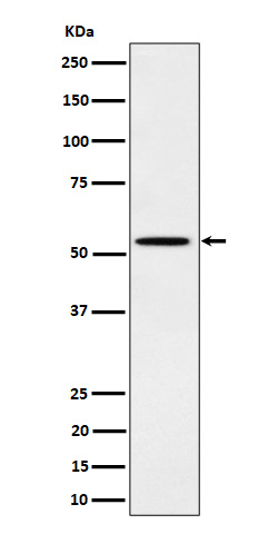
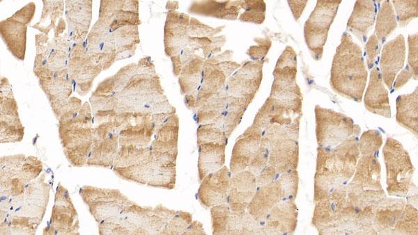
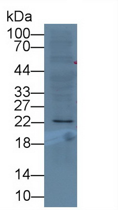
 WB (Western Blot)
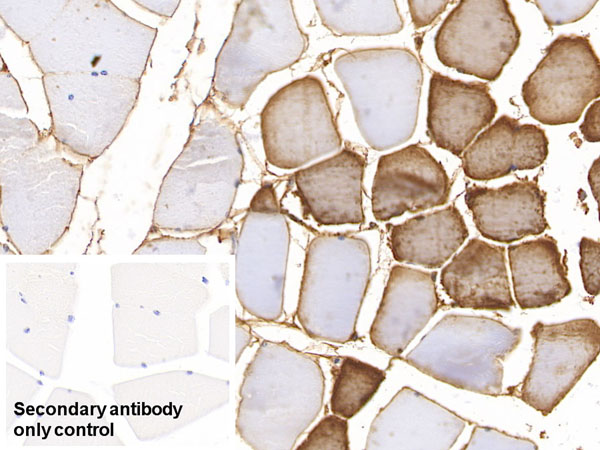
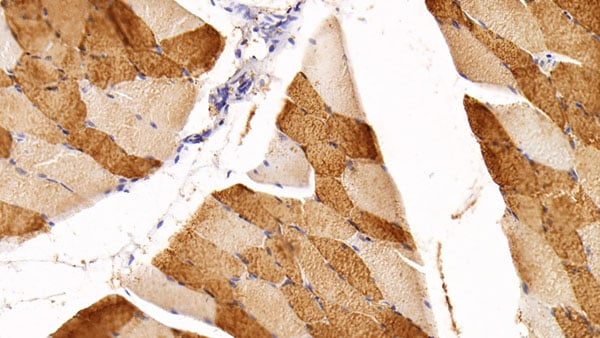
(Western Blot; Sample: Rat Skeletal muscle lysate Primary Ab: 1.5ug/ml Mouse Anti-human SIRT3 Antibody Second Ab: 0.2ug/mL HRP-Linked Caprine Anti-Mouse IgG Polyclonal Antibody)
WB (Western Blot)
(Western Blot; Sample: Rat Skeletal muscle lysate Primary Ab: 1.5ug/ml Mouse Anti-human SIRT3 Antibody Second Ab: 0.2ug/mL HRP-Linked Caprine Anti-Mouse IgG Polyclonal Antibody)
Sirtuin 3 (SIRT3), Monoclonal Antibody (Cat# AAA152711)
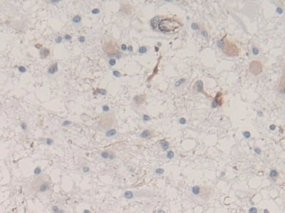
 IHC (Immunohistochemisry)
(DAB staining on IHC-P;Sample: human CerebuM Tissue; Primary Ab: 20ug/ml Mouse Anti-human MAPT AntibodySecond Ab: 2ug/mL HRP-Linked Caprine Anti-Mouse IgG Polyclonal Antibody)
IHC (Immunohistochemisry)
(DAB staining on IHC-P;Sample: human CerebuM Tissue; Primary Ab: 20ug/ml Mouse Anti-human MAPT AntibodySecond Ab: 2ug/mL HRP-Linked Caprine Anti-Mouse IgG Polyclonal Antibody)
Tau Protein (MAPT), Monoclonal Antibody (Cat# AAA152773)
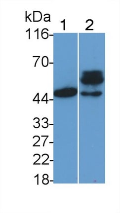
 WB (Western Blot)
(Western Blot; Sample: Lane1: human Saliva; Lane2: human PlasmaPrimary Ab: 2ug/ml Mouse Anti-human SAA AntibodySecond Ab: 0.2ug/mL HRP-Linked Caprine Anti-Mouse IgG Polyclonal Antibody)
WB (Western Blot)
(Western Blot; Sample: Lane1: human Saliva; Lane2: human PlasmaPrimary Ab: 2ug/ml Mouse Anti-human SAA AntibodySecond Ab: 0.2ug/mL HRP-Linked Caprine Anti-Mouse IgG Polyclonal Antibody)
SeuM Amyloid A (SAA), Monoclonal Antibody (Cat# AAA152675)
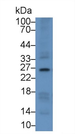
 WB (Western Blot)
(Western Blot; Sample: Hela cell lysate Primary Ab: 2ug/ml Mouse Anti-Mouse DTYMK Antibody Second Ab: 0.2ug/mL HRP-Linked Caprine Anti-Mouse IgG Polyclonal Antibody)
WB (Western Blot)
(Western Blot; Sample: Hela cell lysate Primary Ab: 2ug/ml Mouse Anti-Mouse DTYMK Antibody Second Ab: 0.2ug/mL HRP-Linked Caprine Anti-Mouse IgG Polyclonal Antibody)
Deoxythymidylate Kinase (DTYMK), Monoclonal Antibody (Cat# AAA152677)
CD279, Monoclonal Antibody (Cat# AAA128513)
CD279, Monoclonal Antibody (Cat# AAA128514)
N-Sulfated Heparan Sulfate, Monoclonal Antibody (Cat# AAA128576)
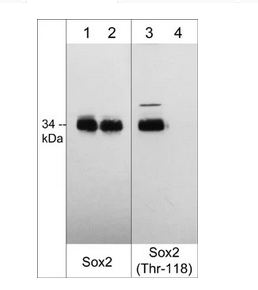
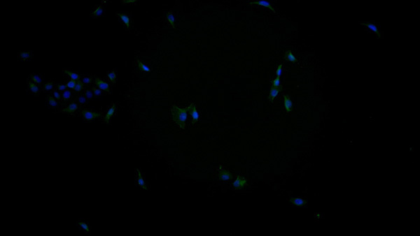
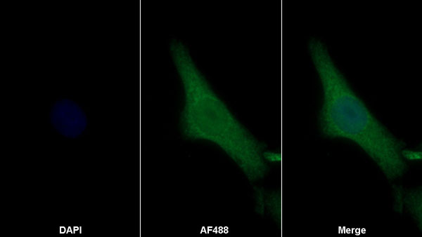
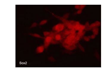 ICC (Immunocytochemistry)
(Immunocytochemical labeling of Sox2 in aldehyde fixed and NP-40 permeabilized human NCI-H446 lung carcinoma cells. The cells were labeled with mouse monoclonal anti-Sox2 (SM5511). The antibody was detected using goat anti-mouse DyLight 594.)
ICC (Immunocytochemistry)
(Immunocytochemical labeling of Sox2 in aldehyde fixed and NP-40 permeabilized human NCI-H446 lung carcinoma cells. The cells were labeled with mouse monoclonal anti-Sox2 (SM5511). The antibody was detected using goat anti-mouse DyLight 594.)
Sox2, Monoclonal Antibody (Cat# AAA71707)
 Application Data
(Fig.2: Specificity testing of TET2. BOSC cells were transiently transfected with expression vectors containing either the cDNA of CEACAM1, 3, 5, 6, 7, 8 or a recombinant transmembrane-anchored PSG1 fusion protein. Recognition of CEACAM4 was tested on CHO cells stably transfected with a CEACAM4 expression vector. Expression of the constructs was confirmed with monoclonal antibodies known to recognise the corresponding proteins (CEACAM1, 3, 4, 5 and 6: D14HD11; CEACAM7: CAC2; CEACAM8: 80H3; PSG1: BAP1; green curves). An irrelevant monoclonal antibody served as a negative control (black curves). For specificity testing, protein G purified TET2 was tested on all CEACAM transfectants. A positive signal was obtained with CEACAM1, CEACAM5, CEACAM6 and CEACAM8 expressing cells (red curves).)
Application Data
(Fig.2: Specificity testing of TET2. BOSC cells were transiently transfected with expression vectors containing either the cDNA of CEACAM1, 3, 5, 6, 7, 8 or a recombinant transmembrane-anchored PSG1 fusion protein. Recognition of CEACAM4 was tested on CHO cells stably transfected with a CEACAM4 expression vector. Expression of the constructs was confirmed with monoclonal antibodies known to recognise the corresponding proteins (CEACAM1, 3, 4, 5 and 6: D14HD11; CEACAM7: CAC2; CEACAM8: 80H3; PSG1: BAP1; green curves). An irrelevant monoclonal antibody served as a negative control (black curves). For specificity testing, protein G purified TET2 was tested on all CEACAM transfectants. A positive signal was obtained with CEACAM1, CEACAM5, CEACAM6 and CEACAM8 expressing cells (red curves).)
CEACAM1,5,6,8, Monoclonal Antibody (Cat# AAA71469)
 Application Data
(Fig.2: SDS-PAGE analysis of purified LV-2A2 monoclonal antibody. Lane 1: molecular weight marker, Lane 2: 2 ?g of purified LV-2A2 antibody. Proteins were separated by SDS-PAGE and stained with RAPID StainTM Reagent.)
Application Data
(Fig.2: SDS-PAGE analysis of purified LV-2A2 monoclonal antibody. Lane 1: molecular weight marker, Lane 2: 2 ?g of purified LV-2A2 antibody. Proteins were separated by SDS-PAGE and stained with RAPID StainTM Reagent.)
Crisp3, Monoclonal Antibody (Cat# AAA71471)

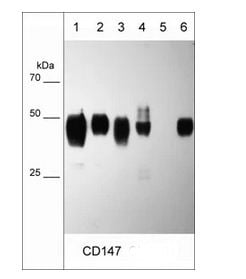
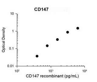 Standard Curve (Sample)
(Representative Standard Curve using mouse monoclonal CD147 (CM0361) for ELISA capture of human recombinant CD147 extracellular region with a His tag. Capture was detected using anti-His tag antibody followed by appropriate secondary antibody HRP conjugate.)
Standard Curve (Sample)
(Representative Standard Curve using mouse monoclonal CD147 (CM0361) for ELISA capture of human recombinant CD147 extracellular region with a His tag. Capture was detected using anti-His tag antibody followed by appropriate secondary antibody HRP conjugate.)
CD147/Emmprin/Basigin, Monoclonal Antibody (Cat# AAA71585)
HIV, gp160,120, Monoclonal Antibody (Cat# AAA71354)
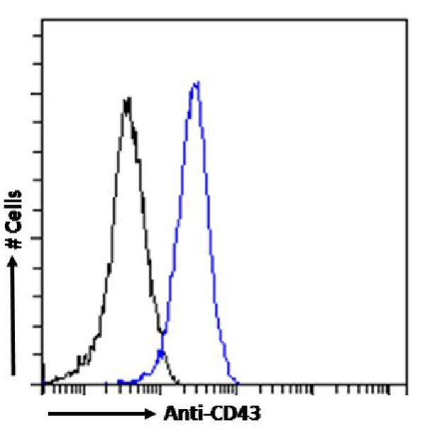
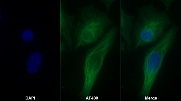
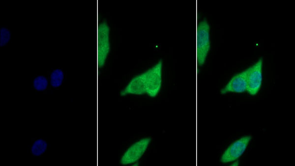
 IF (Immunofluorescence)
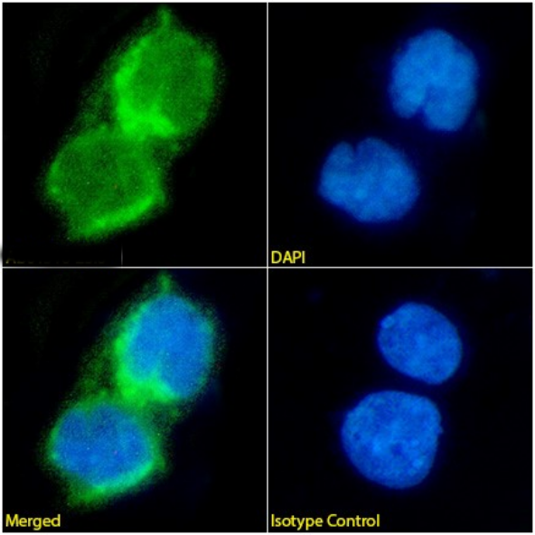
(Immunofluorescencestainingoffixedratsplenocyteswithanti-CD43antibodyOX-58(AAA72485). ImmunofluorescenceanalysisofparaformaldehydefixedratsplenocytesonShi-fixcover-slipsstainedwiththechimericrabbitIgGversionofOX-58(AAA72485)(1:100dilution)for1hfollowedbyAlexaFluor488secondaryantibody(1:1000dilution),showingmembraneandcytoplasmicstaining.ThenuclearstainisDAPI(blue).Panelsshowfromleft-right,top-bottomAAA72485,DAPI,mergedchannelsandanisotypecontrol.TheisotypecontrolwasanunknownspecificityantibodyfollowedbystainingwithAlexaFluor488secondaryantibody.)
IF (Immunofluorescence)
(Immunofluorescencestainingoffixedratsplenocyteswithanti-CD43antibodyOX-58(AAA72485). ImmunofluorescenceanalysisofparaformaldehydefixedratsplenocytesonShi-fixcover-slipsstainedwiththechimericrabbitIgGversionofOX-58(AAA72485)(1:100dilution)for1hfollowedbyAlexaFluor488secondaryantibody(1:1000dilution),showingmembraneandcytoplasmicstaining.ThenuclearstainisDAPI(blue).Panelsshowfromleft-right,top-bottomAAA72485,DAPI,mergedchannelsandanisotypecontrol.TheisotypecontrolwasanunknownspecificityantibodyfollowedbystainingwithAlexaFluor488secondaryantibody.)
CD43, Monoclonal Recombinant Antibody (Cat# AAA72485)
 IF (Immunofluorescence)
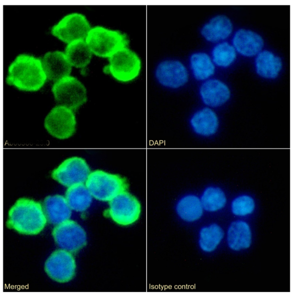
(ImmunofluorescencestainingofJurkatcellswithanti-CD8antibodyBAG45G1(AAA72516). ImmunofluorescenceanalysisofparaformaldehydefixedJurkatcellsonShi-fixcoverslipsstainedwiththechimericrabbitIgGversionofBAG45G1(AAA72516)(1:100dilution)for1hfollowedbyAlexaFluor488secondaryantibody(1:1000dilution),showingmembranestaining.ThenuclearstainisDAPI(blue).Panelsshow,fromleft-right,top-bottom,AAA72516,DAPI,mergedchannelsandanisotypecontrol.TheisotypecontrolwasanunknownspecificityantibodyfollowedbystainingwithAlexaFluor488secondaryantibody.)
IF (Immunofluorescence)
(ImmunofluorescencestainingofJurkatcellswithanti-CD8antibodyBAG45G1(AAA72516). ImmunofluorescenceanalysisofparaformaldehydefixedJurkatcellsonShi-fixcoverslipsstainedwiththechimericrabbitIgGversionofBAG45G1(AAA72516)(1:100dilution)for1hfollowedbyAlexaFluor488secondaryantibody(1:1000dilution),showingmembranestaining.ThenuclearstainisDAPI(blue).Panelsshow,fromleft-right,top-bottom,AAA72516,DAPI,mergedchannelsandanisotypecontrol.TheisotypecontrolwasanunknownspecificityantibodyfollowedbystainingwithAlexaFluor488secondaryantibody.)
CD8, Monoclonal Recombinant Antibody (Cat# AAA72516)
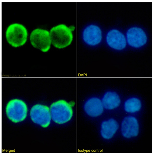 IF (Immunofluorescence)
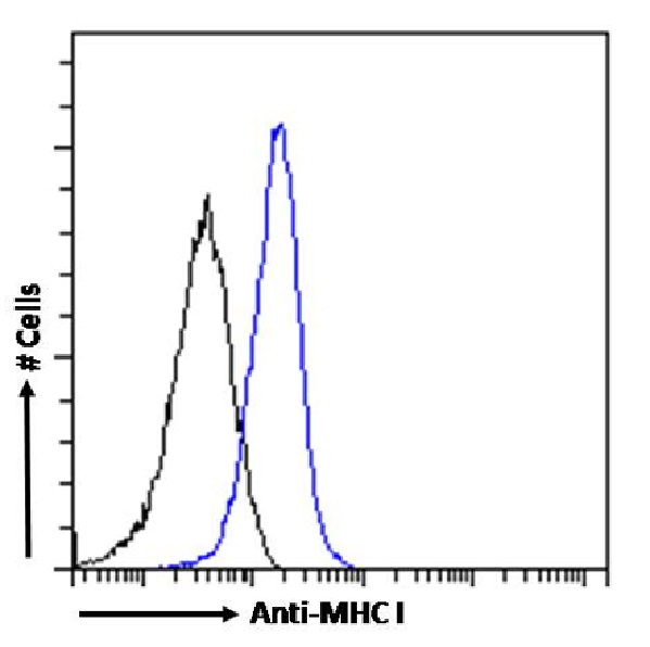
(Immunofluorescencestainingoffixedratsplenocyteswithanti-MHCI(RT1A)antibodyOX-18(AAA72456). ImmunofluorescenceanalysisofparaformaldehydefixedratsplenocytesonShi-fixcover-slipsstainedwiththechimericrabbitIgGversionofOX-18(AAA72456)(1:100dilution)for1hfollowedbyAlexaFluor488secondaryantibody(1:1000dilution),showingmembraneandcytoplasmicstaining.ThenuclearstainisDAPI(blue).Panelsshowfromleft-right,top-bottomAAA72456,DAPI,mergedchannelsandanisotypecontrol.TheisotypecontrolwasanunknownspecificityantibodyfollowedbystainingwithAlexaFluor488secondaryantibody.)
IF (Immunofluorescence)
(Immunofluorescencestainingoffixedratsplenocyteswithanti-MHCI(RT1A)antibodyOX-18(AAA72456). ImmunofluorescenceanalysisofparaformaldehydefixedratsplenocytesonShi-fixcover-slipsstainedwiththechimericrabbitIgGversionofOX-18(AAA72456)(1:100dilution)for1hfollowedbyAlexaFluor488secondaryantibody(1:1000dilution),showingmembraneandcytoplasmicstaining.ThenuclearstainisDAPI(blue).Panelsshowfromleft-right,top-bottomAAA72456,DAPI,mergedchannelsandanisotypecontrol.TheisotypecontrolwasanunknownspecificityantibodyfollowedbystainingwithAlexaFluor488secondaryantibody.)
MHC I (RT1A), Monoclonal Recombinant Antibody (Cat# AAA72456)

 FCM/FACS (Flow Cytometry)
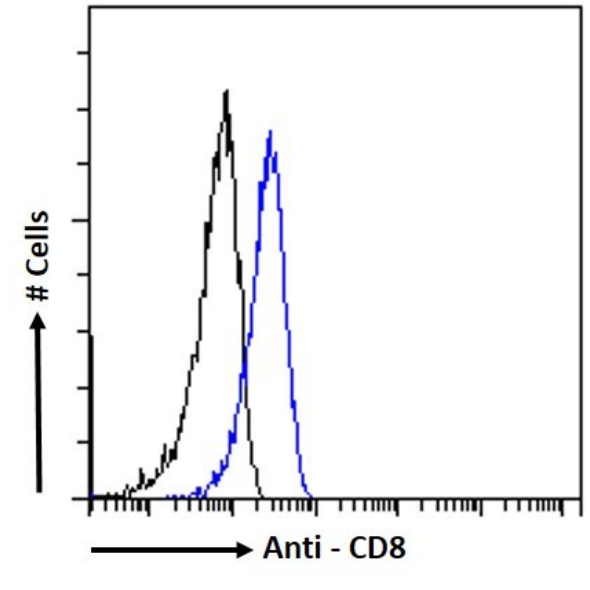
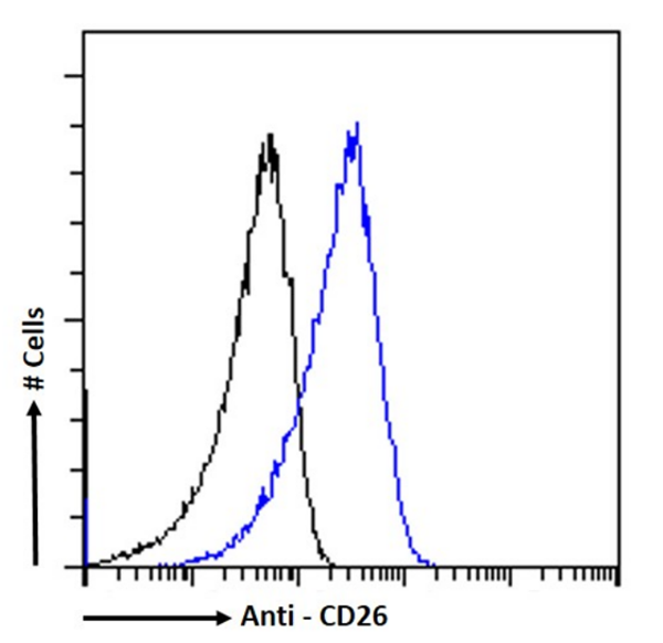
(Flowcytometryusingtheanti-CD26antibodyOX-61(AAA72464). Paraformaldehydefixedratsplenocyteseswerestainedwithanti-unknownspecificityantibodyortherabbitIgGversionofOX-61(AAA72464,blueline)atadilutionof1:100for1hatRT.Afterwashing,theboundantibodywasdetectedusingagoatanti-rabbitIgGAlexaFluor488antibodyatadilutionof1:1000andcellsanalyzedusingaFACSCantoflow-cytometer.)
FCM/FACS (Flow Cytometry)
(Flowcytometryusingtheanti-CD26antibodyOX-61(AAA72464). Paraformaldehydefixedratsplenocyteseswerestainedwithanti-unknownspecificityantibodyortherabbitIgGversionofOX-61(AAA72464,blueline)atadilutionof1:100for1hatRT.Afterwashing,theboundantibodywasdetectedusingagoatanti-rabbitIgGAlexaFluor488antibodyatadilutionof1:1000andcellsanalyzedusingaFACSCantoflow-cytometer.)
CD26, Monoclonal Recombinant Antibody (Cat# AAA72464)
What are Monoclonal Antibodies?
Monoclonal antibodies are specialized laboratory-produced proteins developed for binding to specific biological antigens or other molecular targets. Since they come from a single cell (or clone), they are especially consistent and accurate in the data they are involved in producing.
This type of antibody material has been shown to be a powerful tool in finding and subsequently destroying harmful cells in an organism, such as those found in cancers or various autoimmune diseases. This makes them excellent aids in medical testing and research, which is why they are so widely used.
AAA Biotech offers a comprehensive range of high-quality monoclonal antibodies that perform effectively in various laboratory tests, including (amongst others) ELISA, western blotting, immunohistochemistry, and flow cytometry. All of the products in our catalog are thoroughly quality tested to make sure that they are reliable and will consistently perform well in your research.
What Are The Uses of Monoclonal Antibodies
Monoclonal antibodies are used in many lab tests, including (amongst others) ELISA, western blotting, immunohistochemistry, and flow cytometry.
ELISA is a test that helps detect a specific substance/analyte in a sample. It uses antibodies (often monoclonal) bound to a solid surface (such as the well of a microplate) to “capture” the substance/analyte in the sample and immobilize it so that the detection antibody component can then bind to it and produce a signal, which can then be measured.
Western blotting identifies specific proteins in a sample. The sample is first separated on a gel, and then antibodies are applied that will typically bind to the target, which will all be localized to a single band in a lane.
Immunohistochemistry helps locate specific proteins in cells or tissue samples using antibodies.
Flow cytometry looks at and sorts cells. It uses antibodies that are conjugated to reporter molecules called “fluorophores”, which, under special lights, emit light themselves, which can then be measured by a detector instrument.
How Monoclonal Antibodies Are Used as Medicine?
Please note that all of the products listed in AAA Biotech’s also known as AAA Bio or AAABio catalog are strictly for research-use only (RUO).
Monoclonal antibodies can also be used as therapeutic/medical treatments, particularly in the context of cancers. They are designed to find and bind to specific cells or proteins, helping the immune system recognize and attack the cancer. These treatments work in different ways, such as:
- Radioimmunotherapy attaches a small amount of radioactive molecule to the antibody, so it delivers the radiation directly to the cancer cells that the antibody is specifically binding to.
- Antibody-directed enzyme prodrug therapy uses antibodies that are specifically bound to special enzymes. These enzymes activate a harmless drug in the body and turn it into a cancer-killing drug only near the cancer cells—this helps avoid harming healthy cells.
- Immunoliposomes are tiny “bubbles” filled with medicine/drug and coated with antibodies. They carry the drug straight to the cancer cells.
Why Buy Monoclonal Antibodies From Us?
At AAA Biotech, we provide high-performance monoclonal antibodies designed to support a wide range of research needs.
1. Validated for Versatile Applications
The antibodies in our catalog are extensively validated and compatible with multiple techniques, including (but not limited to) ELISA, flow cytometry (FC), immunocytochemistry (ICC), immunofluorescence (IF), immunohistochemistry (IHC), immunoprecipitation (IP), and western blotting (WB).
2. Wide Selection & Specialized Options
We offer antibodies for common and rare species, that are available in various conjugated forms, and also in recombinant formats. Essentially, there is almost anything one might need to meet their experimental model’s requirements.
3. High-Quality Proteins
Our proteins meet high purity standards—90% or more as confirmed by SDS-PAGE. Many are available with tags like His, Flag, GST, or MBP, and we also supply native and biologically active proteins for functional studies.
Frequently Asked Questions
1. Are your monoclonal antibodies validated for specific applications?
Yes, our antibodies are tested and validated for use in methods such as ELISA, western blot, IHC, flow cytometry, and more. Refer to specific product pages or datasheets for individual product information.
2. How do I choose the right monoclonal antibody for my application?
Review the product details directly for application validation, species reactivity, and target information. You may also contact our support team at any time for help.
3. How quickly can I receive my order?
Most orders are processed and shipped within 1–3 business days, depending on product availability and your shipping location.