Filters
▼Clonality
▼Type
▼Reactivity
▼Gene Name
▼Isotype
▼Host
▼Application
▼Clone
▼Monoclonal Antibodies
Get accurate results in your research with our Monoclonal Antibodies, which are specially made to target exactly what you require for your research, and will produce consistent, reliable performance in lab tests.
Viewing 7250-7300 of 27645 product results
 IF (Immunofluorescence)
(Immunofluorescent staining of COS7 cells transiently transfected with recombinant PECR protein using PECR antibody)
IF (Immunofluorescence)
(Immunofluorescent staining of COS7 cells transiently transfected with recombinant PECR protein using PECR antibody)
PECR, Monoclonal Antibody (Cat# AAA106724)
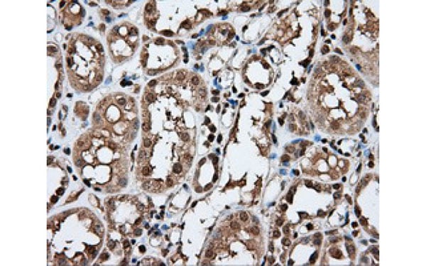
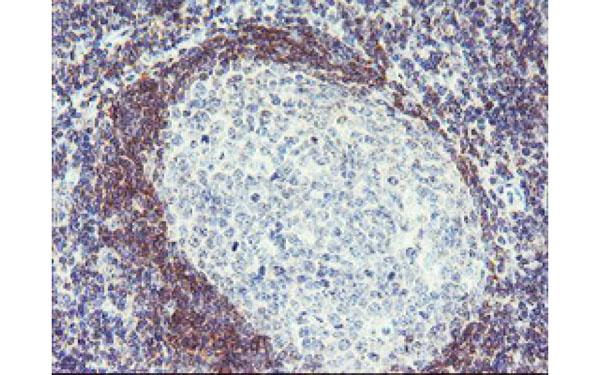 IHC (Immunohistochemisry)
(Immunohistochemical analysis of PLDN protein in paraffin embedded Human tonsil tissue using PLDN antibody)
IHC (Immunohistochemisry)
(Immunohistochemical analysis of PLDN protein in paraffin embedded Human tonsil tissue using PLDN antibody)
PLDN, Monoclonal Antibody (Cat# AAA106987)
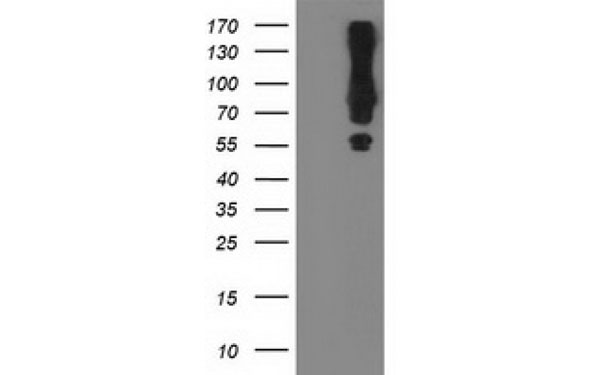
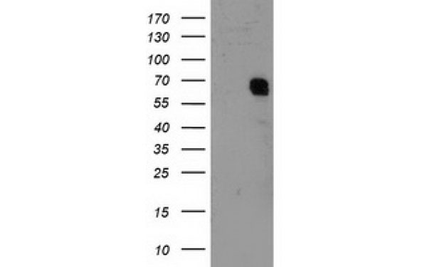
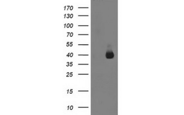
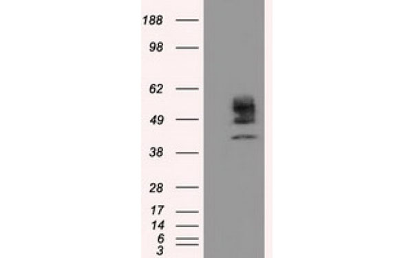
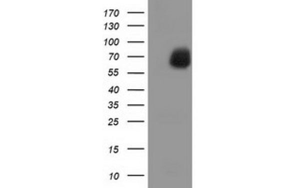
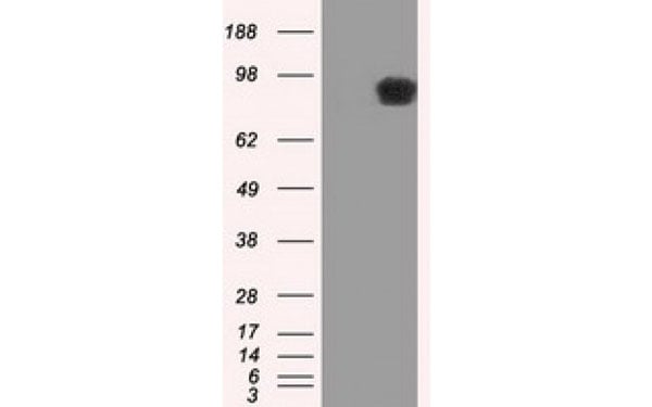
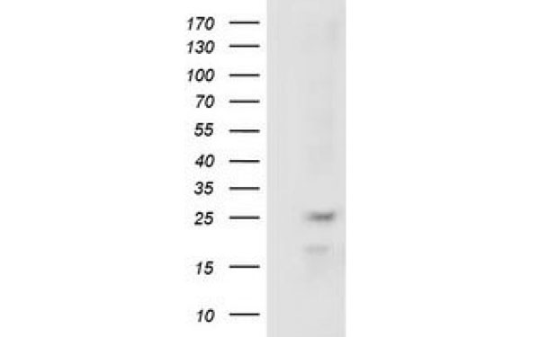 WB (Western Blot)
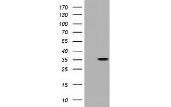
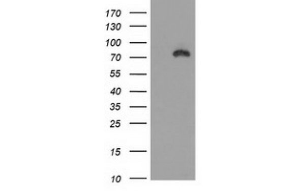
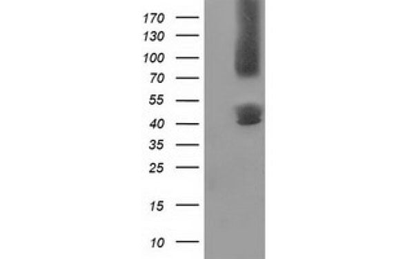
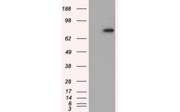
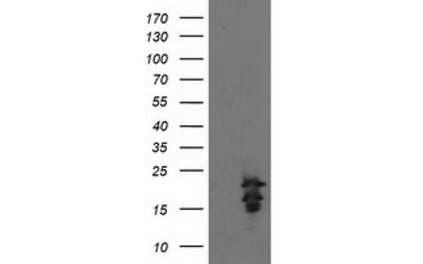
(Western Blot analysis of HEK293T cell lysates (5 ug) transfected with either recombinant RABL2A protein (Right) or empty vector (Left) detected with RABL2A antibody)
WB (Western Blot)
(Western Blot analysis of HEK293T cell lysates (5 ug) transfected with either recombinant RABL2A protein (Right) or empty vector (Left) detected with RABL2A antibody)
RABL2A, Monoclonal Antibody (Cat# AAA107016)
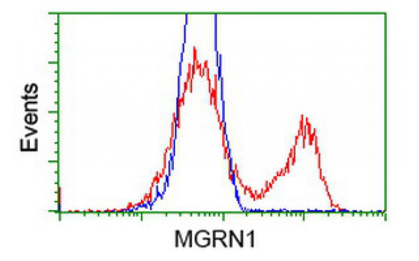
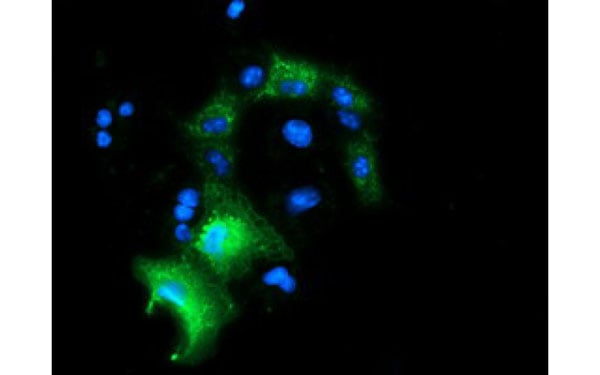
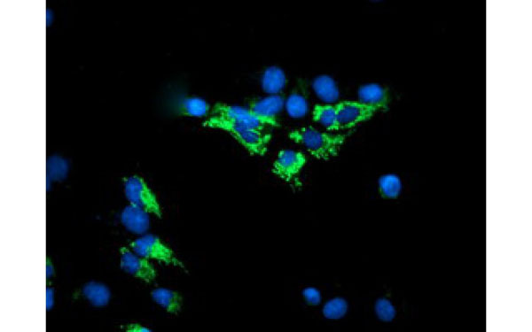
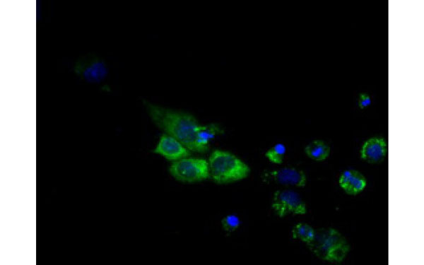
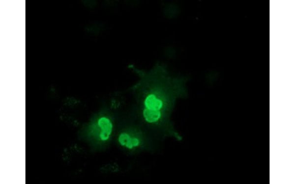
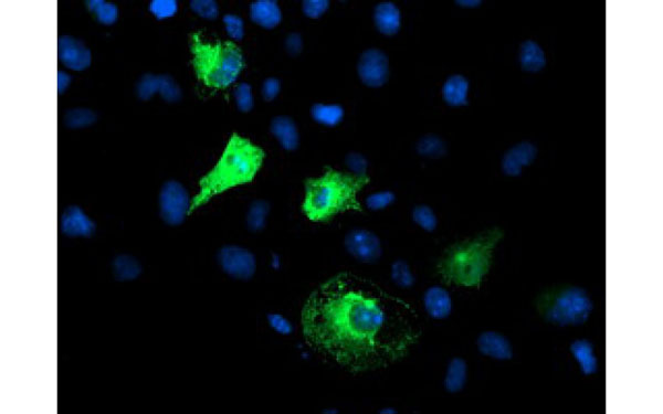
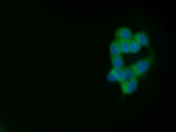
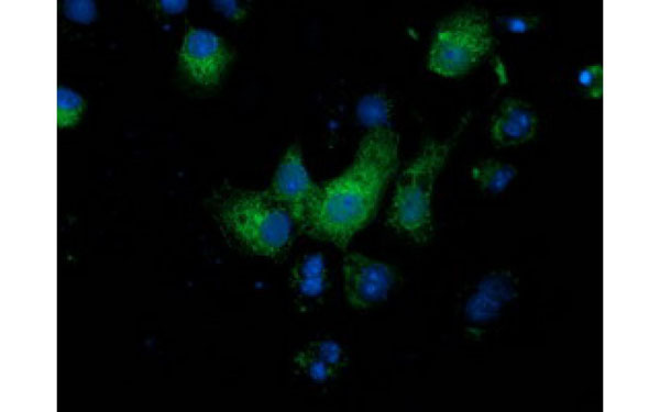 IF (Immunofluorescence)
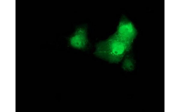
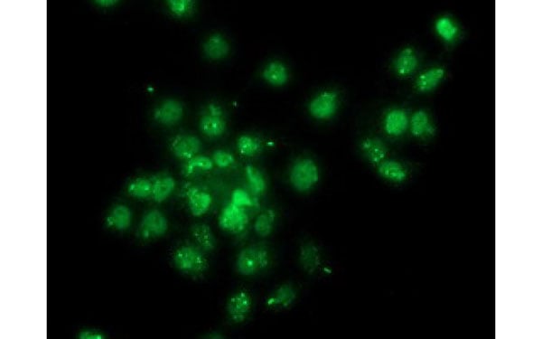
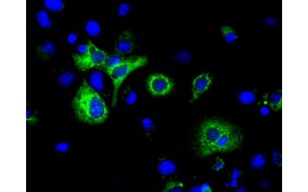
(Immunofluorescent staining of COS7 cells transiently transfected with recombinant MGRN1 protein using MGRN1 antibody)
IF (Immunofluorescence)
(Immunofluorescent staining of COS7 cells transiently transfected with recombinant MGRN1 protein using MGRN1 antibody)
MGRN1, Monoclonal Antibody (Cat# AAA107018)
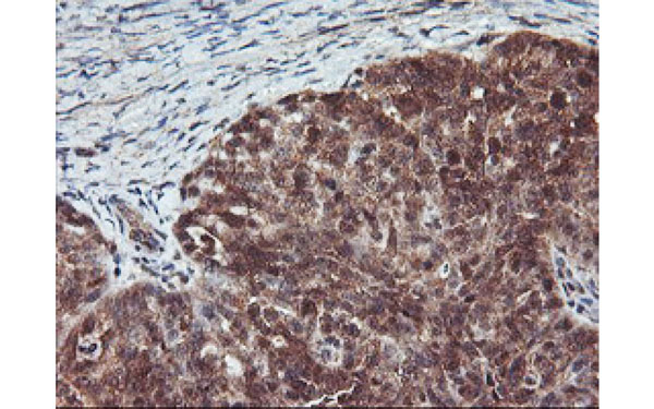
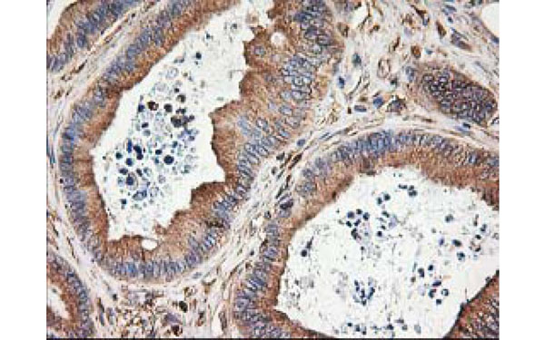
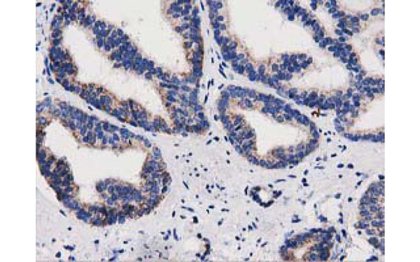
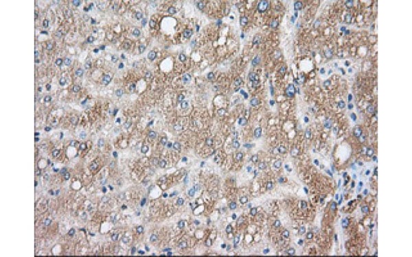
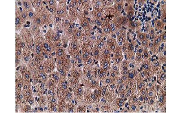
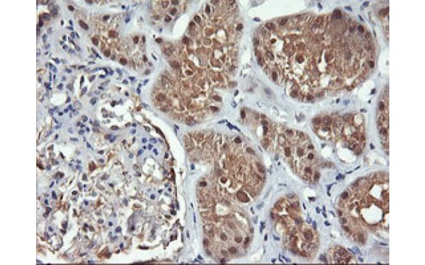
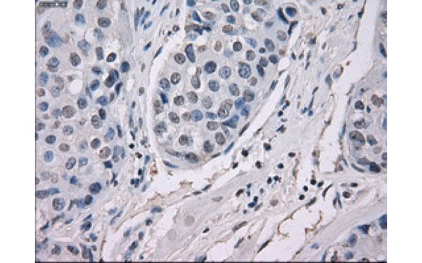
 IHC (Immunohistochemisry)
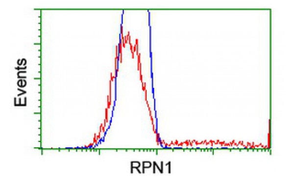
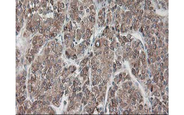
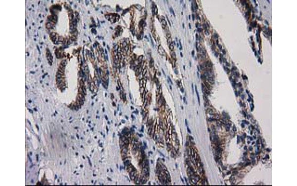
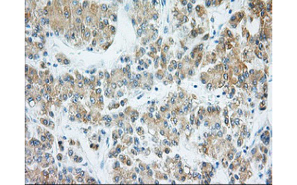
(Immunohistochemical analysis of RPN1 protein in paraffin embedded Carcinoma of Human liver tissue using RPN1 antibody)
IHC (Immunohistochemisry)
(Immunohistochemical analysis of RPN1 protein in paraffin embedded Carcinoma of Human liver tissue using RPN1 antibody)
RPN1, Monoclonal Antibody (Cat# AAA107023)
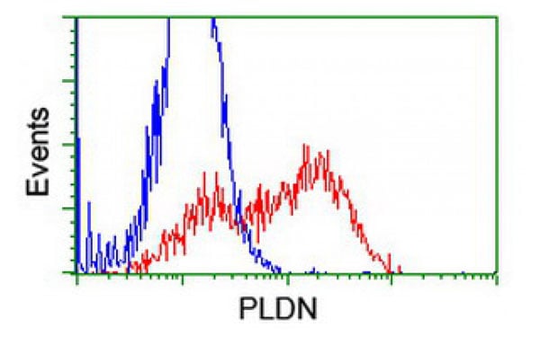
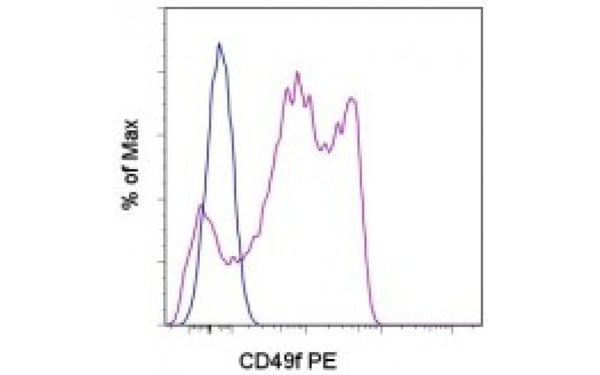 FCM/FACS (Flow Cytometry)
(Staining of normal human peripheral blood cells with Rat IgG2a kappa Isotype Control (PE) (blue histogram) or CD49f antibody (PE) (purple histogram). Cells in the lymphocyte gate were used for analysis.)
FCM/FACS (Flow Cytometry)
(Staining of normal human peripheral blood cells with Rat IgG2a kappa Isotype Control (PE) (blue histogram) or CD49f antibody (PE) (purple histogram). Cells in the lymphocyte gate were used for analysis.)
CD49f, Monoclonal Antibody (Cat# AAA107028)
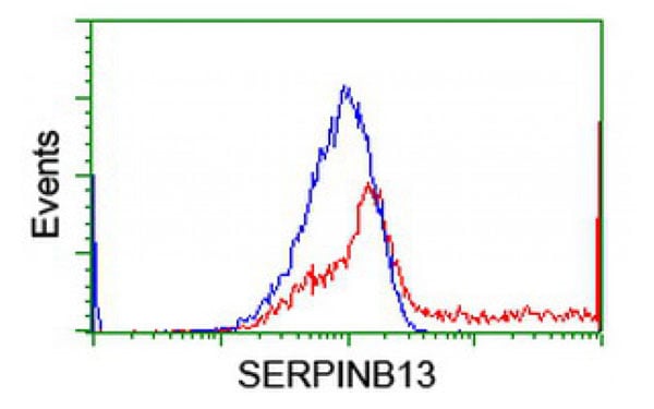
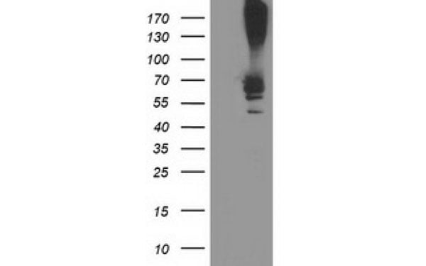
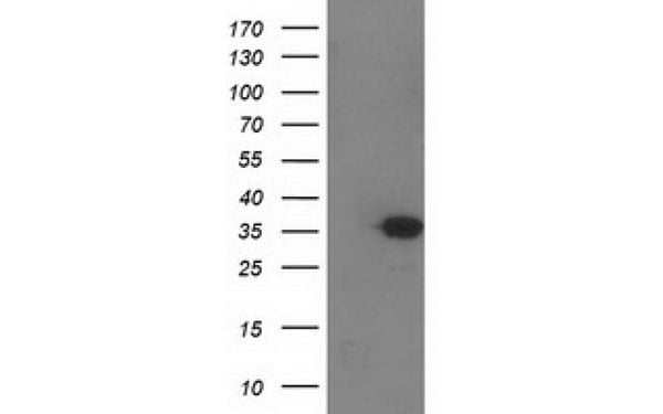
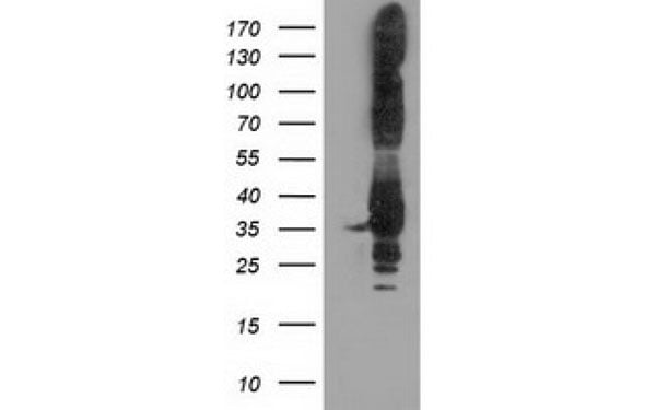
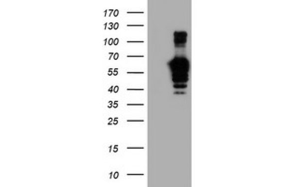
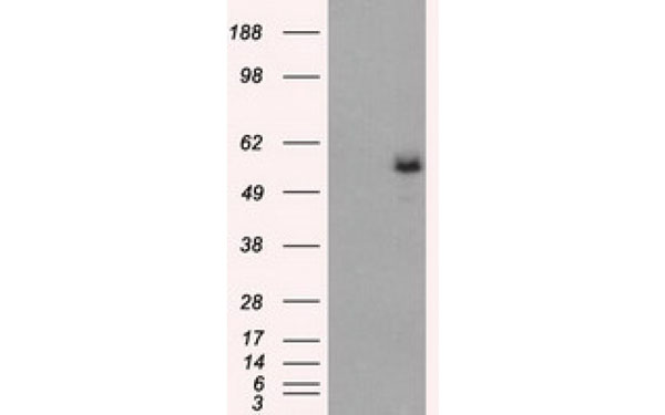
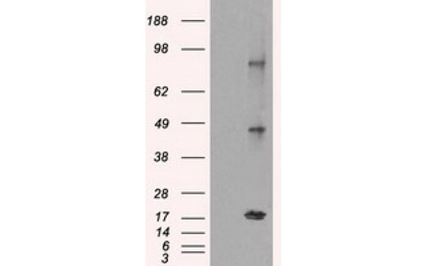
 WB (Western Blot)
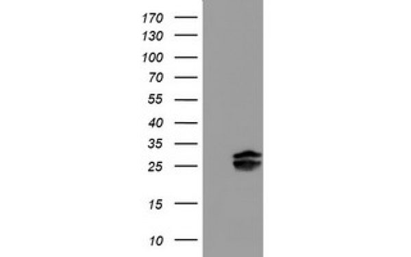
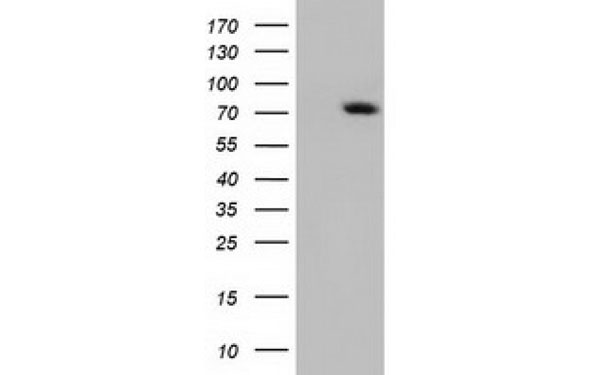
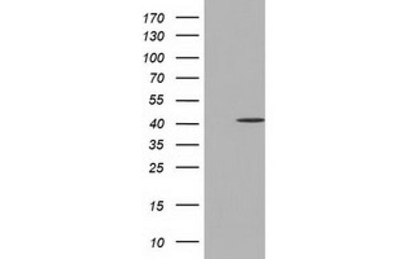
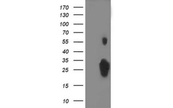
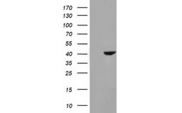
(Western Blot analysis of HEK293T cell lysates (5 ug) transfected with either recombinant SERPINB13 protein (Right) or empty vector (Left) detected with SERPINB13 antibody)
WB (Western Blot)
(Western Blot analysis of HEK293T cell lysates (5 ug) transfected with either recombinant SERPINB13 protein (Right) or empty vector (Left) detected with SERPINB13 antibody)
SERPINB13, Monoclonal Antibody (Cat# AAA107034)
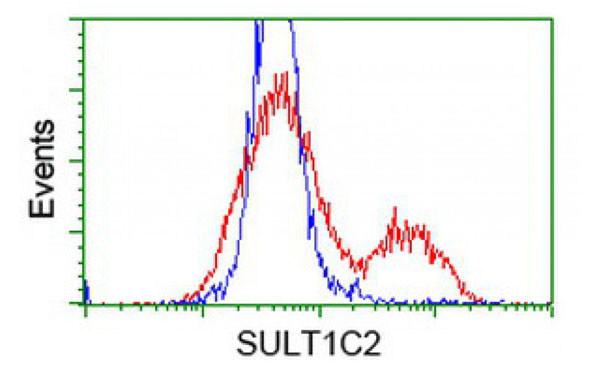
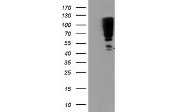
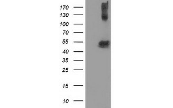
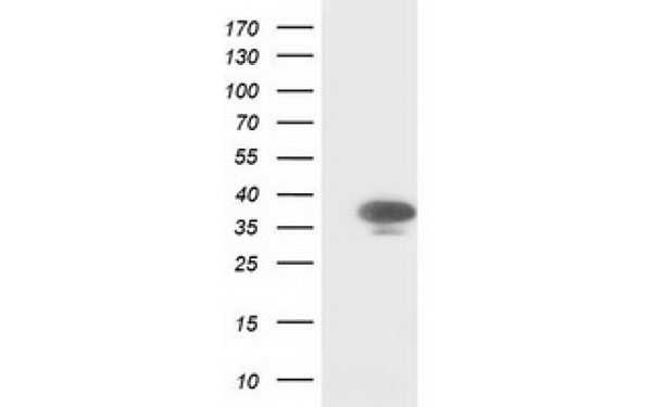
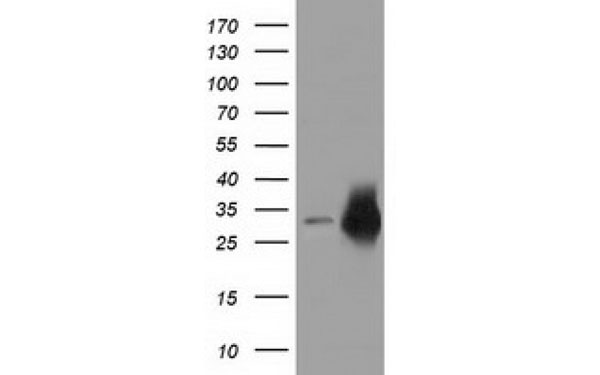
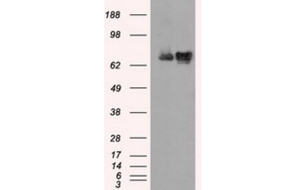
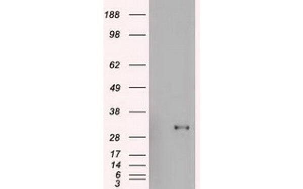
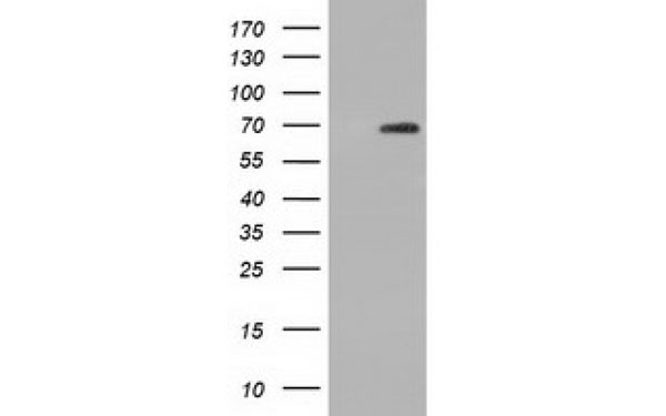
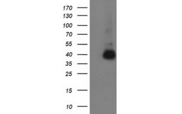
 WB (Western Blot)
(Western Blot analysis of HEK293T cell lysates (5 ug) transfected with either recombinant SulT1C2 protein (Right) or empty vector (Left) detected with SulT1C2 antibody)
WB (Western Blot)
(Western Blot analysis of HEK293T cell lysates (5 ug) transfected with either recombinant SulT1C2 protein (Right) or empty vector (Left) detected with SulT1C2 antibody)
SulT1C2, Monoclonal Antibody (Cat# AAA107044)
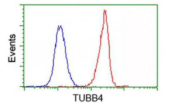
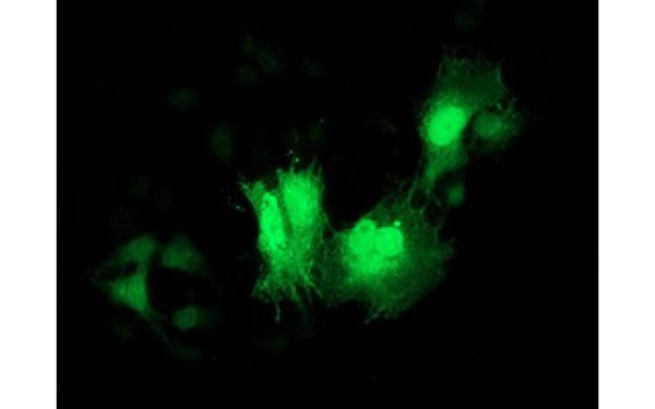
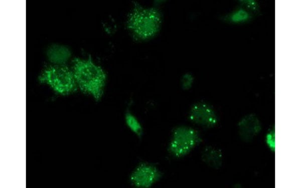
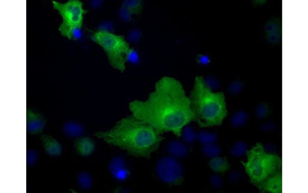
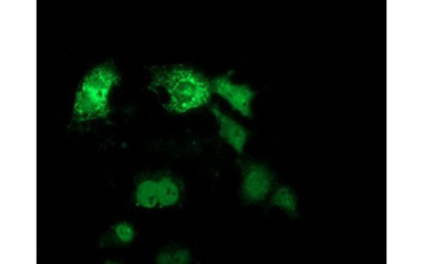
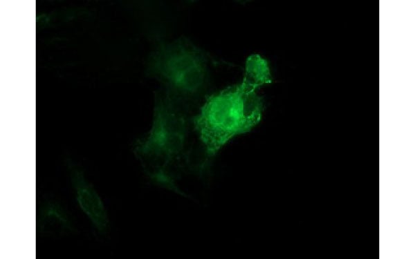 IF (Immunofluorescence)
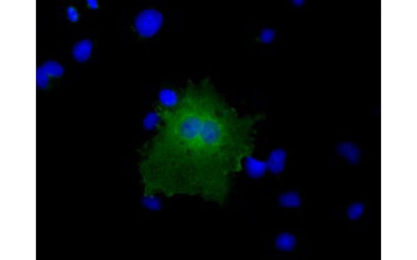
(Immunofluorescent staining of COS7 cells transiently transfected with recombinant TUBB4 protein using TUBB4 antibody)
IF (Immunofluorescence)
(Immunofluorescent staining of COS7 cells transiently transfected with recombinant TUBB4 protein using TUBB4 antibody)
TUBB4, Monoclonal Antibody (Cat# AAA107045)
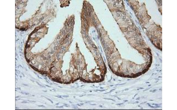
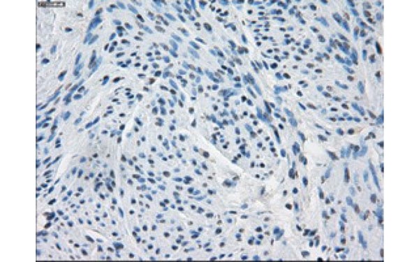
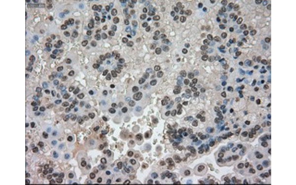
 IHC (Immunohistochemisry)
(Immunohistochemical analysis of PYCR2 protein in paraffin embedded Carcinoma of Human prostate tissue using PYCR2 antibody)
IHC (Immunohistochemisry)
(Immunohistochemical analysis of PYCR2 protein in paraffin embedded Carcinoma of Human prostate tissue using PYCR2 antibody)
PYCR2, Monoclonal Antibody (Cat# AAA107046)
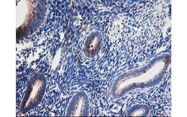
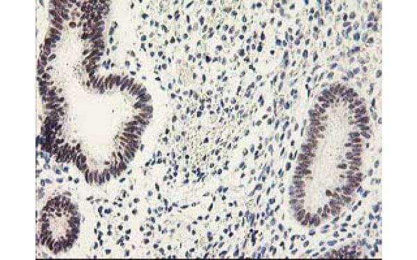 IHC (Immunohiostchemistry)
(Immunohistochemical analysis of GTF2F1 protein in paraffin embedded Human endometrium tissue using GTF2F1 antibody)
IHC (Immunohiostchemistry)
(Immunohistochemical analysis of GTF2F1 protein in paraffin embedded Human endometrium tissue using GTF2F1 antibody)
GTF2F1, Monoclonal Antibody (Cat# AAA106463)
 IF (Immunofluorescence)
(Immunofluorescent staining of COS7 cells transiently transfected with recombinant CTNNB1 protein using CTNNB1 antibody)
IF (Immunofluorescence)
(Immunofluorescent staining of COS7 cells transiently transfected with recombinant CTNNB1 protein using CTNNB1 antibody)
CTNNB1, Monoclonal Antibody (Cat# AAA106342)

 IHC (Immunohiostchemistry)
(Immunohistochemical analysis of ACY3 protein in paraffin embedded Carcinoma of Human prostate tissue using ACY3 antibody)
IHC (Immunohiostchemistry)
(Immunohistochemical analysis of ACY3 protein in paraffin embedded Carcinoma of Human prostate tissue using ACY3 antibody)
ACY3, Monoclonal Antibody (Cat# AAA106347)
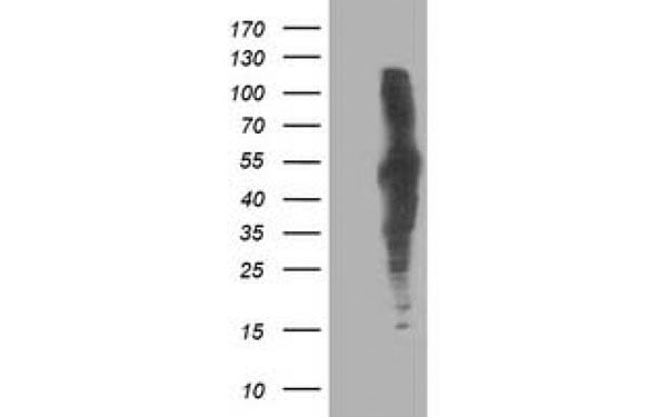
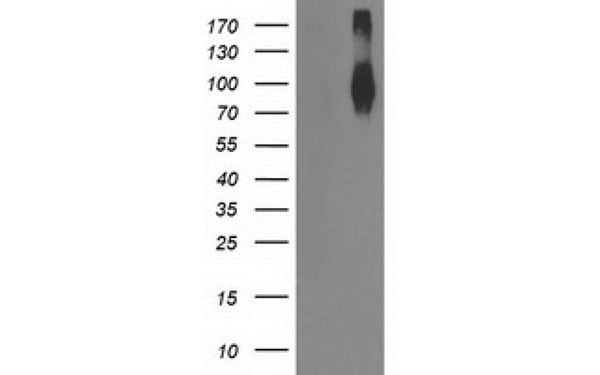
 WB (Western Blot)
(Western Blot analysis of HEK293T cell lysates (5 ug) transfected with either recombinant ARHGAP25 protein (Right) or empty vector (Left) detected with ARHGAP25 antibody)
WB (Western Blot)
(Western Blot analysis of HEK293T cell lysates (5 ug) transfected with either recombinant ARHGAP25 protein (Right) or empty vector (Left) detected with ARHGAP25 antibody)
ARHGAP25, Monoclonal Antibody (Cat# AAA106350)
 IF (Immunofluorescence)
(Immunofluorescent staining of COS7 cells transiently transfected with recombinant CDK2 protein using CDK2 antibody)
IF (Immunofluorescence)
(Immunofluorescent staining of COS7 cells transiently transfected with recombinant CDK2 protein using CDK2 antibody)
CDK2, Monoclonal Antibody (Cat# AAA106351)
 IF (Immunofluorescence)
(Immunofluorescent staining of COS7 cells transiently transfected with recombinant DLD protein using DLD antibody)
IF (Immunofluorescence)
(Immunofluorescent staining of COS7 cells transiently transfected with recombinant DLD protein using DLD antibody)
DLD, Monoclonal Antibody (Cat# AAA106364)
 IF (Immunofluorescence)
(Immunofluorescent staining of COS7 cells transiently transfected with recombinant ANKRD53 protein using ANKRD53 antibody)
IF (Immunofluorescence)
(Immunofluorescent staining of COS7 cells transiently transfected with recombinant ANKRD53 protein using ANKRD53 antibody)
ANKRD53, Monoclonal Antibody (Cat# AAA106372)
 IHC (Immunohiostchemistry)
(Immunohistochemical analysis of CRYM protein in paraffin embedded Human prostate tissue using CRYM antibody)
IHC (Immunohiostchemistry)
(Immunohistochemical analysis of CRYM protein in paraffin embedded Human prostate tissue using CRYM antibody)
CRYM, Monoclonal Antibody (Cat# AAA106379)
 IF (Immunofluorescence)
(Immunofluorescent staining of COS7 cells transiently transfected with recombinant CYP1A2 protein using CYP1A2 antibody)
IF (Immunofluorescence)
(Immunofluorescent staining of COS7 cells transiently transfected with recombinant CYP1A2 protein using CYP1A2 antibody)
CYP1A2, Monoclonal Antibody (Cat# AAA106392)
 IF (Immunofluorescence)
(Immunofluorescent staining of COS7 cells transiently transfected with recombinant EPM2AIP1 protein using EPM2AIP1 antibody)
IF (Immunofluorescence)
(Immunofluorescent staining of COS7 cells transiently transfected with recombinant EPM2AIP1 protein using EPM2AIP1 antibody)
EPM2AIP1, Monoclonal Antibody (Cat# AAA106395)
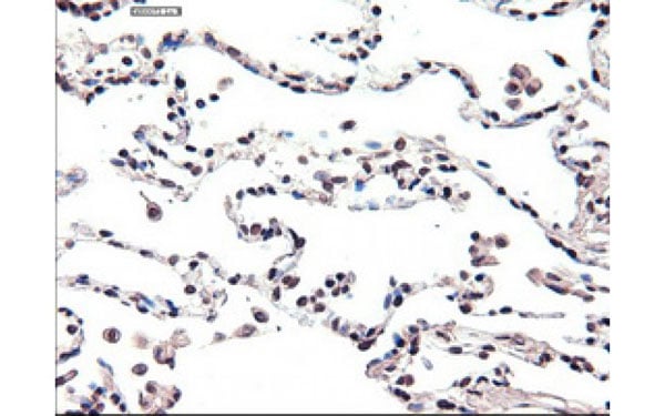
 IHC (Immunohiostchemistry)
(Immunohistochemical analysis of GOLM1 protein in paraffin embedded Carcinoma of Human lung tissue using GOLM1 antibody)
IHC (Immunohiostchemistry)
(Immunohistochemical analysis of GOLM1 protein in paraffin embedded Carcinoma of Human lung tissue using GOLM1 antibody)
GOLM1, Monoclonal Antibody (Cat# AAA106399)
 IF (Immunofluorescence)
(Immunofluorescent staining of COS7 cells transiently transfected with recombinant GPHN protein using GPHN antibody)
IF (Immunofluorescence)
(Immunofluorescent staining of COS7 cells transiently transfected with recombinant GPHN protein using GPHN antibody)
GPHN, Monoclonal Antibody (Cat# AAA106409)
 IF (Immunofluorescence)
(Immunofluorescent staining of COS7 cells transiently transfected with recombinant CBWD1 protein using CBWD1 antibody)
IF (Immunofluorescence)
(Immunofluorescent staining of COS7 cells transiently transfected with recombinant CBWD1 protein using CBWD1 antibody)
CBWD1, Monoclonal Antibody (Cat# AAA106410)
 IF (Immunofluorescence)
(Immunofluorescent staining of COS7 cells transiently transfected with recombinant FH protein using FH antibody)
IF (Immunofluorescence)
(Immunofluorescent staining of COS7 cells transiently transfected with recombinant FH protein using FH antibody)
FH, Monoclonal Antibody (Cat# AAA106417)
 IF (Immunofluorescence)
(Immunofluorescent staining of COS7 cells transiently transfected with recombinant HSPA1A protein using HSPA1A antibody)
IF (Immunofluorescence)
(Immunofluorescent staining of COS7 cells transiently transfected with recombinant HSPA1A protein using HSPA1A antibody)
HSPA1A, Monoclonal Antibody (Cat# AAA106192)
 IF (Immunofluorescence)
(Immunofluorescent staining of COS7 cells transiently transfected with recombinant H6PD protein using H6PD antibody)
IF (Immunofluorescence)
(Immunofluorescent staining of COS7 cells transiently transfected with recombinant H6PD protein using H6PD antibody)
H6PD, Monoclonal Antibody (Cat# AAA106200)
 IF (Immunofluorescence)
(Immunofluorescent staining of COS7 cells transiently transfected with recombinant C1orf50 protein using C1orf50 antibody)
IF (Immunofluorescence)
(Immunofluorescent staining of COS7 cells transiently transfected with recombinant C1orf50 protein using C1orf50 antibody)
C1orf50, Monoclonal Antibody (Cat# AAA106272)
 IHC (Immunohiostchemistry)
(Immunohistochemical analysis of GSC protein in paraffin embedded Carcinoma of Human kidney tissue using GSC antibody)
IHC (Immunohiostchemistry)
(Immunohistochemical analysis of GSC protein in paraffin embedded Carcinoma of Human kidney tissue using GSC antibody)
GSC, Monoclonal Antibody (Cat# AAA106275)
 IF (Immunofluorescence)
(Immunofluorescent staining of COS7 cells transiently transfected with recombinant FBXO21 protein using FBXO21 antibody)
IF (Immunofluorescence)
(Immunofluorescent staining of COS7 cells transiently transfected with recombinant FBXO21 protein using FBXO21 antibody)
FBXO21, Monoclonal Antibody (Cat# AAA106279)
Chlamydia, Monoclonal Antibody (Cat# AAA106285)
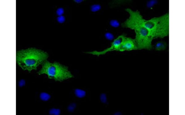
 IHC (Immunohiostchemistry)
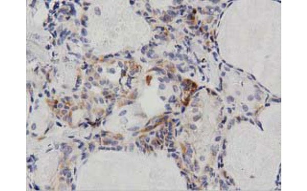
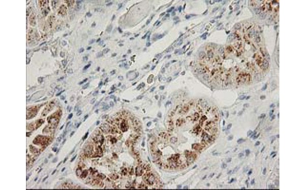
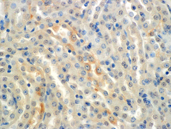
(Immunohistochemical analysis of APP protein in paraffin embedded Human Kidney tissue using APP antibody)
IHC (Immunohiostchemistry)
(Immunohistochemical analysis of APP protein in paraffin embedded Human Kidney tissue using APP antibody)
APP, Monoclonal Antibody (Cat# AAA106335)
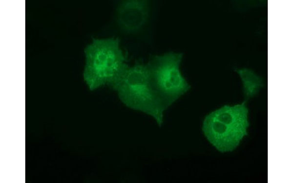
 IF (Immunofluorescence)
(Immunofluorescent staining of COS7 cells transiently transfected with recombinant AK5 protein using AK5 antibody)
IF (Immunofluorescence)
(Immunofluorescent staining of COS7 cells transiently transfected with recombinant AK5 protein using AK5 antibody)
AK5, Monoclonal Antibody (Cat# AAA106207)
 IF (Immunofluorescence)
(Immunofluorescent staining of COS7 cells transiently transfected with recombinant BTK protein using BTK antibody)
IF (Immunofluorescence)
(Immunofluorescent staining of COS7 cells transiently transfected with recombinant BTK protein using BTK antibody)
BTK, Monoclonal Antibody (Cat# AAA106221)
 IF (Immunofluorescence)
(Immunofluorescent staining of COS7 cells transiently transfected with recombinant ANKRD53 protein using ANKRD53 antibody)
IF (Immunofluorescence)
(Immunofluorescent staining of COS7 cells transiently transfected with recombinant ANKRD53 protein using ANKRD53 antibody)
ANKRD53, Monoclonal Antibody (Cat# AAA106241)
 IHC (Immunohiostchemistry)
(Immunohistochemical analysis of CTAG1B protein in paraffin embedded Carcinoma of Human kidney tissue using CTAG1B antibody)
IHC (Immunohiostchemistry)
(Immunohistochemical analysis of CTAG1B protein in paraffin embedded Carcinoma of Human kidney tissue using CTAG1B antibody)
CTAG1B, Monoclonal Antibody (Cat# AAA106244)
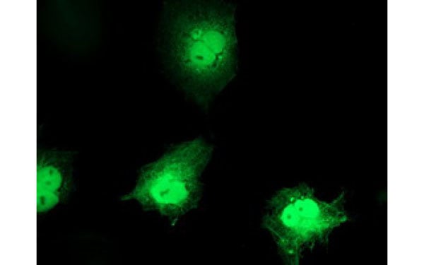
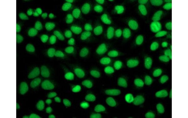
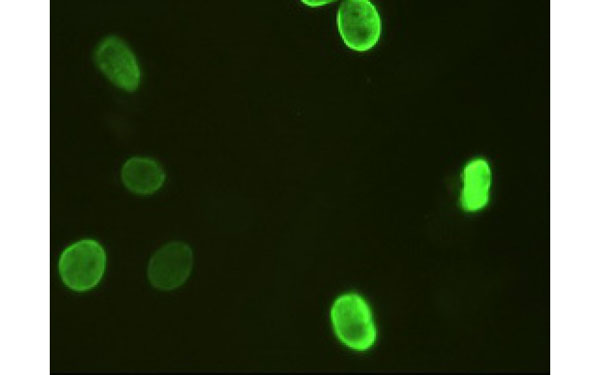 IF (Immunofluorescence)
(Immunofluorescent staining of Hela cells transiently transfected with recombinant FOXA2 protein using FOXA2 antibody)
IF (Immunofluorescence)
(Immunofluorescent staining of Hela cells transiently transfected with recombinant FOXA2 protein using FOXA2 antibody)
FOXA2, Monoclonal Antibody (Cat# AAA106254)
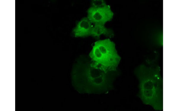
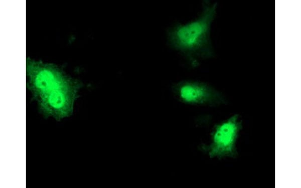
 IF (Immunofluorescence)
(Immunofluorescent staining of endogenous FGFR2 protein in Hela cells using FGFR2 antibody)
IF (Immunofluorescence)
(Immunofluorescent staining of endogenous FGFR2 protein in Hela cells using FGFR2 antibody)
FGFR2, Monoclonal Antibody (Cat# AAA106259)
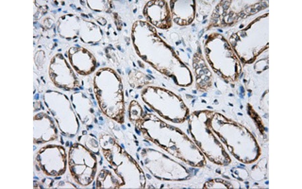
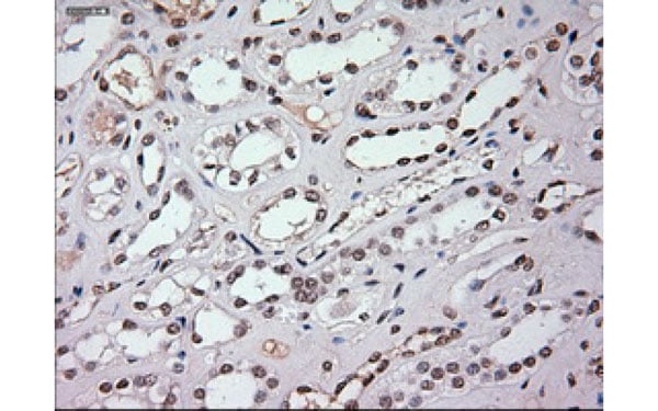 IHC (Immunohiostchemistry)
(Immunohistochemical analysis of EpCAM protein in paraffin embedded Human kidney using EpCAM antibody)
IHC (Immunohiostchemistry)
(Immunohistochemical analysis of EpCAM protein in paraffin embedded Human kidney using EpCAM antibody)
EpCAM, Monoclonal Antibody (Cat# AAA106434)
 FCM/FACS (Flow Cytometry)
FGF21, Monoclonal Antibody (Cat# AAA106450)
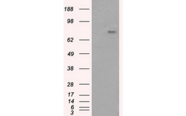
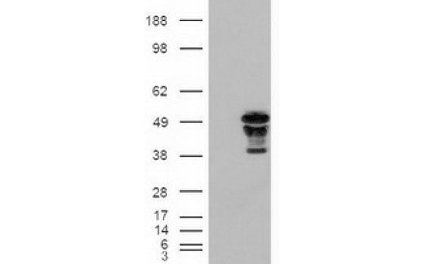
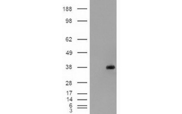
 WB (Western Blot)
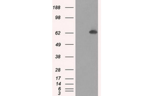
(Western Blot analysis of HEK293T cell lysates (5 ug) transfected with either recombinant HAO1 protein (Right) or empty vector (Left) detected with HAO1 antibody)
WB (Western Blot)
(Western Blot analysis of HEK293T cell lysates (5 ug) transfected with either recombinant HAO1 protein (Right) or empty vector (Left) detected with HAO1 antibody)
HAO1, Monoclonal Antibody (Cat# AAA106453)
 IHC (Immunohiostchemistry)
(Immunohistochemical analysis of GBP2 protein in paraffin embedded Adenocarcinoma of Human breast tissue using GBP2 antibody)
IHC (Immunohiostchemistry)
(Immunohistochemical analysis of GBP2 protein in paraffin embedded Adenocarcinoma of Human breast tissue using GBP2 antibody)
GBP2, Monoclonal Antibody (Cat# AAA106458)
ALDOSTERONE, Monoclonal Antibody (Cat# AAA49153)
PROLACTIN, Monoclonal Antibody (Cat# AAA49161)
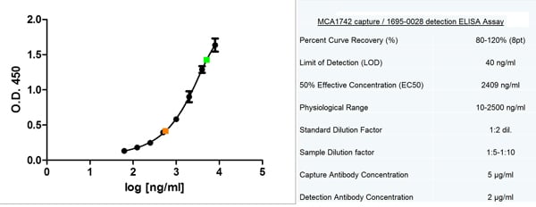
 Application Data
(Sandwich ELISA analysis of CD227 expression using Mouse anti Human CD227, clone C595 as a capture reagent and biotinylated Mouse anti Human CD227, clone 6A4 as a detection reagent with purified breast cancer antigen as antigen for the generation of the standard curve. Detection is by HRP conjugated streptavidin and substrate. Microtitre plate is read at O.D. 450 nm on the iMark Microplate Absorbance Reader . Plasma and serum samples diluted 1:5 are indicated in green and orange respectively)
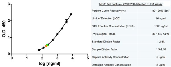
Application Data
(Sandwich ELISA analysis of CD227 expression using Mouse anti Human CD227, clone C595 as a capture reagent and biotinylated Mouse anti Human CD227, clone 6A4 as a detection reagent with purified breast cancer antigen as antigen for the generation of the standard curve. Detection is by HRP conjugated streptavidin and substrate. Microtitre plate is read at O.D. 450 nm on the iMark Microplate Absorbance Reader . Plasma and serum samples diluted 1:5 are indicated in green and orange respectively)
CD227, Monoclonal Antibody (Cat# AAA49064)
 Application Data
(Staining of human peripheral blood granulocytes with Mouse anti Human CD95:Alexa Fluor 647 (AAA49267A647))
Application Data
(Staining of human peripheral blood granulocytes with Mouse anti Human CD95:Alexa Fluor 647 (AAA49267A647))
CD95, Monoclonal Antibody (Cat# AAA49267)

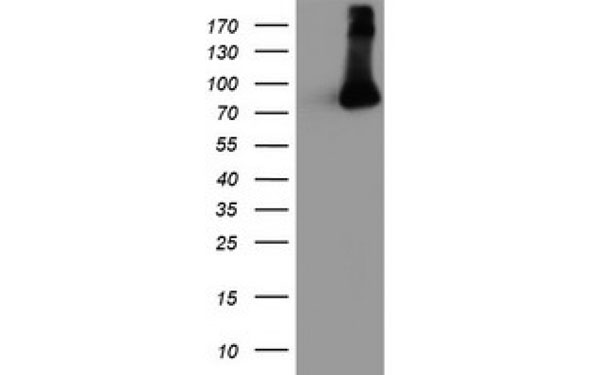
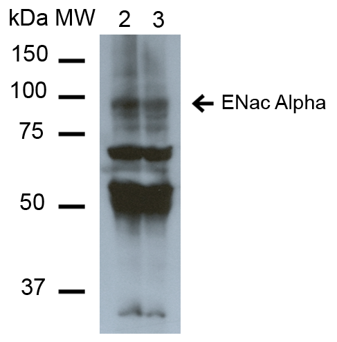
 WB (Western Blot)
(Western Blot analysis of Mouse Whole kidney homogenates showing detection of ~85kDa ENaC alpha protein using Mouse Anti-ENaC alpha Monoclonal Antibody, Clone 2G4. Lane 1: Molecular Weight Ladder (MW). Lane 2: Low-salt diet. Lane 3: Normal-salt diet. Load: 20 ug. Primary Antibody: Mouse Anti-ENaC alpha Monoclonal Antibody at 1:1000. Predicted/Observed Size: ~85kDa.)
WB (Western Blot)
(Western Blot analysis of Mouse Whole kidney homogenates showing detection of ~85kDa ENaC alpha protein using Mouse Anti-ENaC alpha Monoclonal Antibody, Clone 2G4. Lane 1: Molecular Weight Ladder (MW). Lane 2: Low-salt diet. Lane 3: Normal-salt diet. Load: 20 ug. Primary Antibody: Mouse Anti-ENaC alpha Monoclonal Antibody at 1:1000. Predicted/Observed Size: ~85kDa.)
ENaC alpha, Monoclonal Antibody (Cat# AAA103891)
 WB (Western Blot)
(Western Blot analysis of Mouse Whole kidney homogenates showing detection of ~85kDa ENaC alpha protein using Mouse Anti-ENaC alpha Monoclonal Antibody, Clone 2G4. Lane 1: Molecular Weight Ladder (MW). Lane 2: Low-salt diet. Lane 3: Normal-salt diet. Load: 20 ug. Primary Antibody: Mouse Anti-ENaC alpha Monoclonal Antibody at 1:1000. Predicted/Observed Size: ~85kDa.)
WB (Western Blot)
(Western Blot analysis of Mouse Whole kidney homogenates showing detection of ~85kDa ENaC alpha protein using Mouse Anti-ENaC alpha Monoclonal Antibody, Clone 2G4. Lane 1: Molecular Weight Ladder (MW). Lane 2: Low-salt diet. Lane 3: Normal-salt diet. Load: 20 ug. Primary Antibody: Mouse Anti-ENaC alpha Monoclonal Antibody at 1:1000. Predicted/Observed Size: ~85kDa.)
ENaC alpha, Monoclonal Antibody (Cat# AAA103893)

 WB (Western Blot)
(Western Blot analysis of Mouse Whole kidney homogenates showing detection of ~85kDa ENaC alpha protein using Mouse Anti-ENaC alpha Monoclonal Antibody, Clone 2G4. Lane 1: Molecular Weight Ladder (MW). Lane 2: Low-salt diet. Lane 3: Normal-salt diet. Load: 20 ug. Primary Antibody: Mouse Anti-ENaC alpha Monoclonal Antibody at 1:1000. Predicted/Observed Size: ~85kDa.)
WB (Western Blot)
(Western Blot analysis of Mouse Whole kidney homogenates showing detection of ~85kDa ENaC alpha protein using Mouse Anti-ENaC alpha Monoclonal Antibody, Clone 2G4. Lane 1: Molecular Weight Ladder (MW). Lane 2: Low-salt diet. Lane 3: Normal-salt diet. Load: 20 ug. Primary Antibody: Mouse Anti-ENaC alpha Monoclonal Antibody at 1:1000. Predicted/Observed Size: ~85kDa.)
ENaC alpha, Monoclonal Antibody (Cat# AAA103894)
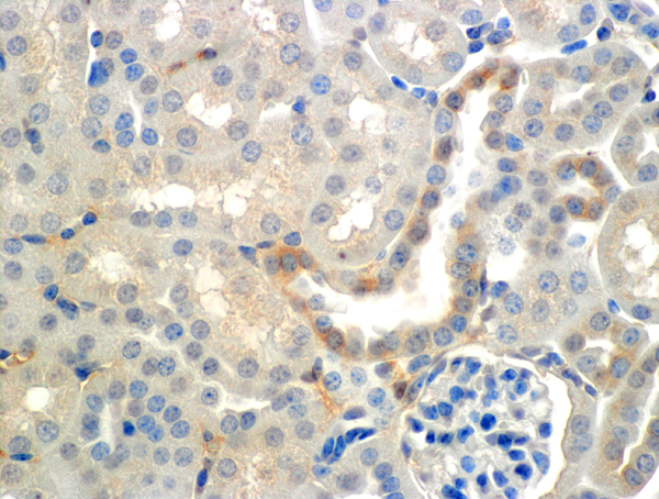
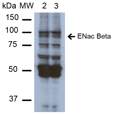 WB (Western Blot)
(Western Blot analysis of Mouse Whole kidney homogenates showing detection of ~87kDa ENaC beta protein using Mouse Anti-ENaC beta Monoclonal Antibody, Clone 7B8. Lane 1: Molecular Weight Ladder (MW). Lane 2: Low-salt diet. Lane 3: Normal-salt diet. Load: 20 ug. Primary Antibody: Mouse Anti-ENaC beta Monoclonal Antibody at 1:1000. Predicted/Observed Size: ~87kDa.)
WB (Western Blot)
(Western Blot analysis of Mouse Whole kidney homogenates showing detection of ~87kDa ENaC beta protein using Mouse Anti-ENaC beta Monoclonal Antibody, Clone 7B8. Lane 1: Molecular Weight Ladder (MW). Lane 2: Low-salt diet. Lane 3: Normal-salt diet. Load: 20 ug. Primary Antibody: Mouse Anti-ENaC beta Monoclonal Antibody at 1:1000. Predicted/Observed Size: ~87kDa.)
ENaC beta, Monoclonal Antibody (Cat# AAA103903)
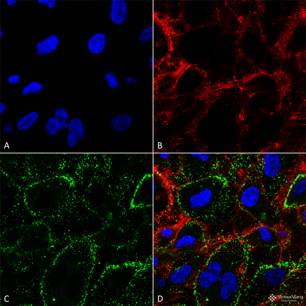
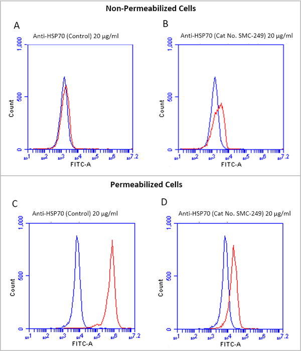 IF (Immunofluorescence)
(Fluorescence-activated cell sorting analysis using Mouse Anti-HSP70 Monoclonal Antibody, Clone 1H11. Tissue: Jurkat E6.1 cells. Species: Human. Fixation: No fixation. Primary Antibody: Mouse Anti-HSP70 Monoclonal Antibody at 20 ug/ml for 40 min at 4 degree C. Counterstain: Propidium Iodide nuclear stain at 2.5 ug/ml for 5 min at RT. Isotype Control: Anti-mouse FITC at 1:32 for 15 min at RT (blue line). Courtesy of: Dr. Elyse Ireland, Institute of Medicine, University of Chester.)
IF (Immunofluorescence)
(Fluorescence-activated cell sorting analysis using Mouse Anti-HSP70 Monoclonal Antibody, Clone 1H11. Tissue: Jurkat E6.1 cells. Species: Human. Fixation: No fixation. Primary Antibody: Mouse Anti-HSP70 Monoclonal Antibody at 20 ug/ml for 40 min at 4 degree C. Counterstain: Propidium Iodide nuclear stain at 2.5 ug/ml for 5 min at RT. Isotype Control: Anti-mouse FITC at 1:32 for 15 min at RT (blue line). Courtesy of: Dr. Elyse Ireland, Institute of Medicine, University of Chester.)
HSP70, Monoclonal Antibody (Cat# AAA103908)
What are Monoclonal Antibodies?
Monoclonal antibodies are specialized laboratory-produced proteins developed for binding to specific biological antigens or other molecular targets. Since they come from a single cell (or clone), they are especially consistent and accurate in the data they are involved in producing.
This type of antibody material has been shown to be a powerful tool in finding and subsequently destroying harmful cells in an organism, such as those found in cancers or various autoimmune diseases. This makes them excellent aids in medical testing and research, which is why they are so widely used.
AAA Biotech offers a comprehensive range of high-quality monoclonal antibodies that perform effectively in various laboratory tests, including (amongst others) ELISA, western blotting, immunohistochemistry, and flow cytometry. All of the products in our catalog are thoroughly quality tested to make sure that they are reliable and will consistently perform well in your research.
What Are The Uses of Monoclonal Antibodies
Monoclonal antibodies are used in many lab tests, including (amongst others) ELISA, western blotting, immunohistochemistry, and flow cytometry.
ELISA is a test that helps detect a specific substance/analyte in a sample. It uses antibodies (often monoclonal) bound to a solid surface (such as the well of a microplate) to “capture” the substance/analyte in the sample and immobilize it so that the detection antibody component can then bind to it and produce a signal, which can then be measured.
Western blotting identifies specific proteins in a sample. The sample is first separated on a gel, and then antibodies are applied that will typically bind to the target, which will all be localized to a single band in a lane.
Immunohistochemistry helps locate specific proteins in cells or tissue samples using antibodies.
Flow cytometry looks at and sorts cells. It uses antibodies that are conjugated to reporter molecules called “fluorophores”, which, under special lights, emit light themselves, which can then be measured by a detector instrument.
How Monoclonal Antibodies Are Used as Medicine?
Please note that all of the products listed in AAA Biotech’s also known as AAA Bio or AAABio catalog are strictly for research-use only (RUO).
Monoclonal antibodies can also be used as therapeutic/medical treatments, particularly in the context of cancers. They are designed to find and bind to specific cells or proteins, helping the immune system recognize and attack the cancer. These treatments work in different ways, such as:
- Radioimmunotherapy attaches a small amount of radioactive molecule to the antibody, so it delivers the radiation directly to the cancer cells that the antibody is specifically binding to.
- Antibody-directed enzyme prodrug therapy uses antibodies that are specifically bound to special enzymes. These enzymes activate a harmless drug in the body and turn it into a cancer-killing drug only near the cancer cells—this helps avoid harming healthy cells.
- Immunoliposomes are tiny “bubbles” filled with medicine/drug and coated with antibodies. They carry the drug straight to the cancer cells.
Why Buy Monoclonal Antibodies From Us?
At AAA Biotech, we provide high-performance monoclonal antibodies designed to support a wide range of research needs.
1. Validated for Versatile Applications
The antibodies in our catalog are extensively validated and compatible with multiple techniques, including (but not limited to) ELISA, flow cytometry (FC), immunocytochemistry (ICC), immunofluorescence (IF), immunohistochemistry (IHC), immunoprecipitation (IP), and western blotting (WB).
2. Wide Selection & Specialized Options
We offer antibodies for common and rare species, that are available in various conjugated forms, and also in recombinant formats. Essentially, there is almost anything one might need to meet their experimental model’s requirements.
3. High-Quality Proteins
Our proteins meet high purity standards—90% or more as confirmed by SDS-PAGE. Many are available with tags like His, Flag, GST, or MBP, and we also supply native and biologically active proteins for functional studies.
Frequently Asked Questions
1. Are your monoclonal antibodies validated for specific applications?
Yes, our antibodies are tested and validated for use in methods such as ELISA, western blot, IHC, flow cytometry, and more. Refer to specific product pages or datasheets for individual product information.
2. How do I choose the right monoclonal antibody for my application?
Review the product details directly for application validation, species reactivity, and target information. You may also contact our support team at any time for help.
3. How quickly can I receive my order?
Most orders are processed and shipped within 1–3 business days, depending on product availability and your shipping location.