Filters
▼Clonality
▼Type
▼Reactivity
▼Gene Name
▼Isotype
▼Host
▼Application
▼Clone
▼Monoclonal Antibodies
Get accurate results in your research with our Monoclonal Antibodies, which are specially made to target exactly what you require for your research, and will produce consistent, reliable performance in lab tests.
Viewing 7450-7500 of 27645 product results

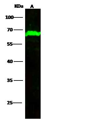
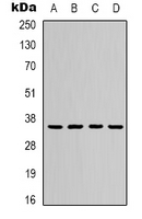
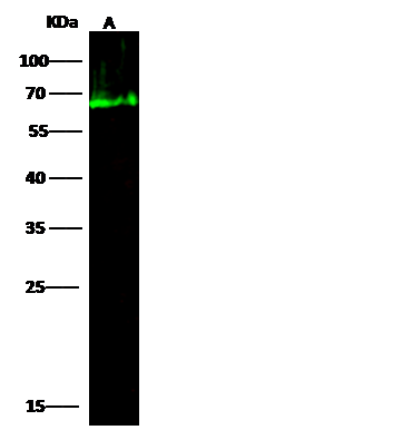 WB (Western Blot)
(Anti-HAS rabbit monoclonal antibody at 1:500 dilutionLane A: HepG2 Whole Cell LysateLysates/proteins at 30 ug per lane.SecondaryGoat Anti-Rabbit IgG H&L (Dylight800) at 1/10000 dilution.Developed using the Odyssey technique.Performed under reducing conditions.Predicted band size:68 kDaObserved band size:68 kDa)
WB (Western Blot)
(Anti-HAS rabbit monoclonal antibody at 1:500 dilutionLane A: HepG2 Whole Cell LysateLysates/proteins at 30 ug per lane.SecondaryGoat Anti-Rabbit IgG H&L (Dylight800) at 1/10000 dilution.Developed using the Odyssey technique.Performed under reducing conditions.Predicted band size:68 kDaObserved band size:68 kDa)
Serum Albumin, Monoclonal Antibody (Cat# AAA257272)
TIM-1/KIM-1, Monoclonal Antibody (Cat# AAA257276)
TIM-1/KIM-1, Monoclonal Antibody (Cat# AAA257278)
IL-33, Monoclonal Antibody (Cat# AAA257279)

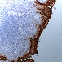
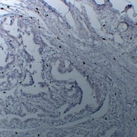
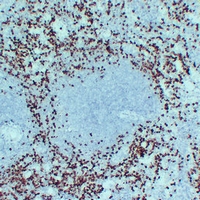
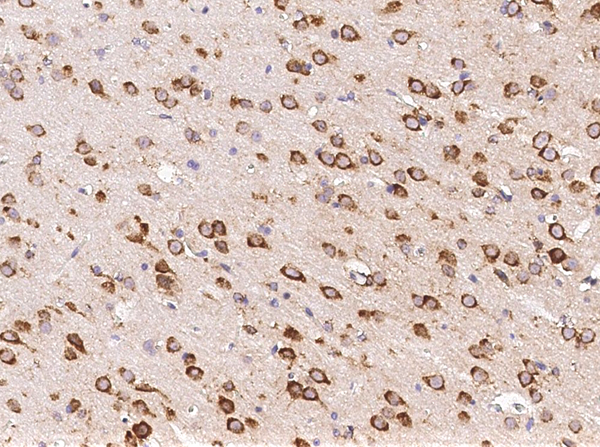 IHC (Immunohiostchemistry)
(Immunochemical staining of rat NXPH1 in rat brain with rabbit monoclonal antibody (1:20000, formalin-fixed paraffin embedded sections).)
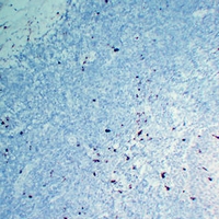
IHC (Immunohiostchemistry)
(Immunochemical staining of rat NXPH1 in rat brain with rabbit monoclonal antibody (1:20000, formalin-fixed paraffin embedded sections).)
NXPH1, Monoclonal Antibody (Cat# AAA257294)
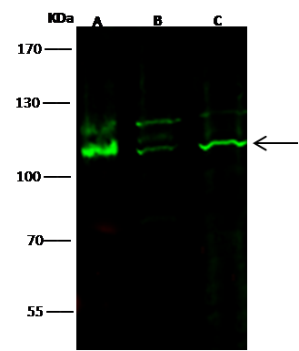
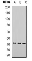
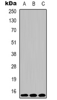
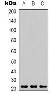
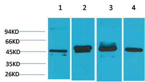
 IP (Immunoprecipitation)
(rat UCHL3 was immunoprecipitated using:Lane A:0.5 mg k562 Whole Cell LysateLane B:0.5 mg Jurkat Whole Cell LysateLane C:0.5 mg SH-SY5Y Whole Cell Lysate2 uL anti-rat UCHL3 rabbit monoclonal antibody and 15 ul of 50 % Protein G agarose.Primary antibody:Anti-rat UCHL3 rabbit monoclonal antibody,at 1:200 dilutionSecondary antibody:Dylight 800-labeled antibody to rabbit IgG (H+L), at 1:5000 dilutionDeveloped using the odssey technique.Performed under reducing conditions.Predicted band size: 26 kDaObserved band size: 26 kDa)
IP (Immunoprecipitation)
(rat UCHL3 was immunoprecipitated using:Lane A:0.5 mg k562 Whole Cell LysateLane B:0.5 mg Jurkat Whole Cell LysateLane C:0.5 mg SH-SY5Y Whole Cell Lysate2 uL anti-rat UCHL3 rabbit monoclonal antibody and 15 ul of 50 % Protein G agarose.Primary antibody:Anti-rat UCHL3 rabbit monoclonal antibody,at 1:200 dilutionSecondary antibody:Dylight 800-labeled antibody to rabbit IgG (H+L), at 1:5000 dilutionDeveloped using the odssey technique.Performed under reducing conditions.Predicted band size: 26 kDaObserved band size: 26 kDa)
UCHL3, Monoclonal Antibody (Cat# AAA257298)
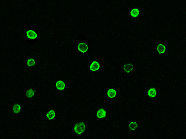
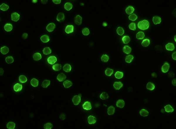 IF (Immunofluorescence)
(Immunofluorescence staining of mouse Ly9 in mouse splenocytes. Cells were fixed with 4% PFA, blocked with 10% serum, and incubated with rabbit anti-mouse Ly9 monoclonal antibody (1:60) at 37 degree C 1 hour. Then cells were stained with the Alexa Fluor 488-conjugated Goat Anti-rabbit IgG secondary antibody (green).)
IF (Immunofluorescence)
(Immunofluorescence staining of mouse Ly9 in mouse splenocytes. Cells were fixed with 4% PFA, blocked with 10% serum, and incubated with rabbit anti-mouse Ly9 monoclonal antibody (1:60) at 37 degree C 1 hour. Then cells were stained with the Alexa Fluor 488-conjugated Goat Anti-rabbit IgG secondary antibody (green).)
CD229, Monoclonal Antibody (Cat# AAA257169)
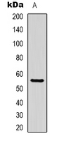
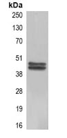
 IP (Immunoprecipitation)
(Mouse METAP2 was immunoprecipitated using:Lane A:0.5 mg HepG2 Whole Cell Lysate2 uL anti-Mouse METAP2 rabbit monoclonal antibody and 15 ul of 50 % Protein G agarose.Primary antibody:Anti-Mouse METAP2 rabbit monoclonal antibody,at 1:200 dilutionSecondary antibody:Clean-Blot IP Detection Reagent (HRP) at 1:1000 dilutionDeveloped using the DAB staining technique.Performed under reducing conditions.Predicted band size: 52 kDaObserved band size: 72 kDa)
IP (Immunoprecipitation)
(Mouse METAP2 was immunoprecipitated using:Lane A:0.5 mg HepG2 Whole Cell Lysate2 uL anti-Mouse METAP2 rabbit monoclonal antibody and 15 ul of 50 % Protein G agarose.Primary antibody:Anti-Mouse METAP2 rabbit monoclonal antibody,at 1:200 dilutionSecondary antibody:Clean-Blot IP Detection Reagent (HRP) at 1:1000 dilutionDeveloped using the DAB staining technique.Performed under reducing conditions.Predicted band size: 52 kDaObserved band size: 72 kDa)
methionyl aminopeptidase 2/METAP2, Monoclonal Antibody (Cat# AAA257171)
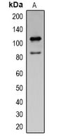
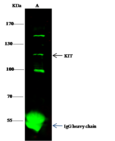 IP (Immunoprecipitation)
(Mouse KIT was immunoprecipitated using:Lane A:0.5 mg K562 Whole Cell Lysate1 uL anti-Mouse KIT rabbit monoclonal antibody and 15 ul of 50 % Protein G agarose.Primary antibody:Anti-Mouse KIT rabbit monoclonal antibody,at 1:500 dilutionSecondary antibody:Dylight 800-labeled antibody to rabbit IgG (H+L), at 1:5000 dilutionDeveloped using the odssey technique.Performed under reducing conditions.Predicted band size: 110 kDaObserved band size: 110 kDa)
IP (Immunoprecipitation)
(Mouse KIT was immunoprecipitated using:Lane A:0.5 mg K562 Whole Cell Lysate1 uL anti-Mouse KIT rabbit monoclonal antibody and 15 ul of 50 % Protein G agarose.Primary antibody:Anti-Mouse KIT rabbit monoclonal antibody,at 1:500 dilutionSecondary antibody:Dylight 800-labeled antibody to rabbit IgG (H+L), at 1:5000 dilutionDeveloped using the odssey technique.Performed under reducing conditions.Predicted band size: 110 kDaObserved band size: 110 kDa)
c-Kit, Monoclonal Antibody (Cat# AAA257173)
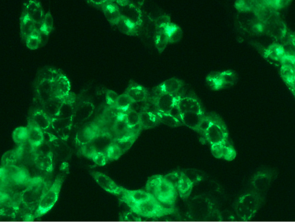
 IF (Immunofluorescence)
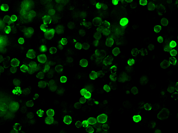
(Immunofluorescence analysis of SARS-COV-2 Nucleocapsid overexpressed HEK293 Cells were stained with purified anti-SARS-CoV-2 Nucleocapsid Mouse Mab,then a Alexa Fluor-488-conjugated second step antibody.)
IF (Immunofluorescence)
(Immunofluorescence analysis of SARS-COV-2 Nucleocapsid overexpressed HEK293 Cells were stained with purified anti-SARS-CoV-2 Nucleocapsid Mouse Mab,then a Alexa Fluor-488-conjugated second step antibody.)
SARS-CoV-2 (2019-nCoV) Nucleocapsid, Monoclonal Antibody (Cat# AAA257784)
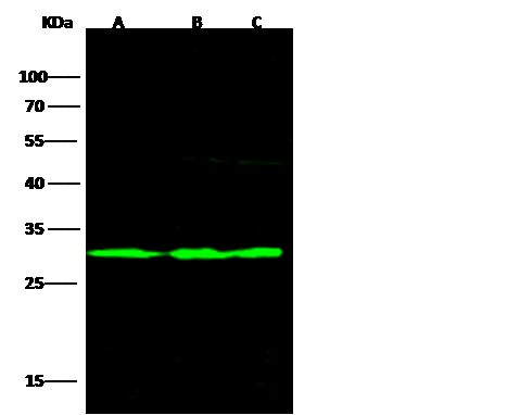
 WB (Western Blot)
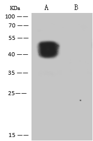
(Anti-SARS-CoV-2 (2019-nCoV) Nucleocapsid rabbit monoclonal antibody at 1:5000 dilution.Lane A: SARS-COV-2 Nucleocapsid overexpressed HEK293 Whole Cell LysateLane B: HEK293 Whole Cell LysateLysates/proteins at 30 ug per lane.SecondaryGoat Anti-Rabbit IgG (H+L)/HRP at 1/10000 dilutionDeveloped using the ECL technique.Performed under reducing conditions.)
WB (Western Blot)
(Anti-SARS-CoV-2 (2019-nCoV) Nucleocapsid rabbit monoclonal antibody at 1:5000 dilution.Lane A: SARS-COV-2 Nucleocapsid overexpressed HEK293 Whole Cell LysateLane B: HEK293 Whole Cell LysateLysates/proteins at 30 ug per lane.SecondaryGoat Anti-Rabbit IgG (H+L)/HRP at 1/10000 dilutionDeveloped using the ECL technique.Performed under reducing conditions.)
SARS-CoV-2 (2019-nCoV) Nucleocapsid, Monoclonal Antibody (Cat# AAA257785)

 IF (Immunofluorescence)
(Immunofluorescence analysis of SARS-COV-2 Nucleocapsid overexpressed HEK293 Cells were stained with purified anti-SARS-CoV-2 Nucleocapsid Rabbit Mab,then a Alexa Fluor-488-conjugated second step antibody.)
IF (Immunofluorescence)
(Immunofluorescence analysis of SARS-COV-2 Nucleocapsid overexpressed HEK293 Cells were stained with purified anti-SARS-CoV-2 Nucleocapsid Rabbit Mab,then a Alexa Fluor-488-conjugated second step antibody.)
SARS-CoV-2 (2019-nCoV) Nucleocapsid, Monoclonal Antibody (Cat# AAA257786)
 IF (Immunofluorescence)
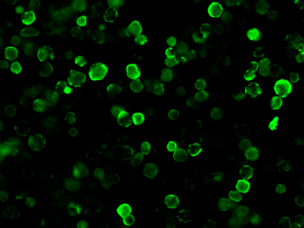
(Immunofluorescence analysis of SARS-COV-2 Spike overexpressed HEK293 Cells were stained with purified anti-SARS-CoV-2 Spike Chimera Mab,then a FITC-conjugated second step antibody.)
IF (Immunofluorescence)
(Immunofluorescence analysis of SARS-COV-2 Spike overexpressed HEK293 Cells were stained with purified anti-SARS-CoV-2 Spike Chimera Mab,then a FITC-conjugated second step antibody.)
SARS-CoV-2 (2019-nCoV) Spike S2, Monoclonal Antibody (Cat# AAA257787)

 IF (Immunofluorescence)
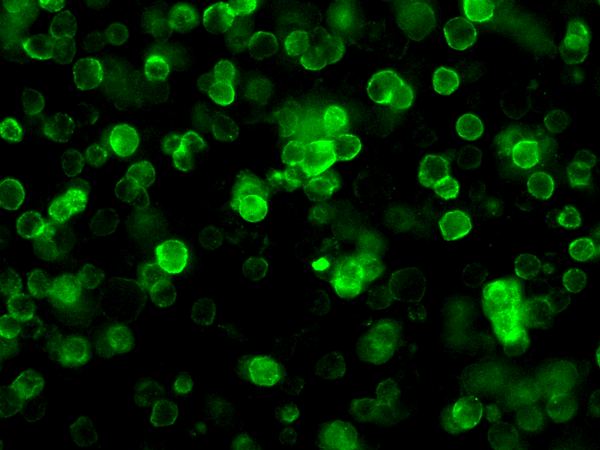
(Immunofluorescence analysis of SARS-COV-2 Spike overexpressed HEK293 Cells were stained with purified anti-SARS-CoV-2 Spike Mouse Mab,then a Alexa Fluor-488-conjugated second step antibody.)
IF (Immunofluorescence)
(Immunofluorescence analysis of SARS-COV-2 Spike overexpressed HEK293 Cells were stained with purified anti-SARS-CoV-2 Spike Mouse Mab,then a Alexa Fluor-488-conjugated second step antibody.)
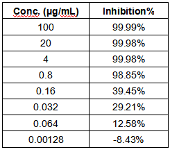
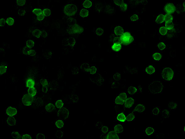
SARS-CoV-2 Spike Neutralizing, Monoclonal Antibody (Cat# AAA257791)

 IF (Immunofluorescence)
(Immunofluorescence analysis of SARS-COV-2 Spike overexpressed HEK293 Cells were stained with purified anti-SARS-CoV-2 Spike Rabbit Mab,then a Alexa Fluor-488-conjugated second step antibody.)
IF (Immunofluorescence)
(Immunofluorescence analysis of SARS-COV-2 Spike overexpressed HEK293 Cells were stained with purified anti-SARS-CoV-2 Spike Rabbit Mab,then a Alexa Fluor-488-conjugated second step antibody.)
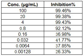
SARS-CoV-2 (2019-nCoV) Spike Neutralizing, Monoclonal Antibody (Cat# AAA257792)
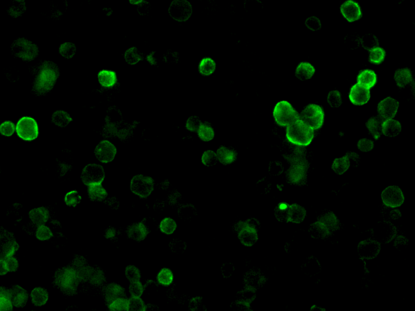
 IF (Immunofluorescence)
(Immunofluorescence staining of ratCD5 in rat spleen cells. Cells were fixed with 4% PFA,blocked with 10% serum, and incubated with mouse anti- ratCD5 monoclonal antibody (dilution ratio 1:60) at 4 degree C overnight. Then cells were stained with the Alexa Fluor488-conjugated Goat Anti-mouse IgG secondary antibody (green). Positive staining was localized to Cell membrane.)
IF (Immunofluorescence)
(Immunofluorescence staining of ratCD5 in rat spleen cells. Cells were fixed with 4% PFA,blocked with 10% serum, and incubated with mouse anti- ratCD5 monoclonal antibody (dilution ratio 1:60) at 4 degree C overnight. Then cells were stained with the Alexa Fluor488-conjugated Goat Anti-mouse IgG secondary antibody (green). Positive staining was localized to Cell membrane.)
CD5, Monoclonal Antibody (Cat# AAA257323)
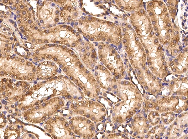
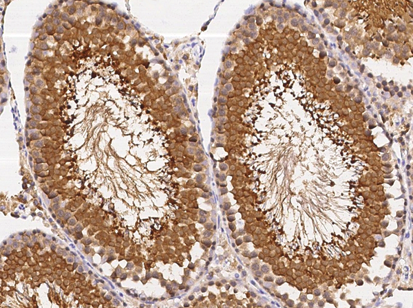
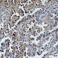
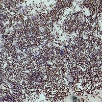
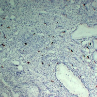
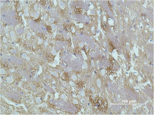
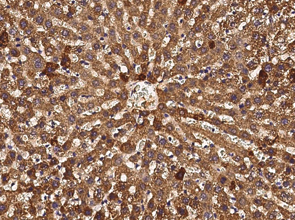 IHC (Immunohiostchemistry)
(Immunochemical staining of rat ART4 in rat liver with mouse monoclonal antibody (1:60, formalin-fixed paraffin embedded sections).)
IHC (Immunohiostchemistry)
(Immunochemical staining of rat ART4 in rat liver with mouse monoclonal antibody (1:60, formalin-fixed paraffin embedded sections).)
ART4/CD297, Monoclonal Antibody (Cat# AAA257326)

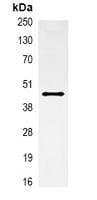
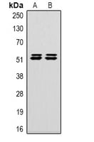
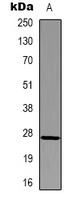
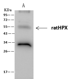 IP (Immunoprecipitation)
(ratHPX was immunoprecipitated using:Lane A:0.5 mg HepG2 Whole Cell Lysate2 uL anti-ratHPX rabbit monoclonal antibody and 60 ug of Immunomagnetic beads Protein A/G.Primary antibody:Anti-ratHPX rabbit monoclonal antibody,at 1:100 dilutionSecondary antibody:Clean-Blot IP Detection Reagent (HRP) at 1:1000dilutionDeveloped using the ECL technique.Performed under reducing conditions.Predicted band size: 52 kDaObserved band size :52 kDa)
IP (Immunoprecipitation)
(ratHPX was immunoprecipitated using:Lane A:0.5 mg HepG2 Whole Cell Lysate2 uL anti-ratHPX rabbit monoclonal antibody and 60 ug of Immunomagnetic beads Protein A/G.Primary antibody:Anti-ratHPX rabbit monoclonal antibody,at 1:100 dilutionSecondary antibody:Clean-Blot IP Detection Reagent (HRP) at 1:1000dilutionDeveloped using the ECL technique.Performed under reducing conditions.Predicted band size: 52 kDaObserved band size :52 kDa)
Hemopexin, Monoclonal Antibody (Cat# AAA257344)
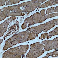
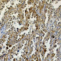
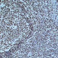
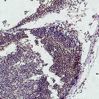
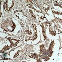
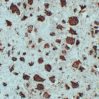
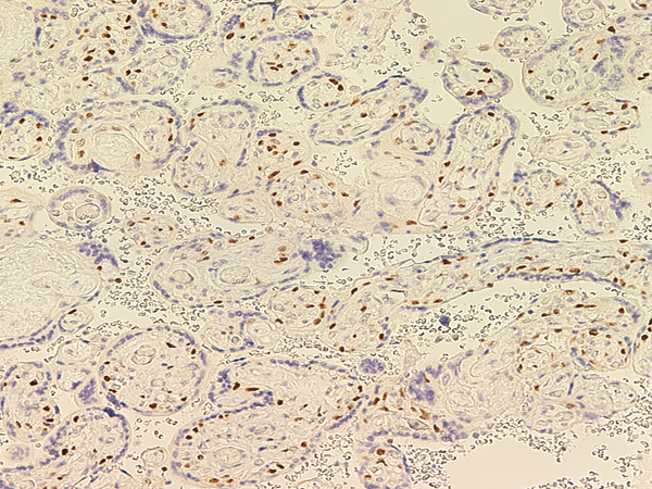 IHC (Immunohiostchemistry)
(Immunochemical staining of human PARP in human placenta with mouse monoclonal antibody at 1:500 dilution, formalin-fixed paraffin embedded sections.)
IHC (Immunohiostchemistry)
(Immunochemical staining of human PARP in human placenta with mouse monoclonal antibody at 1:500 dilution, formalin-fixed paraffin embedded sections.)
PARP, Monoclonal Antibody (Cat# AAA257734)


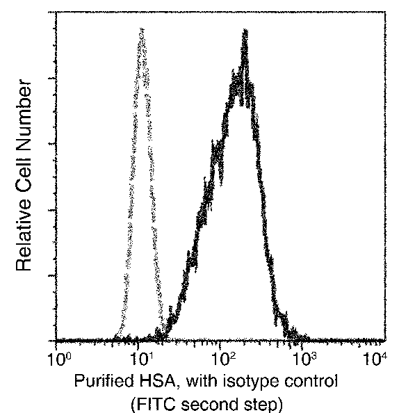
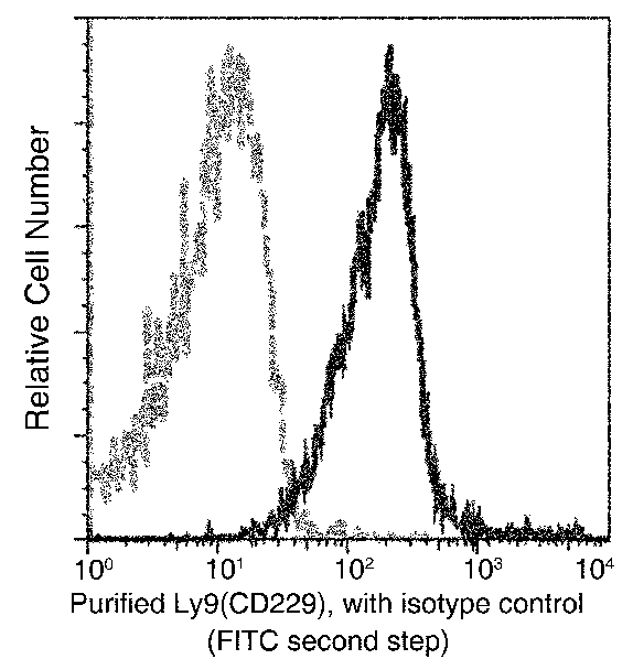
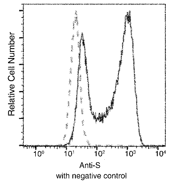
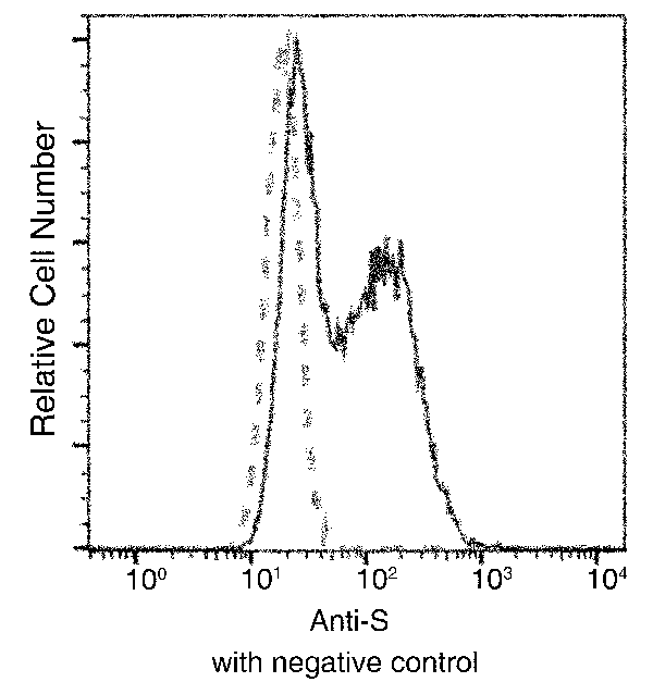
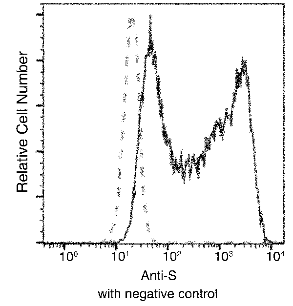
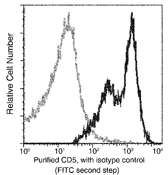
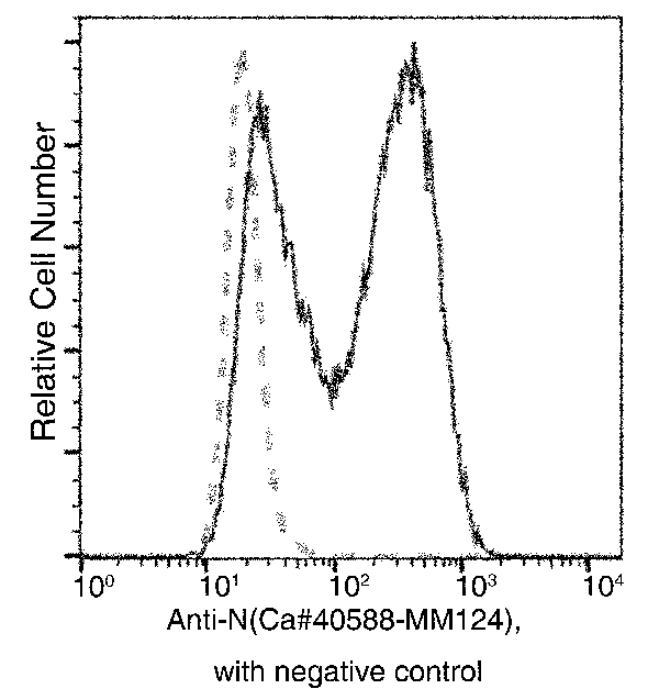 FCM/FACS (Flow Cytometry)
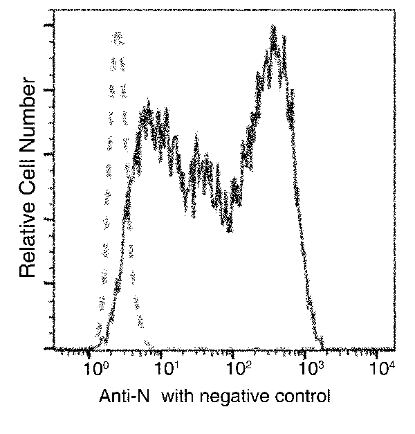
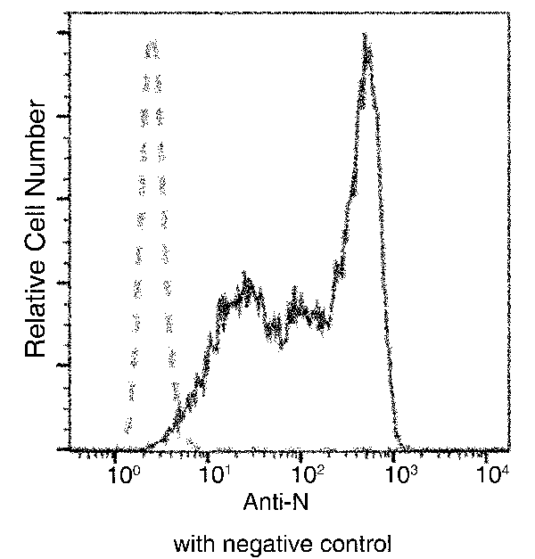
(Flow cytometric analysis of SARS-COV-2 Nucleocapsid overexpressed HEK293 Cells were stained with purified anti-SARS-COV-2 Nucleocapsid Mouse MAb, then a FITC-conjugated second step antibody. The fluorescence histograms were derived from gated events with the forward and side light-scatter characteristics of intact cells.(Validation Experiment))
FCM/FACS (Flow Cytometry)
(Flow cytometric analysis of SARS-COV-2 Nucleocapsid overexpressed HEK293 Cells were stained with purified anti-SARS-COV-2 Nucleocapsid Mouse MAb, then a FITC-conjugated second step antibody. The fluorescence histograms were derived from gated events with the forward and side light-scatter characteristics of intact cells.(Validation Experiment))
COVID 19 Nucleocapsid Coronavirus, Monoclonal Antibody (Cat# AAA258293)
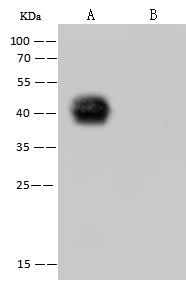
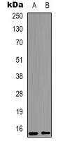
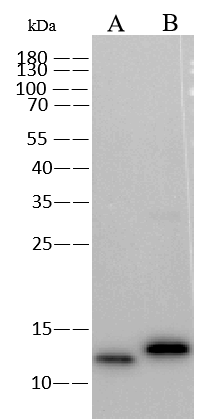 WB (Western Blot)
(Anti-Monkeypox Virus (MPXV) A29 mouse monoclonal antibody at 1:1000 dilution.; Lane A: Monkeypox Virus (MPXV) Protein A29 10ng; Lane B: Vaccinia virus (VACV)(strain Copenhagen) A27L Protein 10ng; Secondary; Goat Anti-Mouse IgG (H+L)/HRP at 1/10000 dilution; Developed using the ECL technique.; Performed under reducing conditions.)
WB (Western Blot)
(Anti-Monkeypox Virus (MPXV) A29 mouse monoclonal antibody at 1:1000 dilution.; Lane A: Monkeypox Virus (MPXV) Protein A29 10ng; Lane B: Vaccinia virus (VACV)(strain Copenhagen) A27L Protein 10ng; Secondary; Goat Anti-Mouse IgG (H+L)/HRP at 1/10000 dilution; Developed using the ECL technique.; Performed under reducing conditions.)
Monkeypox Virus (MPXV) A29, Monoclonal Recombinant Antibody (Cat# AAA258683)
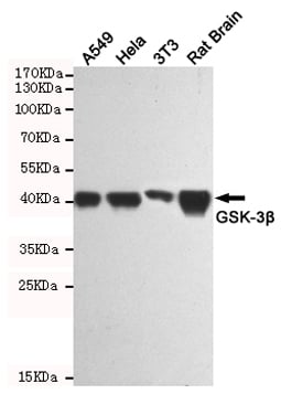
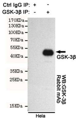 IP (Immunoprecipitation)
(Immunoprecipitation analysis of Hela cell lysates using GSK-3beta.GSK-3beta Rabbit mAb was used for the western blot analysis (1:1000 diluted).)
IP (Immunoprecipitation)
(Immunoprecipitation analysis of Hela cell lysates using GSK-3beta.GSK-3beta Rabbit mAb was used for the western blot analysis (1:1000 diluted).)
GSK3 beta, Monoclonal Antibody (Cat# AAA290858)
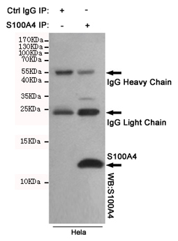 IP (Immunoprecipitation)
(Immunoprecipitation analysis of Hela cell lysates using S100A4 mouse mAb.)
IP (Immunoprecipitation)
(Immunoprecipitation analysis of Hela cell lysates using S100A4 mouse mAb.)
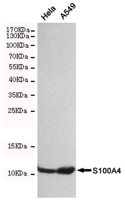
S100A4, Monoclonal Antibody (Cat# AAA290861)
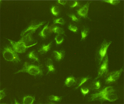
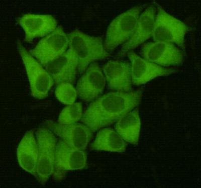
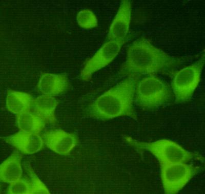 ICC (Immunocytochemistry)
(Immunocytochemistry staining of Hela cells fixed with 4% Paraformaldehyde and using anti-PKM2 mouse mAb (dilution 1:400).)
ICC (Immunocytochemistry)
(Immunocytochemistry staining of Hela cells fixed with 4% Paraformaldehyde and using anti-PKM2 mouse mAb (dilution 1:400).)
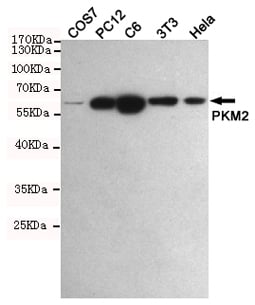
PKM2, Monoclonal Antibody (Cat# AAA290864)
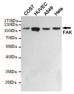
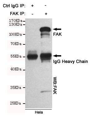 IP (Immunoprecipitation)
(Immunoprecipitation analysis of Hela cell lysates using FAK mouse mAb.)
IP (Immunoprecipitation)
(Immunoprecipitation analysis of Hela cell lysates using FAK mouse mAb.)
FAK, Monoclonal Antibody (Cat# AAA290866)
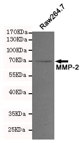
 WB (Western Blot)
(Western blot analysis of extracts from Raw264.7 cell lysates using MMP-2 mouse mAb (1:200 diluted). Predicted band size:64,72KDa. Observed band size:72KDa.)
WB (Western Blot)
(Western blot analysis of extracts from Raw264.7 cell lysates using MMP-2 mouse mAb (1:200 diluted). Predicted band size:64,72KDa. Observed band size:72KDa.)
MMP-2, Monoclonal Antibody (Cat# AAA290868)
 IP (Immunoprecipitation)
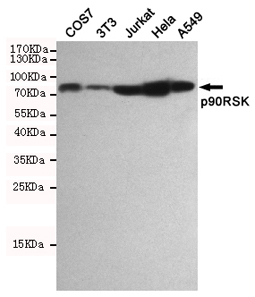
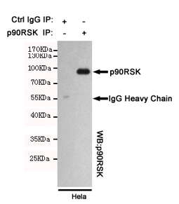
(Immunoprecipitation analysis of Hela cell lysates using p90RSK mouse mAb.)
IP (Immunoprecipitation)
(Immunoprecipitation analysis of Hela cell lysates using p90RSK mouse mAb.)
p90RSK, Monoclonal Antibody (Cat# AAA290870)
 IP (Immunoprecipitation)
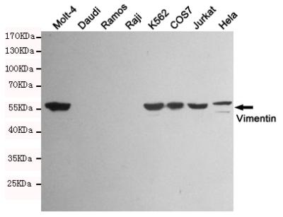
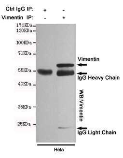
(Immunoprecipitation analysis of Hela cell lysates using Vimentin mouse mAb.)
IP (Immunoprecipitation)
(Immunoprecipitation analysis of Hela cell lysates using Vimentin mouse mAb.)
Vimentin, Monoclonal Antibody (Cat# AAA290872)
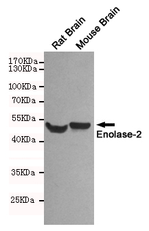

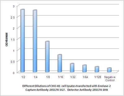 ELISA
(Observed Enolase-2 levels in CHO-K1 cell lysates transfected with Enolase-2 at different dilution.)
ELISA
(Observed Enolase-2 levels in CHO-K1 cell lysates transfected with Enolase-2 at different dilution.)
Enolase-2, Monoclonal Antibody (Cat# AAA290875)
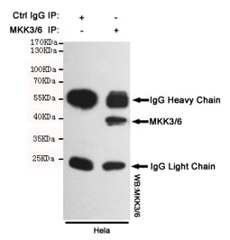 IP (Immunoprecipitation)
(Immunoprecipitation analysis of Hela cell lysates using MKK3/6 mouse mAb.)
IP (Immunoprecipitation)
(Immunoprecipitation analysis of Hela cell lysates using MKK3/6 mouse mAb.)
MKK3/6, Monoclonal Antibody (Cat# AAA290876)
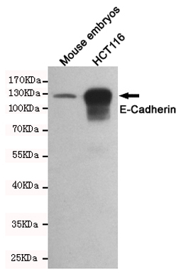
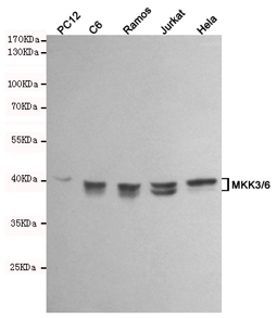
 WB (Western Blot)
(Western blot detection of E-Cadherin in Mouse embryos and HCT116 cell lysates using E-Cadherin mouse mAb (dilution 1:1000).Predicted band size:135kDa.Observed band size:135kDa.)
WB (Western Blot)
(Western blot detection of E-Cadherin in Mouse embryos and HCT116 cell lysates using E-Cadherin mouse mAb (dilution 1:1000).Predicted band size:135kDa.Observed band size:135kDa.)
E-Cadherin, Monoclonal Antibody (Cat# AAA290878)
 IP (Immunoprecipitation)
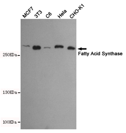
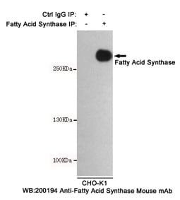
(Immunoprecipitation analysis of CHO-K1 cell lysates using Fatty Acid Synthase mouse mAb.)
IP (Immunoprecipitation)
(Immunoprecipitation analysis of CHO-K1 cell lysates using Fatty Acid Synthase mouse mAb.)
Fatty Acid Synthase, Monoclonal Antibody (Cat# AAA290879)
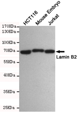
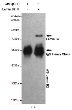 IP (Immunoprecipitation)
(Immunoprecipitation analysis of Hela cell lysates using Lamin B2 mouse mAb.)
IP (Immunoprecipitation)
(Immunoprecipitation analysis of Hela cell lysates using Lamin B2 mouse mAb.)
Lamin B2, Monoclonal Antibody (Cat# AAA290880)

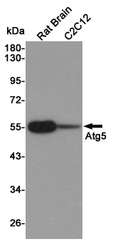 WB (Western Blot)
(Western blot detection of Atg5 in Rat Brain and C2C12 cell lysates using Atg5 mouse mAb (1:1000 diluted).Predicted band size:55KDa.Observed band size:55KDa.)
WB (Western Blot)
(Western blot detection of Atg5 in Rat Brain and C2C12 cell lysates using Atg5 mouse mAb (1:1000 diluted).Predicted band size:55KDa.Observed band size:55KDa.)
Atg5, Monoclonal Antibody (Cat# AAA290884)
 IP (Immunoprecipitation)
(Dilution: IP WB (1/5000 - 1/10000), IP (1/100 - 1/200)Immunoprecipitation of TAP-tagged protein from HEK293T cells transfected with vector overexpressing TAP tag, using Anti-TAP-tag Antibody.)
IP (Immunoprecipitation)
(Dilution: IP WB (1/5000 - 1/10000), IP (1/100 - 1/200)Immunoprecipitation of TAP-tagged protein from HEK293T cells transfected with vector overexpressing TAP tag, using Anti-TAP-tag Antibody.)
TAP-tag, Monoclonal Antibody (Cat# AAA293368)
 IP (Immunoprecipitation)
(Dilution: WB (1/5000 - 1/10000), IH (1/200 - 1/500), IP (1/100 - 1/200))
IP (Immunoprecipitation)
(Dilution: WB (1/5000 - 1/10000), IH (1/200 - 1/500), IP (1/100 - 1/200))
Alpha-actin-1, Monoclonal Antibody (Cat# AAA293372)
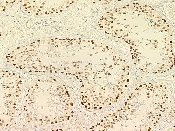
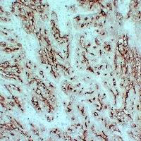
 IHC (Immunohiostchemistry)
(Dilution: WB: (1/20000 - 1/600000), IH (1/1000 - 1/2000)Immunohistochemical analysis of Beta-tubulin-HRP labled staining in human breast cancer formalin fixed paraffin embedded tissue section. The section was pre-treated using heat mediated antigen retrieval with sodium citrate buffer (pH 6.0). The section was then incubated with the antibody at room temperature and detected using an HRP conjugated compact polymer system. DAB was used as the chromogen. The section was then counterstained with haematoxylin and mounted with DPX.)
IHC (Immunohiostchemistry)
(Dilution: WB: (1/20000 - 1/600000), IH (1/1000 - 1/2000)Immunohistochemical analysis of Beta-tubulin-HRP labled staining in human breast cancer formalin fixed paraffin embedded tissue section. The section was pre-treated using heat mediated antigen retrieval with sodium citrate buffer (pH 6.0). The section was then incubated with the antibody at room temperature and detected using an HRP conjugated compact polymer system. DAB was used as the chromogen. The section was then counterstained with haematoxylin and mounted with DPX.)
Beta-tubulin, Monoclonal Antibody (Cat# AAA293375)
 IP (Immunoprecipitation)
(Dilution: WB (1/1000 - 1/3000), IH (1/100 - 1/200), IP (1/50 - 1/200))
IP (Immunoprecipitation)
(Dilution: WB (1/1000 - 1/3000), IH (1/100 - 1/200), IP (1/50 - 1/200))
Carbonic Anhydrase 9, Monoclonal Antibody (Cat# AAA293376)
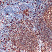
 IHC (Immunohiostchemistry)
(Dilution: WB: (1/2000 - 1/5000), IH (1/200 - 1/500)Immunohistochemical analysis of Cleaved PARP1 staining in human tonsil formalin fixed paraffin embedded tissue section. The section was pre-treated using heat mediated antigen retrieval with sodium citrate buffer (pH 6.0). The section was then incubated with the antibody at room temperature and detected using an HRP conjugated compact polymer system. DAB was used as the chromogen. The section was then counterstained with haematoxylin and mounted with DPX.)
IHC (Immunohiostchemistry)
(Dilution: WB: (1/2000 - 1/5000), IH (1/200 - 1/500)Immunohistochemical analysis of Cleaved PARP1 staining in human tonsil formalin fixed paraffin embedded tissue section. The section was pre-treated using heat mediated antigen retrieval with sodium citrate buffer (pH 6.0). The section was then incubated with the antibody at room temperature and detected using an HRP conjugated compact polymer system. DAB was used as the chromogen. The section was then counterstained with haematoxylin and mounted with DPX.)
Cleaved PARP1, Monoclonal Antibody (Cat# AAA293378)
 IHC (Immunohiostchemistry)
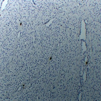
(Dilution: WB: (1/1000 - 1/2000), IH (1/100 - 1/200)Immunohistochemical analysis of ERCC1 staining in human lung cancer formalin fixed paraffin embedded tissue section. The section was pre-treated using heat mediated antigen retrieval with sodium citrate buffer (pH 6.0). The section was then incubated with the antibody at room temperature and detected using an HRP conjugated compact polymer system. DAB was used as the chromogen. The section was then counterstained with haematoxylin and mounted with DPX.)
IHC (Immunohiostchemistry)
(Dilution: WB: (1/1000 - 1/2000), IH (1/100 - 1/200)Immunohistochemical analysis of ERCC1 staining in human lung cancer formalin fixed paraffin embedded tissue section. The section was pre-treated using heat mediated antigen retrieval with sodium citrate buffer (pH 6.0). The section was then incubated with the antibody at room temperature and detected using an HRP conjugated compact polymer system. DAB was used as the chromogen. The section was then counterstained with haematoxylin and mounted with DPX.)
ERCC1, Monoclonal Antibody (Cat# AAA293381)
 IHC (Immunohiostchemistry)
(Dilution: WB: (1/1000 - 1/3000), IH (1/200 - 1/500)Immunohistochemical analysis of Histone H3 (MonoMethyl K9) staining in human colon formalin fixed paraffin embedded tissue section. The section was pre-treated using heat mediated antigen retrieval with sodium citrate buffer (pH 6.0). The section was then incubated with the antibody at room temperature and detected using an HRP conjugated compact polymer system. DAB was used as the chromogen. The section was then counterstained with haematoxylin and mounted with DPX.)
IHC (Immunohiostchemistry)
(Dilution: WB: (1/1000 - 1/3000), IH (1/200 - 1/500)Immunohistochemical analysis of Histone H3 (MonoMethyl K9) staining in human colon formalin fixed paraffin embedded tissue section. The section was pre-treated using heat mediated antigen retrieval with sodium citrate buffer (pH 6.0). The section was then incubated with the antibody at room temperature and detected using an HRP conjugated compact polymer system. DAB was used as the chromogen. The section was then counterstained with haematoxylin and mounted with DPX.)
Histone H3, Monoclonal Antibody (Cat# AAA293387)
 IHC (Immunohiostchemistry)
(Dilution: WB: (1/500 - 1/1000), IH (1/200 - 1/500)Immunohistochemical analysis of Histone H3 (TriMethyl K4) staining in human colon formalin fixed paraffin embedded tissue section. The section was pre-treated using heat mediated antigen retrieval with sodium citrate buffer (pH 6.0). The section was then incubated with the antibody at room temperature and detected using an HRP conjugated compact polymer system. DAB was used as the chromogen. The section was then counterstained with haematoxylin and mounted with DPX.)
IHC (Immunohiostchemistry)
(Dilution: WB: (1/500 - 1/1000), IH (1/200 - 1/500)Immunohistochemical analysis of Histone H3 (TriMethyl K4) staining in human colon formalin fixed paraffin embedded tissue section. The section was pre-treated using heat mediated antigen retrieval with sodium citrate buffer (pH 6.0). The section was then incubated with the antibody at room temperature and detected using an HRP conjugated compact polymer system. DAB was used as the chromogen. The section was then counterstained with haematoxylin and mounted with DPX.)
Histone H3, Monoclonal Antibody (Cat# AAA293389)
 IHC (Immunohiostchemistry)
(Dilution: WB: (1/1000 - 1/2000), IH (1/100 - 1/200)Immunohistochemical analysis of HSP27 staining in human breast cancer formalin fixed paraffin embedded tissue section. The section was pre-treated using heat mediated antigen retrieval with sodium citrate buffer (pH 6.0). The section was then incubated with the antibody at room temperature and detected using an HRP conjugated compact polymer system. DAB was used as the chromogen. The section was then counterstained with haematoxylin and mounted with DPX.)
IHC (Immunohiostchemistry)
(Dilution: WB: (1/1000 - 1/2000), IH (1/100 - 1/200)Immunohistochemical analysis of HSP27 staining in human breast cancer formalin fixed paraffin embedded tissue section. The section was pre-treated using heat mediated antigen retrieval with sodium citrate buffer (pH 6.0). The section was then incubated with the antibody at room temperature and detected using an HRP conjugated compact polymer system. DAB was used as the chromogen. The section was then counterstained with haematoxylin and mounted with DPX.)
HSP27, Monoclonal Antibody (Cat# AAA293390)
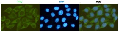
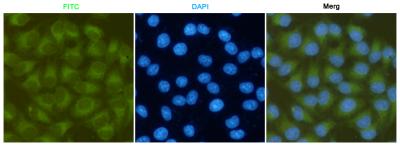
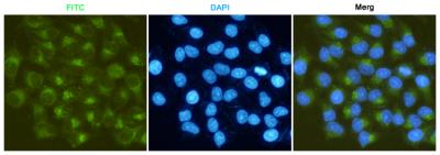
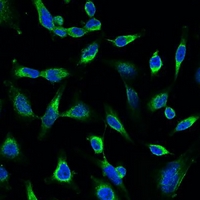 IF (Immunofluorescence)
(Dilution: IF WB (1/1000 - 1/3000), IF/IC (1/100 - 1/200)Immunofluorescent analysis of Peroxiredoxin 1 staining in Hela cells. Formalin-fixed cells were permeabilized with 0.1% Triton X-100 in TBS for 5-10 minutes and blocked with 3% BSA-PBS for 30 minutes at room temperature. Cells were probed with the primary antibody in 3% BSA-PBS and incubated overnight at 4 degree C in a hidified chamber. Cells were washed with PBST and incubated with a FITC-conjugated secondary antibody (green) in PBS at room temperature in the dark. DAPI was used to stain the cell nuclei (blue).)
IF (Immunofluorescence)
(Dilution: IF WB (1/1000 - 1/3000), IF/IC (1/100 - 1/200)Immunofluorescent analysis of Peroxiredoxin 1 staining in Hela cells. Formalin-fixed cells were permeabilized with 0.1% Triton X-100 in TBS for 5-10 minutes and blocked with 3% BSA-PBS for 30 minutes at room temperature. Cells were probed with the primary antibody in 3% BSA-PBS and incubated overnight at 4 degree C in a hidified chamber. Cells were washed with PBST and incubated with a FITC-conjugated secondary antibody (green) in PBS at room temperature in the dark. DAPI was used to stain the cell nuclei (blue).)
Peroxiredoxin 1, Monoclonal Antibody (Cat# AAA293392)
 IF (Immunofluorescence)
(Dilution: IF IH (1/100 - 1/300)Immunohistochemical analysis of CD66e staining in human colon carcinoma formalin fixed paraffin embedded tissue section. The section was pre-treated using heat mediated antigen retrieval with sodium citrate buffer (pH 6.0). The section was then incubated with the antibody at room temperature and detected using an HRP conjugated compact polymer system. DAB was used as the chromogen. The section was then counterstained with haematoxylin and mounted with DPX.)
IF (Immunofluorescence)
(Dilution: IF IH (1/100 - 1/300)Immunohistochemical analysis of CD66e staining in human colon carcinoma formalin fixed paraffin embedded tissue section. The section was pre-treated using heat mediated antigen retrieval with sodium citrate buffer (pH 6.0). The section was then incubated with the antibody at room temperature and detected using an HRP conjugated compact polymer system. DAB was used as the chromogen. The section was then counterstained with haematoxylin and mounted with DPX.)
CD66e, Monoclonal Antibody (Cat# AAA293401)
 IF (Immunofluorescence)
(Dilution: IF IH (1/100 - 1/300)Immunohistochemical analysis of BOB1 staining in human tonsil formalin fixed paraffin embedded tissue section. The section was pre-treated using heat mediated antigen retrieval with sodium citrate buffer (pH 6.0). The section was then incubated with the antibody at room temperature and detected using an HRP conjugated compact polymer system. DAB was used as the chromogen. The section was then counterstained with haematoxylin and mounted with DPX.)
IF (Immunofluorescence)
(Dilution: IF IH (1/100 - 1/300)Immunohistochemical analysis of BOB1 staining in human tonsil formalin fixed paraffin embedded tissue section. The section was pre-treated using heat mediated antigen retrieval with sodium citrate buffer (pH 6.0). The section was then incubated with the antibody at room temperature and detected using an HRP conjugated compact polymer system. DAB was used as the chromogen. The section was then counterstained with haematoxylin and mounted with DPX.)
BOB1, Monoclonal Antibody (Cat# AAA293404)

 FCM/FACS (Flow Cytometry)
(Dilution: IH (1/100 - 1/300)Immunohistochemical analysis of TPSAB1 staining in human uterus formalin fixed paraffin embedded tissue section. The section was pre-treated using heat mediated antigen retrieval with sodium citrate buffer (pH 6.0). The section was then incubated with the antibody at room temperature and detected using an HRP conjugated compact polymer system. DAB was used as the chromogen. The section was then counterstained with haematoxylin and mounted with DPX.)
FCM/FACS (Flow Cytometry)
(Dilution: IH (1/100 - 1/300)Immunohistochemical analysis of TPSAB1 staining in human uterus formalin fixed paraffin embedded tissue section. The section was pre-treated using heat mediated antigen retrieval with sodium citrate buffer (pH 6.0). The section was then incubated with the antibody at room temperature and detected using an HRP conjugated compact polymer system. DAB was used as the chromogen. The section was then counterstained with haematoxylin and mounted with DPX.)
TPSAB1, Monoclonal Antibody (Cat# AAA293406)
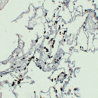 IF (Immunofluorescence)
(Dilution: IF IH (1/100 - 1/300)Immunohistochemical analysis of ACP5 staining in human lung formalin fixed paraffin embedded tissue section. The section was pre-treated using heat mediated antigen retrieval with sodium citrate buffer (pH 6.0). The section was then incubated with the antibody at room temperature and detected using an HRP conjugated compact polymer system. DAB was used as the chromogen. The section was then counterstained with haematoxylin and mounted with DPX.)
IF (Immunofluorescence)
(Dilution: IF IH (1/100 - 1/300)Immunohistochemical analysis of ACP5 staining in human lung formalin fixed paraffin embedded tissue section. The section was pre-treated using heat mediated antigen retrieval with sodium citrate buffer (pH 6.0). The section was then incubated with the antibody at room temperature and detected using an HRP conjugated compact polymer system. DAB was used as the chromogen. The section was then counterstained with haematoxylin and mounted with DPX.)
ACP5, Monoclonal Antibody (Cat# AAA293407)
 IF (Immunofluorescence)
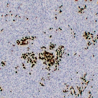
(Dilution: IF IH (1/100 - 1/300)Immunohistochemical analysis of Myeloperoxidase staining in human tonsil formalin fixed paraffin embedded tissue section. The section was pre-treated using heat mediated antigen retrieval with sodium citrate buffer (pH 6.0). The section was then incubated with the antibody at room temperature and detected using an HRP conjugated compact polymer system. DAB was used as the chromogen. The section was then counterstained with haematoxylin and mounted with DPX.)
IF (Immunofluorescence)
(Dilution: IF IH (1/100 - 1/300)Immunohistochemical analysis of Myeloperoxidase staining in human tonsil formalin fixed paraffin embedded tissue section. The section was pre-treated using heat mediated antigen retrieval with sodium citrate buffer (pH 6.0). The section was then incubated with the antibody at room temperature and detected using an HRP conjugated compact polymer system. DAB was used as the chromogen. The section was then counterstained with haematoxylin and mounted with DPX.)
Myeloperoxidase, Monoclonal Antibody (Cat# AAA293410)


 IHC (Immunohistochemistry)
(Dilution: WB: 1:1000-2000 IHC:1:200-500)
IHC (Immunohistochemistry)
(Dilution: WB: 1:1000-2000 IHC:1:200-500)
GAP-43, Monoclonal Antibody (Cat# AAA293654)
What are Monoclonal Antibodies?
Monoclonal antibodies are specialized laboratory-produced proteins developed for binding to specific biological antigens or other molecular targets. Since they come from a single cell (or clone), they are especially consistent and accurate in the data they are involved in producing.
This type of antibody material has been shown to be a powerful tool in finding and subsequently destroying harmful cells in an organism, such as those found in cancers or various autoimmune diseases. This makes them excellent aids in medical testing and research, which is why they are so widely used.
AAA Biotech offers a comprehensive range of high-quality monoclonal antibodies that perform effectively in various laboratory tests, including (amongst others) ELISA, western blotting, immunohistochemistry, and flow cytometry. All of the products in our catalog are thoroughly quality tested to make sure that they are reliable and will consistently perform well in your research.
What Are The Uses of Monoclonal Antibodies
Monoclonal antibodies are used in many lab tests, including (amongst others) ELISA, western blotting, immunohistochemistry, and flow cytometry.
ELISA is a test that helps detect a specific substance/analyte in a sample. It uses antibodies (often monoclonal) bound to a solid surface (such as the well of a microplate) to “capture” the substance/analyte in the sample and immobilize it so that the detection antibody component can then bind to it and produce a signal, which can then be measured.
Western blotting identifies specific proteins in a sample. The sample is first separated on a gel, and then antibodies are applied that will typically bind to the target, which will all be localized to a single band in a lane.
Immunohistochemistry helps locate specific proteins in cells or tissue samples using antibodies.
Flow cytometry looks at and sorts cells. It uses antibodies that are conjugated to reporter molecules called “fluorophores”, which, under special lights, emit light themselves, which can then be measured by a detector instrument.
How Monoclonal Antibodies Are Used as Medicine?
Please note that all of the products listed in AAA Biotech’s also known as AAA Bio or AAABio catalog are strictly for research-use only (RUO).
Monoclonal antibodies can also be used as therapeutic/medical treatments, particularly in the context of cancers. They are designed to find and bind to specific cells or proteins, helping the immune system recognize and attack the cancer. These treatments work in different ways, such as:
- Radioimmunotherapy attaches a small amount of radioactive molecule to the antibody, so it delivers the radiation directly to the cancer cells that the antibody is specifically binding to.
- Antibody-directed enzyme prodrug therapy uses antibodies that are specifically bound to special enzymes. These enzymes activate a harmless drug in the body and turn it into a cancer-killing drug only near the cancer cells—this helps avoid harming healthy cells.
- Immunoliposomes are tiny “bubbles” filled with medicine/drug and coated with antibodies. They carry the drug straight to the cancer cells.
Why Buy Monoclonal Antibodies From Us?
At AAA Biotech, we provide high-performance monoclonal antibodies designed to support a wide range of research needs.
1. Validated for Versatile Applications
The antibodies in our catalog are extensively validated and compatible with multiple techniques, including (but not limited to) ELISA, flow cytometry (FC), immunocytochemistry (ICC), immunofluorescence (IF), immunohistochemistry (IHC), immunoprecipitation (IP), and western blotting (WB).
2. Wide Selection & Specialized Options
We offer antibodies for common and rare species, that are available in various conjugated forms, and also in recombinant formats. Essentially, there is almost anything one might need to meet their experimental model’s requirements.
3. High-Quality Proteins
Our proteins meet high purity standards—90% or more as confirmed by SDS-PAGE. Many are available with tags like His, Flag, GST, or MBP, and we also supply native and biologically active proteins for functional studies.
Frequently Asked Questions
1. Are your monoclonal antibodies validated for specific applications?
Yes, our antibodies are tested and validated for use in methods such as ELISA, western blot, IHC, flow cytometry, and more. Refer to specific product pages or datasheets for individual product information.
2. How do I choose the right monoclonal antibody for my application?
Review the product details directly for application validation, species reactivity, and target information. You may also contact our support team at any time for help.
3. How quickly can I receive my order?
Most orders are processed and shipped within 1–3 business days, depending on product availability and your shipping location.