Filters
▼Clonality
▼Type
▼Reactivity
▼Gene Name
▼Isotype
▼Host
▼Application
▼Clone
▼Monoclonal Antibodies
Get accurate results in your research with our Monoclonal Antibodies, which are specially made to target exactly what you require for your research, and will produce consistent, reliable performance in lab tests.
Viewing 7500-7550 of 27597 product results
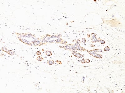
 IHC (Immunohiostchemistry)
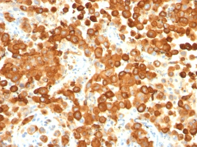
(Formalin-fixed, paraffin-embedded human Testis stained with gp100 / Melanosome Monoclonal Antibody (HMB45 + PMEL/783).)
IHC (Immunohiostchemistry)
(Formalin-fixed, paraffin-embedded human Testis stained with gp100 / Melanosome Monoclonal Antibody (HMB45 + PMEL/783).)
gp100 / Melanosome / PMEL17 / SILV, Monoclonal Antibody (Cat# AAA62851)
Others not tested
T1/ST2 (IL-33 R), Monoclonal Antibody (Cat# AAA63034)
 WB (Western Blot)
(Western Blot Analysis of Raji Cell Lysate using Kappa Light Chain Monoclonal Antibody (HP6053 + L1C1).)
WB (Western Blot)
(Western Blot Analysis of Raji Cell Lysate using Kappa Light Chain Monoclonal Antibody (HP6053 + L1C1).)
Kappa Light Chain, Monoclonal Antibody (Cat# AAA62510)
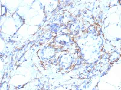
 IHC (Immunohistochemistry)
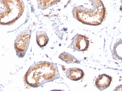
(Formalin-fixed, paraffin-embedded human Colon Carcinoma stained with SM-MHC Monoclonal Antibody (MYH11/923 + SMMS-1).)
IHC (Immunohistochemistry)
(Formalin-fixed, paraffin-embedded human Colon Carcinoma stained with SM-MHC Monoclonal Antibody (MYH11/923 + SMMS-1).)
Smooth Muscle Myosin Heavy Chain (SM-MHC), Monoclonal Antibody (Cat# AAA62748)
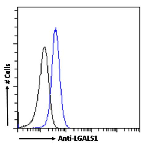
 WB (Western Blot)
(Western Blot using anti-LGALS1 antibody SAIC-25B-112 (AAA72075). U251 cell lysates (35ug protein in RIPA buffer) were resolved on a SDS PAGE gel and blots were probed with the chimeric rabbit version of SAIC-25B-112 () at 0.1ug/ml before detection using an anti-rabbit secondary antibody. A primary incubation of 1h was used and protein was detected by chemiluminescence.)
WB (Western Blot)
(Western Blot using anti-LGALS1 antibody SAIC-25B-112 (AAA72075). U251 cell lysates (35ug protein in RIPA buffer) were resolved on a SDS PAGE gel and blots were probed with the chimeric rabbit version of SAIC-25B-112 () at 0.1ug/ml before detection using an anti-rabbit secondary antibody. A primary incubation of 1h was used and protein was detected by chemiluminescence.)
LGALS1, Monoclonal Antibody (Cat# AAA72075)
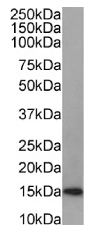
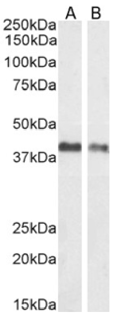
 WB (Western Blot)
(Western Blot using anti-TAGLN antibody SAIC-33B-129 (AAA72078). HeLa(A), Caco-2(B), U2OS(C) cell lysate and human placenta(D), muscle(E) tissue lysates (35ug protein in RIPA buffer) were resolved on a SDS PAGE gel and blots were probed with the chimeric rabbit version of SAIC-33B-129 () at 0.0003ug/ml before detection using an anti-rabbit secondary antibody. A primary incubation of 1h was used and protein was detected by chemiluminescence.)
WB (Western Blot)
(Western Blot using anti-TAGLN antibody SAIC-33B-129 (AAA72078). HeLa(A), Caco-2(B), U2OS(C) cell lysate and human placenta(D), muscle(E) tissue lysates (35ug protein in RIPA buffer) were resolved on a SDS PAGE gel and blots were probed with the chimeric rabbit version of SAIC-33B-129 () at 0.0003ug/ml before detection using an anti-rabbit secondary antibody. A primary incubation of 1h was used and protein was detected by chemiluminescence.)
TAGLN, Monoclonal Antibody (Cat# AAA72078)
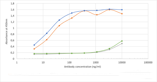
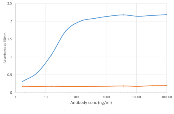 ELISA
(ELISA of anti-VISTA antibody on VISTA-Fc fusion protein. Binding curve of the rabbit chimeric version of the anti-VISTA antibody MH5A (; blue line) to an ELISA plate coated with mouse VISTA-Fc fusion protein (Pr00164-1.9) at a concentration of 5ug/ml. A 3-fold serial dilution from 10000 to 1.7 ng/ml was performed using . For signal detection, a 1:4000 dilution of anti-rabbit IgG1 HRP (BioRad) antibody was used.)
ELISA
(ELISA of anti-VISTA antibody on VISTA-Fc fusion protein. Binding curve of the rabbit chimeric version of the anti-VISTA antibody MH5A (; blue line) to an ELISA plate coated with mouse VISTA-Fc fusion protein (Pr00164-1.9) at a concentration of 5ug/ml. A 3-fold serial dilution from 10000 to 1.7 ng/ml was performed using . For signal detection, a 1:4000 dilution of anti-rabbit IgG1 HRP (BioRad) antibody was used.)
PD-1H, Monoclonal Antibody (Cat# AAA72099)
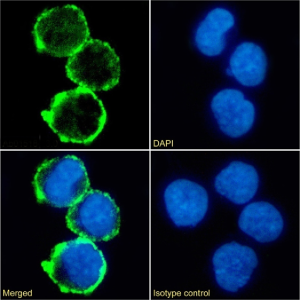
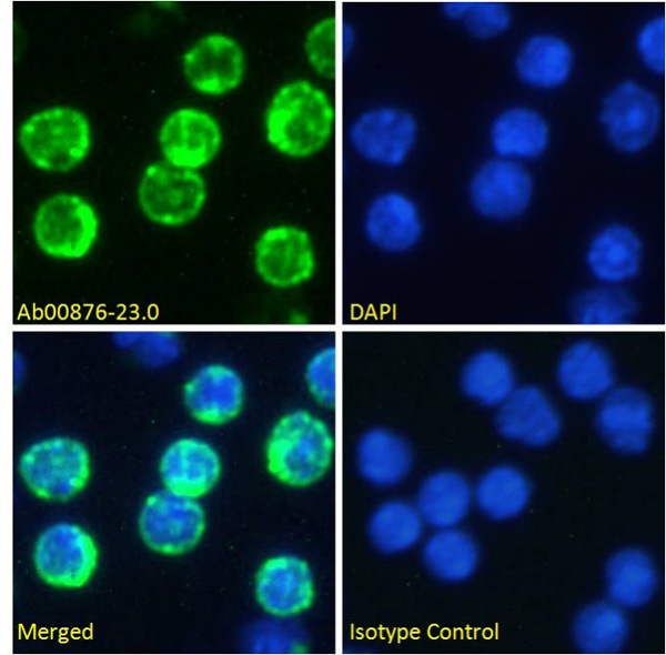
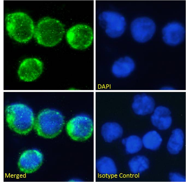
 IF (Immunofluorescence)
(Immunofluorescence staining of fixed mouse splenocytes with anti- CD226 antibody 10E5 Immunofluorescence analysis of paraformaldehyde fixed mouse splenocytes on Shi-fix coverslips stained with the chimeric rabbit IgG version of 10E5 at 10ug/ml for 1h followed by Alexa Fluor 488 secondary antibody (2ug/ml), showing membrane staining. The nuclear stain is DAPI (blue). Panels show from left-right, top-bottom DAPI, merged channels and an isotype control. The isotype control was an unknown specificity antibody followed by staining with Alexa Fluor 488 secondary antibody.)
IF (Immunofluorescence)
(Immunofluorescence staining of fixed mouse splenocytes with anti- CD226 antibody 10E5 Immunofluorescence analysis of paraformaldehyde fixed mouse splenocytes on Shi-fix coverslips stained with the chimeric rabbit IgG version of 10E5 at 10ug/ml for 1h followed by Alexa Fluor 488 secondary antibody (2ug/ml), showing membrane staining. The nuclear stain is DAPI (blue). Panels show from left-right, top-bottom DAPI, merged channels and an isotype control. The isotype control was an unknown specificity antibody followed by staining with Alexa Fluor 488 secondary antibody.)
CD226, Monoclonal Antibody (Cat# AAA72126)

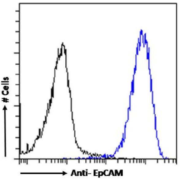
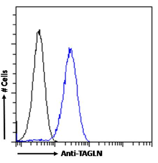
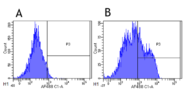
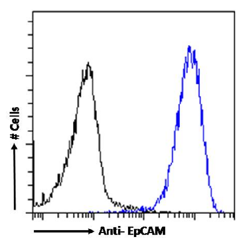 FCM/FACS (Flow Cytometry)
(Flow cytometry using the Anti-EpCAM antibody HEA125 (AAA72133). Caco-2 cells were stained with anti-unknown specificity antibody or the mouse IgG1 version of HEA125 at a dilution of 1:100 for 1h at RT. After washing, the bound antibody was detected using a goat anti-mouse IgG AlexaFluor 488 antibody at a dilution of 1:1000 and cells analyzed using a FACSCanto flow-cytometer.)
FCM/FACS (Flow Cytometry)
(Flow cytometry using the Anti-EpCAM antibody HEA125 (AAA72133). Caco-2 cells were stained with anti-unknown specificity antibody or the mouse IgG1 version of HEA125 at a dilution of 1:100 for 1h at RT. After washing, the bound antibody was detected using a goat anti-mouse IgG AlexaFluor 488 antibody at a dilution of 1:1000 and cells analyzed using a FACSCanto flow-cytometer.)
EpCAM, Monoclonal Antibody (Cat# AAA72133)
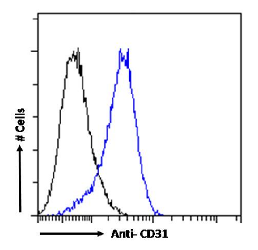 FCM/FACS (Flow Cytometry)
(Flow cytometry using the Anti-CD31 antibody BAG-85D10 (AAA72148). Paraformaldehyde fixed Jurkat cells were stained with anti-unknown specificity antibody or the rabbit IgG version of BAG-85D10 (, blue line) at a dilution of 1:100 for 1h at RT. After washing, the bound antibody was detected using a goat anti-rabbit IgG AlexaFluor 488 antibody at a dilution of 1:1000 and cells analyzed using a FACSCanto flow-cytometer.)
FCM/FACS (Flow Cytometry)
(Flow cytometry using the Anti-CD31 antibody BAG-85D10 (AAA72148). Paraformaldehyde fixed Jurkat cells were stained with anti-unknown specificity antibody or the rabbit IgG version of BAG-85D10 (, blue line) at a dilution of 1:100 for 1h at RT. After washing, the bound antibody was detected using a goat anti-rabbit IgG AlexaFluor 488 antibody at a dilution of 1:1000 and cells analyzed using a FACSCanto flow-cytometer.)
CD31, Monoclonal Antibody (Cat# AAA72148)
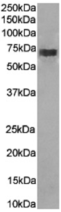
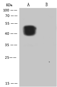
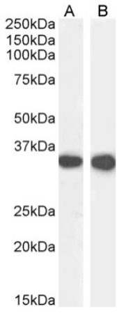 WB (Western Blot)
( Western Blot using anti-CD74 antibody In-1 Mouse spleen (A) and mouse lymph node (B) lysate samples (35ug protein in RIPA buffer) were resolved on a 10% SDS PAGE gel and blots probed with the chimeric rabbit version of In-1 at 0.0001 ug/ml before detection using an anti-rabbit secondary antibody. A primary incubation of 1h was used and protein was detected by chemiluminescence. The expected running size for unmodified CD74 is 31.6kDa, and this protein is glycosylated at several residues. successfully detected CD74 in mouse spleen and mouse lymph node lysates.)
WB (Western Blot)
( Western Blot using anti-CD74 antibody In-1 Mouse spleen (A) and mouse lymph node (B) lysate samples (35ug protein in RIPA buffer) were resolved on a 10% SDS PAGE gel and blots probed with the chimeric rabbit version of In-1 at 0.0001 ug/ml before detection using an anti-rabbit secondary antibody. A primary incubation of 1h was used and protein was detected by chemiluminescence. The expected running size for unmodified CD74 is 31.6kDa, and this protein is glycosylated at several residues. successfully detected CD74 in mouse spleen and mouse lymph node lysates.)
CD74, Monoclonal Recombinant Antibody (Cat# AAA72009)
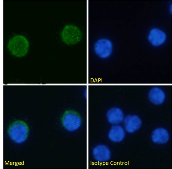
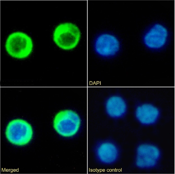
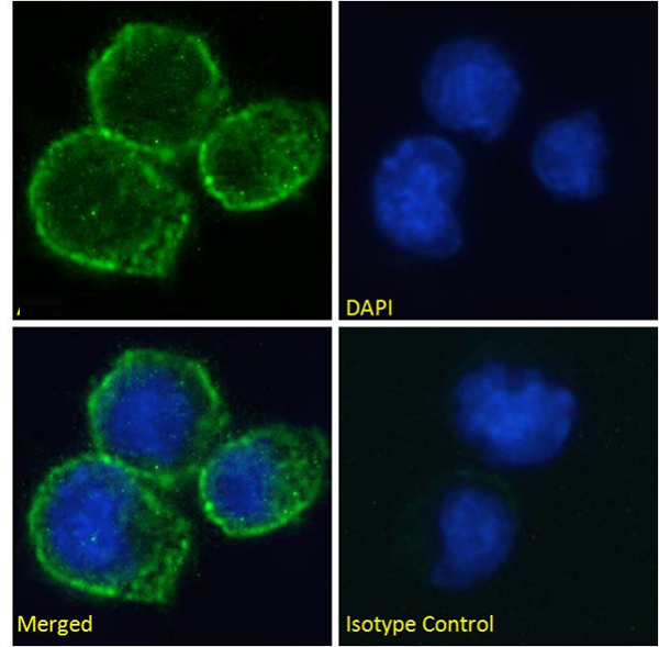
 IF (Immunofluorescence)
(Immunofluorescence staining of fixed Molt4 cells with anti-CD2 antibody YTH 655 Immunofluorescence analysis of paraformaldehyde fixed Molt4 cells on Shi-fix coverslips, permeabilized with 0.15% Triton and stained with the chimeric rabbit IgG version of YTH 655 (AAA72014) at 10 ug/ml for 1h followed by Alexa Fluor 488 secondary antibody (1 ug/ml), showing membrane, and some cytoplasmic staining. The nuclear stain is DAPI (blue). Panels show from left-right, top-bottom AAA72014, DAPI, merged channels and an isotype control. The isotype control was stained with an anti-Fluorescein antibody followed by Alexa Fluor 488 secondary antibody.)
IF (Immunofluorescence)
(Immunofluorescence staining of fixed Molt4 cells with anti-CD2 antibody YTH 655 Immunofluorescence analysis of paraformaldehyde fixed Molt4 cells on Shi-fix coverslips, permeabilized with 0.15% Triton and stained with the chimeric rabbit IgG version of YTH 655 (AAA72014) at 10 ug/ml for 1h followed by Alexa Fluor 488 secondary antibody (1 ug/ml), showing membrane, and some cytoplasmic staining. The nuclear stain is DAPI (blue). Panels show from left-right, top-bottom AAA72014, DAPI, merged channels and an isotype control. The isotype control was stained with an anti-Fluorescein antibody followed by Alexa Fluor 488 secondary antibody.)
CD2, Monoclonal Recombinant Antibody (Cat# AAA72014)
 IF (Immunofluorescence)
(Immunofluorescence staining of fixed Molt4 cells with anti-CD2 antibody YTH 655 Immunofluorescence analysis of paraformaldehyde fixed Molt4 cells on Shi-fix coverslips, permeabilized with 0.15% Triton and stained with the chimeric rabbit IgG version of YTH 655 at 10 ug/ml for 1h followed by Alexa Fluor 488 secondary antibody (1 ug/ml), showing membrane, and some cytoplasmic staining. The nuclear stain is DAPI (blue). Panels show from left-right, top-bottom, DAPI, merged channels and an isotype control. The isotype control was stained with an anti-Fluorescein antibody followed by Alexa Fluor 488 secondary antibody.)
IF (Immunofluorescence)
(Immunofluorescence staining of fixed Molt4 cells with anti-CD2 antibody YTH 655 Immunofluorescence analysis of paraformaldehyde fixed Molt4 cells on Shi-fix coverslips, permeabilized with 0.15% Triton and stained with the chimeric rabbit IgG version of YTH 655 at 10 ug/ml for 1h followed by Alexa Fluor 488 secondary antibody (1 ug/ml), showing membrane, and some cytoplasmic staining. The nuclear stain is DAPI (blue). Panels show from left-right, top-bottom, DAPI, merged channels and an isotype control. The isotype control was stained with an anti-Fluorescein antibody followed by Alexa Fluor 488 secondary antibody.)
CD2, Monoclonal Recombinant Antibody (Cat# AAA72015)
 IF (Immunofluorescence)
(Immunofluorescence staining of fixed Daudi cells with anti-CD40 antibody A54 scFv Immunofluorescence analysis of paraformaldehyde fixed Daudi cells on Shi-fix coverslips, permeabilized with 0.15% Triton and stained with the chimeric rabbit IgG version of A54 scFv (AAA72042) at 10 ug/ml for 1h followed by Alexa Fluor 488 secondary antibody (1 ug/ml), showing membrane staining. The nuclear stain is DAPI (blue). Panels show from left-right, top-bottom AAA72042, DAPI, merged channels and an isotype control. The isotype control was stained with an anti-Fluorescein antibody followed by Alexa Fluor 488 secondary antibody.)
IF (Immunofluorescence)
(Immunofluorescence staining of fixed Daudi cells with anti-CD40 antibody A54 scFv Immunofluorescence analysis of paraformaldehyde fixed Daudi cells on Shi-fix coverslips, permeabilized with 0.15% Triton and stained with the chimeric rabbit IgG version of A54 scFv (AAA72042) at 10 ug/ml for 1h followed by Alexa Fluor 488 secondary antibody (1 ug/ml), showing membrane staining. The nuclear stain is DAPI (blue). Panels show from left-right, top-bottom AAA72042, DAPI, merged channels and an isotype control. The isotype control was stained with an anti-Fluorescein antibody followed by Alexa Fluor 488 secondary antibody.)
CD40, Monoclonal Recombinant Antibody (Cat# AAA72042)
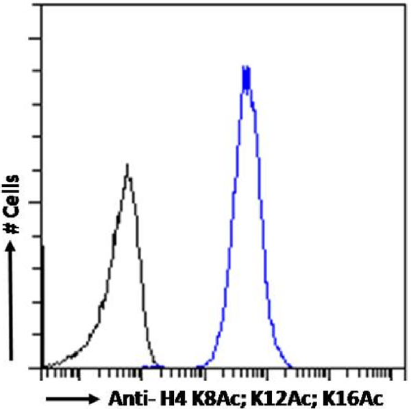 FCM/FACS (Flow Cytometry)
(Flow cytometry using the anti-H4 K8Ac; K12Ac; K16Ac antibody KM-2 (AAA72187). HeLa cells were fixed using 2% PFA and stained with anti-unknown specificity antibody or the rabbit IgG1 version of KM-2 (, blue line) at a dilution of 1:100 for 1h at RT. After washing, the bound antibody was detected using a goat anti-rabbit IgG AlexaFluor 488 antibody at a dilution of 1:1000 and cells analyzed using a FACSCanto flow-cytometer.)
FCM/FACS (Flow Cytometry)
(Flow cytometry using the anti-H4 K8Ac; K12Ac; K16Ac antibody KM-2 (AAA72187). HeLa cells were fixed using 2% PFA and stained with anti-unknown specificity antibody or the rabbit IgG1 version of KM-2 (, blue line) at a dilution of 1:100 for 1h at RT. After washing, the bound antibody was detected using a goat anti-rabbit IgG AlexaFluor 488 antibody at a dilution of 1:1000 and cells analyzed using a FACSCanto flow-cytometer.)
H4 K8Ac; K12Ac; K16Ac, Monoclonal Antibody (Cat# AAA72187)
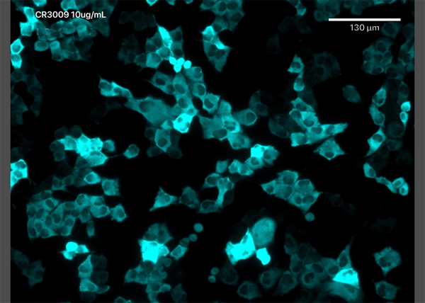 IF (Immunofluorescence)
(Immunofluorescence staining of MDCK-SIAT1 cells transfected with SARS-CoV-2 NP with anti-Covid-19 & SARS-CoV Nucleoprotein antibody CR3009 (03-009) Immunofluorescence analysis of MDCK-SIAT1 cells stably transfected with SARS-CoV-2 NP. The cells were seeded in a flat bottomed 96 well plate overnight, fixed in 10% formalin at 4C for 30min, permeabilised for 20min at RT and then stained with the human IgG1 version of CR3009 (03-009) in PBS/0.1% BSA at 10ug/ml for 1 hour followed by a goat anti-human Alexa Fluor 647 (Invitrogen) secondary antibody. The image is courtesy of Jack Tan, Radcliffe Department of Medicine, University of Oxford.)
IF (Immunofluorescence)
(Immunofluorescence staining of MDCK-SIAT1 cells transfected with SARS-CoV-2 NP with anti-Covid-19 & SARS-CoV Nucleoprotein antibody CR3009 (03-009) Immunofluorescence analysis of MDCK-SIAT1 cells stably transfected with SARS-CoV-2 NP. The cells were seeded in a flat bottomed 96 well plate overnight, fixed in 10% formalin at 4C for 30min, permeabilised for 20min at RT and then stained with the human IgG1 version of CR3009 (03-009) in PBS/0.1% BSA at 10ug/ml for 1 hour followed by a goat anti-human Alexa Fluor 647 (Invitrogen) secondary antibody. The image is courtesy of Jack Tan, Radcliffe Department of Medicine, University of Oxford.)
COVID 19 Nucleocapsid (NP) Coronavirus, Monoclonal Antibody (Cat# AAA72198)
CD33, Monoclonal Recombinant Antibody (Cat# AAA71970)
Protein A Affinity Purified
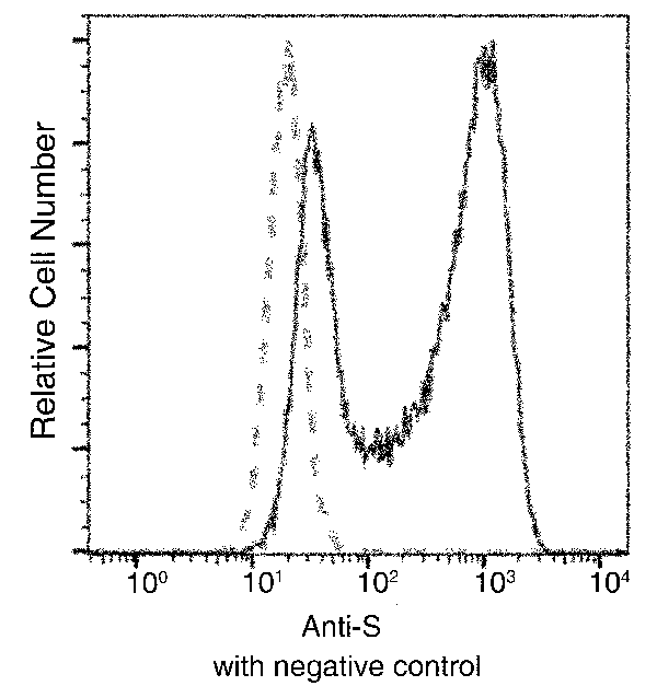
 FCM/FACS (Flow Cytometry)
( Flow-cytometry using anti-CD22 antibody Epratuzumab Cynomolgus monkey lymphocytes were stained with an isotype control or the rabbit-chimeric version of Epratuzumab at a concentration of 1 ug/ml for 30 mins at RT. After washing, bound antibody was detected using a AF488 conjugated donkey anti-rabbit antibody and cells analysed on a FACSCanto flow-cytometer.)
FCM/FACS (Flow Cytometry)
( Flow-cytometry using anti-CD22 antibody Epratuzumab Cynomolgus monkey lymphocytes were stained with an isotype control or the rabbit-chimeric version of Epratuzumab at a concentration of 1 ug/ml for 30 mins at RT. After washing, bound antibody was detected using a AF488 conjugated donkey anti-rabbit antibody and cells analysed on a FACSCanto flow-cytometer.)
CD22, Monoclonal Recombinant Antibody (Cat# AAA71980)
PAT mAb3, Monoclonal Antibody (Cat# AAA71735)
Protein A affinity chromatography purified
HCG-beta mAb-labeling, Monoclonal Antibody (Cat# AAA71754)
 WB (Western Blot)
(Anti-Zika virus(ZIKV)(strain Zika SPH2015)ZIKV-SN1 mouse monoclonal antibody at 1:1000 dilution.Sample:Zika virus(ZIKV)(strain Zika SPH2015)ZIKV-SN1 Recombinant ProteinLane A: 30ngLane B: 10ngSecondaryRabbit Anti-Mouse IgG F(ab)2/HRP at 1/10000 dilution.Developed using the ECL technique.Performed under reducing conditions.)
WB (Western Blot)
(Anti-Zika virus(ZIKV)(strain Zika SPH2015)ZIKV-SN1 mouse monoclonal antibody at 1:1000 dilution.Sample:Zika virus(ZIKV)(strain Zika SPH2015)ZIKV-SN1 Recombinant ProteinLane A: 30ngLane B: 10ngSecondaryRabbit Anti-Mouse IgG F(ab)2/HRP at 1/10000 dilution.Developed using the ECL technique.Performed under reducing conditions.)
Zika virus (ZIKV) ZIKV-NS1, Monoclonal Antibody (Cat# AAA257045)
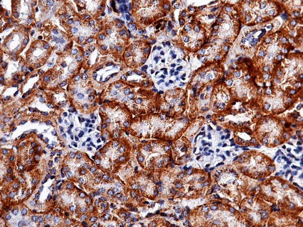
 IHC (Immunohiostchemistry)
(Immunochemical staining of mouse CA12 in mouse intestine with rabbit monoclonal antibody (1:200, formalin-fixed paraffin embedded sections).)
IHC (Immunohiostchemistry)
(Immunochemical staining of mouse CA12 in mouse intestine with rabbit monoclonal antibody (1:200, formalin-fixed paraffin embedded sections).)
carbonic anhydrase XII, Monoclonal Antibody (Cat# AAA257051)
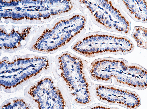
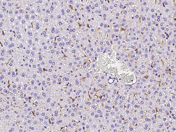
 IHC (Immunohiostchemistry)
(Immunochemical staining of mouse CXADR in mouse liver with rabbit monoclonal antibody (1:200, formalin-fixed paraffin embedded sections).)
IHC (Immunohiostchemistry)
(Immunochemical staining of mouse CXADR in mouse liver with rabbit monoclonal antibody (1:200, formalin-fixed paraffin embedded sections).)
CAR/CXADR, Monoclonal Antibody (Cat# AAA257054)
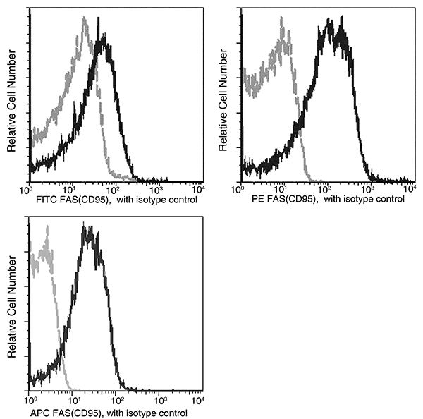
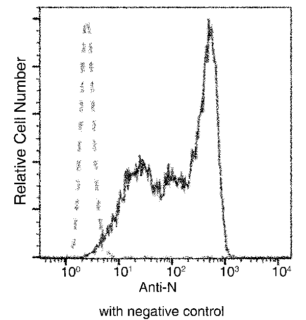
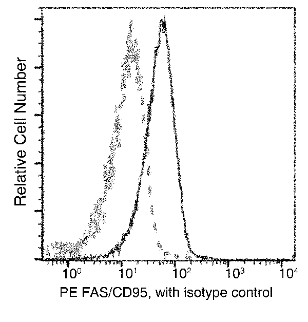 FCM/FACS (Flow Cytometry)
(Profile of anti-FAS (CD95) reactivity on mouse splenocytes analyzed by flow cytometry. Cells should be Fc-blocked by treatment with Mouse BD Fc Block' purified anti-CD16/CD32 mAb 2.4G2 prior to staining, washed, then stained with PE Rabbit anti-FAS(CD95).)
FCM/FACS (Flow Cytometry)
(Profile of anti-FAS (CD95) reactivity on mouse splenocytes analyzed by flow cytometry. Cells should be Fc-blocked by treatment with Mouse BD Fc Block' purified anti-CD16/CD32 mAb 2.4G2 prior to staining, washed, then stained with PE Rabbit anti-FAS(CD95).)
Fas/CD95, Monoclonal Antibody (Cat# AAA257058)
FSTL3, Monoclonal Antibody (Cat# AAA257059)
 FCM/FACS (Flow Cytometry)
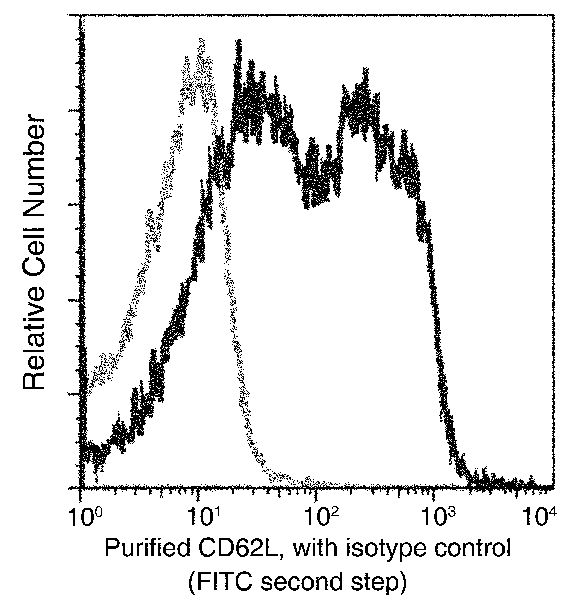
(Flow cytometric analysis of Mouse CD62L expression on BABL/c splenocytes. Cells were stained with purified anti-Mouse CD62L, then a FITC-conjugated second step antibody. The fluorescence histograms were derived from gated events with the forward and side light-scatter characteristics of intact cells.)
FCM/FACS (Flow Cytometry)
(Flow cytometric analysis of Mouse CD62L expression on BABL/c splenocytes. Cells were stained with purified anti-Mouse CD62L, then a FITC-conjugated second step antibody. The fluorescence histograms were derived from gated events with the forward and side light-scatter characteristics of intact cells.)
SELL/CD62L, Monoclonal Antibody (Cat# AAA257068)
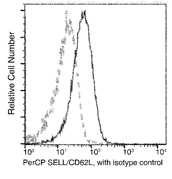 FCM/FACS (Flow Cytometry)
(Flow cytometric analysis of Mouse SELL(CD62L) expression on BABL/c splenocytes. Cells were stained with PerCP-conjugated anti-Mouse SELL(CD62L). The fluorescence histograms were derived from gated events with the forward and side light-scatter characteristics of intact cells.)
FCM/FACS (Flow Cytometry)
(Flow cytometric analysis of Mouse SELL(CD62L) expression on BABL/c splenocytes. Cells were stained with PerCP-conjugated anti-Mouse SELL(CD62L). The fluorescence histograms were derived from gated events with the forward and side light-scatter characteristics of intact cells.)
SELL/CD62L, Monoclonal Antibody (Cat# AAA257069)
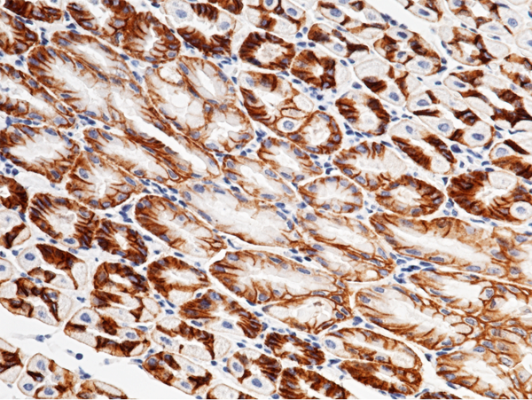
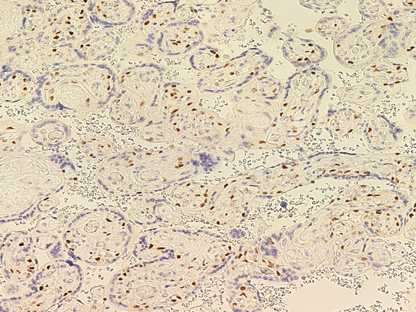
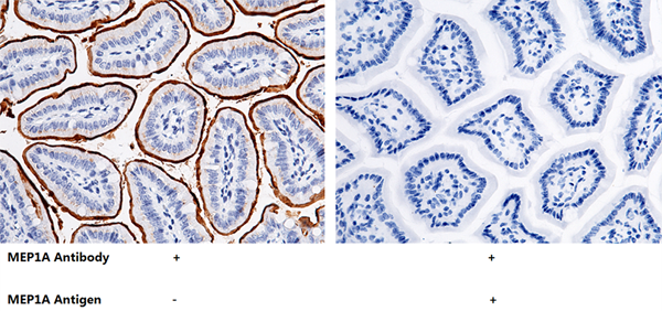 IHC (Immunohiostchemistry)
(Immunochemical staining of mouse MEP1A in mouse intestine with rabbit monoclonal antibody (1:200, formalin-fixed paraffin embedded sections). The left panel: tissue incubated with primary antibody; The right panel: tissue incubated with the mixture of primary antibody and antigen (recombinant protein).)
IHC (Immunohiostchemistry)
(Immunochemical staining of mouse MEP1A in mouse intestine with rabbit monoclonal antibody (1:200, formalin-fixed paraffin embedded sections). The left panel: tissue incubated with primary antibody; The right panel: tissue incubated with the mixture of primary antibody and antigen (recombinant protein).)
Meprin alpha/MEP1A, Monoclonal Antibody (Cat# AAA257072)
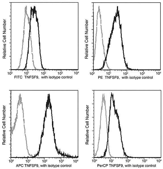
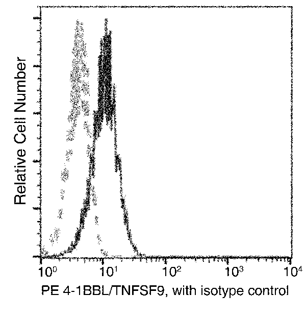 FCM/FACS (Flow Cytometry)
(Flow cytometric analysis of Mouse 4-1BBL(TNFSF9) expression on RAW264.7 cells. Cells were stained with PE-conjugated anti-Mouse 4-1BBL(TNFSF9). The fluorescence histograms were derived from gated events with the forward and side light-scatter characteristics of intact cells.)
FCM/FACS (Flow Cytometry)
(Flow cytometric analysis of Mouse 4-1BBL(TNFSF9) expression on RAW264.7 cells. Cells were stained with PE-conjugated anti-Mouse 4-1BBL(TNFSF9). The fluorescence histograms were derived from gated events with the forward and side light-scatter characteristics of intact cells.)
4-1BBL/TNFSF9, Monoclonal Antibody (Cat# AAA257077)
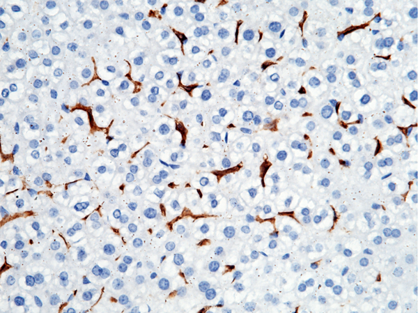
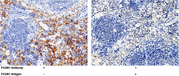
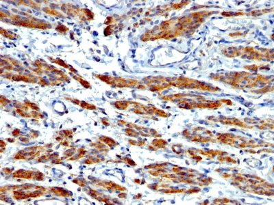
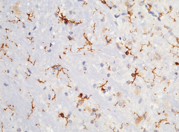
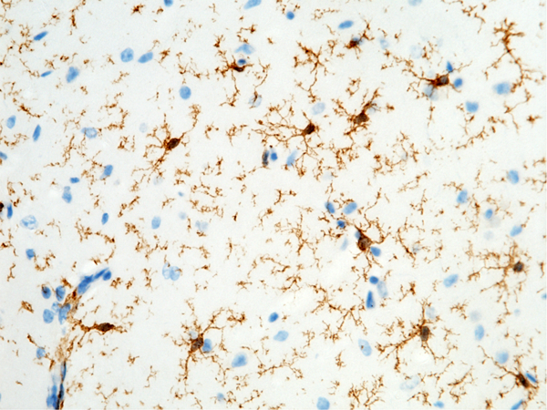 IHC (Immunohistochemisry)
(Immunochemical staining of mouse FCGR1 in mouse brain with rabbit monoclonal antibody (1:200, formalin-fixed paraffin embedded sections). Positive staining was localized to microglia.)
IHC (Immunohistochemisry)
(Immunochemical staining of mouse FCGR1 in mouse brain with rabbit monoclonal antibody (1:200, formalin-fixed paraffin embedded sections). Positive staining was localized to microglia.)
CD64, Monoclonal Antibody (Cat# AAA257081)
 IHC (Immunohistochemisry)
(Immunochemical staining of mouse FCGR1 in mouse brain with rabbit monoclonal antibody (1:200, formalin-fixed paraffin embedded sections). Positive staining was localized to microglia.)
IHC (Immunohistochemisry)
(Immunochemical staining of mouse FCGR1 in mouse brain with rabbit monoclonal antibody (1:200, formalin-fixed paraffin embedded sections). Positive staining was localized to microglia.)
CD64, Monoclonal Antibody (Cat# AAA257082)
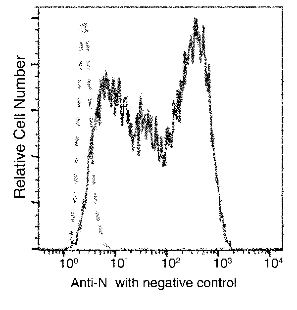
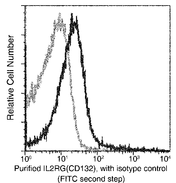 FCM/FACS (Flow Cytometry)
(Flow cytometric analysis of Mouse IL2RG(CD132) expression on BABL/c splenocytes. Cells were stained with purified anti-Mouse IL2RG(CD132), then a FITC-conjugated second step antibody. The fluorescence histograms were derived from gated events with the forward and side light-scatter characteristics of intact cells.)
FCM/FACS (Flow Cytometry)
(Flow cytometric analysis of Mouse IL2RG(CD132) expression on BABL/c splenocytes. Cells were stained with purified anti-Mouse IL2RG(CD132), then a FITC-conjugated second step antibody. The fluorescence histograms were derived from gated events with the forward and side light-scatter characteristics of intact cells.)
IL2RG, Monoclonal Antibody (Cat# AAA257084)
PIGR, Monoclonal Antibody (Cat# AAA257088)
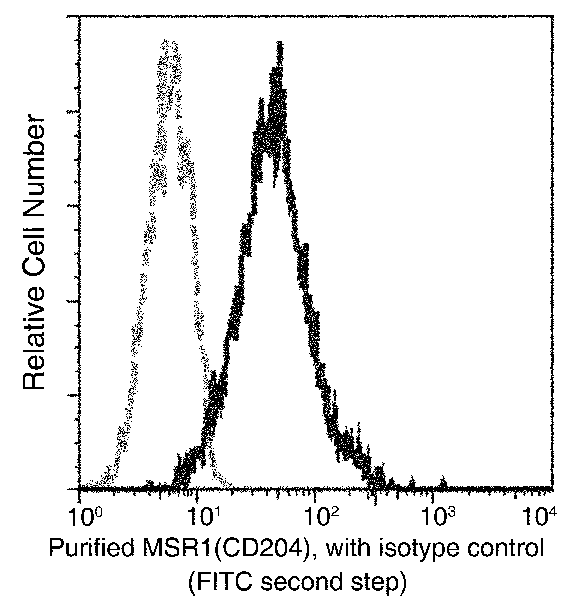 FCM/FACS (Flow Cytometry)
(Flow cytometric analysis of Mouse MSR1(CD204) expression on Raw264.7 cells. Cells were stained with purified anti-Mouse MSR1(CD204), then a FITC-conjugated second step antibody. The fluorescence histograms were derived from gated events with the forward and side light-scatter characteristics of intact cells.)
FCM/FACS (Flow Cytometry)
(Flow cytometric analysis of Mouse MSR1(CD204) expression on Raw264.7 cells. Cells were stained with purified anti-Mouse MSR1(CD204), then a FITC-conjugated second step antibody. The fluorescence histograms were derived from gated events with the forward and side light-scatter characteristics of intact cells.)
CD204/MSR1, Monoclonal Antibody (Cat# AAA257093)
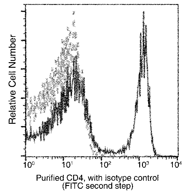
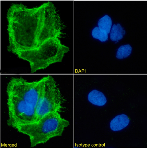
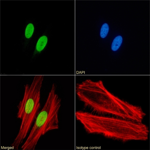
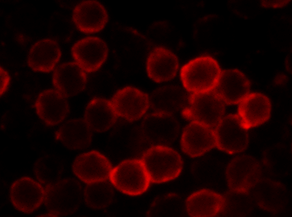
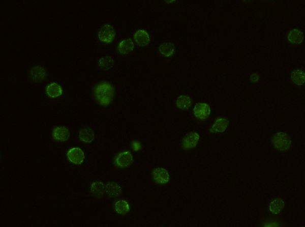
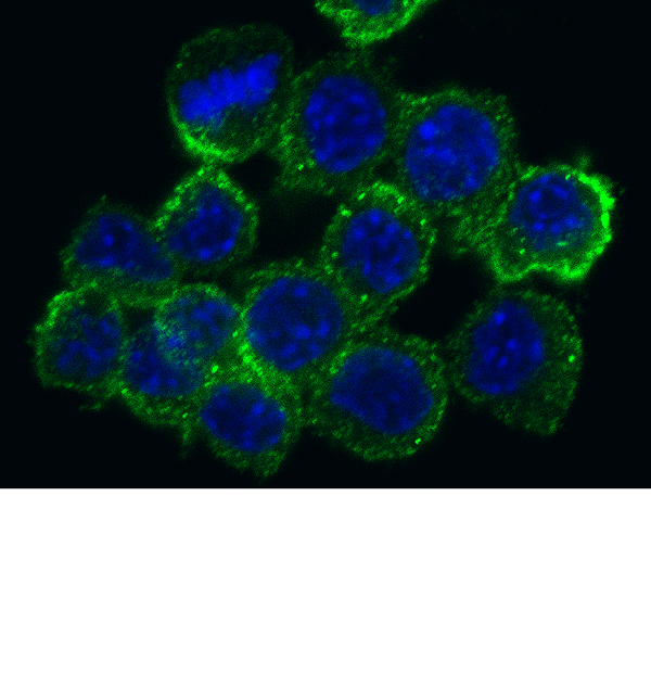
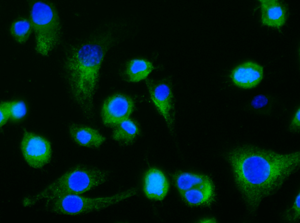
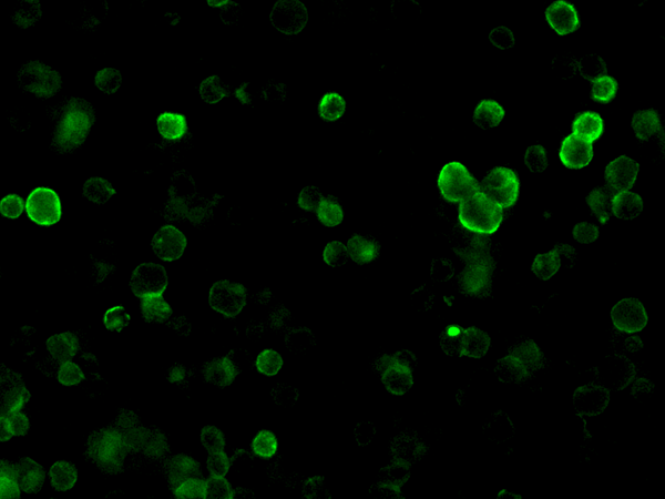
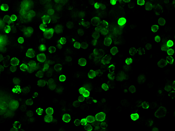
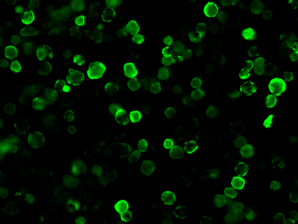
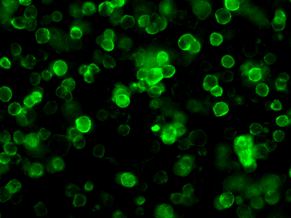
 IF (Immunofluorescence)
(Immunofluorescence staining of mouse CD4 in mouse spleen cells. Cells were fixed with 4% PFA,blocked with 10% serum, and incubated with rabbit anti-mouse CD4 monoclonal antibody (dilution ratio 1:300) at 4 degree C overnight. Then cells were stained with the Alexa Fluor488-conjugated Goat Anti-rabbit IgG secondary antibody (green). Positive staining was localized to Cell membrane.)
IF (Immunofluorescence)
(Immunofluorescence staining of mouse CD4 in mouse spleen cells. Cells were fixed with 4% PFA,blocked with 10% serum, and incubated with rabbit anti-mouse CD4 monoclonal antibody (dilution ratio 1:300) at 4 degree C overnight. Then cells were stained with the Alexa Fluor488-conjugated Goat Anti-rabbit IgG secondary antibody (green). Positive staining was localized to Cell membrane.)
CD4, Monoclonal Antibody (Cat# AAA257094)
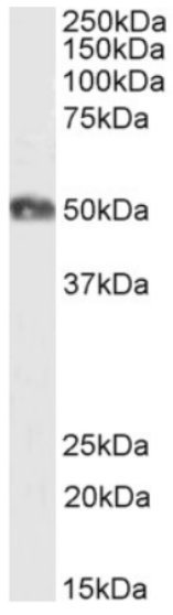
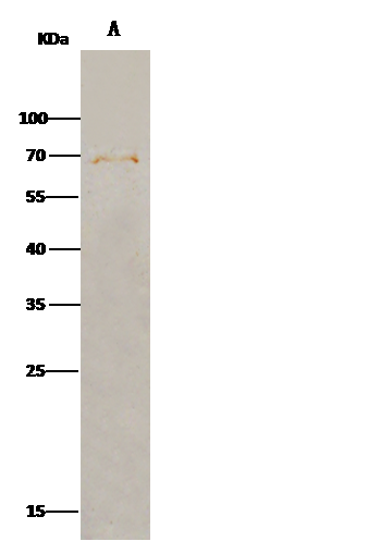 IP (Immunoprecipitation)
(Mouse FGFRL1 was immunoprecipitated using:Lane A:0.5 mg Hela Whole Cell Lysate2 uL anti-Mouse FGFRL1 rabbit monoclonal antibody and 15 ul of 50 % Protein G agarose.Primary antibody:Anti-Mouse FGFRL1 rabbit monoclonal antibody,at 1:100 dilutionSecondary antibody:Clean-Blot IP Detection Reagent (HRP) at 1:1000 dilutionDeveloped using the DAB staining technique.Performed under reducing conditions.Predicted band size: 55 kDaObserved band size: 70 kDa)
IP (Immunoprecipitation)
(Mouse FGFRL1 was immunoprecipitated using:Lane A:0.5 mg Hela Whole Cell Lysate2 uL anti-Mouse FGFRL1 rabbit monoclonal antibody and 15 ul of 50 % Protein G agarose.Primary antibody:Anti-Mouse FGFRL1 rabbit monoclonal antibody,at 1:100 dilutionSecondary antibody:Clean-Blot IP Detection Reagent (HRP) at 1:1000 dilutionDeveloped using the DAB staining technique.Performed under reducing conditions.Predicted band size: 55 kDaObserved band size: 70 kDa)
FGFRL1, Monoclonal Antibody (Cat# AAA257105)
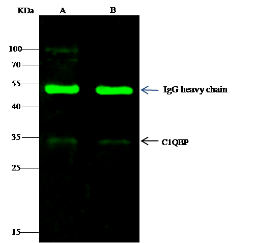
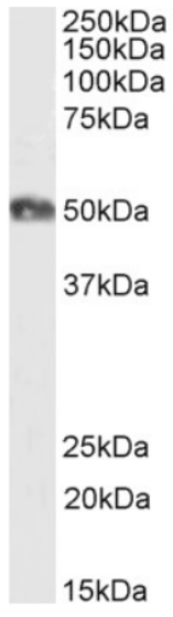
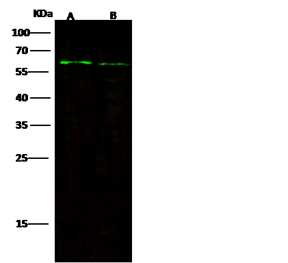
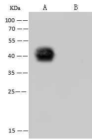
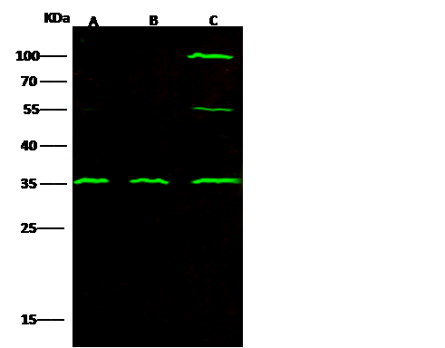 WB (Western Blot)
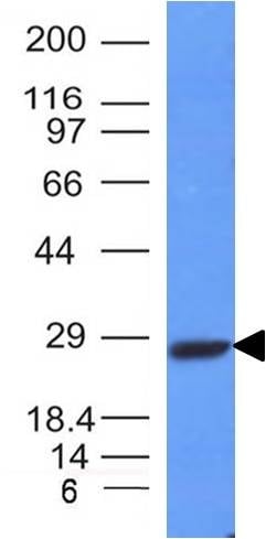
(Anti-C1QBP rabbit monoclonal antibody at 1:500 dilutionLane A: HepG2 Whole Cell LysateLane B: Jurkat Whole Cell LysateLane C: Raji Whole Cell LysateLysates/proteins at 30 ug per lane.SecondaryGoat Anti-Rabbit IgG H&L (Dylight800) at 1/10000 dilution.Developed using the Odyssey technique.Performed under reducing conditions.Predicted band size:31 kDaObserved band size:35 kDa)
WB (Western Blot)
(Anti-C1QBP rabbit monoclonal antibody at 1:500 dilutionLane A: HepG2 Whole Cell LysateLane B: Jurkat Whole Cell LysateLane C: Raji Whole Cell LysateLysates/proteins at 30 ug per lane.SecondaryGoat Anti-Rabbit IgG H&L (Dylight800) at 1/10000 dilution.Developed using the Odyssey technique.Performed under reducing conditions.Predicted band size:31 kDaObserved band size:35 kDa)
C1QBP, Monoclonal Antibody (Cat# AAA257109)
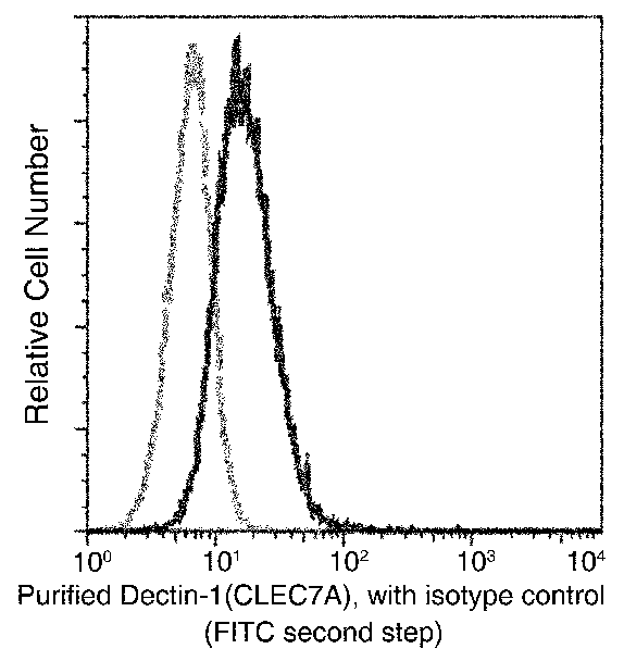 FCM/FACS (Flow Cytometry)
(Flow cytometric analysis of Mouse Dectin-1(CLEC7A) expression on Raw264.7 cells. Cells were stained with purified anti-Mouse Dectin-1(CLEC7A), then a FITC-conjugated second step antibody. The fluorescence histograms were derived from gated events with the forward and side light-scatter characteristics of intact cells.)
FCM/FACS (Flow Cytometry)
(Flow cytometric analysis of Mouse Dectin-1(CLEC7A) expression on Raw264.7 cells. Cells were stained with purified anti-Mouse Dectin-1(CLEC7A), then a FITC-conjugated second step antibody. The fluorescence histograms were derived from gated events with the forward and side light-scatter characteristics of intact cells.)
Dectin-1, Monoclonal Antibody (Cat# AAA257114)
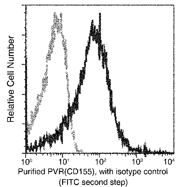 FCM/FACS (Flow Cytometry)
(Flow cytometric analysis of Mouse PVR(CD155) expression on BABL/c splenocytes. Cells were stained with purified anti-Mouse PVR(CD155), then a FITC-conjugated second step antibody. The fluorescence histograms were derived from gated events with the forward and side light-scatter characteristics of intact cells.)
FCM/FACS (Flow Cytometry)
(Flow cytometric analysis of Mouse PVR(CD155) expression on BABL/c splenocytes. Cells were stained with purified anti-Mouse PVR(CD155), then a FITC-conjugated second step antibody. The fluorescence histograms were derived from gated events with the forward and side light-scatter characteristics of intact cells.)
CD155/PVR, Monoclonal Antibody (Cat# AAA257120)
Influenza A H1N1 Matrix protein 1/M1 Protein 1, Monoclonal Antibody (Cat# AAA257032)
Influenza A H1N1 Matrix protein 1/M1 Protein 1, Monoclonal Antibody (Cat# AAA257033)

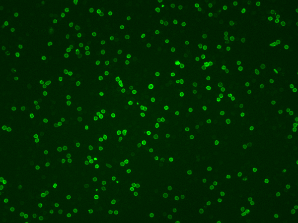
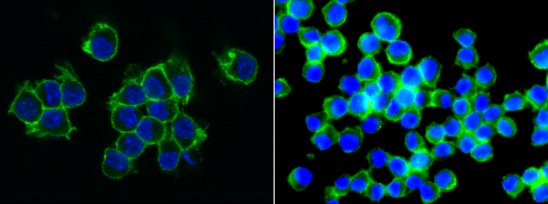
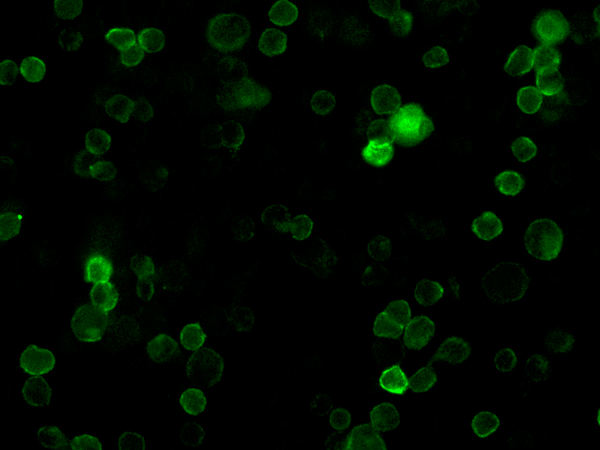 IF (Immunofluorescence)
(Immunofluorescence analysis of SARS-COV-2 Spike overexpressed HEK293 Cells were stained with purified anti-SARS-CoV-2 Spike Mouse Mab,then a Alexa Fluor-488-conjugated second step antibody.)
IF (Immunofluorescence)
(Immunofluorescence analysis of SARS-COV-2 Spike overexpressed HEK293 Cells were stained with purified anti-SARS-CoV-2 Spike Mouse Mab,then a Alexa Fluor-488-conjugated second step antibody.)
SARS-CoV-2 Spike Neutralizing, Monoclonal Antibody (Cat# AAA257791)

 IF (Immunofluorescence)
(Immunofluorescence analysis of SARS-COV-2 Spike overexpressed HEK293 Cells were stained with purified anti-SARS-CoV-2 Spike Rabbit Mab,then a Alexa Fluor-488-conjugated second step antibody.)
IF (Immunofluorescence)
(Immunofluorescence analysis of SARS-COV-2 Spike overexpressed HEK293 Cells were stained with purified anti-SARS-CoV-2 Spike Rabbit Mab,then a Alexa Fluor-488-conjugated second step antibody.)
SARS-CoV-2 (2019-nCoV) Spike Neutralizing, Monoclonal Antibody (Cat# AAA257792)


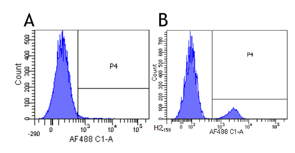
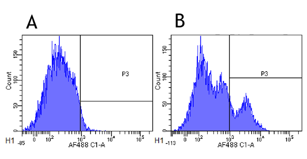
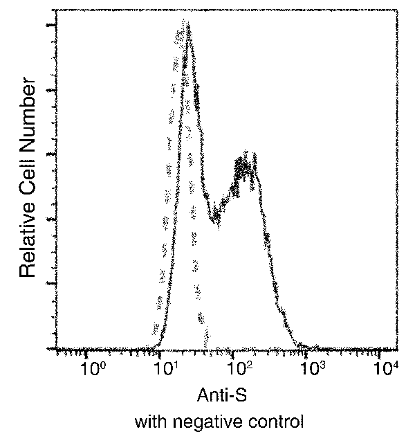
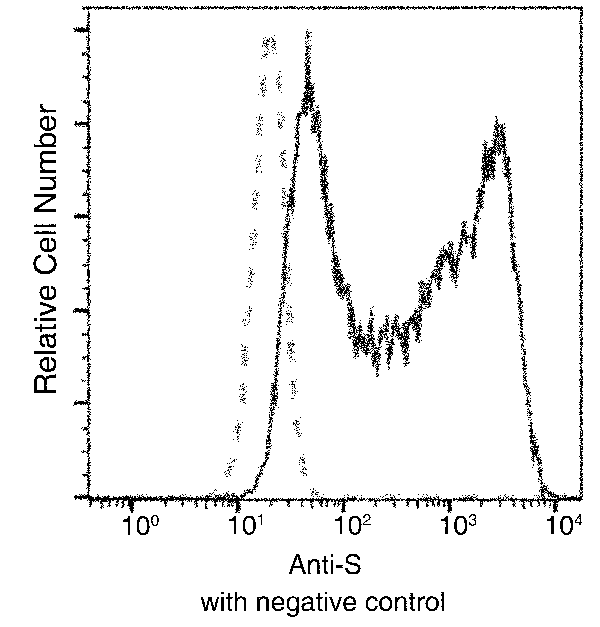
 FCM/FACS (Flow Cytometry)
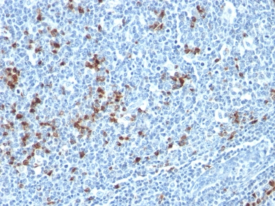
(Flow cytometric analysis of SARS-COV-2 Nucleocapsid overexpressed HEK293 Cells were stained with purified anti-SARS-COV-2 Nucleocapsid Mouse MAb, then a FITC-conjugated second step antibody. The fluorescence histograms were derived from gated events with the forward and side light-scatter characteristics of intact cells.(Validation Experiment))
FCM/FACS (Flow Cytometry)
(Flow cytometric analysis of SARS-COV-2 Nucleocapsid overexpressed HEK293 Cells were stained with purified anti-SARS-COV-2 Nucleocapsid Mouse MAb, then a FITC-conjugated second step antibody. The fluorescence histograms were derived from gated events with the forward and side light-scatter characteristics of intact cells.(Validation Experiment))
COVID 19 Nucleocapsid Coronavirus, Monoclonal Antibody (Cat# AAA258293)
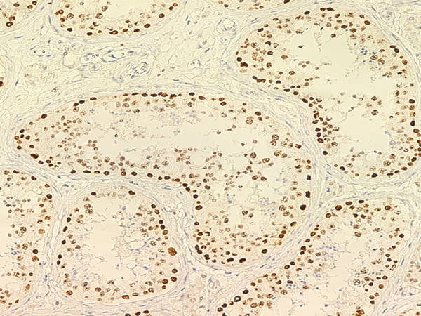
 IHC (Immunohiostchemistry)
(Immunochemical staining of human PARP in human placenta with mouse monoclonal antibody at 1:500 dilution, formalin-fixed paraffin embedded sections.)
IHC (Immunohiostchemistry)
(Immunochemical staining of human PARP in human placenta with mouse monoclonal antibody at 1:500 dilution, formalin-fixed paraffin embedded sections.)
PARP, Monoclonal Antibody (Cat# AAA257734)
 IF (Immunofluorescence)
(Immunofluorescence analysis of SARS-COV-2 Nucleocapsid overexpressed HEK293 Cells were stained with purified anti-SARS-CoV-2 Nucleocapsid Mouse Mab,then a Alexa Fluor-488-conjugated second step antibody.)
IF (Immunofluorescence)
(Immunofluorescence analysis of SARS-COV-2 Nucleocapsid overexpressed HEK293 Cells were stained with purified anti-SARS-CoV-2 Nucleocapsid Mouse Mab,then a Alexa Fluor-488-conjugated second step antibody.)
SARS-CoV-2 (2019-nCoV) Nucleocapsid, Monoclonal Antibody (Cat# AAA257784)
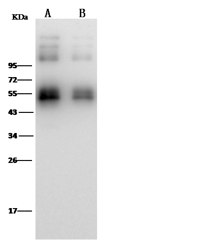
 WB (Western Blot)
(Anti-SARS-CoV-2 (2019-nCoV) Nucleocapsid rabbit monoclonal antibody at 1:5000 dilution.Lane A: SARS-COV-2 Nucleocapsid overexpressed HEK293 Whole Cell LysateLane B: HEK293 Whole Cell LysateLysates/proteins at 30 ug per lane.SecondaryGoat Anti-Rabbit IgG (H+L)/HRP at 1/10000 dilutionDeveloped using the ECL technique.Performed under reducing conditions.)
WB (Western Blot)
(Anti-SARS-CoV-2 (2019-nCoV) Nucleocapsid rabbit monoclonal antibody at 1:5000 dilution.Lane A: SARS-COV-2 Nucleocapsid overexpressed HEK293 Whole Cell LysateLane B: HEK293 Whole Cell LysateLysates/proteins at 30 ug per lane.SecondaryGoat Anti-Rabbit IgG (H+L)/HRP at 1/10000 dilutionDeveloped using the ECL technique.Performed under reducing conditions.)
SARS-CoV-2 (2019-nCoV) Nucleocapsid, Monoclonal Antibody (Cat# AAA257785)

 IF (Immunofluorescence)
(Immunofluorescence analysis of SARS-COV-2 Nucleocapsid overexpressed HEK293 Cells were stained with purified anti-SARS-CoV-2 Nucleocapsid Rabbit Mab,then a Alexa Fluor-488-conjugated second step antibody.)
IF (Immunofluorescence)
(Immunofluorescence analysis of SARS-COV-2 Nucleocapsid overexpressed HEK293 Cells were stained with purified anti-SARS-CoV-2 Nucleocapsid Rabbit Mab,then a Alexa Fluor-488-conjugated second step antibody.)
SARS-CoV-2 (2019-nCoV) Nucleocapsid, Monoclonal Antibody (Cat# AAA257786)
 IF (Immunofluorescence)
(Immunofluorescence analysis of SARS-COV-2 Spike overexpressed HEK293 Cells were stained with purified anti-SARS-CoV-2 Spike Chimera Mab,then a FITC-conjugated second step antibody.)
IF (Immunofluorescence)
(Immunofluorescence analysis of SARS-COV-2 Spike overexpressed HEK293 Cells were stained with purified anti-SARS-CoV-2 Spike Chimera Mab,then a FITC-conjugated second step antibody.)
SARS-CoV-2 (2019-nCoV) Spike S2, Monoclonal Antibody (Cat# AAA257787)
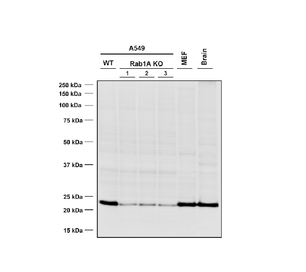
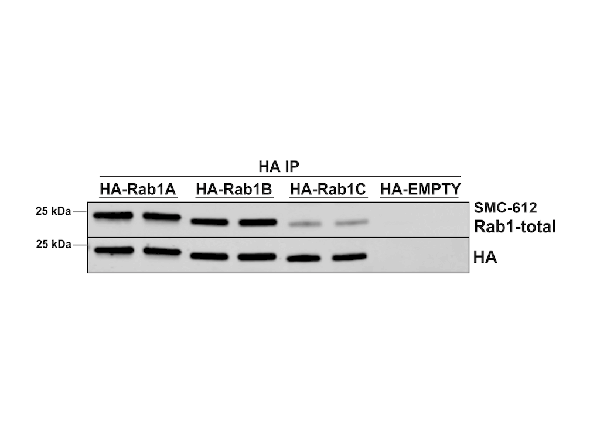 IP (Immunoprecipitation)
(Immunoprecipitation analysis using Mouse Anti-RAB1A Monoclonal Antibody, Clone 7H4 . Tissue: HEK293 cells overexpressing RAB1A, RAB1B, and RAB1C. Species: Human. Primary Antibody: Mouse Anti-RAB1A Monoclonal Antibody for 2 hours..)
IP (Immunoprecipitation)
(Immunoprecipitation analysis using Mouse Anti-RAB1A Monoclonal Antibody, Clone 7H4 . Tissue: HEK293 cells overexpressing RAB1A, RAB1B, and RAB1C. Species: Human. Primary Antibody: Mouse Anti-RAB1A Monoclonal Antibody for 2 hours..)
RAB1A, Monoclonal Antibody (Cat# AAA254032)
What are Monoclonal Antibodies?
Monoclonal antibodies are specialized laboratory-produced proteins developed for binding to specific biological antigens or other molecular targets. Since they come from a single cell (or clone), they are especially consistent and accurate in the data they are involved in producing.
This type of antibody material has been shown to be a powerful tool in finding and subsequently destroying harmful cells in an organism, such as those found in cancers or various autoimmune diseases. This makes them excellent aids in medical testing and research, which is why they are so widely used.
AAA Biotech offers a comprehensive range of high-quality monoclonal antibodies that perform effectively in various laboratory tests, including (amongst others) ELISA, western blotting, immunohistochemistry, and flow cytometry. All of the products in our catalog are thoroughly quality tested to make sure that they are reliable and will consistently perform well in your research.
What Are The Uses of Monoclonal Antibodies
Monoclonal antibodies are used in many lab tests, including (amongst others) ELISA, western blotting, immunohistochemistry, and flow cytometry.
ELISA is a test that helps detect a specific substance/analyte in a sample. It uses antibodies (often monoclonal) bound to a solid surface (such as the well of a microplate) to “capture” the substance/analyte in the sample and immobilize it so that the detection antibody component can then bind to it and produce a signal, which can then be measured.
Western blotting identifies specific proteins in a sample. The sample is first separated on a gel, and then antibodies are applied that will typically bind to the target, which will all be localized to a single band in a lane.
Immunohistochemistry helps locate specific proteins in cells or tissue samples using antibodies.
Flow cytometry looks at and sorts cells. It uses antibodies that are conjugated to reporter molecules called “fluorophores”, which, under special lights, emit light themselves, which can then be measured by a detector instrument.
How Monoclonal Antibodies Are Used as Medicine?
Please note that all of the products listed in AAA Biotech’s also known as AAA Bio or AAABio catalog are strictly for research-use only (RUO).
Monoclonal antibodies can also be used as therapeutic/medical treatments, particularly in the context of cancers. They are designed to find and bind to specific cells or proteins, helping the immune system recognize and attack the cancer. These treatments work in different ways, such as:
- Radioimmunotherapy attaches a small amount of radioactive molecule to the antibody, so it delivers the radiation directly to the cancer cells that the antibody is specifically binding to.
- Antibody-directed enzyme prodrug therapy uses antibodies that are specifically bound to special enzymes. These enzymes activate a harmless drug in the body and turn it into a cancer-killing drug only near the cancer cells—this helps avoid harming healthy cells.
- Immunoliposomes are tiny “bubbles” filled with medicine/drug and coated with antibodies. They carry the drug straight to the cancer cells.
Why Buy Monoclonal Antibodies From Us?
At AAA Biotech, we provide high-performance monoclonal antibodies designed to support a wide range of research needs.
1. Validated for Versatile Applications
The antibodies in our catalog are extensively validated and compatible with multiple techniques, including (but not limited to) ELISA, flow cytometry (FC), immunocytochemistry (ICC), immunofluorescence (IF), immunohistochemistry (IHC), immunoprecipitation (IP), and western blotting (WB).
2. Wide Selection & Specialized Options
We offer antibodies for common and rare species, that are available in various conjugated forms, and also in recombinant formats. Essentially, there is almost anything one might need to meet their experimental model’s requirements.
3. High-Quality Proteins
Our proteins meet high purity standards—90% or more as confirmed by SDS-PAGE. Many are available with tags like His, Flag, GST, or MBP, and we also supply native and biologically active proteins for functional studies.
Frequently Asked Questions
1. Are your monoclonal antibodies validated for specific applications?
Yes, our antibodies are tested and validated for use in methods such as ELISA, western blot, IHC, flow cytometry, and more. Refer to specific product pages or datasheets for individual product information.
2. How do I choose the right monoclonal antibody for my application?
Review the product details directly for application validation, species reactivity, and target information. You may also contact our support team at any time for help.
3. How quickly can I receive my order?
Most orders are processed and shipped within 1–3 business days, depending on product availability and your shipping location.