Filters
▼Clonality
▼Type
▼Reactivity
▼Gene Name
▼Isotype
▼Host
▼Application
▼Clone
▼Monoclonal Antibodies
Get accurate results in your research with our Monoclonal Antibodies, which are specially made to target exactly what you require for your research, and will produce consistent, reliable performance in lab tests.
Viewing 7150-7200 of 27777 product results
 Application Data
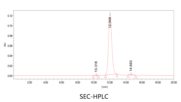
(The purity of this product is >95% as determined by SEC-HPLC.)
Application Data
(The purity of this product is >95% as determined by SEC-HPLC.)
LASV GPC, Monoclonal Recombinant Antibody (Cat# AAA120209)
Blood group H antigen/Globo H, Monoclonal Antibody (Cat# AAA120696)
Protein A or G purified from cell culture supernatant.
INFLUENZA A NUCLEOPROTEIN, Monoclonal Antibody (Cat# AAA49403)
 Application Data
(Staining of human peripheral blood granulocytes with Mouse anti Human CD53)
Application Data
(Staining of human peripheral blood granulocytes with Mouse anti Human CD53)
CD53, Monoclonal Antibody (Cat# AAA49436)
CD8 ALPHA, Monoclonal Antibody (Cat# AAA49526)
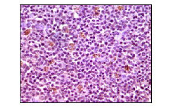 IHC (Immunohiostchemistry)
(Immunohistochemical analysis of paraffin-embedded human lymphnode tissues using MCL1 antibody with DAB staining.)
IHC (Immunohiostchemistry)
(Immunohistochemical analysis of paraffin-embedded human lymphnode tissues using MCL1 antibody with DAB staining.)
MCL1, Monoclonal Antibody (Cat# AAA74819)
FSH, Monoclonal Antibody (Cat# AAA74475)
Serotonin, Monoclonal Antibody (Cat# AAA74310)
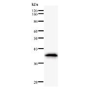
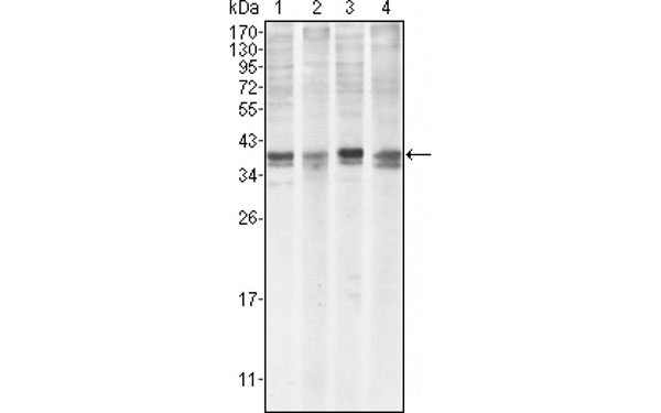
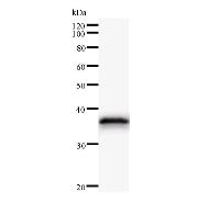
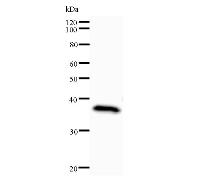
 Quality Control #2
(Arrow indicates the region of immunized recombinant protein carrying 50-200 amino acids.)
Quality Control #2
(Arrow indicates the region of immunized recombinant protein carrying 50-200 amino acids.)
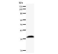
ASH1L, Monoclonal Antibody (Cat# AAA36315)
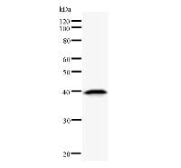
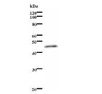
 Quality Control #2
(Arrow indicates the region of immunized recombinant protein carrying 50-200 amino acids.)
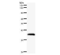
Quality Control #2
(Arrow indicates the region of immunized recombinant protein carrying 50-200 amino acids.)
DBP, Monoclonal Antibody (Cat# AAA36316)

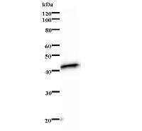
 WB (Western Blot)
(Detection of SCML2 by Western blot.Samples: Whole cell lysate from human HeLa (H, 25 ug), mouse NIH3T3 (M, 25 ug) and rat F2408 (R, 25 ug) cells. [Lot No. SCMAD14A-3]Predicted molecular weight: 77 kDa)
WB (Western Blot)
(Detection of SCML2 by Western blot.Samples: Whole cell lysate from human HeLa (H, 25 ug), mouse NIH3T3 (M, 25 ug) and rat F2408 (R, 25 ug) cells. [Lot No. SCMAD14A-3]Predicted molecular weight: 77 kDa)
SCML2, Monoclonal Antibody (Cat# AAA36329)


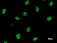
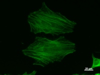 ICC (Immunocytochemistry)
(Immunostaining analysis in HeLa cells. HeLa cells were fixed with 4% paraformaldehyde and permeabilized with 0.1% Triton X-100 in PBS. The cells were immunostained with anti-ACTB mAb. [Lot No. ACTBD11B7-1])
ICC (Immunocytochemistry)
(Immunostaining analysis in HeLa cells. HeLa cells were fixed with 4% paraformaldehyde and permeabilized with 0.1% Triton X-100 in PBS. The cells were immunostained with anti-ACTB mAb. [Lot No. ACTBD11B7-1])
ACTB, Monoclonal Antibody (Cat# AAA36339)
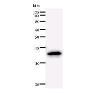
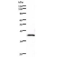
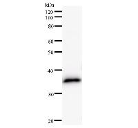
 Quality Control #2
(Arrow indicates the region of immunized recombinant protein carrying 50-200 amino acids.)
Quality Control #2
(Arrow indicates the region of immunized recombinant protein carrying 50-200 amino acids.)
ARID4A, Monoclonal Antibody (Cat# AAA36493)
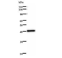
 Quality Control #2
(Arrow indicates the region of immunized recombinant protein carrying 50-200 amino acids.)
Quality Control #2
(Arrow indicates the region of immunized recombinant protein carrying 50-200 amino acids.)
PAX7, Monoclonal Antibody (Cat# AAA36500)
 Quality Control #2
(Arrow indicates the region of immunized recombinant protein carrying 50-200 amino acids.)
Quality Control #2
(Arrow indicates the region of immunized recombinant protein carrying 50-200 amino acids.)
EVI1, Monoclonal Antibody (Cat# AAA36503)
 Quality Control #2
(Arrow indicates the region of immunized recombinant protein carrying 50-200 amino acids.)
Quality Control #2
(Arrow indicates the region of immunized recombinant protein carrying 50-200 amino acids.)
MXI1, Monoclonal Antibody (Cat# AAA36506)

 WB (Western Blot)
(Detection of human SECISBP2 by Western blot.Samples: Whole cell lysate (25 ug) from HEK293 cells. [Lot No. 2957C2a-1]Predicted molecular weight: 95 kDa)
WB (Western Blot)
(Detection of human SECISBP2 by Western blot.Samples: Whole cell lysate (25 ug) from HEK293 cells. [Lot No. 2957C2a-1]Predicted molecular weight: 95 kDa)
SECISBP2, Monoclonal Antibody (Cat# AAA36514)

 ICC (Immunocytochemistry)
(Immunostaining analysis in HeLa cells. HeLa cells were fixed with 4% paraformaldehyde and permeabilized with 0.1% Triton X-100 in PBS. The cells were immunostained with anti-ISL2 mAb. [Lot No.203C5a-1])
ICC (Immunocytochemistry)
(Immunostaining analysis in HeLa cells. HeLa cells were fixed with 4% paraformaldehyde and permeabilized with 0.1% Triton X-100 in PBS. The cells were immunostained with anti-ISL2 mAb. [Lot No.203C5a-1])
ISL2, Monoclonal Antibody (Cat# AAA36521)

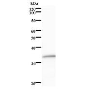
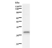
 WB (Western Blot)
(Detection of human HOXB7 by Western blot.Samples: Whole cell lysate (50 ug) from A2058 cells. [Lot No. 747C4a-1]Predicted molecular weight: 25 kDa)
WB (Western Blot)
(Detection of human HOXB7 by Western blot.Samples: Whole cell lysate (50 ug) from A2058 cells. [Lot No. 747C4a-1]Predicted molecular weight: 25 kDa)
HOXB7, Monoclonal Antibody (Cat# AAA36371)

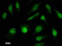
 ICC (Immunocytochemistry)
(Immunostaining analysis in HeLa cells. HeLa cells were fixed with 4% paraformaldehyde and permeabilized with 0.1% Triton X-100 in PBS. The cells were immunostained with anti-ASH2L mAb. [Lot No. 2046D2a-1])
ICC (Immunocytochemistry)
(Immunostaining analysis in HeLa cells. HeLa cells were fixed with 4% paraformaldehyde and permeabilized with 0.1% Triton X-100 in PBS. The cells were immunostained with anti-ASH2L mAb. [Lot No. 2046D2a-1])
ASH2L, Monoclonal Antibody (Cat# AAA36379)
 Quality Control #2
(Arrow indicates the region of immunized recombinant protein carrying 50-200 amino acids.)
Quality Control #2
(Arrow indicates the region of immunized recombinant protein carrying 50-200 amino acids.)
SUPT4H1, Monoclonal Antibody (Cat# AAA36422)

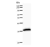
 WB (Western Blot)
(Detection of ZNF354A by Western blot.Samples: Whole cell lysate from human HeLa (H, 25 ug), mouse NIH3T3 (M, 25 ug) and rat F2408 (R, 25 ug) cells. [Lot No. 149C1a-1]Predicted molecular weight: 69 kDa)
WB (Western Blot)
(Detection of ZNF354A by Western blot.Samples: Whole cell lysate from human HeLa (H, 25 ug), mouse NIH3T3 (M, 25 ug) and rat F2408 (R, 25 ug) cells. [Lot No. 149C1a-1]Predicted molecular weight: 69 kDa)
ZNF354A, Monoclonal Antibody (Cat# AAA36444)
 Quality Control #2
(Arrow indicates the region of immunized recombinant protein carrying 50-200 amino acids.)
Quality Control #2
(Arrow indicates the region of immunized recombinant protein carrying 50-200 amino acids.)
HEYL, Monoclonal Antibody (Cat# AAA36446)
Mitragynine, Monoclonal Antibody (Cat# AAA36584)
Vitamin B12, Monoclonal Antibody (Cat# AAA36589)
>90% by SDS-PAGE
Usutu Virus, Monoclonal Antibody (Cat# AAA36591)
Ion Exchange
Nipah Virus Glycoprotein F, Monoclonal Antibody (Cat# AAA36598)
Purification: Ion exchange
African Swine Fever (ASF) Virus P30 protein, Monoclonal Antibody (Cat# AAA36605)
Frataxin, Monoclonal Antibody (Cat# AAA39134)
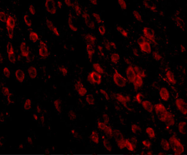
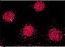
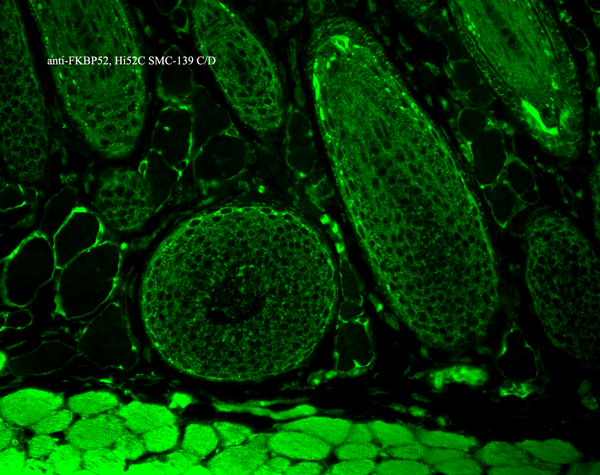
 IF (Immunofluorescence)
(Immunofluorescence of PD-1 in human spleen tissue with PD-1 antibody at 20 μg/ml.Green: PD-1 Antibody [7A11B1]Blue: DAPI staining)
IF (Immunofluorescence)
(Immunofluorescence of PD-1 in human spleen tissue with PD-1 antibody at 20 μg/ml.Green: PD-1 Antibody [7A11B1]Blue: DAPI staining)
PD-1, Monoclonal Antibody (Cat# AAA39194)
 Application Data
(Fig. 2: BOSC23 cells were transiently transfected with expression vectors containing either the cDNA of CEACAM1, CEACAM3-8 or PSG. The latter expressed as a membrane bound fusion protein. Expression of the constructs was tested with monoclonal antibodies known to recognize the corresponding proteins (CEACAM1,3,4,5 and 6: D14HD11; CEACAM7: BAC2; CEACAM8: Tet2; PSG: BAP3; green curves). An irrelevant monoclonal antibody served as a negative control (black curves). For specificity testing, protein G-purified GM-2H6 was tested on all CEACAM transfectants. A positive signal was obtained only with CEACAM8 transfected cells (red curve).)
Application Data
(Fig. 2: BOSC23 cells were transiently transfected with expression vectors containing either the cDNA of CEACAM1, CEACAM3-8 or PSG. The latter expressed as a membrane bound fusion protein. Expression of the constructs was tested with monoclonal antibodies known to recognize the corresponding proteins (CEACAM1,3,4,5 and 6: D14HD11; CEACAM7: BAC2; CEACAM8: Tet2; PSG: BAP3; green curves). An irrelevant monoclonal antibody served as a negative control (black curves). For specificity testing, protein G-purified GM-2H6 was tested on all CEACAM transfectants. A positive signal was obtained only with CEACAM8 transfected cells (red curve).)
CEACAM8, Monoclonal Antibody (Cat# AAA71467)
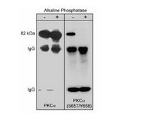
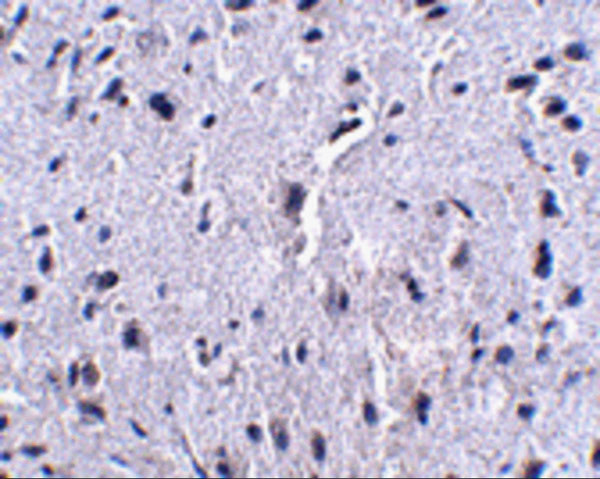
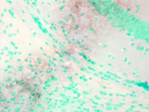
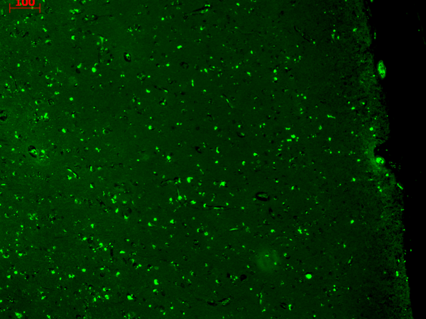
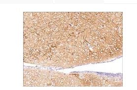 IHC (Immunohiostchemistry)
(Formalin fixed, citric acid treated parafin sections of adult mouse brain. Sections were probed with anti-PKCalpha (PM2371) then anti-mouse:HRP before detection using DAB. (Image provided by Carl Hobbs and Dr. Pat Doherty at Wolfson Centre for Age-Related Diseases, King's College London).)
IHC (Immunohiostchemistry)
(Formalin fixed, citric acid treated parafin sections of adult mouse brain. Sections were probed with anti-PKCalpha (PM2371) then anti-mouse:HRP before detection using DAB. (Image provided by Carl Hobbs and Dr. Pat Doherty at Wolfson Centre for Age-Related Diseases, King's College London).)
PKCalpha, Monoclonal Antibody (Cat# AAA71684)
TAR DNA, Monoclonal Antibody (Cat# AAA37960)
Streptavidin, Monoclonal Antibody (Cat# AAA37962)
Anti BSA, Clone 9E2C2, Monoclonal Antibody (Cat# AAA37652)
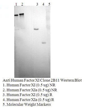 Application Data
Factor XI, Clone 2B11, Monoclonal Antibody (Cat# AAA37670)
HIV-1 gp41, Monoclonal Antibody (Cat# AAA37913)
IgA 1&2, Monoclonal Antibody (Cat# AAA37925)
CD28, Monoclonal Antibody (Cat# AAA37938)
Clusterin, Monoclonal Antibody (Cat# AAA37946)
Herpesvirus 8, Monoclonal Antibody (Cat# AAA37955)

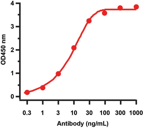 ELISA
(Figure 2 ELISA Validation of HIV-1 p24 Antibody with HIV-1 p24 Protein Coating Antigen: HIV-1 p24 recombinant protein, 1ug/mL, incubated at 4 degree C overnight. Detection Antibodies: HIV-1 p24 antibody, AAA41004, dilution: 0.3-1000 ng/mL, incubated at RT for 1 hr. Secondary Antibodies: Goat anti-mouse HRP at 1:5,000, incubated at RT for 1 hr.)
ELISA
(Figure 2 ELISA Validation of HIV-1 p24 Antibody with HIV-1 p24 Protein Coating Antigen: HIV-1 p24 recombinant protein, 1ug/mL, incubated at 4 degree C overnight. Detection Antibodies: HIV-1 p24 antibody, AAA41004, dilution: 0.3-1000 ng/mL, incubated at RT for 1 hr. Secondary Antibodies: Goat anti-mouse HRP at 1:5,000, incubated at RT for 1 hr.)
HIV-1 p24, Monoclonal Antibody (Cat# AAA41004)
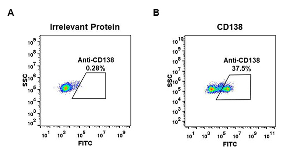
 FCM/FACS (Flow Cytometry)
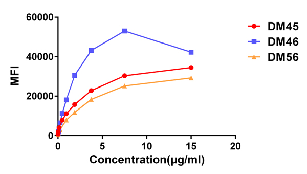
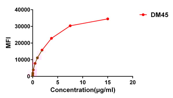
(Figure 3. Affinity ranking of different Rabbit anti-CD138 mAb clones by titration of different concentration onto H929 cells. The Y-axis represents the mean fluorescence intensity (MFI) while the X-axis represents the concentration of IgG used.)
FCM/FACS (Flow Cytometry)
(Figure 3. Affinity ranking of different Rabbit anti-CD138 mAb clones by titration of different concentration onto H929 cells. The Y-axis represents the mean fluorescence intensity (MFI) while the X-axis represents the concentration of IgG used.)
CD138, Monoclonal Antibody (Cat# AAA47286)
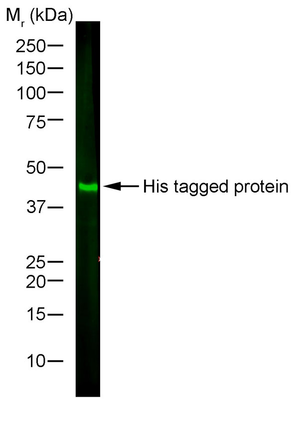
 Application Data
(His-Ubiquitin recombinant protein probed with Mouse anti Histidine Tag)
Application Data
(His-Ubiquitin recombinant protein probed with Mouse anti Histidine Tag)
HISTIDINE TAG, Monoclonal Antibody (Cat# AAA49054)
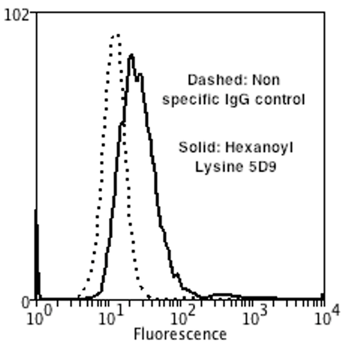
 WB (Western Blot)
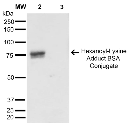
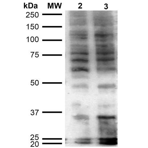
(Western Blot analysis of Human Cervical cancer cell line (HeLa) lysate showing detection of Hexanoyl-Lysine adduct protein using Mouse Anti-Hexanoyl-Lysine adduct Monoclonal Antibody, Clone 5D9. Lane 1: Molecular Weight Ladder (MW). Lane 2: HeLa cell lysate. Lane 3: H2O2 treated HeLa cell lysate. Load: 12 ug. Block: 5% Skim Milk in TBST. Primary Antibody: Mouse Anti-Hexanoyl-Lysine adduct Monoclonal Antibody at 1:1000 for 2 hours at RT. Secondary Antibody: Goat Anti-Mouse IgG: HRP at 1:2000 for 60 min at RT. Color Development: ECL solution for 5 min in RT.)
WB (Western Blot)
(Western Blot analysis of Human Cervical cancer cell line (HeLa) lysate showing detection of Hexanoyl-Lysine adduct protein using Mouse Anti-Hexanoyl-Lysine adduct Monoclonal Antibody, Clone 5D9. Lane 1: Molecular Weight Ladder (MW). Lane 2: HeLa cell lysate. Lane 3: H2O2 treated HeLa cell lysate. Load: 12 ug. Block: 5% Skim Milk in TBST. Primary Antibody: Mouse Anti-Hexanoyl-Lysine adduct Monoclonal Antibody at 1:1000 for 2 hours at RT. Secondary Antibody: Goat Anti-Mouse IgG: HRP at 1:2000 for 60 min at RT. Color Development: ECL solution for 5 min in RT.)
Hexanoyl-Lysine adduct, Monoclonal Antibody (Cat# AAA104014)
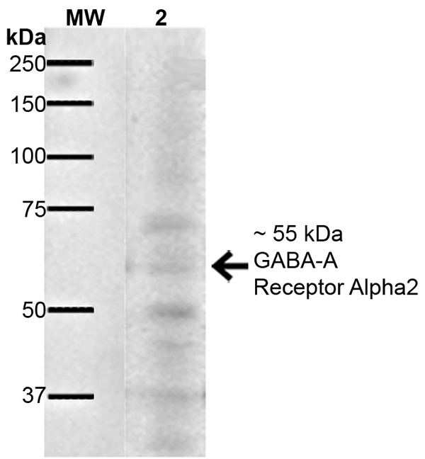
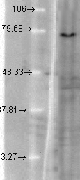
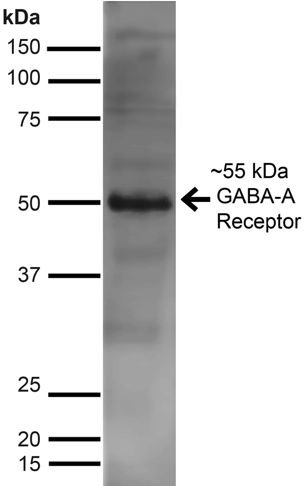 WB (Western Blot)
(Western Blot analysis of Rat Brain showing detection of ~55 kDa GABA A Receptor Alpha 2 protein using Mouse Anti-GABA A Receptor Alpha 2 Monoclonal Antibody, Clone S399-19. Lane 1: MW Ladder. Lane 2: Rat Brain. Load: 10 ug. Block: 5% Skim Milk for 1 hour at RT. Primary Antibody: Mouse Anti-GABA A Receptor Alpha 2 Monoclonal Antibody at 1:1000 for 1 hour at RT. Secondary Antibody: Goat Anti-Mouse IgG: HRP at 1:100 for 1 hour at RT. Color Development: ECL solution for 6 min at RT. Predicted/Observed Size: ~55 kDa.)
WB (Western Blot)
(Western Blot analysis of Rat Brain showing detection of ~55 kDa GABA A Receptor Alpha 2 protein using Mouse Anti-GABA A Receptor Alpha 2 Monoclonal Antibody, Clone S399-19. Lane 1: MW Ladder. Lane 2: Rat Brain. Load: 10 ug. Block: 5% Skim Milk for 1 hour at RT. Primary Antibody: Mouse Anti-GABA A Receptor Alpha 2 Monoclonal Antibody at 1:1000 for 1 hour at RT. Secondary Antibody: Goat Anti-Mouse IgG: HRP at 1:100 for 1 hour at RT. Color Development: ECL solution for 6 min at RT. Predicted/Observed Size: ~55 kDa.)
GABA-A Receptor Alpha2, Monoclonal Antibody (Cat# AAA103792)
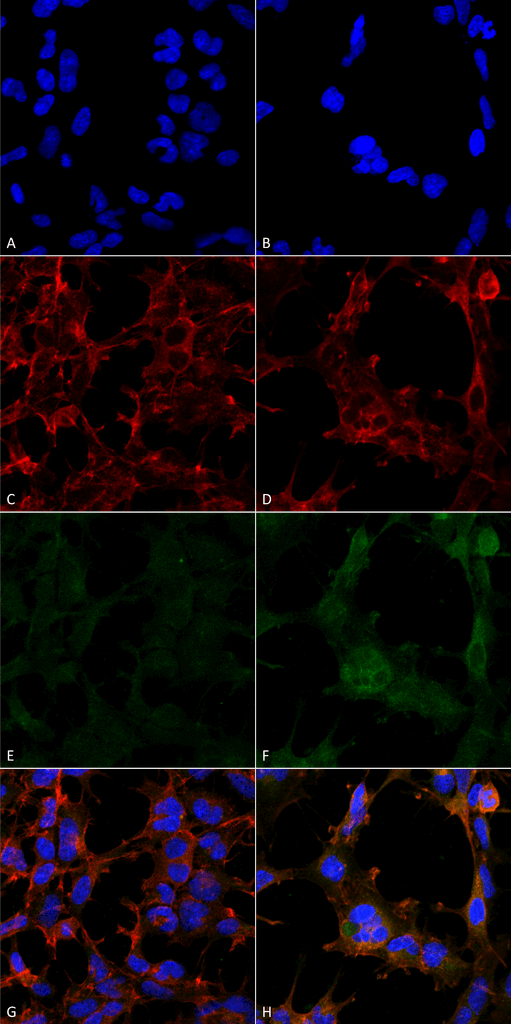
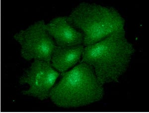 IF (Immunofluorescence)
(ICC/IF analysis of BMP2 in Hep3B cells line, stained with monoclonal anti-human BMP2 antibody (1:100) with goat anti-mouse IgG-Alexa fluor 488 conjugate (Green).)
IF (Immunofluorescence)
(ICC/IF analysis of BMP2 in Hep3B cells line, stained with monoclonal anti-human BMP2 antibody (1:100) with goat anti-mouse IgG-Alexa fluor 488 conjugate (Green).)
BMP2, Monoclonal Antibody (Cat# AAA48133)
Hsp90, Monoclonal Antibody (Cat# AAA47749)
 WB (Western Blot)
(Western Blot analysis of hamster T-CHO cell lysate showing detection of KCNQ1 protein using Mouse Anti-KCNQ1 Monoclonal Antibody, Clone S37A-10. Load: 15 ug. Block: 1.5% BSA for 30 minutes at RT. Primary Antibody: Mouse Anti-KCNQ1 Monoclonal Antibody at 1:1000 for 2 hours at RT. Secondary Antibody: Sheep Anti-Mouse IgG: HRP for 1 hour at RT.)
WB (Western Blot)
(Western Blot analysis of hamster T-CHO cell lysate showing detection of KCNQ1 protein using Mouse Anti-KCNQ1 Monoclonal Antibody, Clone S37A-10. Load: 15 ug. Block: 1.5% BSA for 30 minutes at RT. Primary Antibody: Mouse Anti-KCNQ1 Monoclonal Antibody at 1:1000 for 2 hours at RT. Secondary Antibody: Sheep Anti-Mouse IgG: HRP for 1 hour at RT.)
KCNQ1, Monoclonal Antibody (Cat# AAA102891)


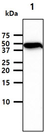
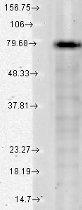
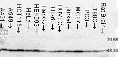 WB (Western Blot)
(Western Blot analysis of Human Cell lysates showing detection of FKBP52 protein using Mouse Anti-FKBP52 Monoclonal Antibody, Clone Hi52C. Load: 15 ug. Block: 1.5% BSA for 30 minutes at RT. Primary Antibody: Mouse Anti-FKBP52 Monoclonal Antibody at 1.5 ug/mL for 2 hours at RT. Secondary Antibody: Sheep Anti-Mouse IgG: HRP for 1 hour at RT.)
WB (Western Blot)
(Western Blot analysis of Human Cell lysates showing detection of FKBP52 protein using Mouse Anti-FKBP52 Monoclonal Antibody, Clone Hi52C. Load: 15 ug. Block: 1.5% BSA for 30 minutes at RT. Primary Antibody: Mouse Anti-FKBP52 Monoclonal Antibody at 1.5 ug/mL for 2 hours at RT. Secondary Antibody: Sheep Anti-Mouse IgG: HRP for 1 hour at RT.)
FKBP52, Monoclonal Antibody (Cat# AAA102898)
What are Monoclonal Antibodies?
Monoclonal antibodies are specialized laboratory-produced proteins developed for binding to specific biological antigens or other molecular targets. Since they come from a single cell (or clone), they are especially consistent and accurate in the data they are involved in producing.
This type of antibody material has been shown to be a powerful tool in finding and subsequently destroying harmful cells in an organism, such as those found in cancers or various autoimmune diseases. This makes them excellent aids in medical testing and research, which is why they are so widely used.
AAA Biotech offers a comprehensive range of high-quality monoclonal antibodies that perform effectively in various laboratory tests, including (amongst others) ELISA, western blotting, immunohistochemistry, and flow cytometry. All of the products in our catalog are thoroughly quality tested to make sure that they are reliable and will consistently perform well in your research.
What Are The Uses of Monoclonal Antibodies
Monoclonal antibodies are used in many lab tests, including (amongst others) ELISA, western blotting, immunohistochemistry, and flow cytometry.
ELISA is a test that helps detect a specific substance/analyte in a sample. It uses antibodies (often monoclonal) bound to a solid surface (such as the well of a microplate) to “capture” the substance/analyte in the sample and immobilize it so that the detection antibody component can then bind to it and produce a signal, which can then be measured.
Western blotting identifies specific proteins in a sample. The sample is first separated on a gel, and then antibodies are applied that will typically bind to the target, which will all be localized to a single band in a lane.
Immunohistochemistry helps locate specific proteins in cells or tissue samples using antibodies.
Flow cytometry looks at and sorts cells. It uses antibodies that are conjugated to reporter molecules called “fluorophores”, which, under special lights, emit light themselves, which can then be measured by a detector instrument.
How Monoclonal Antibodies Are Used as Medicine?
Please note that all of the products listed in AAA Biotech’s catalog are strictly for research-use only (RUO).
Monoclonal antibodies can also be used as therapeutic/medical treatments, particularly in the context of cancers. They are designed to find and bind to specific cells or proteins, helping the immune system recognize and attack the cancer. These treatments work in different ways, such as:
- Radioimmunotherapy attaches a small amount of radioactive molecule to the antibody, so it delivers the radiation directly to the cancer cells that the antibody is specifically binding to.
- Antibody-directed enzyme prodrug therapy uses antibodies that are specifically bound to special enzymes. These enzymes activate a harmless drug in the body and turn it into a cancer-killing drug only near the cancer cells—this helps avoid harming healthy cells.
- Immunoliposomes are tiny “bubbles” filled with medicine/drug and coated with antibodies. They carry the drug straight to the cancer cells.
Why Buy Monoclonal Antibodies From Us?
At AAA Biotech, we provide high-performance monoclonal antibodies designed to support a wide range of research needs.
1. Validated for Versatile Applications
The antibodies in our catalog are extensively validated and compatible with multiple techniques, including (but not limited to) ELISA, flow cytometry (FC), immunocytochemistry (ICC), immunofluorescence (IF), immunohistochemistry (IHC), immunoprecipitation (IP), and western blotting (WB).
2. Wide Selection & Specialized Options
We offer antibodies for common and rare species, that are available in various conjugated forms, and also in recombinant formats. Essentially, there is almost anything one might need to meet their experimental model’s requirements.
3. High-Quality Proteins
Our proteins meet high purity standards—90% or more as confirmed by SDS-PAGE. Many are available with tags like His, Flag, GST, or MBP, and we also supply native and biologically active proteins for functional studies.
Frequently Asked Questions
1. Are your monoclonal antibodies validated for specific applications?
Yes, our antibodies are tested and validated for use in methods such as ELISA, western blot, IHC, flow cytometry, and more. Refer to specific product pages or datasheets for individual product information.
2. How do I choose the right monoclonal antibody for my application?
Review the product details directly for application validation, species reactivity, and target information. You may also contact our support team at any time for help.
3. How quickly can I receive my order?
Most orders are processed and shipped within 1–3 business days, depending on product availability and your shipping location.