Filters
▼Clonality
▼Type
▼Reactivity
▼Gene Name
▼Isotype
▼Host
▼Application
▼Clone
▼Secondary Antibodies
At AAA Biotech also known as AAA Bio or AAABio, we provide a vast collection of high-quality, purified, conjugated and unconjugated secondary antibodies to use in various applications, such as Western Blotting, Flow Cytometry, Cell Imaging, or CUT & Tag.
Our secondary antibodies are available with a wide range of labels—such as HRP, AP, FITC, and biotin—to match your detection system and enhance signal sensitivity. Each antibody is rigorously tested to ensure high specificity and minimal cross-reactivity, delivering reliable performance across multiple species and assay types.
Whether you're conducting qualitative imaging or quantitative analysis, AAA Biotech has the right secondary antibody to support your research with consistency and precision. Our entire catalog of secondary antibodies is available for browsing through our main website.
Viewing 400-450 of 552 product results
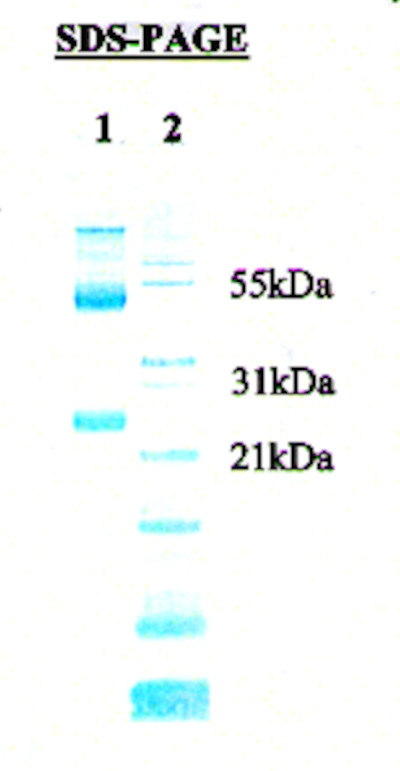
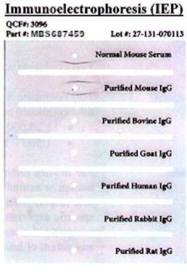
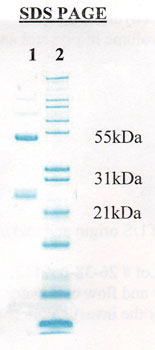
 IEP (Immunoelectrophoresis)
IgG (H&L), Secondary Antibody (Cat# AAA79060)
Affinity purified using solid phase Mouse IgG.
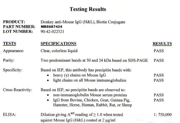
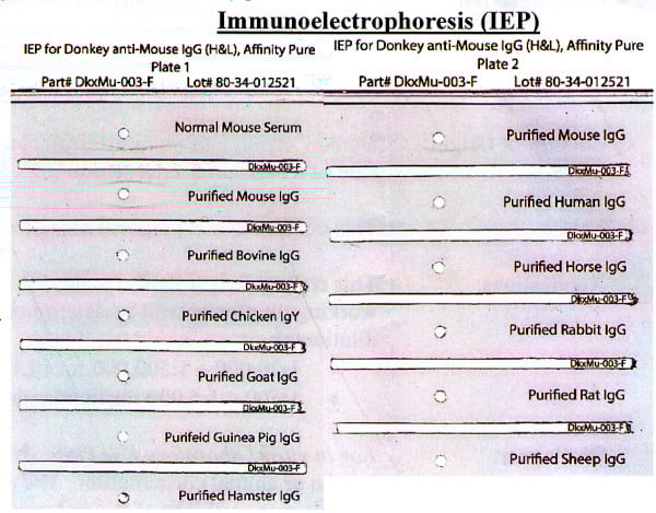
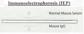
IgG (H&L), Secondary Antibody (Cat# AAA79061)
Cross Reactivity: Based on IEP, no reactivity is observed to:
non-immunoglobulin mouse serum immunoglobulins
Human IgG or serum proteins
Affinity purified using solid phase Mouse IgG.
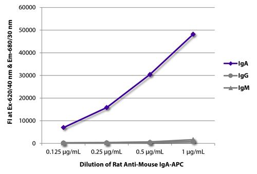
 Application Data
IgG, Secondary Antibody (Cat# AAA79083)
Goat anti Pig IgM, Secondary Antibody (Cat# AAA75104)
Goat anti Human IgG (HRP), Secondary Antibody (Cat# AAA75482)
 Application Data
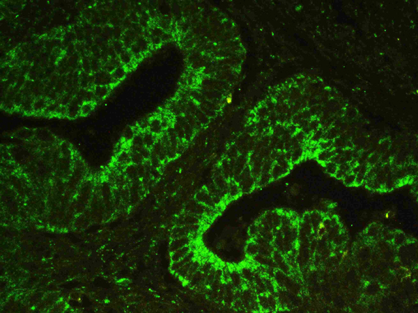
(Lamin B1 was detected in paraffin-embedded sections of human lung cancer tissues using rabbit anti- Lamin B1 Antigen Affinity purified polyclonal antibody at 1 ug/mL. DyLight 488 Conjugated Goat Anti-rabbit IgG secondary antibody was used to detect the primary antibody at 10 ug/mL.)
Application Data
(Lamin B1 was detected in paraffin-embedded sections of human lung cancer tissues using rabbit anti- Lamin B1 Antigen Affinity purified polyclonal antibody at 1 ug/mL. DyLight 488 Conjugated Goat Anti-rabbit IgG secondary antibody was used to detect the primary antibody at 10 ug/mL.)
DyLight488 Conjugated Goat Anti-rabbit IgG, Polyclonal Secondary Antibody (Cat# AAA46012)
 Application Data
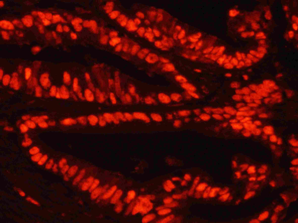
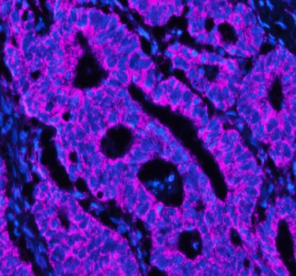
(MCM2 was detected in paraffin-embedded sections of human intestinal cancer tissues using rabbit anti- MCM2 Antigen Affinity purified polyclonal antibody at 1 ug/mL. TRITC Conjugated Goat Anti-rabbit IgG secondary antibody was used to detect the primary antibody at 30ug/mL.)
Application Data
(MCM2 was detected in paraffin-embedded sections of human intestinal cancer tissues using rabbit anti- MCM2 Antigen Affinity purified polyclonal antibody at 1 ug/mL. TRITC Conjugated Goat Anti-rabbit IgG secondary antibody was used to detect the primary antibody at 30ug/mL.)
TRITC Conjugated Goat Anti-rabbit IgG, Polyclonal Secondary Antibody (Cat# AAA45596)
HRP Conjugated Goat Anti-mouse IgG, Polyclonal Secondary Antibody (Cat# AAA45605)
HRP Conjugated Goat Anti-rabbit IgG, Polyclonal Secondary Antibody (Cat# AAA45616)
IgG+IgM+IgA, Polyclonal Secondary Antibody (Cat# AAA312912)
IgY, Polyclonal Secondary Antibody (Cat# AAA312916)
IgY, Polyclonal Secondary Antibody (Cat# AAA312917)
IgG, Secondary Antibody (Cat# AAA62292)
Goat Anti-Mouse IgG Heavy and Light Chain Antibody, Alkaline Phosphatase Conjugated, Secondary Antibody (Cat# AAA47965)
Goat anti Rabbit IgG, Fc Fragment Specific Antibody, Peroxidase Conjugated, Secondary Antibody (Cat# AAA47968)
Streptavidin, Secondary Antibody (Cat# AAA77536)
Rabbit anti Monkey IgG Fab, Polyclonal Secondary Antibody (Cat# AAA77588)
Goat anti Chicken IgM (Fc specific), Polyclonal Secondary Antibody (Cat# AAA77594)
Goat anti Human IgE (epsilon chain) (HRP), Secondary Antibody (Cat# AAA75527)
Donkey anti Rabbit IgG (H + L) (Fab'2) (FITC), Secondary Antibody (Cat# AAA75530)
Goat anti Rabbit IgG (H + L) (HRP), Secondary Antibody (Cat# AAA75539)
Goat anti Bird IgG (H + L), Secondary Antibody (Cat# AAA75546)
Goat anti Mouse IgG (H + L) (Fab'2) (FITC), Secondary Antibody (Cat# AAA75499)
Mouse Anti-Human IgG2 (gamma 2 chain specific), Monoclonal Secondary Antibody (Cat# AAA78729)
Mouse Anti-Human IgG4 (gamma 4 chain specific), Monoclonal Secondary Antibody (Cat# AAA78732)
Goat Anti-Human IgG, Bovine/Horse/Mouse ads, Secondary Antibody (Cat# AAA78874)
Rabbit Anti-Human IgG (H+L)-PE, Secondary Antibody (Cat# AAA78876)
 Application Data
IgAC, Secondary Antibody (Cat# AAA78920)
 IEP (Immunoelectrophoresis)
IgG (H&L), Secondary Antibody (Cat# AAA79064)
Affinity purified using solid phase Mouse IgG.
 IEP (Immunoelectrophoresis)
IgG (H&L), Secondary Antibody (Cat# AAA79074)
Cross Reactivity: Based on IEP, no reactivity is observed to non-immunoglobulin mouse serum immunoglobulins
Affinity purified using solid phase Mouse IgG.
IgY (H&L), Secondary Antibody (Cat# AAA79078)
Affinity purified using solid phase Turkey IgY.
Goat F(ab')2 Anti-Human IgM (u chain specific), Polyclonal Secondary Antibody (Cat# AAA78790)
Goat F(ab')2 Anti-Human IgG (gamma chain specific), Polyclonal Secondary Antibody (Cat# AAA78795)
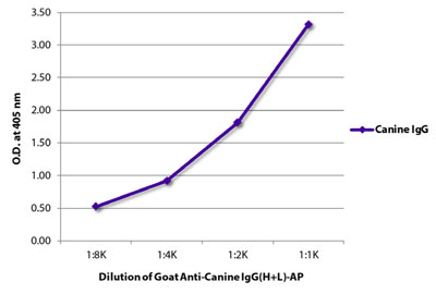 ELISA
(ELISA plate was coated with purified canine IgG. Immunoglobulin was detected with Goat Anti-Canine IgG(H+L)-HRP (SB Cat. No. AAA78834).)
ELISA
(ELISA plate was coated with purified canine IgG. Immunoglobulin was detected with Goat Anti-Canine IgG(H+L)-HRP (SB Cat. No. AAA78834).)
Goat Anti-Canine IgG (H+L), Secondary Antibody (Cat# AAA78834)
Donkey Anti-Goat IgG (H+L), Polyclonal Secondary Antibody (Cat# AAA78859)
IgG, Secondary Antibody (Cat# AAA81724)
anti-human IgG, Secondary Antibody (Cat# AAA59095)
Goat anti Armenian Hamster IgG (H + L) (FITC), Secondary Antibody (Cat# AAA75519)
Minimal cross-reactivity with Bovine, Human, Mouse, Rabbit, and Rat serum proteins.
Goat Anti-Chicken IgY(H+L), Multi-Species SP ads, Secondary Antibody (Cat# AAA78641)
Goat anti Rabbit IgG, Secondary Antibody (Cat# AAA78821)
HRP Conjugated Goat Anti-mouse IgM, Polyclonal Secondary Antibody (Cat# AAA45603)
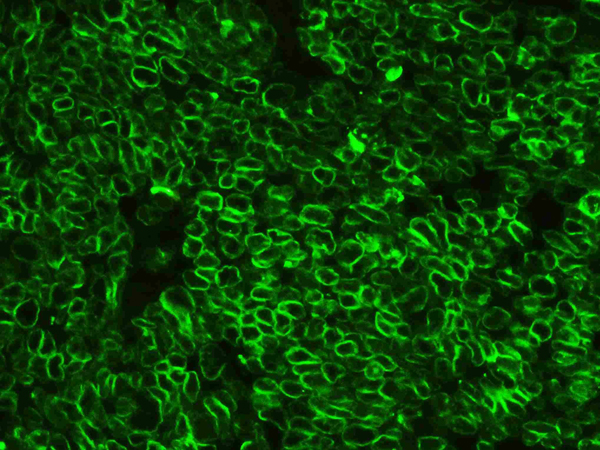
 IF (Immunofluorescence)
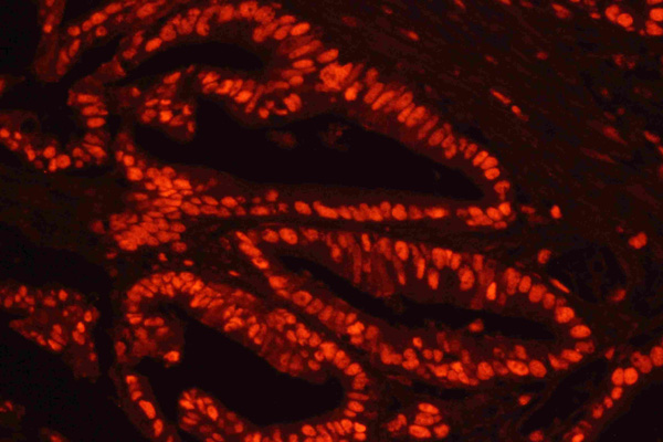
(Figure 2. IF analysis of COX5B using anti-COX5B antibody (A06090-2).COX5B was detected in a paraffin-embedded section of human intestinal cancer tissue. Heat mediated antigen retrieval was performed in EDTA buffer (pH 8.0, epitope retrieval solution). The tissue section was blocked with 10% goat serum. The tissue section was then incubated with 5ug/mL rabbit anti-COX5B Antibody (A06090-2) overnight at 4 degree C. DyLight 647 Conjugated Goat Anti-Rabbit IgG (AAA127948) was used as secondary antibody at 1:500 dilution and incubated for 30 minutes at 37 degree C. The section was counterstained with DAPI. Visualize using a fluorescence microscope and filter sets appropriate for the label used.)
IF (Immunofluorescence)
(Figure 2. IF analysis of COX5B using anti-COX5B antibody (A06090-2).COX5B was detected in a paraffin-embedded section of human intestinal cancer tissue. Heat mediated antigen retrieval was performed in EDTA buffer (pH 8.0, epitope retrieval solution). The tissue section was blocked with 10% goat serum. The tissue section was then incubated with 5ug/mL rabbit anti-COX5B Antibody (A06090-2) overnight at 4 degree C. DyLight 647 Conjugated Goat Anti-Rabbit IgG (AAA127948) was used as secondary antibody at 1:500 dilution and incubated for 30 minutes at 37 degree C. The section was counterstained with DAPI. Visualize using a fluorescence microscope and filter sets appropriate for the label used.)
IgG, Polyclonal Secondary Antibody (Cat# AAA127948)
IgG, Secondary Antibody (Cat# AAA60524)
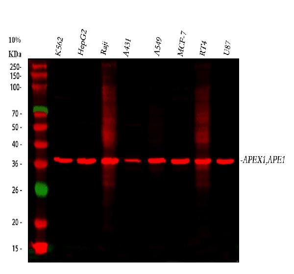
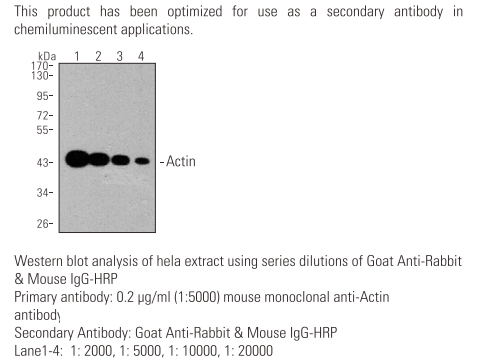 WB (Western Blot)
IgG, Secondary Antibody (Cat# AAA62290)
IgG, Secondary Antibody (Cat# AAA62293)
Biotin Conjugated Goat Anti-rabbit IgG, Polyclonal Secondary Antibody (Cat# AAA45604)
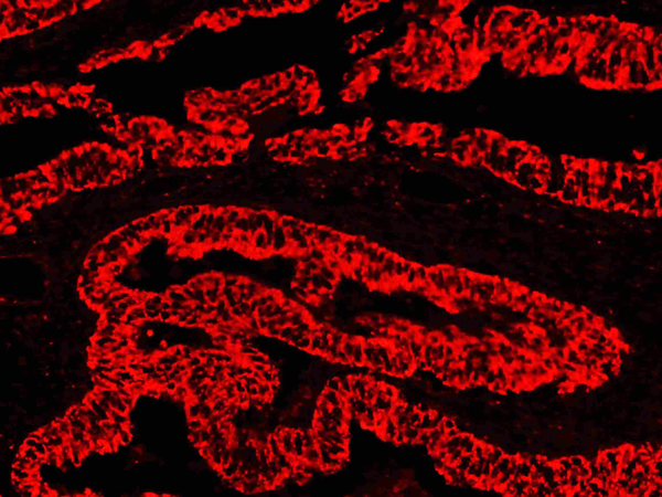
 IHC (Immunohiostchemistry)
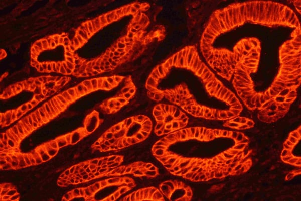
(Ki67 was detected in paraffin-embedded sections of human intestinal cancer tissues using rabbit anti- Ki67 Antigen Affinity purified polyclonal antibody at 1 ug/mL. Cy3 Conjugated Avidin was used for visualization at 10ug/mL.)
IHC (Immunohiostchemistry)
(Ki67 was detected in paraffin-embedded sections of human intestinal cancer tissues using rabbit anti- Ki67 Antigen Affinity purified polyclonal antibody at 1 ug/mL. Cy3 Conjugated Avidin was used for visualization at 10ug/mL.)
Cy3 Conjugated Avidin, Polyclonal Secondary Antibody (Cat# AAA45606)
Biotin Conjugated Mouse Anti-rabbit IgG, Polyclonal Secondary Antibody (Cat# AAA45607)
unconjugated Rabbit Anti-rat IgG, Polyclonal Secondary Antibody (Cat# AAA45615)
HRP Conjugated Rabbit Anti-human IgG, Polyclonal Secondary Antibody (Cat# AAA45620)
What Are Secondary Antibodies?
Secondary antibodies are immunoglobulins that bind to primary antibodies, which are directly bound to the target antigen. They are conjugated with either an enzyme, fluorophore, or biotin, and they are almost always only targeting the constant region (Fc) of the primary antibody. As a result, this amplifies the signal in various assays and helps researchers detect, quantify, and visualize specific antigens in a complex biological sample.
Key Applications of Secondary Antibodies
- Western blotting for protein detection and quantification.
- ELISA (Enzyme-Linked Immunosorbent Assay) for antigen-antibody interaction studies and quantification.
- Immunohistochemistry (IHC) and immunofluorescence imaging for tissue and cell analysis.
- Flow cytometry for cell surface and intracellular marker detection.
- Immunoprecipitation (IP) and chromatin immunoprecipitation (ChIP) assays.
- Signal amplification in various immunoassays.
- Multiplex assays using fluorophore-conjugated secondary antibodies.
Advantages of Using Secondary Antibodies
-
High sensitivity
Multiple secondary antibodies can bind to a single primary antibody and amplify the signal. This is especially useful for detecting antigens even if they are in low concentrations.
-
Increased flexibility in assay design
Secondary antibodies come pre-conjugated with labels, such as enzymes, fluorophores, biotin, and more. This provides researchers with flexibility to use in multiple applications. They also support multiplexing for the simultaneous detection of multiple targets.
-
Cost-effective
Since the Fc domain remains constant within the same animal class, only one type of secondary antibody is needed to bind many types of primary antibodies. This reduces the cost of labeling multiple primary antibodies.
-
Better signal-to-noise ratio
Secondary antibodies minimize the background noise. As a result, they provide clearer and more accurate results.
-
Versatility
A single species- and isotype-specific secondary antibody can be used with any compatible primary antibody. This can help streamline reagent inventory and reduce cost and preparation time.
-
Easier detection
Unlike primary antibodies, secondary antibodies’ main characteristic is that they will almost always be partnered with a component that can be used for detection. This can make detection and visualization easy.
Why Buy Secondary Antibodies from AAA Biotech?
- Highly Validated: Most of our secondary antibodies are thoroughly tested to ensure they work reliably across different experiments.
- Versatile Applications: Our secondary antibodies can be used in a wide range of research techniques, including (but not limited to) immunocytochemistry (ICC), ELISA, immunofluorescence (IF), immunohistochemistry (IHC), flow cytometry (FC), immunoprecipitation (IP), and Western blotting (WB).
- Support for Rare Species: We offer secondary antibodies specifically designed for rare or less common species—something many other suppliers don’t provide.
- Affordable Prices: Our secondary antibodies are available at competitive prices to support all research budgets.
- Quick and Convenient Ordering: Simple online ordering process with responsive customer support.
- Privacy: We respect your privacy and ensure your information is protected at every step of the ordering process, and in all other communication with our team members.
FAQ
1. Can I reuse a secondary antibody?
Not recommended for critical experiments, as reusing may compromise sensitivity and specificity.
2. What is the difference between primary and secondary antibodies?
Primary antibodies bind directly to the antigen. Secondary antibodies bind to the primary and are usually labeled for detection.
3. How long do secondary antibodies last?
When stored properly (usually at –20°C or 4°C, depending on formulation), they can last for years. Always refer to the product datasheet.
4. What happens if you use too much secondary antibody?
Excess can increase background noise, leading to a poor signal-to-noise ratio. Titrate to find the optimal concentration.
5. Can secondary antibodies be monoclonal?
Yes. While many are polyclonal, monoclonal secondary antibodies are available for consistent batch-to-batch performance.
6. How to find secondary antibodies?
Use our catalog filters to search by host species, conjugate type, target species, and application.
7. How to store secondary antibodies?
Store at recommended temperatures away from light. Avoid repeated freeze-thaw cycles.