Filters
▼Clonality
▼Type
▼Reactivity
▼Gene Name
▼Isotype
▼Host
▼Application
▼Clone
▼Secondary Antibodies
At AAA Biotech also known as AAA Bio or AAABio, we provide a vast collection of high-quality, purified, conjugated and unconjugated secondary antibodies to use in various applications, such as Western Blotting, Flow Cytometry, Cell Imaging, or CUT & Tag.
Our secondary antibodies are available with a wide range of labels—such as HRP, AP, FITC, and biotin—to match your detection system and enhance signal sensitivity. Each antibody is rigorously tested to ensure high specificity and minimal cross-reactivity, delivering reliable performance across multiple species and assay types.
Whether you're conducting qualitative imaging or quantitative analysis, AAA Biotech has the right secondary antibody to support your research with consistency and precision. Our entire catalog of secondary antibodies is available for browsing through our main website.
Viewing 500-550 of 552 product results
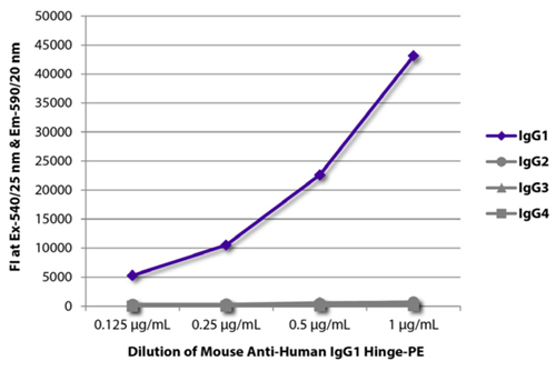 Application Data
(FLISA plate was coated with purified human IgG1, IgG2, IgG3, and IgG4. Immunoglobulins were detected with serially diluted Mouse Anti-Human IgG1 Hinge-PE (Cat. No. AAA78722).)
Application Data
(FLISA plate was coated with purified human IgG1, IgG2, IgG3, and IgG4. Immunoglobulins were detected with serially diluted Mouse Anti-Human IgG1 Hinge-PE (Cat. No. AAA78722).)
Mouse Anti-Human IgG1 (gamma 1 chain specific), Monoclonal Secondary Antibody (Cat# AAA78722)
Rat Anti-Mouse IgG2a (gamma 2a chain specific), Monoclonal Secondary Antibody (Cat# AAA78650)
Rabbit F(ab')2 Anti-Mouse IgG (H+L), Polyclonal Secondary Antibody (Cat# AAA78840)
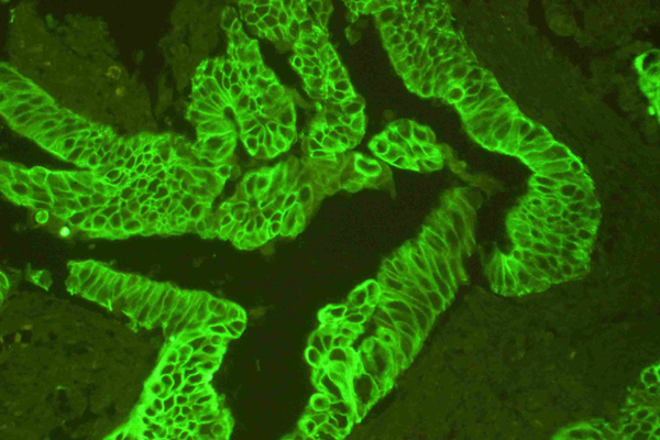 Application Data
(PCK was detected in paraffin-embedded sections of human intestinal cancer tissues using mouse anti- PCK Antigen Affinity purified monoclonal antibody at 1 ug/mL. DyLight-488 Conjugated Goat Anti-mouse IgG secondary antibody was used to detect the primary antibody at 10ug/mL.)
Application Data
(PCK was detected in paraffin-embedded sections of human intestinal cancer tissues using mouse anti- PCK Antigen Affinity purified monoclonal antibody at 1 ug/mL. DyLight-488 Conjugated Goat Anti-mouse IgG secondary antibody was used to detect the primary antibody at 10ug/mL.)
DyLight488 Conjugated Goat Anti-mouse IgG, Polyclonal Secondary Antibody (Cat# AAA46013)
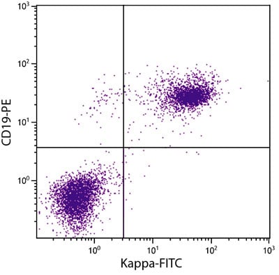 Application Data
(BALB/c mouse splenocytes were stained with Rat anti-Mouse Kappa-BIOT and Rat Anti-Mouse CD19-PE followed by Streptavidin-FITC.)
Application Data
(BALB/c mouse splenocytes were stained with Rat anti-Mouse Kappa-BIOT and Rat Anti-Mouse CD19-PE followed by Streptavidin-FITC.)
Rat Anti-Mouse Kappa (kappa chain specific), Monoclonal Secondary Antibody (Cat# AAA78652)
IgG, Polyclonal Secondary Antibody (Cat# AAA312915)
 ELISA
ELISA
hFab (CH1), Secondary Antibody (Cat# AAA268874)
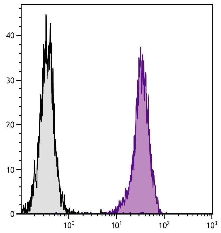 FCM/FACS (Flow Cytometry)
(BALB/c mouse splenocytes were stained with Rat Anti-Mouse CD45-UNLB (SB Cat. No. 1660-01) followed by Mouse Anti-Rat IgG2b-BIOT and Streptavidin-FITC)
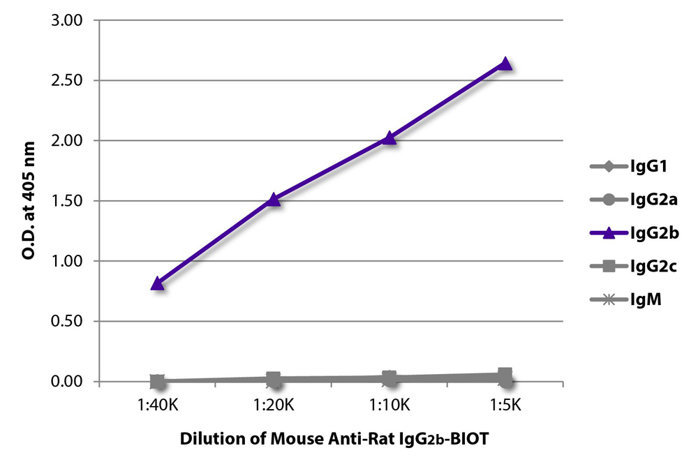
FCM/FACS (Flow Cytometry)
(BALB/c mouse splenocytes were stained with Rat Anti-Mouse CD45-UNLB (SB Cat. No. 1660-01) followed by Mouse Anti-Rat IgG2b-BIOT and Streptavidin-FITC)
IgG2b, Monoclonal Secondary Antibody (Cat# AAA78643)
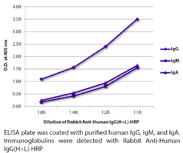 ELISA
ELISA
Rabbit Anti-Human IgG (H+L), Polyclonal Secondary Antibody (Cat# AAA78841)
Donkey Anti-Goat IgG (H+L), Polyclonal Secondary Antibody (Cat# AAA78862)
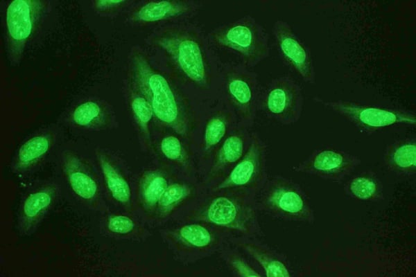
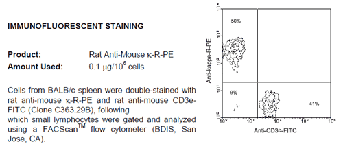 IF (Immunofluorescence)
Rat Anti-Mouse Kappa (kappa chain specific), Monoclonal Secondary Antibody (Cat# AAA78656)
Mouse anti Human IgG1, Secondary Antibody (Cat# AAA74573)
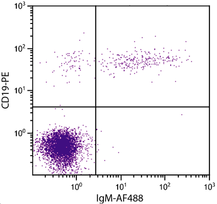
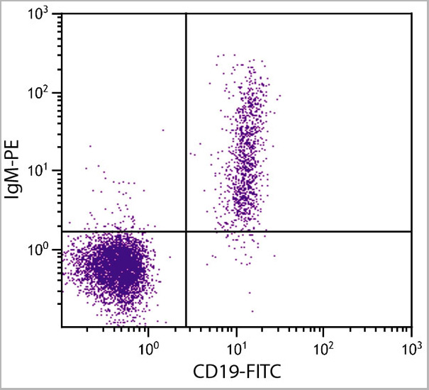 Application Data
(Human peripheral blodd lymohocytes were stained with Goat Anti-Human IgM-PE and Mouse Anti-Human CD19-FITC .)
Application Data
(Human peripheral blodd lymohocytes were stained with Goat Anti-Human IgM-PE and Mouse Anti-Human CD19-FITC .)
Goat Anti-Human IgM (u chain specific), Polyclonal Secondary Antibody (Cat# AAA14900)
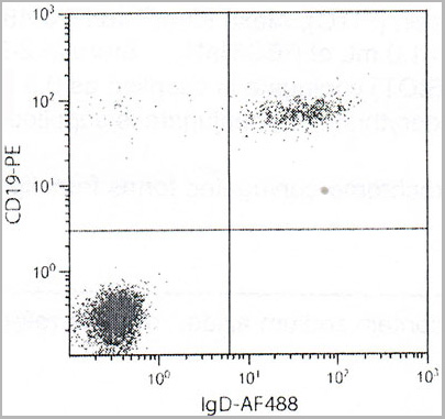 Application Data
(Human peripheral blood lymphocytes were stained with Goat F(ab’)2 Anti-Human IgD-AF488 and Mouse Anti-Human CD19-PE.)
Application Data
(Human peripheral blood lymphocytes were stained with Goat F(ab’)2 Anti-Human IgD-AF488 and Mouse Anti-Human CD19-PE.)
Goat F(ab')2 Anti-Human IgD (delta chain specific), Polyclonal Secondary Antibody (Cat# AAA14902)
 Application Data
Mouse Anti-Human IgG (Fc) (gamma chain specific), Monoclonal Secondary Antibody (Cat# AAA14897)
Rabbit anti Monkey IgG Fab, Polyclonal Secondary Antibody (Cat# AAA14570)
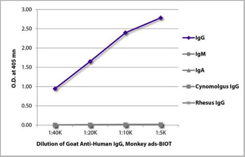 ELISA
(ELISA plate was coated with purified human IgG, IgM, and IgA, cynomolgus IgG, and rhesus IgG. Immunoglobulins were detected with serially diluted Goat Anti-Human IgG, Monkey ads-BIOT (AAA14911) followed by Streptavidin-HRP (please inquire).)
ELISA
(ELISA plate was coated with purified human IgG, IgM, and IgA, cynomolgus IgG, and rhesus IgG. Immunoglobulins were detected with serially diluted Goat Anti-Human IgG, Monkey ads-BIOT (AAA14911) followed by Streptavidin-HRP (please inquire).)
Goat Anti-Human IgG, Monkey ads, Secondary Antibody (Cat# AAA14911)
IgG, Polyclonal Secondary Antibody (Cat# AAA30581)
 Application Data
(Human peripheral blood lymphocytes were stained with mouse Anti-Human IgM-AF488 and mouse Anti-human CD-PE)
Application Data
(Human peripheral blood lymphocytes were stained with mouse Anti-Human IgM-AF488 and mouse Anti-human CD-PE)
Mouse Anti-Human IgM (u chain specific), Monoclonal Secondary Antibody (Cat# AAA14896)
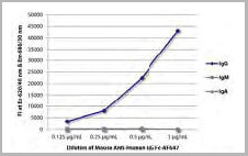
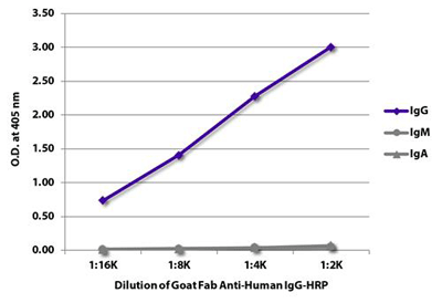 Application Data
(ELISA plate was coated with purified human IgG, IgM, and IgA. Immunoglobulins were detected with serially diluted Goat Fab Anti-Human IgG-HRP (AAA14906))
Application Data
(ELISA plate was coated with purified human IgG, IgM, and IgA. Immunoglobulins were detected with serially diluted Goat Fab Anti-Human IgG-HRP (AAA14906))
Goat F(ab) Anti-Human IgG (gamma chain specific), Secondary Antibody (Cat# AAA14906)
IgG, Polyclonal Secondary Antibody (Cat# AAA14879)
anti-Human IgG+HRP, Polyclonal Secondary Antibody (Cat# AAA13852)
anti-Mouse IgG+HRP, Secondary Antibody (Cat# AAA13853)
 Application Data
(Human peripheral blood lymphocytes were stained with Goat F(ab')2 antihuman IgM-PE and mouse anti-human CD19-FITC .)
Application Data
(Human peripheral blood lymphocytes were stained with Goat F(ab')2 antihuman IgM-PE and mouse anti-human CD19-FITC .)
Goat F(ab')2 Anti-Human IgM, Secondary Antibody (Cat# AAA14912)
Goat Anti-Human IgG (γ chain specific), Secondary Antibody (Cat# AAA14903)
Goat anti Human IgG IgA IgM (Fc specific), Polyclonal Secondary Antibody (Cat# AAA14569)
 Standard Curve (Sample)
Mouse Anti-Human IgG (Fc) (gamma chain specific), Monoclonal Secondary Antibody (Cat# AAA14898)
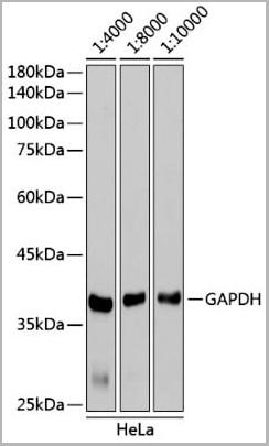
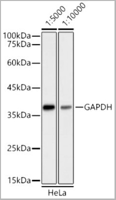 WB (Western Blot)
(Western blot analysis of extracts of HeLa cells, using GAPDH antibody as the primary antibody. Secondary antibody: HRP Goat Anti-Mouse IgG (H+L) antibody (AAA10764) at 1:5000/1:10000 dilution. Lysates/proteins: 25ug per lane. Blocking buffer: 3% nonfat dry milk in TBST. Detection: ECL Basic Kit. Exposure Time: 60s.)
WB (Western Blot)
(Western blot analysis of extracts of HeLa cells, using GAPDH antibody as the primary antibody. Secondary antibody: HRP Goat Anti-Mouse IgG (H+L) antibody (AAA10764) at 1:5000/1:10000 dilution. Lysates/proteins: 25ug per lane. Blocking buffer: 3% nonfat dry milk in TBST. Detection: ECL Basic Kit. Exposure Time: 60s.)
IgG (H+L), Secondary Antibody (Cat# AAA10764)
Mouse Anti-Monkey IgG, Monoclonal Secondary Antibody (Cat# AAA14895)
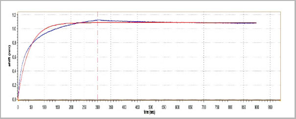 ELISA
(Examples: Interaction against hIgG1 determined by Gator (BLI)*KD=1.2×10⁻¹¹)
ELISA
(Examples: Interaction against hIgG1 determined by Gator (BLI)*KD=1.2×10⁻¹¹)
Fab (CH1), Monoclonal Secondary Antibody (Cat# AAA27978)
Goat anti Monkey IgG (Fc specific), Polyclonal Secondary Antibody (Cat# AAA77592)
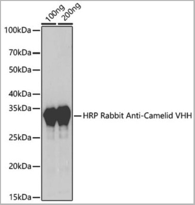 WB (Western Blot)
(Western blot analysis of extracts of various cell lines, using HRP Rabbit Anti-Camelid VHH Antibody antibody (AAA10762) at 1:1000 dilution. Secondary antibody: HRP Goat Anti-Rabbit IgG (H+L) at 1:10000 dilution. Lysates/proteins: 25ug per lane. Blocking buffer: 3% nonfat dry milk in TBST. Detection: ECL Basic Kit. Exposure time: 3s.)
WB (Western Blot)
(Western blot analysis of extracts of various cell lines, using HRP Rabbit Anti-Camelid VHH Antibody antibody (AAA10762) at 1:1000 dilution. Secondary antibody: HRP Goat Anti-Rabbit IgG (H+L) at 1:10000 dilution. Lysates/proteins: 25ug per lane. Blocking buffer: 3% nonfat dry milk in TBST. Detection: ECL Basic Kit. Exposure time: 3s.)
IgG (H+L), Secondary Antibody (Cat# AAA10762)
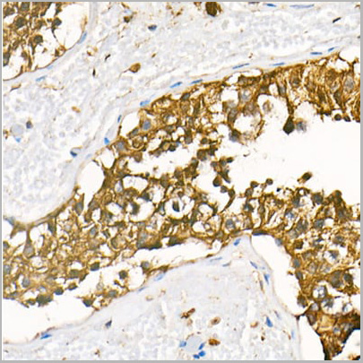 IHC (Immunohistochemistry)
(Immunohistochemistry analysis of HRP Goat Anti-Rabbit IgG (H+L) in paraffin-embedded human testis tissue using HRP Goat Anti-Rabbit IgG (H+L) (AAA10763) at a dilution of 1:100 (40x lens). High pressure antigen retrieval was performed with 0.01 M citrate buffer (pH 6.0) prior to IHC staining.)
IHC (Immunohistochemistry)
(Immunohistochemistry analysis of HRP Goat Anti-Rabbit IgG (H+L) in paraffin-embedded human testis tissue using HRP Goat Anti-Rabbit IgG (H+L) (AAA10763) at a dilution of 1:100 (40x lens). High pressure antigen retrieval was performed with 0.01 M citrate buffer (pH 6.0) prior to IHC staining.)
IgG (H+L), Secondary Antibody (Cat# AAA10763)
Goat F(ab) Anti-Human IgG (gamma chain specific), Secondary Antibody (Cat# AAA14905)
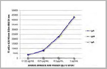
 ELISA
(ELISA analysis was performed by loading 100 uL per well of an anti-Human Fab (CH1) mAb at a concentration of 5, 2.5, 1.25, 0.625, 0.3125, 0.15625, 0.078, and 0.039 ug/mL across 100 ng per well of a human antibody pre-coated plate. The plate was incubated for 1 hour at 37°C. Detection was performed using TMB Substrate for 10 minutes at room temperature in the dark. The plate was stopped with 2M sulfuric acid. Absorbances were read on a spectrophotometer at 450 nm.)
ELISA
(ELISA analysis was performed by loading 100 uL per well of an anti-Human Fab (CH1) mAb at a concentration of 5, 2.5, 1.25, 0.625, 0.3125, 0.15625, 0.078, and 0.039 ug/mL across 100 ng per well of a human antibody pre-coated plate. The plate was incubated for 1 hour at 37°C. Detection was performed using TMB Substrate for 10 minutes at room temperature in the dark. The plate was stopped with 2M sulfuric acid. Absorbances were read on a spectrophotometer at 450 nm.)
Fab (CH1), Monoclonal Secondary Antibody (Cat# AAA27977)
Goat anti Human IgG Fc, Secondary Antibody (Cat# AAA14316)
Goat F(ab')2 Anti-Human IgG (gamma chain specific), Polyclonal Secondary Antibody (Cat# AAA14907)
Goat Anti-Human IgG, Monkey ads, Secondary Antibody (Cat# AAA14913)
anti-Rabbit IgG+HRP, Secondary Antibody (Cat# AAA13854)
Affinity purified and conjugated with horseradish peroxidase (HRP)
IgG (H+L), Secondary Antibody (Cat# AAA26993)
IgG (H&L), Secondary Antibody (Cat# AAA332954)
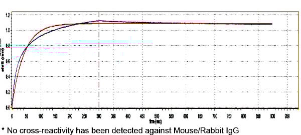
Cross Reactivity: Based on IEP, no reactivity is observed to:
non-immunoglobulin mouse serum proteins
human serum proteins
Affinity purified using solid phase Mouse IgG.
 WB (Western Blot)
(Western BlotPositive WB detected in Recombinantprotein (80ng, 40ng, 20ng, 10ng)All lanes: Human IgG Fc antibody;HRPconjugated at 1:1000Predicted band size: 35 kDaObserved band size: 35 kDa)
WB (Western Blot)
(Western BlotPositive WB detected in Recombinantprotein (80ng, 40ng, 20ng, 10ng)All lanes: Human IgG Fc antibody;HRPconjugated at 1:1000Predicted band size: 35 kDaObserved band size: 35 kDa)
IgG Fc fragment, Polyclonal Secondary Antibody (Cat# AAA15949)
 Application Data
(BALB/c mouse splenocytes were stained with Goat F(ab)2 Anti-Mouse Kappa-BIOT and Rat Anti-Mouse CD19-PE followed by Streptavidin-FITC.)
Application Data
(BALB/c mouse splenocytes were stained with Goat F(ab)2 Anti-Mouse Kappa-BIOT and Rat Anti-Mouse CD19-PE followed by Streptavidin-FITC.)
Goat F(ab')2 Anti-Mouse kappa (kappa chain specific), Polyclonal Secondary Antibody (Cat# AAA14899)
 Application Data
(ELISA plate was coated with purified human IgG, IgM, and IgA. Immunoglobulins were detected with serially diluted Goat Anti-Human IgG-HRP.)
Application Data
(ELISA plate was coated with purified human IgG, IgM, and IgA. Immunoglobulins were detected with serially diluted Goat Anti-Human IgG-HRP.)
Goat Anti-Human IgG (gamma chain specific), Polyclonal Secondary Antibody (Cat# AAA14904)
 Application Data
(FLISA plate was coated with purified rat IgG and IgM. Immunoglobulins were detected with serially diluted Goat Anti-Rat IgG-PE)
Application Data
(FLISA plate was coated with purified rat IgG and IgM. Immunoglobulins were detected with serially diluted Goat Anti-Rat IgG-PE)
Goat Anti-Rat IgG (gamma chain specific), Polyclonal Secondary Antibody (Cat# AAA14908)
Mouse Anti-Monkey IgG, Monoclonal Secondary Antibody (Cat# AAA14894)
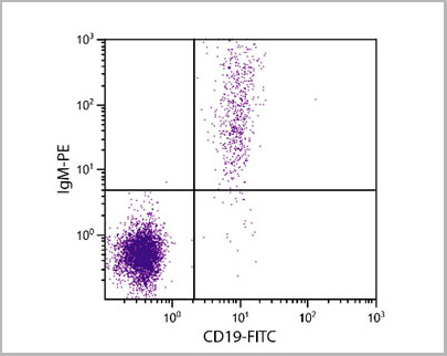
 Application Data
(Human peripheral blood lymphocytes were stained with Goat F(ab')2 Anti-Human IgM-PE and Mouse Anti-Human CD19-FITC .)
Application Data
(Human peripheral blood lymphocytes were stained with Goat F(ab')2 Anti-Human IgM-PE and Mouse Anti-Human CD19-FITC .)
Goat F(ab')2 Anti-Human IgM (u chain specific), Polyclonal Secondary Antibody (Cat# AAA14901)
IgG (H&L), F(ab)'2 fragment, Secondary Antibody (Cat# AAA332955)
Cross Reactivity: Based on IEP, no reactivity is observed to non-immunoglobulin human serum immunoglobulins bovine, goat, human, rabbit or rat IgG
Affinity purified using solid phase Mouse IgG.
anti-human IgG, Monoclonal Secondary Antibody (Cat# AAA13530)
 IF (Immunofluorescence)
(BALB/c mouse splenocytes were stained with Rat Anti-Mouse Lamda-PE and Rat Anti-Mouse CD119-FITC.)
IF (Immunofluorescence)
(BALB/c mouse splenocytes were stained with Rat Anti-Mouse Lamda-PE and Rat Anti-Mouse CD119-FITC.)
Rat Anti-Mouse Lambda (lambda light chain specific), Monoclonal Secondary Antibody (Cat# AAA14893)
What Are Secondary Antibodies?
Secondary antibodies are immunoglobulins that bind to primary antibodies, which are directly bound to the target antigen. They are conjugated with either an enzyme, fluorophore, or biotin, and they are almost always only targeting the constant region (Fc) of the primary antibody. As a result, this amplifies the signal in various assays and helps researchers detect, quantify, and visualize specific antigens in a complex biological sample.
Key Applications of Secondary Antibodies
- Western blotting for protein detection and quantification.
- ELISA (Enzyme-Linked Immunosorbent Assay) for antigen-antibody interaction studies and quantification.
- Immunohistochemistry (IHC) and immunofluorescence imaging for tissue and cell analysis.
- Flow cytometry for cell surface and intracellular marker detection.
- Immunoprecipitation (IP) and chromatin immunoprecipitation (ChIP) assays.
- Signal amplification in various immunoassays.
- Multiplex assays using fluorophore-conjugated secondary antibodies.
Advantages of Using Secondary Antibodies
-
High sensitivity
Multiple secondary antibodies can bind to a single primary antibody and amplify the signal. This is especially useful for detecting antigens even if they are in low concentrations.
-
Increased flexibility in assay design
Secondary antibodies come pre-conjugated with labels, such as enzymes, fluorophores, biotin, and more. This provides researchers with flexibility to use in multiple applications. They also support multiplexing for the simultaneous detection of multiple targets.
-
Cost-effective
Since the Fc domain remains constant within the same animal class, only one type of secondary antibody is needed to bind many types of primary antibodies. This reduces the cost of labeling multiple primary antibodies.
-
Better signal-to-noise ratio
Secondary antibodies minimize the background noise. As a result, they provide clearer and more accurate results.
-
Versatility
A single species- and isotype-specific secondary antibody can be used with any compatible primary antibody. This can help streamline reagent inventory and reduce cost and preparation time.
-
Easier detection
Unlike primary antibodies, secondary antibodies’ main characteristic is that they will almost always be partnered with a component that can be used for detection. This can make detection and visualization easy.
Why Buy Secondary Antibodies from AAA Biotech?
- Highly Validated: Most of our secondary antibodies are thoroughly tested to ensure they work reliably across different experiments.
- Versatile Applications: Our secondary antibodies can be used in a wide range of research techniques, including (but not limited to) immunocytochemistry (ICC), ELISA, immunofluorescence (IF), immunohistochemistry (IHC), flow cytometry (FC), immunoprecipitation (IP), and Western blotting (WB).
- Support for Rare Species: We offer secondary antibodies specifically designed for rare or less common species—something many other suppliers don’t provide.
- Affordable Prices: Our secondary antibodies are available at competitive prices to support all research budgets.
- Quick and Convenient Ordering: Simple online ordering process with responsive customer support.
- Privacy: We respect your privacy and ensure your information is protected at every step of the ordering process, and in all other communication with our team members.
FAQ
1. Can I reuse a secondary antibody?
Not recommended for critical experiments, as reusing may compromise sensitivity and specificity.
2. What is the difference between primary and secondary antibodies?
Primary antibodies bind directly to the antigen. Secondary antibodies bind to the primary and are usually labeled for detection.
3. How long do secondary antibodies last?
When stored properly (usually at –20°C or 4°C, depending on formulation), they can last for years. Always refer to the product datasheet.
4. What happens if you use too much secondary antibody?
Excess can increase background noise, leading to a poor signal-to-noise ratio. Titrate to find the optimal concentration.
5. Can secondary antibodies be monoclonal?
Yes. While many are polyclonal, monoclonal secondary antibodies are available for consistent batch-to-batch performance.
6. How to find secondary antibodies?
Use our catalog filters to search by host species, conjugate type, target species, and application.
7. How to store secondary antibodies?
Store at recommended temperatures away from light. Avoid repeated freeze-thaw cycles.