Filters
▼Clonality
▼Type
▼Reactivity
▼Gene Name
▼Isotype
▼Host
▼Application
▼Clone
▼Polyclonal Antibodies
At AAA Biotech also known as AAA Bio or AAABio, we provide a broad range of purified polyclonal antibodies (pAbs) that are able to all be browsed online through our website. Due to their high specificity and strong binding affinity, these antibodies are ideal for wide swathes of research and experimental applications.
Our polyclonal antibodies can easily support your work, whether you use them for Western Blotting, Immunocytochemistry (with or without Immunofluorescence used in conjunction), Immunohistochemistry, Immunoprecipitation, and ELISA tests. We highly encourage you to browse our range of pAbs and choose the one that best suits your experimental model.
Viewing 2150-2200 of 96805 product results
 IF (Immunofluorescence)
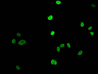
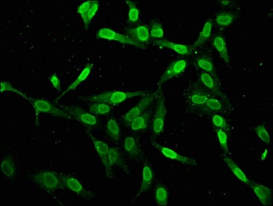
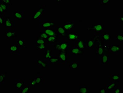
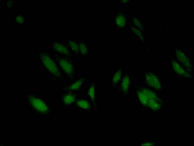
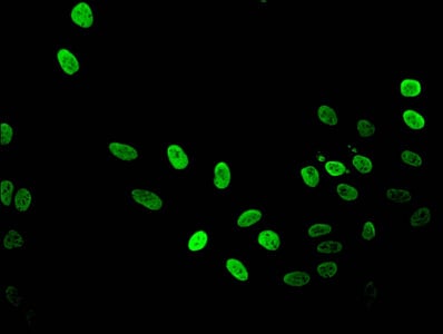
(Immunofluorescent analysis of Hela cells using AAA234330 at a dilution of 1:100 and Alexa Fluor 488-congugated AffiniPure Goat Anti-Rabbit IgG(H+L))
IF (Immunofluorescence)
(Immunofluorescent analysis of Hela cells using AAA234330 at a dilution of 1:100 and Alexa Fluor 488-congugated AffiniPure Goat Anti-Rabbit IgG(H+L))
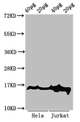
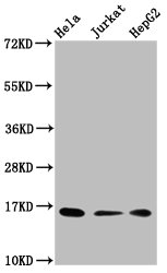
HIST1H1E, Polyclonal Antibody (Cat# AAA234330)
 IF (Immunofluorescence)
(Immunofluorescent analysis of MCF-7 (sodium butyrate, 30 mM, 4h) cells using AAA234334 at a dilution of 1:100 and Alexa Fluor 488-congugated AffiniPure Goat Anti-Rabbit IgG(H+L))
IF (Immunofluorescence)
(Immunofluorescent analysis of MCF-7 (sodium butyrate, 30 mM, 4h) cells using AAA234334 at a dilution of 1:100 and Alexa Fluor 488-congugated AffiniPure Goat Anti-Rabbit IgG(H+L))
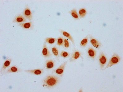
Acetyl-HIST1H1E, Polyclonal Antibody (Cat# AAA234334)
 IHC (Immunohistochemisry)
(Immunofluorescent analysis of MCF-7 cells using AAA234335 at a dilution of 1:100 and Alexa Fluor 488-congugated AffiniPure Goat Anti-Rabbit IgG(H+L))
IHC (Immunohistochemisry)
(Immunofluorescent analysis of MCF-7 cells using AAA234335 at a dilution of 1:100 and Alexa Fluor 488-congugated AffiniPure Goat Anti-Rabbit IgG(H+L))
HIST1H1E, Polyclonal Antibody (Cat# AAA234335)
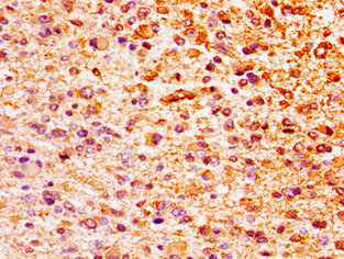
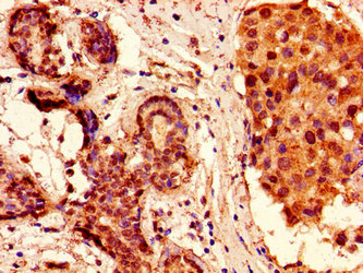
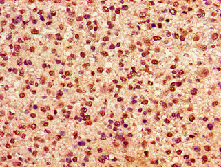
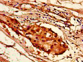
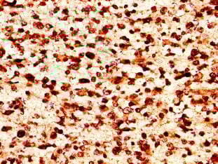
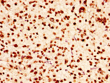
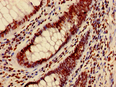
 IHC (Immunohistochemisry)
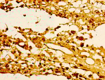
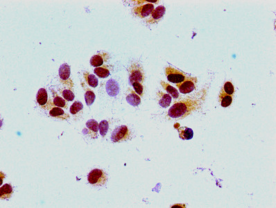
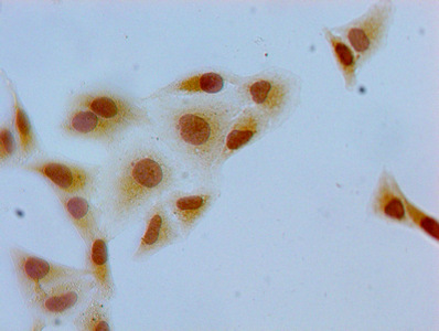
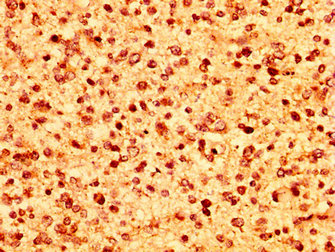
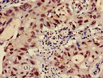
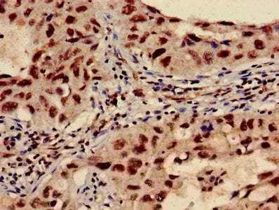
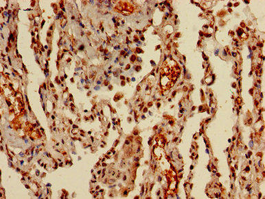
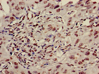
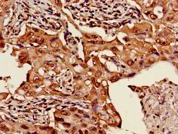
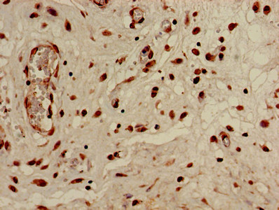
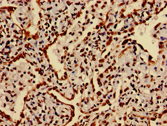
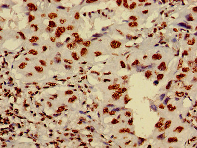
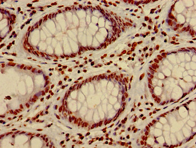
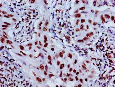
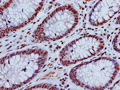
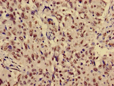
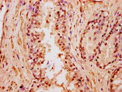
(Immunohistochemistry of paraffin-embedded human melanoma cancer using AAA234342 at dilution of 1:100)
IHC (Immunohistochemisry)
(Immunohistochemistry of paraffin-embedded human melanoma cancer using AAA234342 at dilution of 1:100)
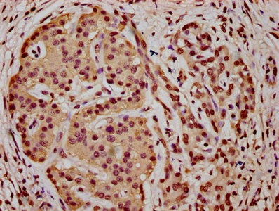
HIST1H1E, Polyclonal Antibody (Cat# AAA234342)
 IF (Immunofluorescence)
(Immunofluorescent analysis of Hela cells (sodium butyrate, 30 mM, 4h) using AAA234343 at a dilution of 1:100 and Alexa Fluor 488-congugated AffiniPure Goat Anti-Rabbit IgG(H+L))
IF (Immunofluorescence)
(Immunofluorescent analysis of Hela cells (sodium butyrate, 30 mM, 4h) using AAA234343 at a dilution of 1:100 and Alexa Fluor 488-congugated AffiniPure Goat Anti-Rabbit IgG(H+L))
Acetyl-HIST1H1C, Polyclonal Antibody (Cat# AAA234343)
 IF (Immunofluorescence)
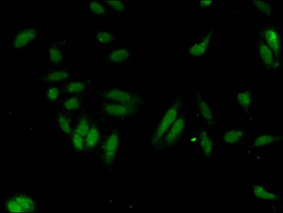
(Immunofluorescent analysis of Hela cells using AAA234344 at a dilution of 1:100 and Alexa Fluor 488-congugated AffiniPure Goat Anti-Rabbit IgG(H+L))
IF (Immunofluorescence)
(Immunofluorescent analysis of Hela cells using AAA234344 at a dilution of 1:100 and Alexa Fluor 488-congugated AffiniPure Goat Anti-Rabbit IgG(H+L))
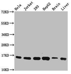
HIST1H1C, Polyclonal Antibody (Cat# AAA234344)
 IHC (Immunohistochemisry)
(Immunofluorescent analysis of Hela cells using AAA234346 at a dilution of 1:100 and Alexa Fluor 488-congugated AffiniPure Goat Anti-Rabbit IgG(H+L))
IHC (Immunohistochemisry)
(Immunofluorescent analysis of Hela cells using AAA234346 at a dilution of 1:100 and Alexa Fluor 488-congugated AffiniPure Goat Anti-Rabbit IgG(H+L))
HIST1H2AG, Polyclonal Antibody (Cat# AAA234346)
 IHC (Immunohistochemisry)
(Immunofluorescent analysis of Hela cells using AAA234347 at a dilution of 1:100 and Alexa Fluor 488-congugated AffiniPure Goat Anti-Rabbit IgG(H+L))
IHC (Immunohistochemisry)
(Immunofluorescent analysis of Hela cells using AAA234347 at a dilution of 1:100 and Alexa Fluor 488-congugated AffiniPure Goat Anti-Rabbit IgG(H+L))
HIST1H1D, Polyclonal Antibody (Cat# AAA234347)
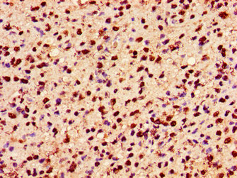
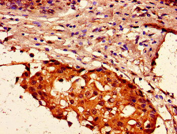
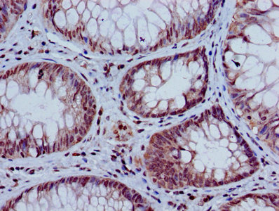
 IHC (Immunohistochemisry)
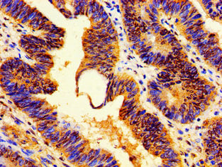
(Immunohistochemistry of paraffin-embedded human breast cancer using AAA234349 at dilution of 1:100)
IHC (Immunohistochemisry)
(Immunohistochemistry of paraffin-embedded human breast cancer using AAA234349 at dilution of 1:100)
HIST1H2AG, Polyclonal Antibody (Cat# AAA234349)
 IHC (Immunohistochemisry)
(Immunofluorescent analysis of Hela cells using AAA234350 at a dilution of 1:100 and Alexa Fluor 488-congugated AffiniPure Goat Anti-Rabbit IgG(H+L))
IHC (Immunohistochemisry)
(Immunofluorescent analysis of Hela cells using AAA234350 at a dilution of 1:100 and Alexa Fluor 488-congugated AffiniPure Goat Anti-Rabbit IgG(H+L))
HIST1H2AG, Polyclonal Antibody (Cat# AAA234350)
 IF (Immunofluorescence)
(Immunofluorescent analysis of Hela cells using AAA234355 at a dilution of 1:100 and Alexa Fluor 488-congugated AffiniPure Goat Anti-Rabbit IgG(H+L))
IF (Immunofluorescence)
(Immunofluorescent analysis of Hela cells using AAA234355 at a dilution of 1:100 and Alexa Fluor 488-congugated AffiniPure Goat Anti-Rabbit IgG(H+L))
Mono-methyl-H2AFZ, Polyclonal Antibody (Cat# AAA234355)
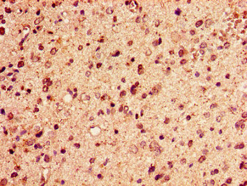
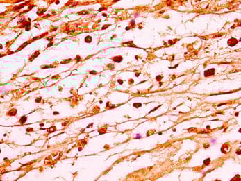
 IHC (Immunohiostchemistry)
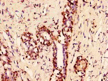
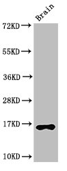
(IHC image of AAA234358 diluted at 1:10 and staining in paraffin-embedded human brain tissue performed on a Leica BondTM system. After dewaxing and hydration, antigen retrieval was mediated by high pressure in a citrate buffer (pH 6.0). Section was blocked with 10% normal goat serum 30min at RT. Then primary antibody (1% BSA) was incubated at 4 degree C overnight. The primary is detected by a biotinylated secondary antibody and visualized using an HRP conjugated SP system.)
IHC (Immunohiostchemistry)
(IHC image of AAA234358 diluted at 1:10 and staining in paraffin-embedded human brain tissue performed on a Leica BondTM system. After dewaxing and hydration, antigen retrieval was mediated by high pressure in a citrate buffer (pH 6.0). Section was blocked with 10% normal goat serum 30min at RT. Then primary antibody (1% BSA) was incubated at 4 degree C overnight. The primary is detected by a biotinylated secondary antibody and visualized using an HRP conjugated SP system.)
H2AFX, Polyclonal Antibody (Cat# AAA234358)
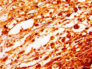
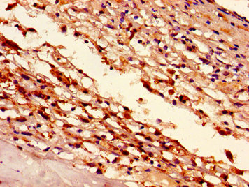
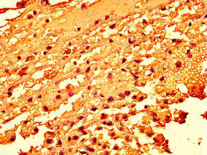
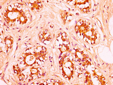
 IHC (Immunohiostchemistry)
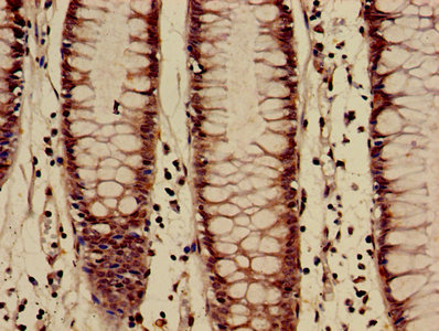
(IHC image of AAA234364 diluted at 1:60 and staining in paraffin-embedded human testis tissue performed on a Leica BondTM system. After dewaxing and hydration, antigen retrieval was mediated by high pressure in a citrate buffer (pH 6.0). Section was blocked with 10% normal goat serum 30min at RT. Then primary antibody (1% BSA) was incubated at 4 degree C overnight. The primary is detected by a biotinylated secondary antibody and visualized using an HRP conjugated SP system.)
IHC (Immunohiostchemistry)
(IHC image of AAA234364 diluted at 1:60 and staining in paraffin-embedded human testis tissue performed on a Leica BondTM system. After dewaxing and hydration, antigen retrieval was mediated by high pressure in a citrate buffer (pH 6.0). Section was blocked with 10% normal goat serum 30min at RT. Then primary antibody (1% BSA) was incubated at 4 degree C overnight. The primary is detected by a biotinylated secondary antibody and visualized using an HRP conjugated SP system.)
HIST1H1C, Polyclonal Antibody (Cat# AAA234364)
 IHC (Immunohistochemisry)
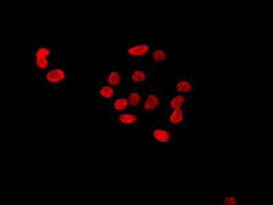
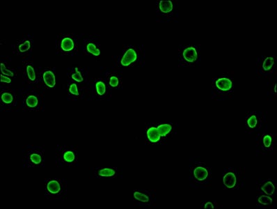
(Immunofluorescence staining of Hela cells with AAA234365 at 1:2.5, counter-stained with DAPI. The cells were fixed in 4% formaldehyde, permeabilized using 0.2% Triton X-100 and blocked in 10% normal Goat Serum. The cells were then incubated with the antibody overnight at 4 degree C.The secondary antibody was Alexa Fluor 488-congugated AffiniPure Goat Anti-Rabbit IgG (H+L).)
IHC (Immunohistochemisry)
(Immunofluorescence staining of Hela cells with AAA234365 at 1:2.5, counter-stained with DAPI. The cells were fixed in 4% formaldehyde, permeabilized using 0.2% Triton X-100 and blocked in 10% normal Goat Serum. The cells were then incubated with the antibody overnight at 4 degree C.The secondary antibody was Alexa Fluor 488-congugated AffiniPure Goat Anti-Rabbit IgG (H+L).)
H2AFZ, Polyclonal Antibody (Cat# AAA234365)
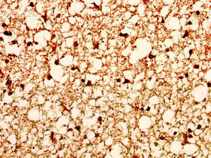
 IHC (Immunohiostchemistry)
(IHC image of AAA234366 diluted at 1:50 and staining in paraffin-embedded human glioma cancer performed on a Leica BondTM system. After dewaxing and hydration, antigen retrieval was mediated by high pressure in a citrate buffer (pH 6.0). Section was blocked with 10% normal goat serum 30min at RT. Then primary antibody (1% BSA) was incubated at 4 degree C overnight. The primary is detected by a biotinylated secondary antibody and visualized using an HRP conjugated SP system.)
IHC (Immunohiostchemistry)
(IHC image of AAA234366 diluted at 1:50 and staining in paraffin-embedded human glioma cancer performed on a Leica BondTM system. After dewaxing and hydration, antigen retrieval was mediated by high pressure in a citrate buffer (pH 6.0). Section was blocked with 10% normal goat serum 30min at RT. Then primary antibody (1% BSA) was incubated at 4 degree C overnight. The primary is detected by a biotinylated secondary antibody and visualized using an HRP conjugated SP system.)
HIST1H1C, Polyclonal Antibody (Cat# AAA234366)
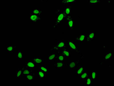
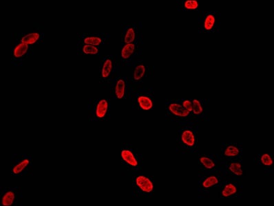
 IF (Immunofluorescence)
(Immunofluorescence staining of hela with AAA234369 at 1:12.5, counter-stained with DAPI. The cells were fixed in 4% formaldehyde, permeabilized using 0.2% Triton X-100 and blocked in 10% normal Goat Serum. The cells were then incubated with the antibody overnight at 4 degree C.The secondary antibody was Alexa Fluor 488-congugated AffiniPure Goat Anti-Rabbit IgG (H+L).)
IF (Immunofluorescence)
(Immunofluorescence staining of hela with AAA234369 at 1:12.5, counter-stained with DAPI. The cells were fixed in 4% formaldehyde, permeabilized using 0.2% Triton X-100 and blocked in 10% normal Goat Serum. The cells were then incubated with the antibody overnight at 4 degree C.The secondary antibody was Alexa Fluor 488-congugated AffiniPure Goat Anti-Rabbit IgG (H+L).)
HIST1H1C, Polyclonal Antibody (Cat# AAA234369)
 IF (Immunofluorescence)
(Immunofluorescence staining of hela with AAA234371, counter-stained with DAPI. The cells were fixed in 4% formaldehyde, permeabilized using 0.2% Triton X-100 and blocked in 10% normal Goat Serum. The cells were then incubated with the antibody overnight at 4 degree C.The secondary antibody was Alexa Fluor 488-congugated AffiniPure Goat Anti-Rabbit IgG (H+L).)
IF (Immunofluorescence)
(Immunofluorescence staining of hela with AAA234371, counter-stained with DAPI. The cells were fixed in 4% formaldehyde, permeabilized using 0.2% Triton X-100 and blocked in 10% normal Goat Serum. The cells were then incubated with the antibody overnight at 4 degree C.The secondary antibody was Alexa Fluor 488-congugated AffiniPure Goat Anti-Rabbit IgG (H+L).)
HIST1H2AG, Polyclonal Antibody (Cat# AAA234371)
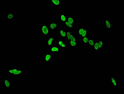
 IF (Immunofluorescence)
(Immunofluorescence staining of hela with AAA234372, counter-stained with DAPI. The cells were fixed in 4% formaldehyde, permeabilized using 0.2% Triton X-100 and blocked in 10% normal Goat Serum. The cells were then incubated with the antibody overnight at 4 degree C.The secondary antibody was Alexa Fluor 488-congugated AffiniPure Goat Anti-Rabbit IgG (H+L).)
IF (Immunofluorescence)
(Immunofluorescence staining of hela with AAA234372, counter-stained with DAPI. The cells were fixed in 4% formaldehyde, permeabilized using 0.2% Triton X-100 and blocked in 10% normal Goat Serum. The cells were then incubated with the antibody overnight at 4 degree C.The secondary antibody was Alexa Fluor 488-congugated AffiniPure Goat Anti-Rabbit IgG (H+L).)
HIST1H2AG, Polyclonal Antibody (Cat# AAA234372)
 IF (Immunofluorescence)
(Immunofluorescence staining of hela with AAA234377, counter-stained with DAPI. The cells were fixed in 4% formaldehyde, permeabilized using 0.2% Triton X-100 and blocked in 10% normal Goat Serum. The cells were then incubated with the antibody overnight at 4 degree C.The secondary antibody was Alexa Fluor 488-congugated AffiniPure Goat Anti-Rabbit IgG (H+L).)
IF (Immunofluorescence)
(Immunofluorescence staining of hela with AAA234377, counter-stained with DAPI. The cells were fixed in 4% formaldehyde, permeabilized using 0.2% Triton X-100 and blocked in 10% normal Goat Serum. The cells were then incubated with the antibody overnight at 4 degree C.The secondary antibody was Alexa Fluor 488-congugated AffiniPure Goat Anti-Rabbit IgG (H+L).)
HIST1H1C, Polyclonal Antibody (Cat# AAA234377)

 IHC (Immunohistochemisry)
(Immunofluorescence staining of hela with AAA234379, counter-stained with DAPI. The cells were fixed in 4% formaldehyde, permeabilized using 0.2% Triton X-100 and blocked in 10% normal Goat Serum. The cells were then incubated with the antibody overnight at 4 degree C.The secondary antibody was Alexa Fluor 488-congugated AffiniPure Goat Anti-Rabbit IgG (H+L).)
IHC (Immunohistochemisry)
(Immunofluorescence staining of hela with AAA234379, counter-stained with DAPI. The cells were fixed in 4% formaldehyde, permeabilized using 0.2% Triton X-100 and blocked in 10% normal Goat Serum. The cells were then incubated with the antibody overnight at 4 degree C.The secondary antibody was Alexa Fluor 488-congugated AffiniPure Goat Anti-Rabbit IgG (H+L).)
HIST1H2AG, Polyclonal Antibody (Cat# AAA234379)
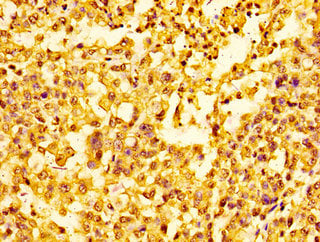
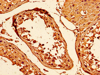
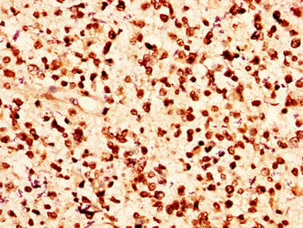
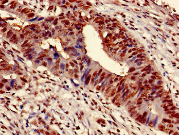
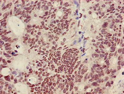
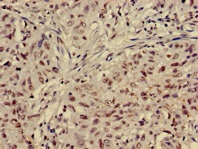
 IHC (Immunohiostchemistry)
(IHC image of AAA234381 diluted at 1:50 and staining in paraffin-embedded human melanoma cancer performed on a Leica BondTM system. After dewaxing and hydration, antigen retrieval was mediated by high pressure in a citrate buffer (pH 6.0). Section was blocked with 10% normal goat serum 30min at RT. Then primary antibody (1% BSA) was incubated at 4 degree C overnight. The primary is detected by a biotinylated secondary antibody and visualized using an HRP conjugated SP system.)
IHC (Immunohiostchemistry)
(IHC image of AAA234381 diluted at 1:50 and staining in paraffin-embedded human melanoma cancer performed on a Leica BondTM system. After dewaxing and hydration, antigen retrieval was mediated by high pressure in a citrate buffer (pH 6.0). Section was blocked with 10% normal goat serum 30min at RT. Then primary antibody (1% BSA) was incubated at 4 degree C overnight. The primary is detected by a biotinylated secondary antibody and visualized using an HRP conjugated SP system.)
HIST1H1C, Polyclonal Antibody (Cat# AAA234381)
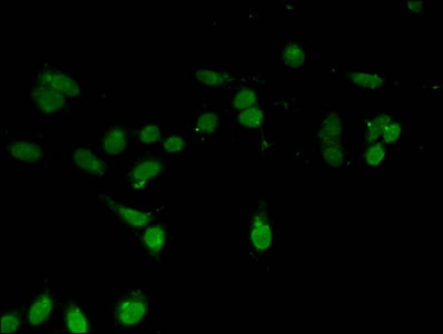
 IF (Immunofluorescence)
(Immunofluorescence staining of A549 cells with AAA234230 at 1:230, counter-stained with DAPI. The cells were fixed in 4% formaldehyde, permeabilized using 0.2% Triton X-100 and blocked in 10% normal Goat Serum. The cells were then incubated with the antibody overnight at 4 degree C.The secondary antibody was Alexa Fluor 488-congugated AffiniPure Goat Anti-Rabbit IgG (H+L).)
IF (Immunofluorescence)
(Immunofluorescence staining of A549 cells with AAA234230 at 1:230, counter-stained with DAPI. The cells were fixed in 4% formaldehyde, permeabilized using 0.2% Triton X-100 and blocked in 10% normal Goat Serum. The cells were then incubated with the antibody overnight at 4 degree C.The secondary antibody was Alexa Fluor 488-congugated AffiniPure Goat Anti-Rabbit IgG (H+L).)
RAMP2, Polyclonal Antibody (Cat# AAA234230)
 IHC (Immunohistochemisry)
(Immunofluorescence staining of U251 cells with AAA234233 at 1:110, counter-stained with DAPI. The cells were fixed in 4% formaldehyde, permeabilized using 0.2% Triton X-100 and blocked in 10% normal Goat Serum. The cells were then incubated with the antibody overnight at 4 degree C.The secondary antibody was Alexa Fluor 488-congugated AffiniPure Goat Anti-Rabbit IgG (H+L).)
IHC (Immunohistochemisry)
(Immunofluorescence staining of U251 cells with AAA234233 at 1:110, counter-stained with DAPI. The cells were fixed in 4% formaldehyde, permeabilized using 0.2% Triton X-100 and blocked in 10% normal Goat Serum. The cells were then incubated with the antibody overnight at 4 degree C.The secondary antibody was Alexa Fluor 488-congugated AffiniPure Goat Anti-Rabbit IgG (H+L).)
SLC25A24, Polyclonal Antibody (Cat# AAA234233)
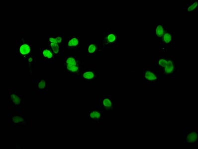
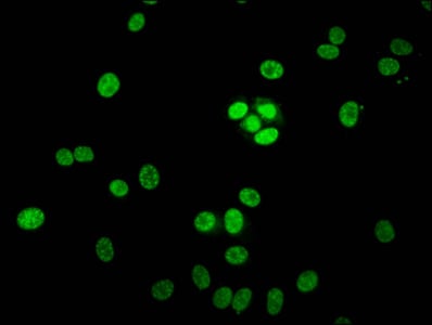
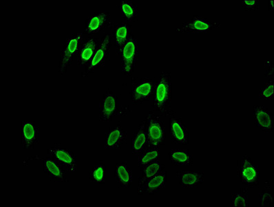
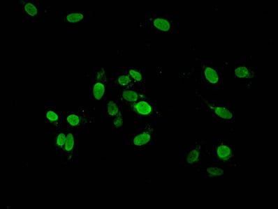
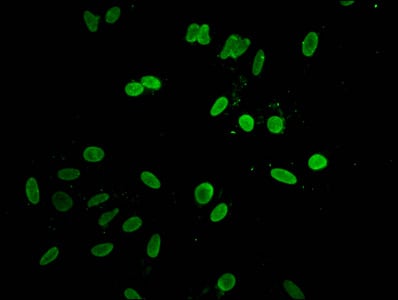
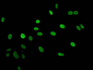
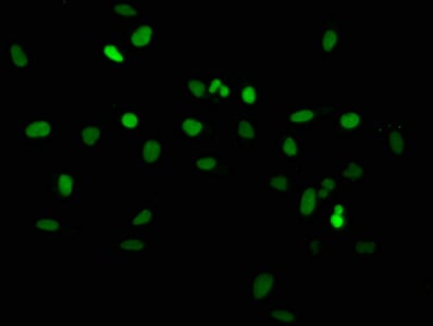
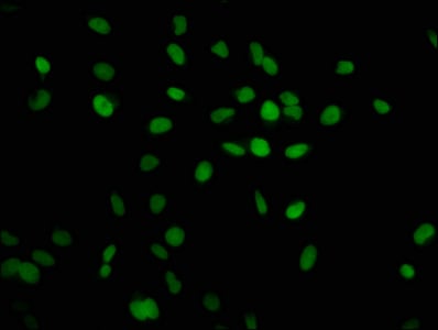
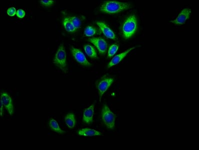
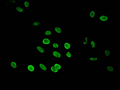
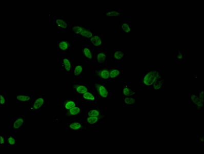
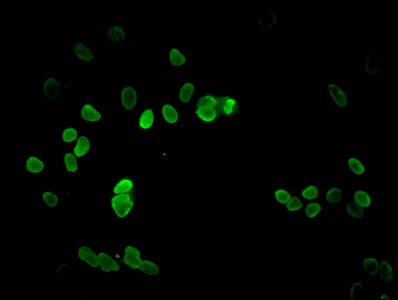
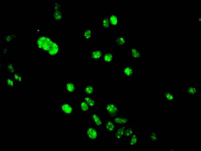
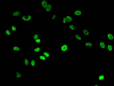
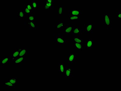 IF (Immunofluorescence)
(Immunofluorescent analysis of Hela cells at a dilution of 1:100 and Alexa Fluor 488-congugated AffiniPure Goat Anti-Rabbit IgG(H+L))
IF (Immunofluorescence)
(Immunofluorescent analysis of Hela cells at a dilution of 1:100 and Alexa Fluor 488-congugated AffiniPure Goat Anti-Rabbit IgG(H+L))
HIST1H3A, Polyclonal Antibody (Cat# AAA234239)

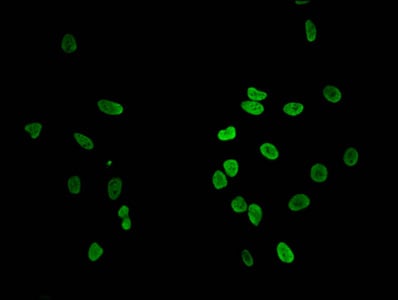
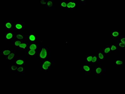
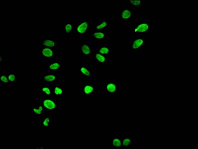
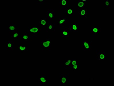
 IF (Immunofluorescence)
(Immunofluorescent analysis of Hela cells at a dilution of 1:100 and Alexa Fluor 488-congugated AffiniPure Goat Anti-Rabbit IgG(H+L))
IF (Immunofluorescence)
(Immunofluorescent analysis of Hela cells at a dilution of 1:100 and Alexa Fluor 488-congugated AffiniPure Goat Anti-Rabbit IgG(H+L))
HIST1H3A, Polyclonal Antibody (Cat# AAA234243)
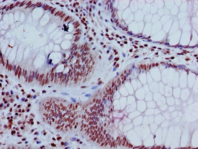
 IHC (Immunohistochemisry)
(Immunofluorescent analysis of Hela cells at a dilution of 1:100 and Alexa Fluor 488-congugated AffiniPure Goat Anti-Rabbit IgG(H+L))
IHC (Immunohistochemisry)
(Immunofluorescent analysis of Hela cells at a dilution of 1:100 and Alexa Fluor 488-congugated AffiniPure Goat Anti-Rabbit IgG(H+L))
HIST1H3A, Polyclonal Antibody (Cat# AAA234244)
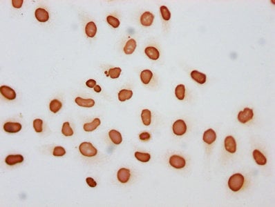
 IF (Immunofluorescence)
(Immunofluorescent analysis of Hela cells at a dilution of 1:100 and Alexa Fluor 488-congugated AffiniPure Goat Anti-Rabbit IgG(H+L))
IF (Immunofluorescence)
(Immunofluorescent analysis of Hela cells at a dilution of 1:100 and Alexa Fluor 488-congugated AffiniPure Goat Anti-Rabbit IgG(H+L))
HIST1H3A, Polyclonal Antibody (Cat# AAA234248)
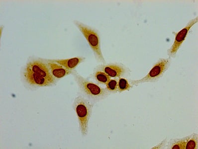
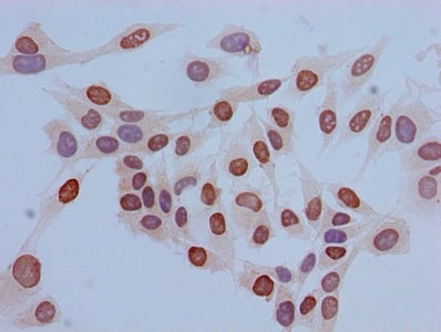
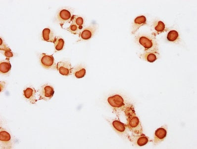
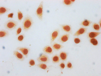
 ICC (Immunocytochemistry)
(Immunocytochemistry analysis of Hela cells using AAA234249 at dilution of 1:100)
ICC (Immunocytochemistry)
(Immunocytochemistry analysis of Hela cells using AAA234249 at dilution of 1:100)
Acetyl-HIST1H2BB, Polyclonal Antibody (Cat# AAA234249)
 IHC (Immunohistochemisry)
(Immunofluorescent analysis of Hela cells using AAA234250 at a dilution of 1:100 and Alexa Fluor 488-congugated AffiniPure Goat Anti-Rabbit IgG(H+L))
IHC (Immunohistochemisry)
(Immunofluorescent analysis of Hela cells using AAA234250 at a dilution of 1:100 and Alexa Fluor 488-congugated AffiniPure Goat Anti-Rabbit IgG(H+L))
HIST1H3A, Polyclonal Antibody (Cat# AAA234250)
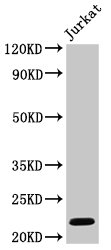
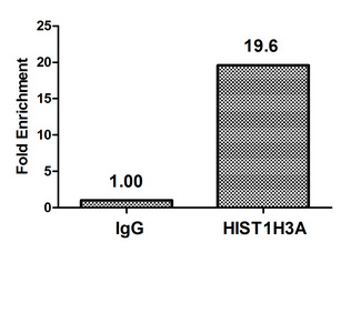
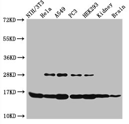
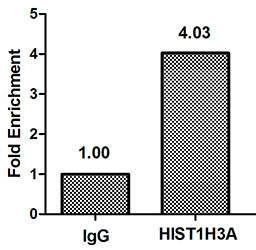
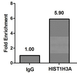
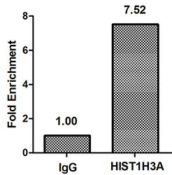
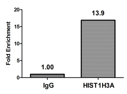 ChIP (Chromatin immunoprecipitation)
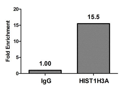
(Chromatin Immunoprecipitation Hela(4^106)were treated with Micrococcal Nuclease, sonicated, and immunoprecipitated with 8ug anti-HIST1H3A or a control normal rabbit IgG. The resulting ChIP DNA was quantified using real-time PCR with primers against the beta-Globin promoter.)
ChIP (Chromatin immunoprecipitation)
(Chromatin Immunoprecipitation Hela(4^106)were treated with Micrococcal Nuclease, sonicated, and immunoprecipitated with 8ug anti-HIST1H3A or a control normal rabbit IgG. The resulting ChIP DNA was quantified using real-time PCR with primers against the beta-Globin promoter.)
HIST1H3A, Polyclonal Antibody (Cat# AAA234257)
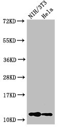
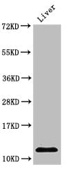
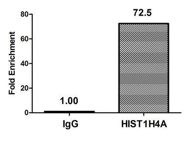
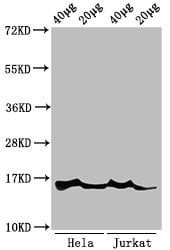
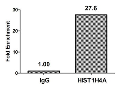
 ChIP (Chromatin Immunoprecipitation)
(Chromatin Immunoprecipitation Hela(4^106)were treated with Micrococcal Nuclease, sonicated, and immunoprecipitated with 8ug anti-HIST1H4A (AAA234259)or a control normal rabbit IgG. The resulting ChIP DNA was quantified using real-time PCR with primers against the beta-Globin promoter.)
ChIP (Chromatin Immunoprecipitation)
(Chromatin Immunoprecipitation Hela(4^106)were treated with Micrococcal Nuclease, sonicated, and immunoprecipitated with 8ug anti-HIST1H4A (AAA234259)or a control normal rabbit IgG. The resulting ChIP DNA was quantified using real-time PCR with primers against the beta-Globin promoter.)
HIST1H4A, Polyclonal Antibody (Cat# AAA234259)
 ChIP (Chromatin Immunoprecipitation)
(Chromatin Immunoprecipitation Hela(4^106)were treated with Micrococcal Nuclease, sonicated, and immunoprecipitated with 8ug anti-HIST1H3A or a control normal rabbit IgG. The resulting ChIP DNA was quantified using real-time PCR with primers against the beta-Globin promoter.)
ChIP (Chromatin Immunoprecipitation)
(Chromatin Immunoprecipitation Hela(4^106)were treated with Micrococcal Nuclease, sonicated, and immunoprecipitated with 8ug anti-HIST1H3A or a control normal rabbit IgG. The resulting ChIP DNA was quantified using real-time PCR with primers against the beta-Globin promoter.)
HIST1H3A, Polyclonal Antibody (Cat# AAA234260)
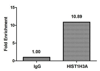
 ChIP (Chromatin immunoprecipitation)
(Chromatin Immunoprecipitation Hela(4^106)were treated with Micrococcal Nuclease, sonicated, and immunoprecipitated with 8ug anti-HIST1H3A (AAA234270)or a control normal rabbit IgG. The resulting ChIP DNA was quantified using real-time PCR with primers against the beta-Globin promoter.)
ChIP (Chromatin immunoprecipitation)
(Chromatin Immunoprecipitation Hela(4^106)were treated with Micrococcal Nuclease, sonicated, and immunoprecipitated with 8ug anti-HIST1H3A (AAA234270)or a control normal rabbit IgG. The resulting ChIP DNA was quantified using real-time PCR with primers against the beta-Globin promoter.)
HIST1H3A, Polyclonal Antibody (Cat# AAA234270)


 ChIP (Chromatin immunoprecipitation)
(Chromatin Immunoprecipitation Hela(4^106)were treated with Micrococcal Nuclease, sonicated, and immunoprecipitated with 8ug anti-HIST1H3A (AAA234272)or a control normal rabbit IgG. The resulting ChIP DNA was quantified using real-time PCR with primers against the beta-Globin promoter.)
ChIP (Chromatin immunoprecipitation)
(Chromatin Immunoprecipitation Hela(4^106)were treated with Micrococcal Nuclease, sonicated, and immunoprecipitated with 8ug anti-HIST1H3A (AAA234272)or a control normal rabbit IgG. The resulting ChIP DNA was quantified using real-time PCR with primers against the beta-Globin promoter.)
HIST1H3A, Polyclonal Antibody (Cat# AAA234272)
 IHC (Immunohistochemisry)
(Immunofluorescent analysis of Hela cells using AAA234274 at a dilution of 1:100 and Alexa Fluor 488-congugated AffiniPure Goat Anti-Rabbit IgG(H+L))
IHC (Immunohistochemisry)
(Immunofluorescent analysis of Hela cells using AAA234274 at a dilution of 1:100 and Alexa Fluor 488-congugated AffiniPure Goat Anti-Rabbit IgG(H+L))
HIST1H4A, Polyclonal Antibody (Cat# AAA234274)
 ChIP (Chromatin immunoprecipitation)
(Chromatin Immunoprecipitation Hela(4^106)were treated with Micrococcal Nuclease, sonicated, and immunoprecipitated with 8ug anti-HIST1H4A (AAA234277)or a control normal rabbit IgG. The resulting ChIP DNA was quantified using real-time PCR with primers against the beta-Globin promoter.)
ChIP (Chromatin immunoprecipitation)
(Chromatin Immunoprecipitation Hela(4^106)were treated with Micrococcal Nuclease, sonicated, and immunoprecipitated with 8ug anti-HIST1H4A (AAA234277)or a control normal rabbit IgG. The resulting ChIP DNA was quantified using real-time PCR with primers against the beta-Globin promoter.)
HIST1H4A, Polyclonal Antibody (Cat# AAA234277)
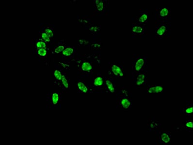
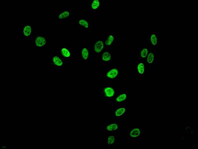
 IF (Immunofluorescence)
(Immunofluorescent analysis of Hela cells using AAA234280 at a dilution of 1:100 and Alexa Fluor 488-congugated AffiniPure Goat Anti-Rabbit IgG(H+L))
IF (Immunofluorescence)
(Immunofluorescent analysis of Hela cells using AAA234280 at a dilution of 1:100 and Alexa Fluor 488-congugated AffiniPure Goat Anti-Rabbit IgG(H+L))
Di-methyl-HIST1H3A, Polyclonal Antibody (Cat# AAA234280)
 IHC (Immunohiostchemistry)
(Immunofluorescent analysis of Hela cells using AAA234283 at a dilution of 1:100 and Alexa Fluor 488-congugated AffiniPure Goat Anti-Rabbit IgG(H+L))
IHC (Immunohiostchemistry)
(Immunofluorescent analysis of Hela cells using AAA234283 at a dilution of 1:100 and Alexa Fluor 488-congugated AffiniPure Goat Anti-Rabbit IgG(H+L))
HIST1H3A, Polyclonal Antibody (Cat# AAA234283)
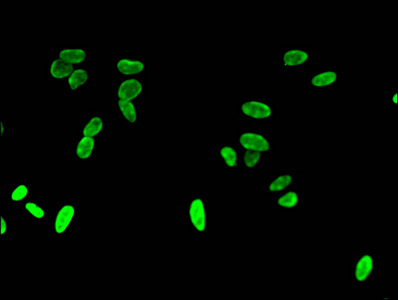
 IF (Immunofluorescence)
(Immunofluorescent analysis of Hela cells using AAA234285 at a dilution of 1:100 and Alexa Fluor 488-congugated AffiniPure Goat Anti-Rabbit IgG(H+L))
IF (Immunofluorescence)
(Immunofluorescent analysis of Hela cells using AAA234285 at a dilution of 1:100 and Alexa Fluor 488-congugated AffiniPure Goat Anti-Rabbit IgG(H+L))
HIST1H4A, Polyclonal Antibody (Cat# AAA234285)
 IHC (Immunohistochemisry)
(Immunofluorescent analysis of Hela cells using AAA234287 at a dilution of 1:100 and Alexa Fluor 488-congugated AffiniPure Goat Anti-Rabbit IgG(H+L))
IHC (Immunohistochemisry)
(Immunofluorescent analysis of Hela cells using AAA234287 at a dilution of 1:100 and Alexa Fluor 488-congugated AffiniPure Goat Anti-Rabbit IgG(H+L))
HIST1H3A, Polyclonal Antibody (Cat# AAA234287)
 ChIP (Chromatin Immunoprecipitation)
(Chromatin Immunoprecipitation Hela(4^106)were treated with Micrococcal Nuclease, sonicated, and immunoprecipitated with 8ug anti-HIST1H3A (AAA234288)or a control normal rabbit IgG. The resulting ChIP DNA was quantified using real-time PCR with primers against the beta-Globin promoter.)
ChIP (Chromatin Immunoprecipitation)
(Chromatin Immunoprecipitation Hela(4^106)were treated with Micrococcal Nuclease, sonicated, and immunoprecipitated with 8ug anti-HIST1H3A (AAA234288)or a control normal rabbit IgG. The resulting ChIP DNA was quantified using real-time PCR with primers against the beta-Globin promoter.)
HIST1H3A, Polyclonal Antibody (Cat# AAA234288)
 IHC (Immunohistochemisry)
(Immunofluorescent analysis of Hela cells using AAA234291 at a dilution of 1:100 and Alexa Fluor 488-congugated AffiniPure Goat Anti-Rabbit IgG(H+L))
IHC (Immunohistochemisry)
(Immunofluorescent analysis of Hela cells using AAA234291 at a dilution of 1:100 and Alexa Fluor 488-congugated AffiniPure Goat Anti-Rabbit IgG(H+L))
HIST1H3A, Polyclonal Antibody (Cat# AAA234291)
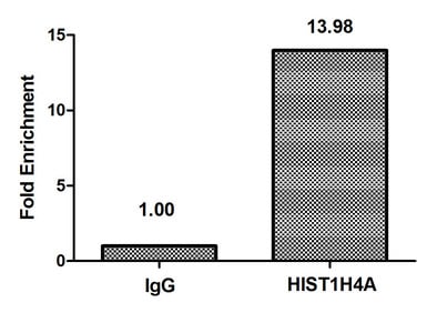
 ChIP (Chromatin Immunoprecipitation)
(Chromatin Immunoprecipitation Hela(4^106)were treated with Micrococcal Nuclease, sonicated, and immunoprecipitated with 8ug anti-HIST1H4A (AAA234293)or a control normal rabbit IgG. The resulting ChIP DNA was quantified using real-time PCR with primers against the beta-Globin promoter.)
ChIP (Chromatin Immunoprecipitation)
(Chromatin Immunoprecipitation Hela(4^106)were treated with Micrococcal Nuclease, sonicated, and immunoprecipitated with 8ug anti-HIST1H4A (AAA234293)or a control normal rabbit IgG. The resulting ChIP DNA was quantified using real-time PCR with primers against the beta-Globin promoter.)
Acetyl-HIST1H4A, Polyclonal Antibody (Cat# AAA234293)
 ChIP (Chromatin immunoprecipitation)
(Chromatin Immunoprecipitation Hela(4^106)were treated with Micrococcal Nuclease, sonicated, and immunoprecipitated with 8ug anti-HIST1H3A (AAA234296)or a control normal rabbit IgG. The resulting ChIP DNA was quantified using real-time PCR with primers against the beta-Globin promoter.)
ChIP (Chromatin immunoprecipitation)
(Chromatin Immunoprecipitation Hela(4^106)were treated with Micrococcal Nuclease, sonicated, and immunoprecipitated with 8ug anti-HIST1H3A (AAA234296)or a control normal rabbit IgG. The resulting ChIP DNA was quantified using real-time PCR with primers against the beta-Globin promoter.)
HIST1H3A, Polyclonal Antibody (Cat# AAA234296)
 ChIP (Chromatin Immunoprecipitation)
(Chromatin Immunoprecipitation Hela(4^106)were treated with Micrococcal Nuclease, sonicated, and immunoprecipitated with 8ug anti-HIST1H3A (AAA234304)or a control normal rabbit IgG. The resulting ChIP DNA was quantified using real-time PCR with primers against the beta-Globin promoter.)
ChIP (Chromatin Immunoprecipitation)
(Chromatin Immunoprecipitation Hela(4^106)were treated with Micrococcal Nuclease, sonicated, and immunoprecipitated with 8ug anti-HIST1H3A (AAA234304)or a control normal rabbit IgG. The resulting ChIP DNA was quantified using real-time PCR with primers against the beta-Globin promoter.)
HIST1H3A, Polyclonal Antibody (Cat# AAA234304)
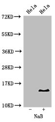
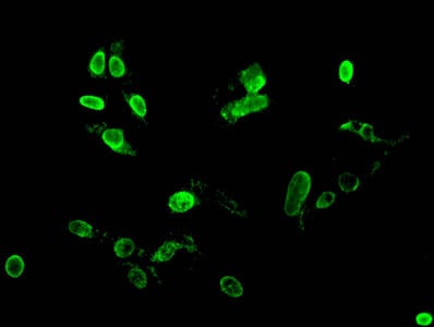
 ICC (Immunocytochemistry)
(Immunocytochemistry analysis of AAA235115 diluted at 1:100 and staining in Hela cells(treated with 30mM sodium crotonylate for 4h) performed on a Leica BondTM system. The cells were fixed in 4% formaldehyde, permeabilized using 0.2% Triton X-100 and blocked with 10% normal goat serum 30min at RT. Then primary antibody (1% BSA) was incubated at 4 degree C overnight. The primary is detected by a biotinylated secondary antibody and visualized using an HRP conjugated SP system.)
ICC (Immunocytochemistry)
(Immunocytochemistry analysis of AAA235115 diluted at 1:100 and staining in Hela cells(treated with 30mM sodium crotonylate for 4h) performed on a Leica BondTM system. The cells were fixed in 4% formaldehyde, permeabilized using 0.2% Triton X-100 and blocked with 10% normal goat serum 30min at RT. Then primary antibody (1% BSA) was incubated at 4 degree C overnight. The primary is detected by a biotinylated secondary antibody and visualized using an HRP conjugated SP system.)
HIST1H3A, Polyclonal Antibody (Cat# AAA235115)
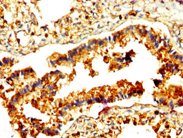
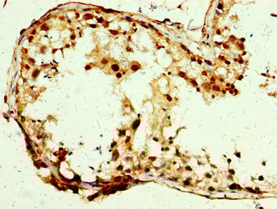
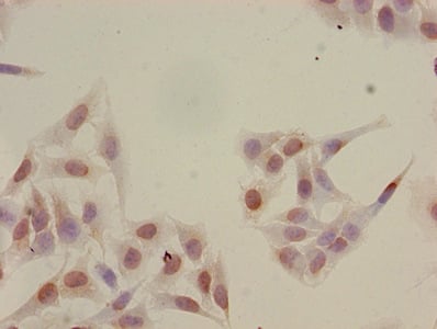
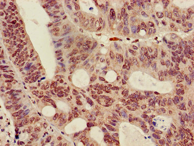
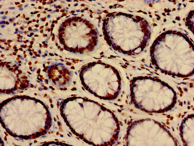
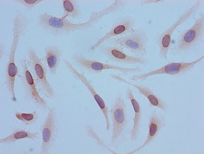
 IHC (Immunohistochemisry)
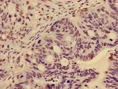
(IHC image of AAA235118 diluted at 1:5 and staining in paraffin-embedded human prostate cancer performed on a Leica BondTM system. After dewaxing and hydration, antigen retrieval was mediated by high pressure in a citrate buffer (pH 6.0). Section was blocked with 10% normal goat serum 30min at RT. Then primary antibody (1% BSA) was incubated at 4 degree C overnight. The primary is detected by a biotinylated secondary antibody and visualized using an HRP conjugated SP system.)
IHC (Immunohistochemisry)
(IHC image of AAA235118 diluted at 1:5 and staining in paraffin-embedded human prostate cancer performed on a Leica BondTM system. After dewaxing and hydration, antigen retrieval was mediated by high pressure in a citrate buffer (pH 6.0). Section was blocked with 10% normal goat serum 30min at RT. Then primary antibody (1% BSA) was incubated at 4 degree C overnight. The primary is detected by a biotinylated secondary antibody and visualized using an HRP conjugated SP system.)
HIST1H3A, Polyclonal Antibody (Cat# AAA235118)
 ICC (Immunocytochemistry)
(Immunocytochemistry analysis of AAA235121 diluted at 1:50 and staining in Hela cells(treated with 10mM sodium propionate for 4h) performed on a Leica BondTM system. The cells were fixed in 4% formaldehyde, permeabilized using 0.2% Triton X-100 and blocked with 10% normal goat serum 30min at RT. Then primary antibody (1% BSA) was incubated at 4 degree C overnight. The primary is detected by a biotinylated secondary antibody and visualized using an HRP conjugated SP system.)
ICC (Immunocytochemistry)
(Immunocytochemistry analysis of AAA235121 diluted at 1:50 and staining in Hela cells(treated with 10mM sodium propionate for 4h) performed on a Leica BondTM system. The cells were fixed in 4% formaldehyde, permeabilized using 0.2% Triton X-100 and blocked with 10% normal goat serum 30min at RT. Then primary antibody (1% BSA) was incubated at 4 degree C overnight. The primary is detected by a biotinylated secondary antibody and visualized using an HRP conjugated SP system.)
HIST1H3A, Polyclonal Antibody (Cat# AAA235121)
 ICC (Immunocytochemistry)
(Immunocytochemistry analysis of AAA235129 diluted at 1:10 and staining in Hela cells(treated with 50mM sodium 3-hydroxybutyrate for 72h) performed on a Leica BondTM system. The cells were fixed in 4% formaldehyde, permeabilized using 0.2% Triton X-100 and blocked with 10% normal goat serum 30min at RT. Then primary antibody (1% BSA) was incubated at 4 degree C overnight. The primary is detected by a biotinylated secondary antibody and visualized using an HRP conjugated SP system.)
ICC (Immunocytochemistry)
(Immunocytochemistry analysis of AAA235129 diluted at 1:10 and staining in Hela cells(treated with 50mM sodium 3-hydroxybutyrate for 72h) performed on a Leica BondTM system. The cells were fixed in 4% formaldehyde, permeabilized using 0.2% Triton X-100 and blocked with 10% normal goat serum 30min at RT. Then primary antibody (1% BSA) was incubated at 4 degree C overnight. The primary is detected by a biotinylated secondary antibody and visualized using an HRP conjugated SP system.)
HIST1H3A, Polyclonal Antibody (Cat# AAA235129)
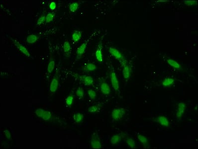
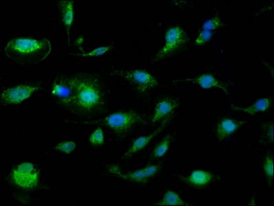
 IHC (Immunohistochemisry)
(Immunofluorescence staining of Hela cells(treated with 30mM sodium butyrate for 4h) with AAA235131 at 1:15, counter-stained with DAPI. The cells were fixed in 4% formaldehyde, permeabilized using 0.2% Triton X-100 and blocked in 10% normal Goat Serum. The cells were then incubated with the antibody overnight at 4 degree C.The secondary antibody was Alexa Fluor 488-congugated AffiniPure Goat Anti-Rabbit IgG (H+L).)
IHC (Immunohistochemisry)
(Immunofluorescence staining of Hela cells(treated with 30mM sodium butyrate for 4h) with AAA235131 at 1:15, counter-stained with DAPI. The cells were fixed in 4% formaldehyde, permeabilized using 0.2% Triton X-100 and blocked in 10% normal Goat Serum. The cells were then incubated with the antibody overnight at 4 degree C.The secondary antibody was Alexa Fluor 488-congugated AffiniPure Goat Anti-Rabbit IgG (H+L).)
HIST1H3A, Polyclonal Antibody (Cat# AAA235131)
What are Polyclonal Antibodies?
Polyclonal antibodies are antibodies that come from multiple B cell clones of a host animal. The typical hosts used for the majority of polyclonal antibody production are rabbits, goats, sheep, and donkeys. These polyclonal antibodies, once having identified their target, will bind to different epitopes located at different regions or sequences on the same protein/antigen. As a result, they are ideal at locating and binding to the target, even if the target is in very low concentrations (due to many different antibodies being able to bind to the same target molecule, which allows for significant amplification of a downstream signal).
Polyclonal antibodies are typically produced by injecting an antigen into a host animal, which causes the animal’s immune system to attack the foreign antigen by mass generating antibodies against it. After a period of time, serum is collected from the animal and purified using physicochemical fractionation, class-specific affinity purification, and/or antigen-affinity purification.
Key Uses of Polyclonal Antibodies
- Western Blotting: This method is used to find specific proteins in biological samples after separating them by size.
- Immunohistochemistry: IHC helps visualize the location of proteins in tissue sections using various staining techniques.
- ELISA: (Enzyme-Linked Immunosorbent Assay) is typically used to identify specific protein quantities in a sample. ELISAs can be either “Quantitative” or “Qualitative”.
- Flow Cytometry: technique that identifies and measures the specific protein on the surface or inside the cells in a fluid suspension.
- Immunoprecipitation: IP isolates and studies a specific protein from a complex mixture using antibodies.
Why Buy Polyclonal Antibodies from AAA Biotech?
1. Ideal for Various Applications
Our antibodies are generally going to be validated for use in multiple types of assays, including ELISA, Western Blotting, Immunohistochemistry, Immunoprecipitation, amongst others. They are ideal for a wide range of research applications.
2. Rigorous Quality Control
All of the antibodies in our catalog undergo strict quality testing to ensure specificity, sensitivity, and consistent performance. We are confident in the ability of our antibodies to provide you with accurate results.
3. Wide Assortment of Antibodies
Antibodies in are catalog can be found for both common and exotic species, and these antibodies are also available in both conjugated and recombinant forms to suit many diverse experimental needs.
4. Highly Purified
Our antibodies are available in purified forms with over 85% purity, as confirmed by SDS-PAGE. They are also available with tags such as His, Flag, GST, or MBP. We cater to customers worldwide.
FAQ
1. How are polyclonal antibodies produced?
Traditionally, polyclonal antibodies are produced by injecting an antigen into a host animal (such as a rabbit or goat), which then triggers an immune response from the host animal. The animal’s B cells produce antibodies that will recognize different parts of the injected antigen. These antibodies are then collected from the animal’s blood and purified for use.
2. How do polyclonal antibodies differ from monoclonal antibodies?
Polyclonal antibodies are a mix of antibodies that bind to different locations (epitopes) of the same antigen, while monoclonal antibodies are identical and bind to just one specific epitope. This makes polyclonal antibodies more versatile and better at detecting proteins that may be present in low quantities or in altered/modified forms.
3. How should I store polyclonal antibodies?
Polyclonal antibodies should be stored at 4°C for short-term use (up to a few weeks) and at -20°C or -80°C for long-term storage. Avoid repeated freeze-thaw cycles by dividing them into small aliquots. Always check the datasheet for specific storage instructions.