Filters
▼Clonality
▼Type
▼Reactivity
▼Gene Name
▼Isotype
▼Host
▼Application
▼Clone
▼Polyclonal Antibodies
At AAA Biotech also known as AAA Bio or AAABio, we provide a broad range of purified polyclonal antibodies (pAbs) that are able to all be browsed online through our website. Due to their high specificity and strong binding affinity, these antibodies are ideal for wide swathes of research and experimental applications.
Our polyclonal antibodies can easily support your work, whether you use them for Western Blotting, Immunocytochemistry (with or without Immunofluorescence used in conjunction), Immunohistochemistry, Immunoprecipitation, and ELISA tests. We highly encourage you to browse our range of pAbs and choose the one that best suits your experimental model.
Viewing 1950-2000 of 96805 product results
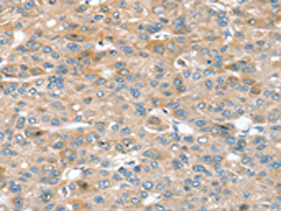
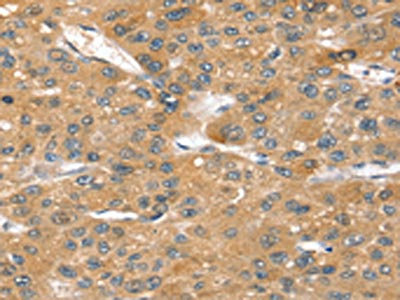
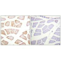
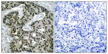
 IHC (Immunohiostchemistry)
(The image on the left is immunohistochemistry of paraffin-embedded Human esophagus cancer tissue using AAA241764(SIK1 Antibody) at dilution 1/30, on the right is treated with synthetic peptide. (Original magnification: ×200))
IHC (Immunohiostchemistry)
(The image on the left is immunohistochemistry of paraffin-embedded Human esophagus cancer tissue using AAA241764(SIK1 Antibody) at dilution 1/30, on the right is treated with synthetic peptide. (Original magnification: ×200))
SIK1, Polyclonal Antibody (Cat# AAA241764)
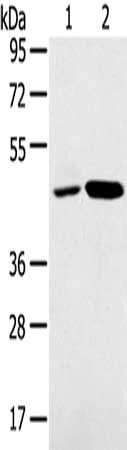
 SDS-PAGE
(Gel: 6%SDS-PAGE, Lysate: 40 ug, Lane 1-5: HT29 cells, hela cells, mouse liver tissue, A549 cells, Jurkat cells, Primary antibody: AAA241765(SIK1 Antibody) at dilution 1/250, Secondary antibody: Goat anti rabbit IgG at 1/8000 dilution, Exposure time: 10 seconds)
SDS-PAGE
(Gel: 6%SDS-PAGE, Lysate: 40 ug, Lane 1-5: HT29 cells, hela cells, mouse liver tissue, A549 cells, Jurkat cells, Primary antibody: AAA241765(SIK1 Antibody) at dilution 1/250, Secondary antibody: Goat anti rabbit IgG at 1/8000 dilution, Exposure time: 10 seconds)
SIK1, Polyclonal Antibody (Cat# AAA241765)
 SDS-PAGE
(Gel: 8%SDS-PAGE, Lysate: 40 ug, Lane 1-2: K562 cells, Jurkat cells, Primary antibody: AAA241771(SNX5 Antibody) at dilution 1/250, Secondary antibody: Goat anti rabbit IgG at 1/8000 dilution, Exposure time: 1 minute)
SDS-PAGE
(Gel: 8%SDS-PAGE, Lysate: 40 ug, Lane 1-2: K562 cells, Jurkat cells, Primary antibody: AAA241771(SNX5 Antibody) at dilution 1/250, Secondary antibody: Goat anti rabbit IgG at 1/8000 dilution, Exposure time: 1 minute)
SNX5, Polyclonal Antibody (Cat# AAA241771)
 SDS-PAGE
(Gel: 6%SDS-PAGE, Lysate: 40 ug, Lane 1-5: Mouse brain tissue, human brain tissue, human prostate tissue, human fat tissue, Human placenta tissue, Primary antibody: AAA241780(SORT1 Antibody) at dilution 1/200, Secondary antibody: Goat anti rabbit IgG at 1/8000 dilution, Exposure time: 40 seconds)
SDS-PAGE
(Gel: 6%SDS-PAGE, Lysate: 40 ug, Lane 1-5: Mouse brain tissue, human brain tissue, human prostate tissue, human fat tissue, Human placenta tissue, Primary antibody: AAA241780(SORT1 Antibody) at dilution 1/200, Secondary antibody: Goat anti rabbit IgG at 1/8000 dilution, Exposure time: 40 seconds)
SORT1, Polyclonal Antibody (Cat# AAA241780)
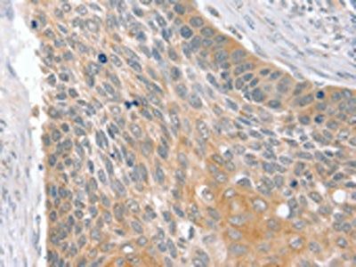
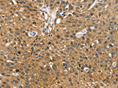
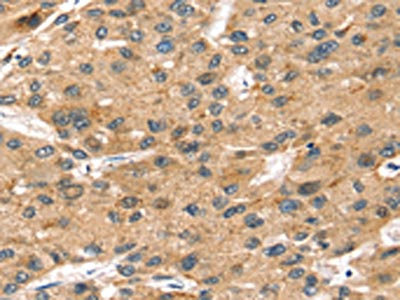
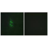
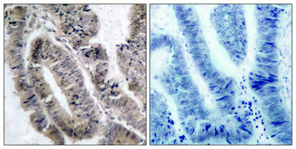
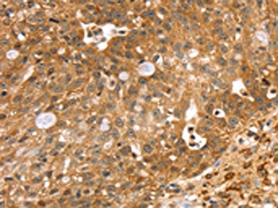 IHC (Immunohiostchemistry)
(The image on the left is immunohistochemistry of paraffin-embedded Human breast cancer tissue using AAA241781(SORT1 Antibody) at dilution 1/40, on the right is treated with synthetic peptide. (Original magnification: ×200))
IHC (Immunohiostchemistry)
(The image on the left is immunohistochemistry of paraffin-embedded Human breast cancer tissue using AAA241781(SORT1 Antibody) at dilution 1/40, on the right is treated with synthetic peptide. (Original magnification: ×200))
SORT1, Polyclonal Antibody (Cat# AAA241781)
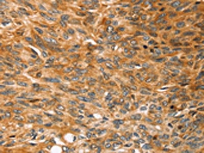
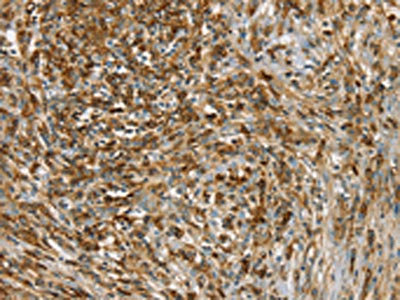
 IHC (Immunohiostchemistry)
(The image on the left is immunohistochemistry of paraffin-embedded Human breast cancer tissue using AAA241783(SOX11 Antibody) at dilution 1/20, on the right is treated with synthetic peptide. (Original magnification: ×200))
IHC (Immunohiostchemistry)
(The image on the left is immunohistochemistry of paraffin-embedded Human breast cancer tissue using AAA241783(SOX11 Antibody) at dilution 1/20, on the right is treated with synthetic peptide. (Original magnification: ×200))
SOX11, Polyclonal Antibody (Cat# AAA241783)
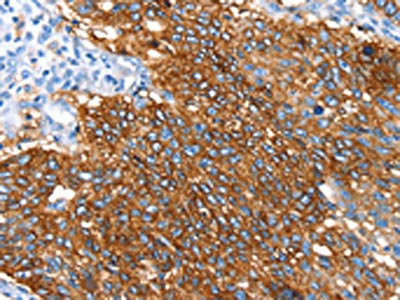
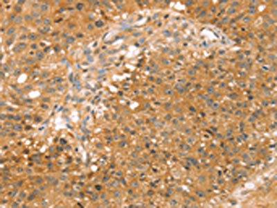
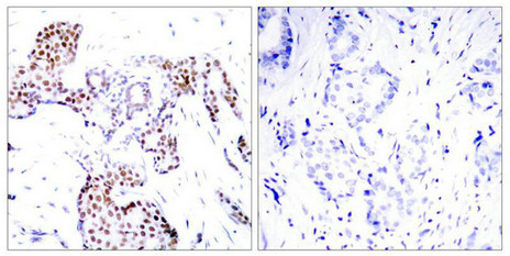
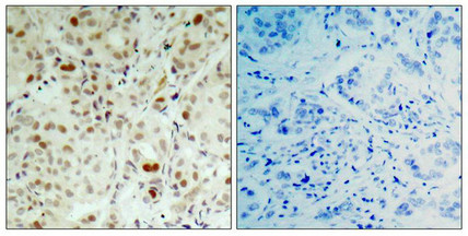
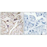
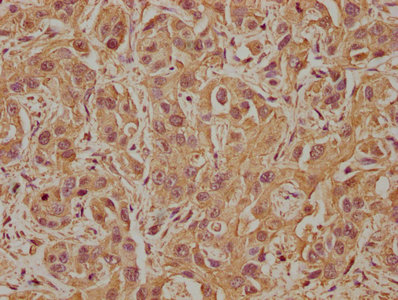
 IHC (Immunohiostchemistry)
(Immunohistochemical analysis of paraffin-embedded human breast carcinoma tissue using Raf1(Phospho-Ser259) Antibody.)
IHC (Immunohiostchemistry)
(Immunohistochemical analysis of paraffin-embedded human breast carcinoma tissue using Raf1(Phospho-Ser259) Antibody.)
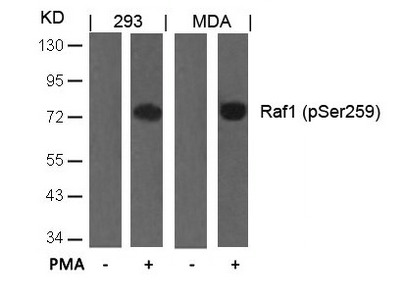
RAF1, Polyclonal Antibody (Cat# AAA243041)
 IHC (Immunohiostchemistry)
(Immunohistochemical analysis of paraffin-embedded human breast carcinoma tissue using Elk1(Phospho-Ser389) Antibody(left) or the same antibody preincubated with blocking peptide(right).)
IHC (Immunohiostchemistry)
(Immunohistochemical analysis of paraffin-embedded human breast carcinoma tissue using Elk1(Phospho-Ser389) Antibody(left) or the same antibody preincubated with blocking peptide(right).)
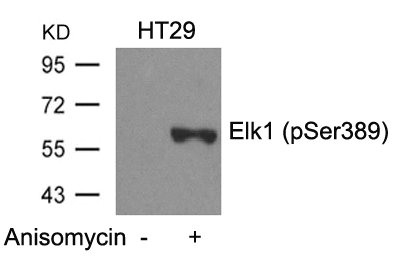
ELK1, Polyclonal Antibody (Cat# AAA243053)
 IF (Immunofluorescence)
(Immunofluorescence staining of methanol-fixed Hela cells using Caveolin-1(Phospho-Tyr14) Antibody.)
IF (Immunofluorescence)
(Immunofluorescence staining of methanol-fixed Hela cells using Caveolin-1(Phospho-Tyr14) Antibody.)
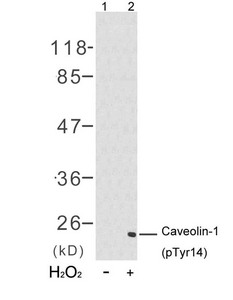
CAV1, Polyclonal Antibody (Cat# AAA243074)
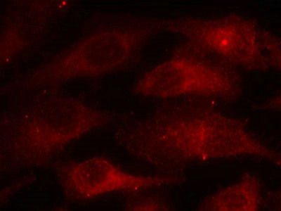
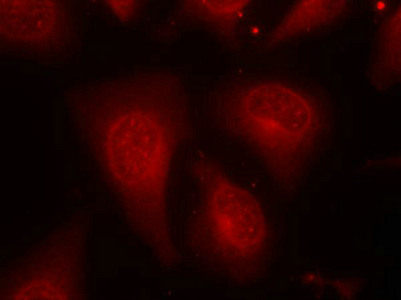
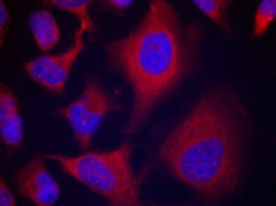
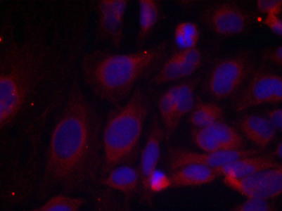
 IF (Immunofluorescence)
(Immunofluorescence staining of methanol-fixed Hela cells using p53(Phospho-Ser37) Antibody.)
IF (Immunofluorescence)
(Immunofluorescence staining of methanol-fixed Hela cells using p53(Phospho-Ser37) Antibody.)
TP53, Polyclonal Antibody (Cat# AAA243077)
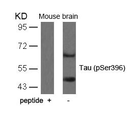
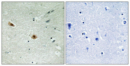
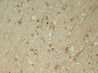 IHC (Immunohiostchemistry)
(Immunohistochemical analysis of paraffin-embedded rat hippocampal region tissue from a model with Alzheimer)
IHC (Immunohiostchemistry)
(Immunohistochemical analysis of paraffin-embedded rat hippocampal region tissue from a model with Alzheimer)
MAPT, Polyclonal Antibody (Cat# AAA243079)
 IF (Immunofluorescence)
(Immunofluorescence staining of methanol-fixed Hela cells using HDAC8(Phospho-Ser39) Antibody.)
IF (Immunofluorescence)
(Immunofluorescence staining of methanol-fixed Hela cells using HDAC8(Phospho-Ser39) Antibody.)
HDAC8, Polyclonal Antibody (Cat# AAA243089)
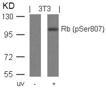
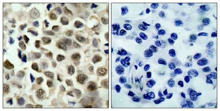
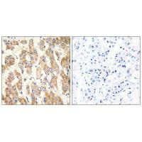
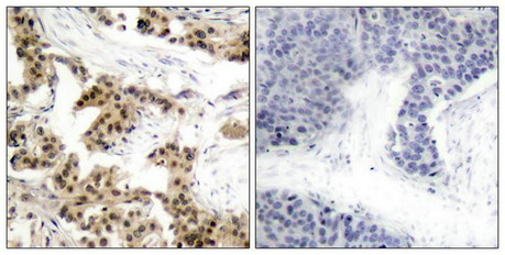
 IHC (Immunohiostchemistry)
(Immunohistochemical analysis of paraffin-embedded human breast carcinoma tissue using Rb(Phospho-Ser807) Antibody(left) or the same antibody preincubated with blocking peptide(right).)
IHC (Immunohiostchemistry)
(Immunohistochemical analysis of paraffin-embedded human breast carcinoma tissue using Rb(Phospho-Ser807) Antibody(left) or the same antibody preincubated with blocking peptide(right).)
RB1, Polyclonal Antibody (Cat# AAA243090)
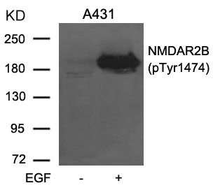 WB (Western Blot)
(Western blot analysis of extracts from A431 cells untreated or treated with EGF using NMDAR2B (phospho-Tyr1474) Antibody.)
WB (Western Blot)
(Western blot analysis of extracts from A431 cells untreated or treated with EGF using NMDAR2B (phospho-Tyr1474) Antibody.)
GRIN2B, Polyclonal Antibody (Cat# AAA243102)
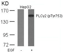
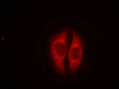
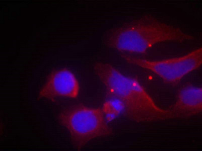
 IF (Immunofluorescence)
(Immunofluorescence staining of methanol-fixed Hela cells using PLCg2(Phospho-Tyr753) Antibody.)
IF (Immunofluorescence)
(Immunofluorescence staining of methanol-fixed Hela cells using PLCg2(Phospho-Tyr753) Antibody.)
PLCG2, Polyclonal Antibody (Cat# AAA243104)
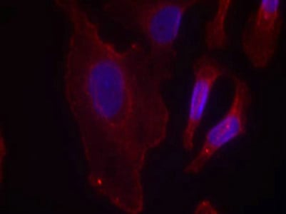
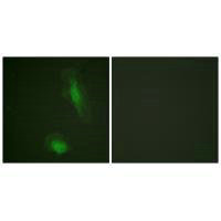
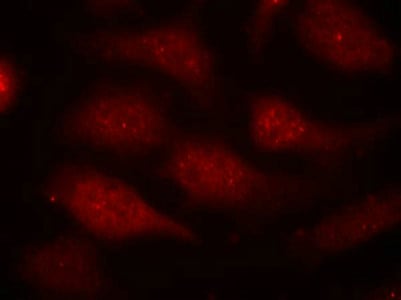
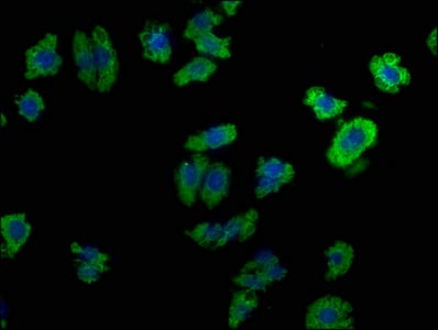
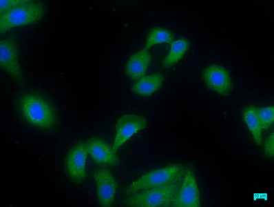
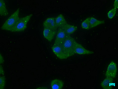
 IF (Immunofluorescence)
(Immunofluorescence analysis of HepG2 cells, using MRC2 antibody.)
IF (Immunofluorescence)
(Immunofluorescence analysis of HepG2 cells, using MRC2 antibody.)
MRC2, Polyclonal Antibody (Cat# AAA242963)
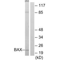
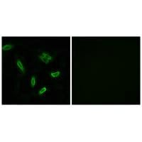
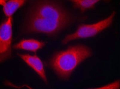
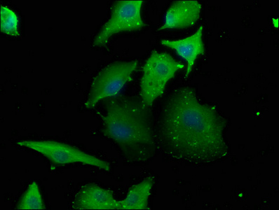
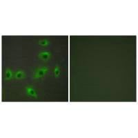 IF (Immunofluorescence)
(Immunofluorescence analysis of HUVEC cells, using BAX antibody.)
IF (Immunofluorescence)
(Immunofluorescence analysis of HUVEC cells, using BAX antibody.)
BAX, Polyclonal Antibody (Cat# AAA242976)
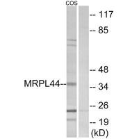
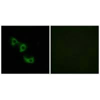 IF (Immunofluorescence)
(Immunofluorescence analysis of HuvEc cells, using MRPL44 antibody.)
IF (Immunofluorescence)
(Immunofluorescence analysis of HuvEc cells, using MRPL44 antibody.)
MRPL44, Polyclonal Antibody (Cat# AAA242984)
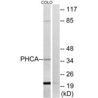
 IF (Immunofluorescence)
(Immunofluorescence analysis of MCF-7 cells, using PHCA antibody.)
IF (Immunofluorescence)
(Immunofluorescence analysis of MCF-7 cells, using PHCA antibody.)
ACER3, Polyclonal Antibody (Cat# AAA242986)
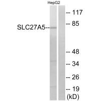
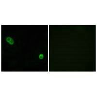
 IF (Immunofluorescence)
(Immunofluorescence analysis of A549 cells, using SLC27A5 antibody.)
IF (Immunofluorescence)
(Immunofluorescence analysis of A549 cells, using SLC27A5 antibody.)
SLC27A5, Polyclonal Antibody (Cat# AAA242990)
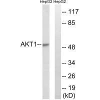
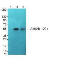 WB (Western Blot)
(Western blot analysis of extracts from HuvEc cells (Lane 2) and JK cells (Lane 3), using Akt (Ab-129) antiobdy. The lane on the left is treated with synthesized peptide.)
WB (Western Blot)
(Western blot analysis of extracts from HuvEc cells (Lane 2) and JK cells (Lane 3), using Akt (Ab-129) antiobdy. The lane on the left is treated with synthesized peptide.)
AKT1, Polyclonal Antibody (Cat# AAA243003)
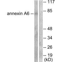
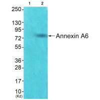 WB (Western Blot)
(Western blot analysis of extracts from HepG2 cells using Annexin A6 antibody. The lane on the left is treated with synthesized peptide.)
WB (Western Blot)
(Western blot analysis of extracts from HepG2 cells using Annexin A6 antibody. The lane on the left is treated with synthesized peptide.)
ANXA6, Polyclonal Antibody (Cat# AAA243007)
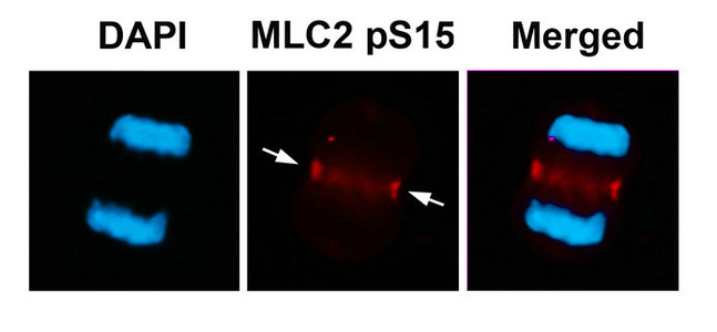
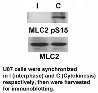 WB (Western Blot)
MYL9, Polyclonal Antibody (Cat# AAA243038)


 IF (Immunofluorescence)
(Immunofluorescence staining of methanol-fixed Hela cells using CDK6(phospho-Tyr24) Antibody.)
IF (Immunofluorescence)
(Immunofluorescence staining of methanol-fixed Hela cells using CDK6(phospho-Tyr24) Antibody.)
CDK6, Polyclonal Antibody (Cat# AAA243328)
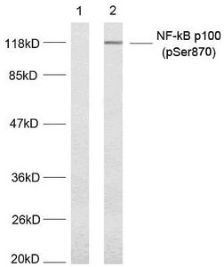 WB (Western Blot)
(Western blot analysis of extract from MDA-MB-435 cells untreated or treated with TNF-alpha; (20ng/ml, 5min) using NF-κB p100(phospho-Ser870) antibody.)
WB (Western Blot)
(Western blot analysis of extract from MDA-MB-435 cells untreated or treated with TNF-alpha; (20ng/ml, 5min) using NF-κB p100(phospho-Ser870) antibody.)
NFKB2, Polyclonal Antibody (Cat# AAA243335)
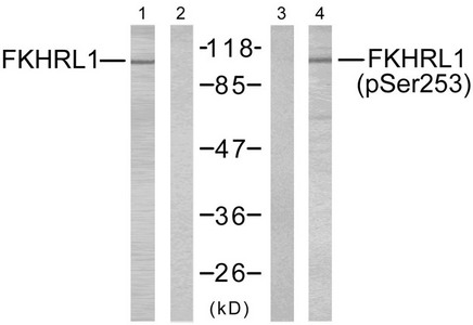
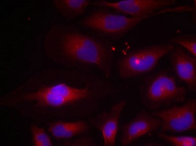
 IF (Immunofluorescence)
(Immunofluorescence staining of methanol-fixed HeLa cells using FKHRL1 (phospho-Ser253) antibody.)
IF (Immunofluorescence)
(Immunofluorescence staining of methanol-fixed HeLa cells using FKHRL1 (phospho-Ser253) antibody.)
FOXO3, Polyclonal Antibody (Cat# AAA243339)
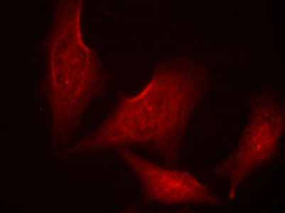
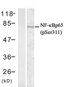
 IHC (Immunohistochemisry)
(Immunohistochemical analysis of paraffin-embedded human breast carcinoma tissue, using NFκB-p65 (phospho-Ser311) antibody.)
IHC (Immunohistochemisry)
(Immunohistochemical analysis of paraffin-embedded human breast carcinoma tissue, using NFκB-p65 (phospho-Ser311) antibody.)
RELA, Polyclonal Antibody (Cat# AAA243340)
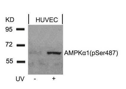
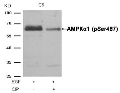 WB (Western Blot)
(Western blot analysis of extracts from C6 cells, treated with EGF or calf intestinal phosphatase (CIP), using AMPKalpha1 (Phospho-Ser487) Antibody.)
WB (Western Blot)
(Western blot analysis of extracts from C6 cells, treated with EGF or calf intestinal phosphatase (CIP), using AMPKalpha1 (Phospho-Ser487) Antibody.)
PRKAA1/PRKAA2, Polyclonal Antibody (Cat# AAA243365)
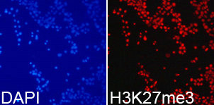 IF (Immunofluorescence)
(Immunofluorescence analysis of 293T cell using H3K27me3 antibody. Blue: DAPI for nuclear staining.)
IF (Immunofluorescence)
(Immunofluorescence analysis of 293T cell using H3K27me3 antibody. Blue: DAPI for nuclear staining.)
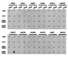
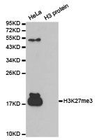
Histone H3K27me3, Polyclonal Antibody (Cat# AAA243392)
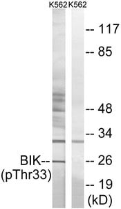
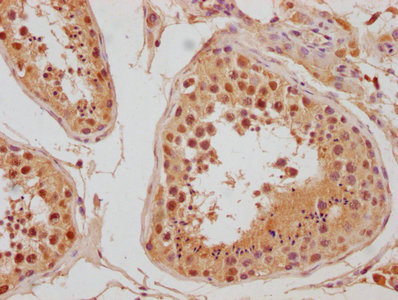
 IHC (Immunohiostchemistry)
(Immunohistochemical analysis of paraffin-embedded human lung carcinoma tissue, using BIK (Phospho-Thr33) antibody. The picture on the right is treated with the synthesized peptide.)
IHC (Immunohiostchemistry)
(Immunohistochemical analysis of paraffin-embedded human lung carcinoma tissue, using BIK (Phospho-Thr33) antibody. The picture on the right is treated with the synthesized peptide.)
BIK, Polyclonal Antibody (Cat# AAA243242)
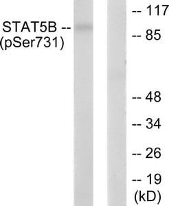
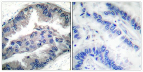
 IHC (Immunohiostchemistry)
(Immunohistochemical analysis of paraffin-embedded human breast carcinoma tissue using STAT5B (phospho-Ser731) antibody. The picture on the right is treated with the synthesized peptide.)
IHC (Immunohiostchemistry)
(Immunohistochemical analysis of paraffin-embedded human breast carcinoma tissue using STAT5B (phospho-Ser731) antibody. The picture on the right is treated with the synthesized peptide.)
STAT5B, Polyclonal Antibody (Cat# AAA243250)
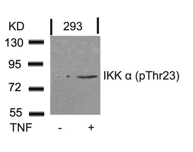
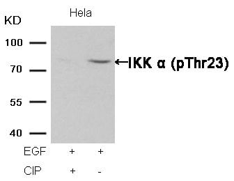 WB (Western Blot)
(Western blot analysis of extracts from Hela cells, treated with EGF or calf intestinal phosphatase (CIP), using IKK alpha (Phospho-Thr23) Antibody.)
WB (Western Blot)
(Western blot analysis of extracts from Hela cells, treated with EGF or calf intestinal phosphatase (CIP), using IKK alpha (Phospho-Thr23) Antibody.)
CHUK, Polyclonal Antibody (Cat# AAA243282)
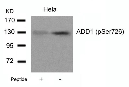
 IF (Immunofluorescence)
(Immunofluorescence staining of methanol-fixed Hela cells using ADD1(Phospho-Ser726) Antibody.)
IF (Immunofluorescence)
(Immunofluorescence staining of methanol-fixed Hela cells using ADD1(Phospho-Ser726) Antibody.)
ADD1, Polyclonal Antibody (Cat# AAA243296)
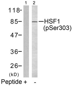
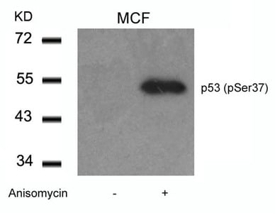
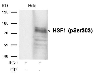 WB (Western Blot)
(Western blot analysis of extracts from Hela cells, treated with IFNa or calf intestinal phosphatase (CIP), using HSF1 (phospho-Ser303) Antibody.)
WB (Western Blot)
(Western blot analysis of extracts from Hela cells, treated with IFNa or calf intestinal phosphatase (CIP), using HSF1 (phospho-Ser303) Antibody.)
HSF1, Polyclonal Antibody (Cat# AAA243310)
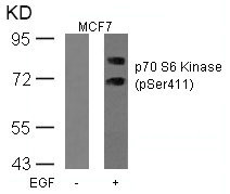
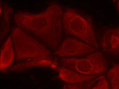 IF (Immunofluorescence)
(Immunofluorescence staining of methanol-fixed MCF7 cells using p70 S6 Kinase(Phospho-Ser411) Antibody.)
IF (Immunofluorescence)
(Immunofluorescence staining of methanol-fixed MCF7 cells using p70 S6 Kinase(Phospho-Ser411) Antibody.)
RPS6KB1, Polyclonal Antibody (Cat# AAA243313)
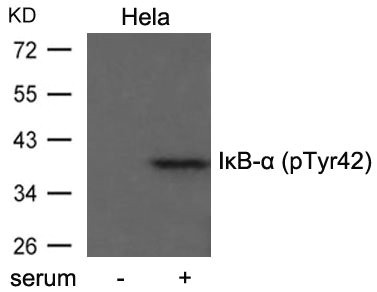
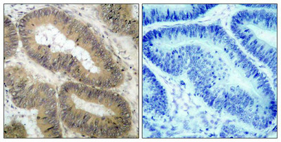
 IHC (Immunohiostchemistry)
(Immunohistochemical analysis of paraffin-embedded human breast carcinoma tissue using IkB-a(phospho-Tyr42) antibody.)
IHC (Immunohiostchemistry)
(Immunohistochemical analysis of paraffin-embedded human breast carcinoma tissue using IkB-a(phospho-Tyr42) antibody.)
NFKBIA, Polyclonal Antibody (Cat# AAA243164)
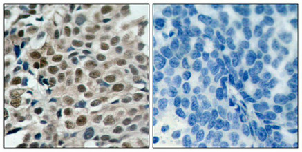
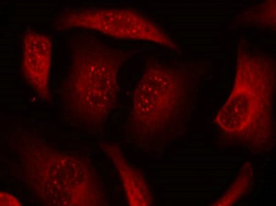
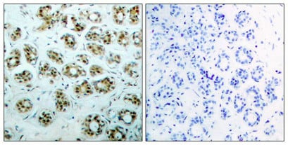
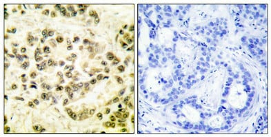
 IHC (Immunohiostchemistry)
(Immunohistochemical analysis of paraffin-embedded human breast carcinoma tissue using NF-κB p105 (phospho-Ser893) antibody.)
IHC (Immunohiostchemistry)
(Immunohistochemical analysis of paraffin-embedded human breast carcinoma tissue using NF-κB p105 (phospho-Ser893) antibody.)
NFKB1, Polyclonal Antibody (Cat# AAA243171)
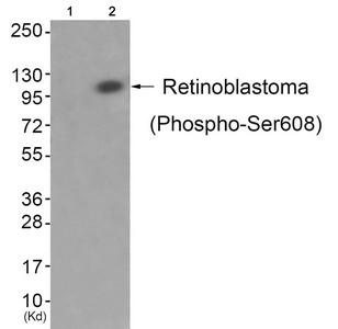
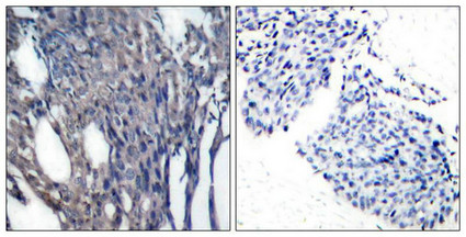
 IHC (Immunohiostchemistry)
(Immunohistochemical analysis of paraffin-embedded human brain tissue using Retinoblastoma (Phospho-Ser608) antibody (left)or the same antibody preincubated with blocking peptide (right).)
IHC (Immunohiostchemistry)
(Immunohistochemical analysis of paraffin-embedded human brain tissue using Retinoblastoma (Phospho-Ser608) antibody (left)or the same antibody preincubated with blocking peptide (right).)
RB1, Polyclonal Antibody (Cat# AAA243204)
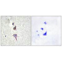
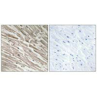
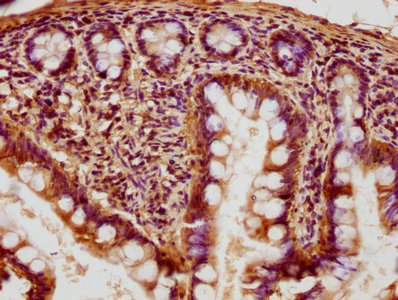
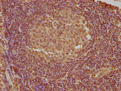
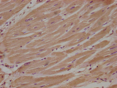 IHC (Immunohiostchemistry)
(IHC image of AAA243530 diluted at 1:100 and staining in paraffin-embedded human heart tissue performed on a Leica BondTM system. After dewaxing and hydration, antigen retrieval was mediated by high pressure in a citrate buffer (pH 6.0). Section was blocked with 10% normal goat serum 30min at RT. Then primary antibody (1% BSA) was incubated at 4 degree C overnight. The primary is detected by a biotinylated secondary antibody and visualized using an HRP conjugated SP system.)
IHC (Immunohiostchemistry)
(IHC image of AAA243530 diluted at 1:100 and staining in paraffin-embedded human heart tissue performed on a Leica BondTM system. After dewaxing and hydration, antigen retrieval was mediated by high pressure in a citrate buffer (pH 6.0). Section was blocked with 10% normal goat serum 30min at RT. Then primary antibody (1% BSA) was incubated at 4 degree C overnight. The primary is detected by a biotinylated secondary antibody and visualized using an HRP conjugated SP system.)
NENF, Polyclonal Antibody (Cat# AAA243530)
 IF (Immunofluorescence)
(Immunofluorescence staining of HepG2 cells with AAA243539 at 1:100, counter-stained with DAPI. The cells were fixed in 4% formaldehyde, permeabilized using 0.2% Triton X-100 and blocked in 10% normal Goat Serum. The cells were then incubated with the antibody overnight at 4 degree C. The secondary antibody was Alexa Fluor 488-congugated AffiniPure Goat Anti-Rabbit IgG(H+L).)
IF (Immunofluorescence)
(Immunofluorescence staining of HepG2 cells with AAA243539 at 1:100, counter-stained with DAPI. The cells were fixed in 4% formaldehyde, permeabilized using 0.2% Triton X-100 and blocked in 10% normal Goat Serum. The cells were then incubated with the antibody overnight at 4 degree C. The secondary antibody was Alexa Fluor 488-congugated AffiniPure Goat Anti-Rabbit IgG(H+L).)
LPAR6, Polyclonal Antibody (Cat# AAA243539)
 IF (Immunofluorescence)
(Immunofluorescence staining of U251 cells with AAA243461 at 1:133, counter-stained with DAPI. The cells were fixed in 4% formaldehyde, permeabilized using 0.2% Triton X-100 and blocked in 10% normal Goat Serum. The cells were then incubated with the antibody overnight at 4 degree C. The secondary antibody was Alexa Fluor 488-congugated AffiniPure Goat Anti-Rabbit IgG(H+L).)
IF (Immunofluorescence)
(Immunofluorescence staining of U251 cells with AAA243461 at 1:133, counter-stained with DAPI. The cells were fixed in 4% formaldehyde, permeabilized using 0.2% Triton X-100 and blocked in 10% normal Goat Serum. The cells were then incubated with the antibody overnight at 4 degree C. The secondary antibody was Alexa Fluor 488-congugated AffiniPure Goat Anti-Rabbit IgG(H+L).)
RABGGTB, Polyclonal Antibody (Cat# AAA243461)
 IF (Immunofluorescence)
(Immunofluorescence staining of HepG2 cells with AAA243471 at 1:50, counter-stained with DAPI. The cells were fixed in 4% formaldehyde, permeabilized using 0.2% Triton X-100 and blocked in 10% normal Goat Serum. The cells were then incubated with the antibody overnight at 4 degree C. The secondary antibody was Alexa Fluor 488-congugated AffiniPure Goat Anti-Rabbit IgG(H+L).)
IF (Immunofluorescence)
(Immunofluorescence staining of HepG2 cells with AAA243471 at 1:50, counter-stained with DAPI. The cells were fixed in 4% formaldehyde, permeabilized using 0.2% Triton X-100 and blocked in 10% normal Goat Serum. The cells were then incubated with the antibody overnight at 4 degree C. The secondary antibody was Alexa Fluor 488-congugated AffiniPure Goat Anti-Rabbit IgG(H+L).)
PDE8A, Polyclonal Antibody (Cat# AAA243471)
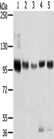
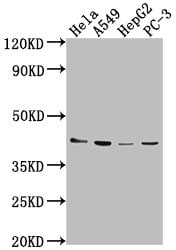
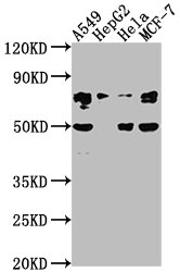 WB (Western Blot)
(Western BlotPositive WB detected in: A549 whole cell lysate, HepG2 whole cell lysate, Hela whole cell lysate, MCF-7 whole cell lysateAll lanes: LEO1 antibody at 1:2000SecondaryGoat polyclonal to rabbit IgG at 1/50000 dilutionPredicted band size: 76, 69 kDaObserved band size: 76 kDa)
WB (Western Blot)
(Western BlotPositive WB detected in: A549 whole cell lysate, HepG2 whole cell lysate, Hela whole cell lysate, MCF-7 whole cell lysateAll lanes: LEO1 antibody at 1:2000SecondaryGoat polyclonal to rabbit IgG at 1/50000 dilutionPredicted band size: 76, 69 kDaObserved band size: 76 kDa)
LEO1, Polyclonal Antibody (Cat# AAA243486)
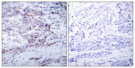
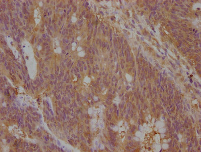
 IHC (Immunohistochemisry)
(IHC image of AAA243494 diluted at 1:740 and staining in paraffin-embedded human breast cancer performed on a Leica BondTM system. After dewaxing and hydration, antigen retrieval was mediated by high pressure in a citrate buffer (pH 6.0). Section was blocked with 10% normal goat serum 30min at RT. Then primary antibody (1% BSA) was incubated at 4 degree C overnight. The primary is detected by a biotinylated secondary antibody and visualized using an HRP conjugated SP system.)
IHC (Immunohistochemisry)
(IHC image of AAA243494 diluted at 1:740 and staining in paraffin-embedded human breast cancer performed on a Leica BondTM system. After dewaxing and hydration, antigen retrieval was mediated by high pressure in a citrate buffer (pH 6.0). Section was blocked with 10% normal goat serum 30min at RT. Then primary antibody (1% BSA) was incubated at 4 degree C overnight. The primary is detected by a biotinylated secondary antibody and visualized using an HRP conjugated SP system.)
PYCARD, Polyclonal Antibody (Cat# AAA243494)
 IF (Immunofluorescence)
(Immunofluorescence staining of Hela cells with AAA243500 at 1:166, counter-stained with DAPI. The cells were fixed in 4% formaldehyde, permeabilized using 0.2% Triton X-100 and blocked in 10% normal Goat Serum. The cells were then incubated with the antibody overnight at 4 degree C. The secondary antibody was Alexa Fluor 488-congugated AffiniPure Goat Anti-Rabbit IgG(H+L).)
IF (Immunofluorescence)
(Immunofluorescence staining of Hela cells with AAA243500 at 1:166, counter-stained with DAPI. The cells were fixed in 4% formaldehyde, permeabilized using 0.2% Triton X-100 and blocked in 10% normal Goat Serum. The cells were then incubated with the antibody overnight at 4 degree C. The secondary antibody was Alexa Fluor 488-congugated AffiniPure Goat Anti-Rabbit IgG(H+L).)
ELF3, Polyclonal Antibody (Cat# AAA243500)
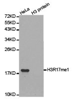
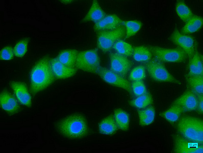
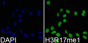 IF (Immunofluorescence)
(Immunofluorescence analysis of 293T cell using H3R17me1 antibody. Blue: DAPI for nuclear staining.)
IF (Immunofluorescence)
(Immunofluorescence analysis of 293T cell using H3R17me1 antibody. Blue: DAPI for nuclear staining.)
Histone H3R17me1, Polyclonal Antibody (Cat# AAA243402)
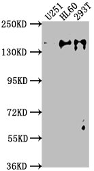 WB (Western Blot)
(Western BlotPositive WB detected in: U251 whole cell lysate, HL60 whole cell lysate, 293T whole cell lysateAll lanes: ADNP antibody at 1:2000SecondaryGoat polyclonal to rabbit IgG at 1/50000 dilutionPredicted band size: 124 kDaObserved band size: 135 kDa)
WB (Western Blot)
(Western BlotPositive WB detected in: U251 whole cell lysate, HL60 whole cell lysate, 293T whole cell lysateAll lanes: ADNP antibody at 1:2000SecondaryGoat polyclonal to rabbit IgG at 1/50000 dilutionPredicted band size: 124 kDaObserved band size: 135 kDa)
ADNP, Polyclonal Antibody (Cat# AAA243441)
 IF (Immunofluorescence)
(Immunofluorescence staining of Hela cells with AAA243447 at 1:200, counter-stained with DAPI. The cells were fixed in 4% formaldehyde, permeabilized using 0.2% Triton X-100 and blocked in 10% normal Goat Serum. The cells were then incubated with the antibody overnight at 4 degree C. The secondary antibody was Alexa Fluor 488-congugated AffiniPure Goat Anti-Rabbit IgG(H+L).)
IF (Immunofluorescence)
(Immunofluorescence staining of Hela cells with AAA243447 at 1:200, counter-stained with DAPI. The cells were fixed in 4% formaldehyde, permeabilized using 0.2% Triton X-100 and blocked in 10% normal Goat Serum. The cells were then incubated with the antibody overnight at 4 degree C. The secondary antibody was Alexa Fluor 488-congugated AffiniPure Goat Anti-Rabbit IgG(H+L).)
EIF2AK2, Polyclonal Antibody (Cat# AAA243447)
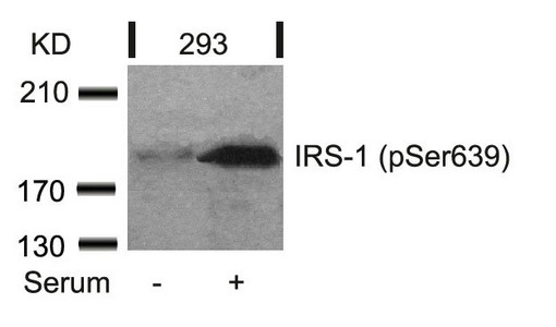
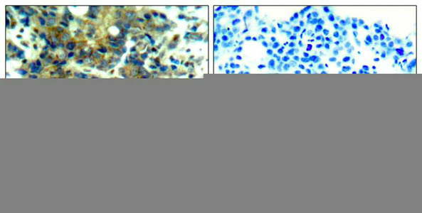
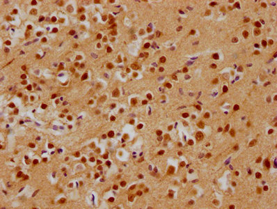
 IHC (Immunohiostchemistry)
(Immunohistochemical analysis of paraffin-embedded human breast carcinoma tissue using IRS-1(Phospho-Ser639) Antibody(left) or the same antibody preincubated with blocking peptide(right).)
IHC (Immunohiostchemistry)
(Immunohistochemical analysis of paraffin-embedded human breast carcinoma tissue using IRS-1(Phospho-Ser639) Antibody(left) or the same antibody preincubated with blocking peptide(right).)
IRS1, Polyclonal Antibody (Cat# AAA243126)
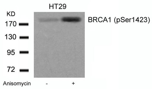
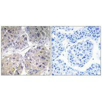
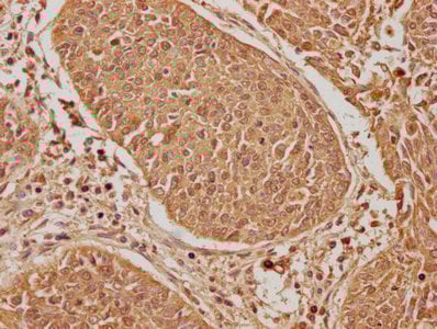
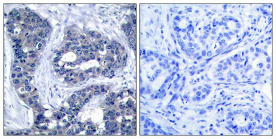
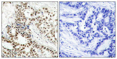 IHC (Immunohiostchemistry)
(Immunohistochemical analysis of paraffin-embedded human breast carcinoma tissue using BRCA1(Phospho-Ser1423) Antibody(left) or the same antibody preincubated with blocking peptide(right).)
IHC (Immunohiostchemistry)
(Immunohistochemical analysis of paraffin-embedded human breast carcinoma tissue using BRCA1(Phospho-Ser1423) Antibody(left) or the same antibody preincubated with blocking peptide(right).)
BRCA1, Polyclonal Antibody (Cat# AAA243128)
What are Polyclonal Antibodies?
Polyclonal antibodies are antibodies that come from multiple B cell clones of a host animal. The typical hosts used for the majority of polyclonal antibody production are rabbits, goats, sheep, and donkeys. These polyclonal antibodies, once having identified their target, will bind to different epitopes located at different regions or sequences on the same protein/antigen. As a result, they are ideal at locating and binding to the target, even if the target is in very low concentrations (due to many different antibodies being able to bind to the same target molecule, which allows for significant amplification of a downstream signal).
Polyclonal antibodies are typically produced by injecting an antigen into a host animal, which causes the animal’s immune system to attack the foreign antigen by mass generating antibodies against it. After a period of time, serum is collected from the animal and purified using physicochemical fractionation, class-specific affinity purification, and/or antigen-affinity purification.
Key Uses of Polyclonal Antibodies
- Western Blotting: This method is used to find specific proteins in biological samples after separating them by size.
- Immunohistochemistry: IHC helps visualize the location of proteins in tissue sections using various staining techniques.
- ELISA: (Enzyme-Linked Immunosorbent Assay) is typically used to identify specific protein quantities in a sample. ELISAs can be either “Quantitative” or “Qualitative”.
- Flow Cytometry: technique that identifies and measures the specific protein on the surface or inside the cells in a fluid suspension.
- Immunoprecipitation: IP isolates and studies a specific protein from a complex mixture using antibodies.
Why Buy Polyclonal Antibodies from AAA Biotech?
1. Ideal for Various Applications
Our antibodies are generally going to be validated for use in multiple types of assays, including ELISA, Western Blotting, Immunohistochemistry, Immunoprecipitation, amongst others. They are ideal for a wide range of research applications.
2. Rigorous Quality Control
All of the antibodies in our catalog undergo strict quality testing to ensure specificity, sensitivity, and consistent performance. We are confident in the ability of our antibodies to provide you with accurate results.
3. Wide Assortment of Antibodies
Antibodies in are catalog can be found for both common and exotic species, and these antibodies are also available in both conjugated and recombinant forms to suit many diverse experimental needs.
4. Highly Purified
Our antibodies are available in purified forms with over 85% purity, as confirmed by SDS-PAGE. They are also available with tags such as His, Flag, GST, or MBP. We cater to customers worldwide.
FAQ
1. How are polyclonal antibodies produced?
Traditionally, polyclonal antibodies are produced by injecting an antigen into a host animal (such as a rabbit or goat), which then triggers an immune response from the host animal. The animal’s B cells produce antibodies that will recognize different parts of the injected antigen. These antibodies are then collected from the animal’s blood and purified for use.
2. How do polyclonal antibodies differ from monoclonal antibodies?
Polyclonal antibodies are a mix of antibodies that bind to different locations (epitopes) of the same antigen, while monoclonal antibodies are identical and bind to just one specific epitope. This makes polyclonal antibodies more versatile and better at detecting proteins that may be present in low quantities or in altered/modified forms.
3. How should I store polyclonal antibodies?
Polyclonal antibodies should be stored at 4°C for short-term use (up to a few weeks) and at -20°C or -80°C for long-term storage. Avoid repeated freeze-thaw cycles by dividing them into small aliquots. Always check the datasheet for specific storage instructions.