Filters
▼Clonality
▼Type
▼Reactivity
▼Gene Name
▼Isotype
▼Host
▼Application
▼Clone
▼Polyclonal Antibodies
At AAA Biotech also known as AAA Bio or AAABio, we provide a broad range of purified polyclonal antibodies (pAbs) that are able to all be browsed online through our website. Due to their high specificity and strong binding affinity, these antibodies are ideal for wide swathes of research and experimental applications.
Our polyclonal antibodies can easily support your work, whether you use them for Western Blotting, Immunocytochemistry (with or without Immunofluorescence used in conjunction), Immunohistochemistry, Immunoprecipitation, and ELISA tests. We highly encourage you to browse our range of pAbs and choose the one that best suits your experimental model.
Viewing 2050-2100 of 96805 product results
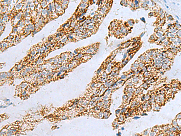
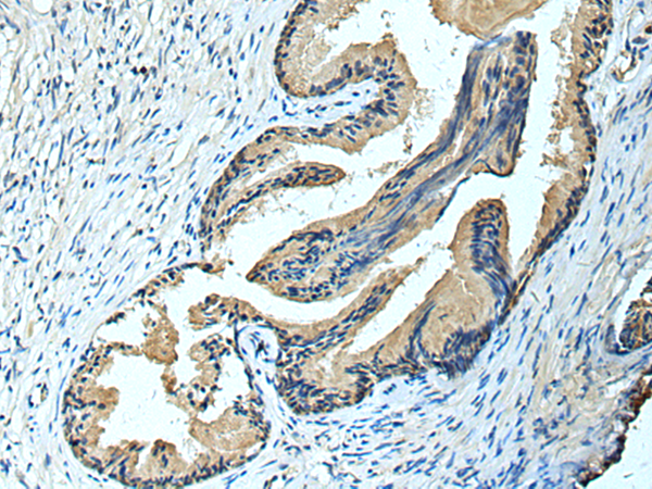
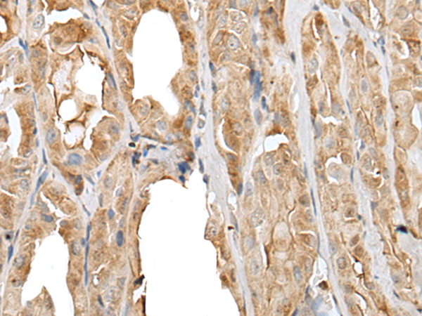
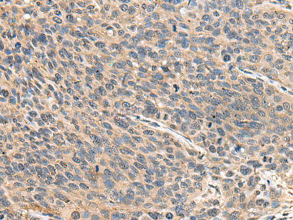
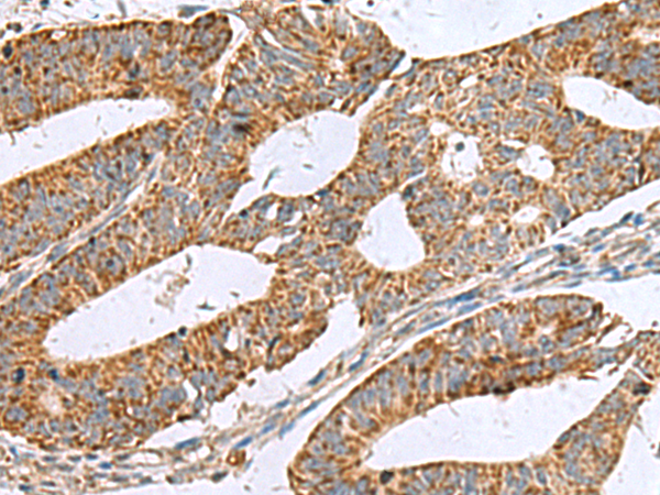
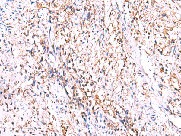
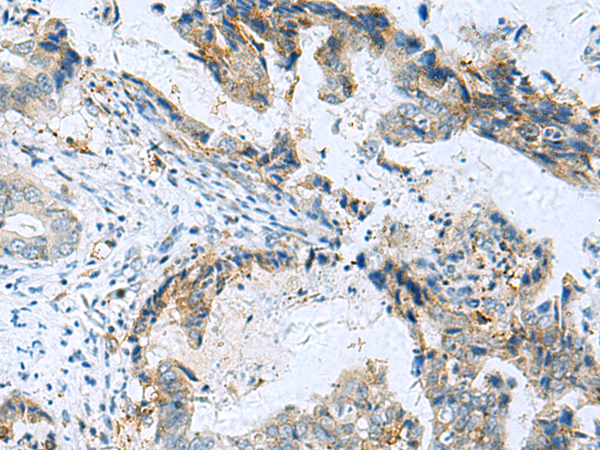
 IHC (Immunohiostchemistry)
(Immunohistochemistry of paraffin-embedded Human tonsil tissue using PYCR2 Polyclonal Antibody at dilution of 1:40(×200))
IHC (Immunohiostchemistry)
(Immunohistochemistry of paraffin-embedded Human tonsil tissue using PYCR2 Polyclonal Antibody at dilution of 1:40(×200))
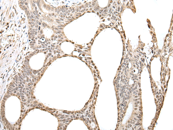
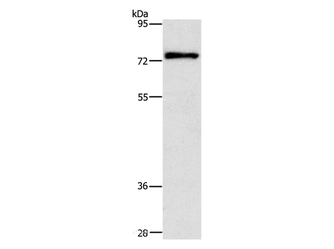
PYCR2, Polyclonal Antibody (Cat# AAA175640)
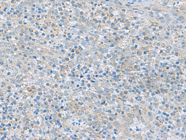
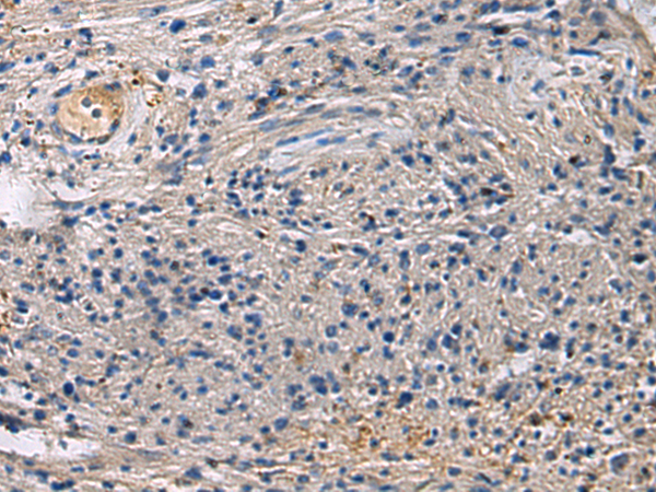
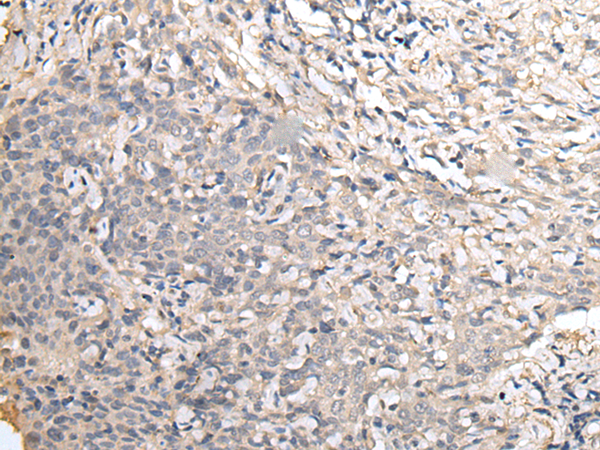
 IHC (Immunohistochemisry)
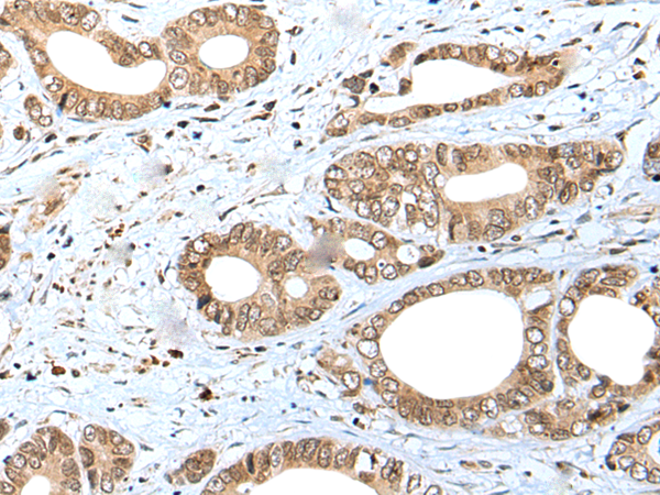
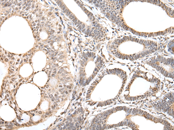
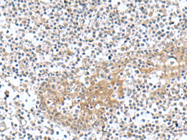
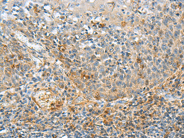
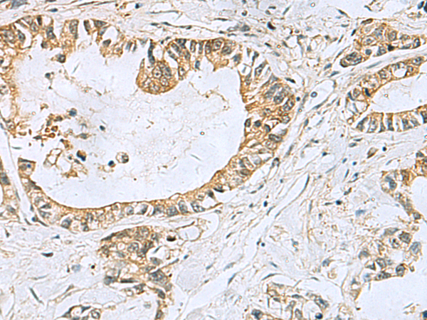
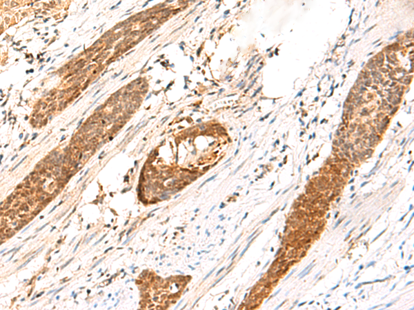
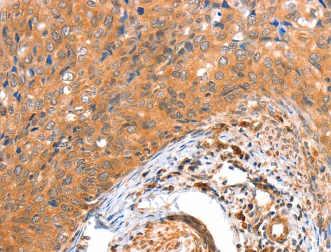
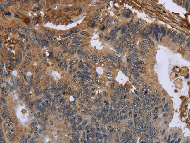
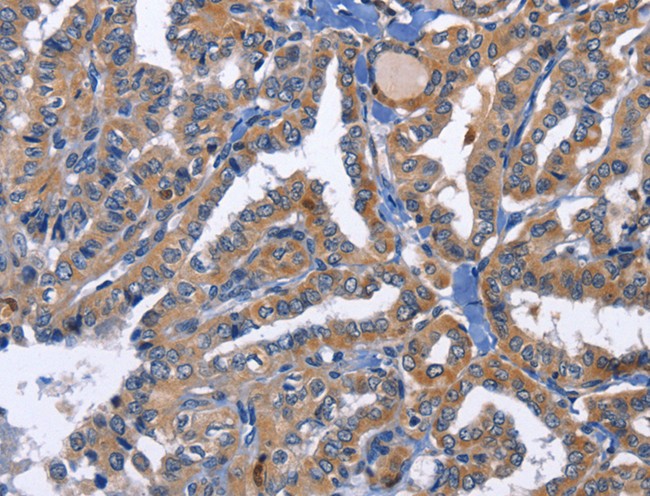
(Immunohistochemistry of paraffin-embedded Human prost ate cancer tissue using FCRLA Polyclonal Antibody at dilution of 1:90(×200))
IHC (Immunohistochemisry)
(Immunohistochemistry of paraffin-embedded Human prost ate cancer tissue using FCRLA Polyclonal Antibody at dilution of 1:90(×200))
FCRLA, Polyclonal Antibody (Cat# AAA175651)
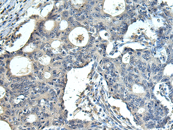
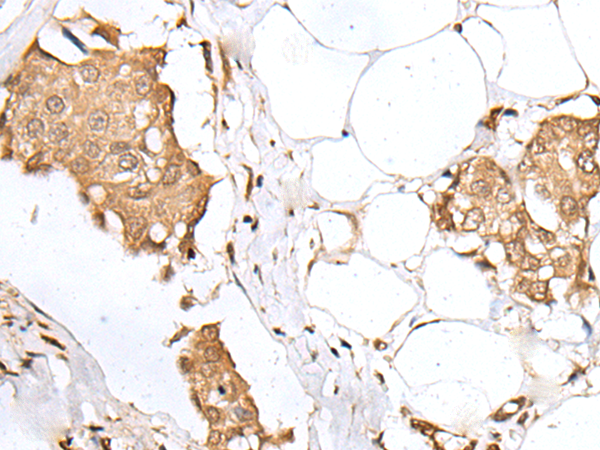
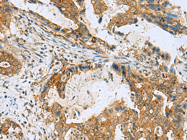
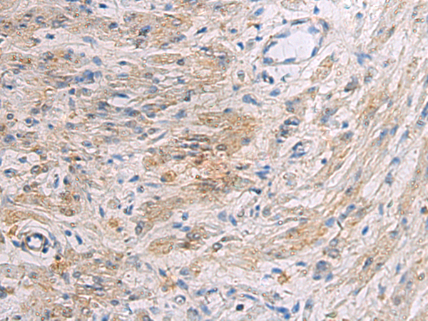 IHC (Immunohiostchemistry)
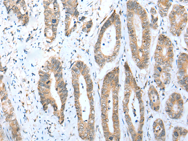
(Immunohistochemistry of paraffin-embedded Human gastric cancer tissue using SLC30A3 Polyclonal Antibody at dilution of 1:55(×200))
IHC (Immunohiostchemistry)
(Immunohistochemistry of paraffin-embedded Human gastric cancer tissue using SLC30A3 Polyclonal Antibody at dilution of 1:55(×200))
SLC30A3, Polyclonal Antibody (Cat# AAA175657)
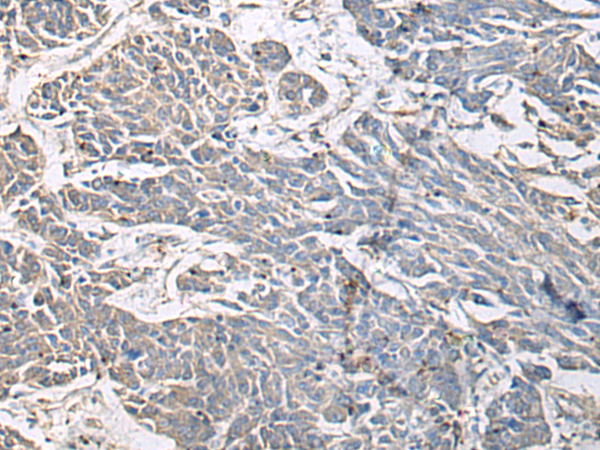
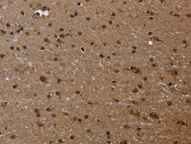
 IHC (Immunohiostchemistry)
(Immunohistochemistry of paraffin-embedded Human brain tissue using PRKCSH Polyclonal Antibody at dilution of 1:65(×200))
IHC (Immunohiostchemistry)
(Immunohistochemistry of paraffin-embedded Human brain tissue using PRKCSH Polyclonal Antibody at dilution of 1:65(×200))
PRKCSH, Polyclonal Antibody (Cat# AAA175661)
 IHC (Immunohiostchemistry)
(Immunohistochemistry of paraffin-embedded Human esophagus cancer tissue using CARTPT Polyclonal Antibody at dilution of 1:25(×200))
IHC (Immunohiostchemistry)
(Immunohistochemistry of paraffin-embedded Human esophagus cancer tissue using CARTPT Polyclonal Antibody at dilution of 1:25(×200))
CARTPT, Polyclonal Antibody (Cat# AAA175114)
 IHC (Immunohiostchemistry)
(Immunohistochemistry of paraffin-embedded Human gastric cancer tissue using ACBD4 Polyclonal Antibody at dilution of 1:30(×200))
IHC (Immunohiostchemistry)
(Immunohistochemistry of paraffin-embedded Human gastric cancer tissue using ACBD4 Polyclonal Antibody at dilution of 1:30(×200))
ACBD4, Polyclonal Antibody (Cat# AAA175116)
 IHC (Immunohistochemisry)
(Immunohistochemistry of paraffin-embedded Human breast cancer tissue using SPCS2 Polyclonal Antibody at dilution of 1:40(×200))
IHC (Immunohistochemisry)
(Immunohistochemistry of paraffin-embedded Human breast cancer tissue using SPCS2 Polyclonal Antibody at dilution of 1:40(×200))
SPCS2, Polyclonal Antibody (Cat# AAA175135)
 IHC (Immunohistochemisry)
(Immunohistochemistry of paraffin-embedded Human lung cancer tissue using NDUFA12 Polyclonal Antibody at dilution of 1:30(×200))
IHC (Immunohistochemisry)
(Immunohistochemistry of paraffin-embedded Human lung cancer tissue using NDUFA12 Polyclonal Antibody at dilution of 1:30(×200))
NDUFA12, Polyclonal Antibody (Cat# AAA175145)
 IHC (Immunohiostchemistry)
(Immunohistochemistry of paraffin-embedded Human thyroid cancer tissue using PRNP Polyclonal Antibody at dilution of 1:45(×200))
IHC (Immunohiostchemistry)
(Immunohistochemistry of paraffin-embedded Human thyroid cancer tissue using PRNP Polyclonal Antibody at dilution of 1:45(×200))
PRNP, Polyclonal Antibody (Cat# AAA175169)
 IHC (Immunohiostchemistry)
(Immunohistochemistry of paraffin-embedded Human breast cancer tissue using SNF8 Polyclonal Antibody at dilution of 1:30(×200))
IHC (Immunohiostchemistry)
(Immunohistochemistry of paraffin-embedded Human breast cancer tissue using SNF8 Polyclonal Antibody at dilution of 1:30(×200))
SNF8, Polyclonal Antibody (Cat# AAA175171)
 IHC (Immunohiostchemistry)
(Immunohistochemistry of paraffin-embedded Human gastric cancer tissue using ZBTB5 Polyclonal Antibody at dilution of 1:40(×200))
IHC (Immunohiostchemistry)
(Immunohistochemistry of paraffin-embedded Human gastric cancer tissue using ZBTB5 Polyclonal Antibody at dilution of 1:40(×200))
ZBTB5, Polyclonal Antibody (Cat# AAA175172)
 IHC (Immunohistochemisry)
(Immunohistochemistry of paraffin-embedded Human prost ate cancer tissue using ZNF281 Polyclonal Antibody at dilution of 1:30(×200))
IHC (Immunohistochemisry)
(Immunohistochemistry of paraffin-embedded Human prost ate cancer tissue using ZNF281 Polyclonal Antibody at dilution of 1:30(×200))
ZNF281, Polyclonal Antibody (Cat# AAA175173)
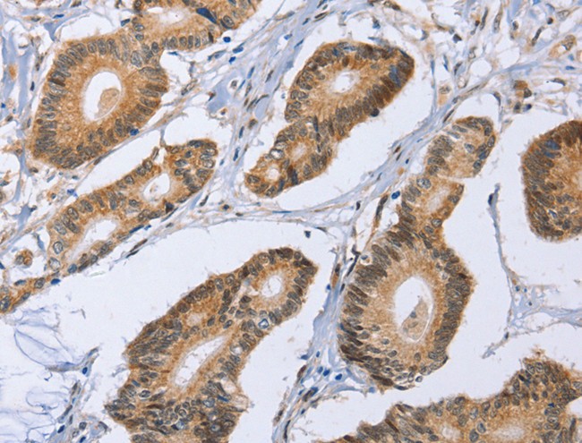
 IHC (Immunohiostchemistry)
(Immunohistochemistry of paraffin-embedded Human colorectal cancer tissue using ZNF213 Polyclonal Antibody at dilution of 1:30(×200))
IHC (Immunohiostchemistry)
(Immunohistochemistry of paraffin-embedded Human colorectal cancer tissue using ZNF213 Polyclonal Antibody at dilution of 1:30(×200))
ZNF213, Polyclonal Antibody (Cat# AAA175179)
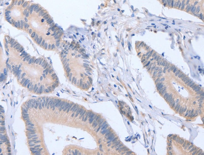
 IHC (Immunohistochemisry)
(Immunohistochemistry of paraffin-embedded Human esophagus cancer tissue using SCCPDH Polyclonal Antibody at dilution of 1:85(×200))
IHC (Immunohistochemisry)
(Immunohistochemistry of paraffin-embedded Human esophagus cancer tissue using SCCPDH Polyclonal Antibody at dilution of 1:85(×200))
SCCPDH, Polyclonal Antibody (Cat# AAA175513)
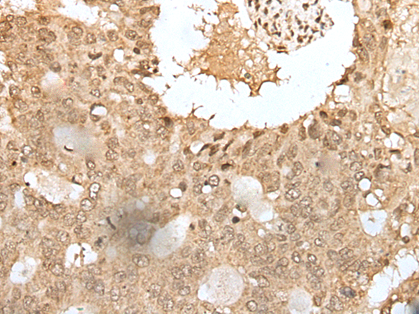
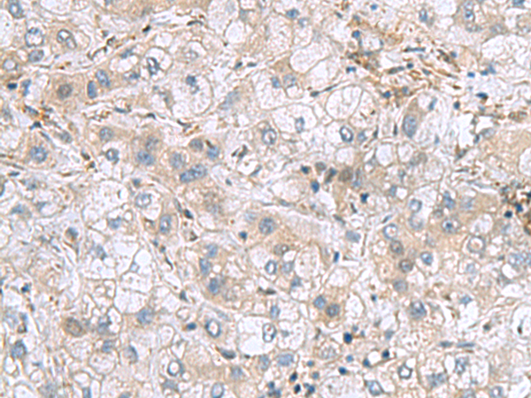
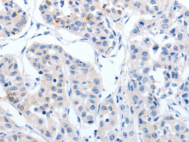
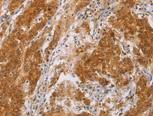
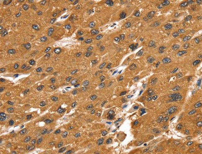
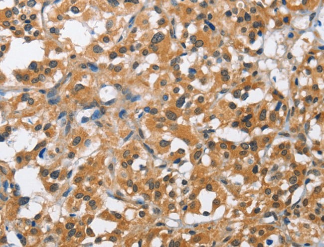
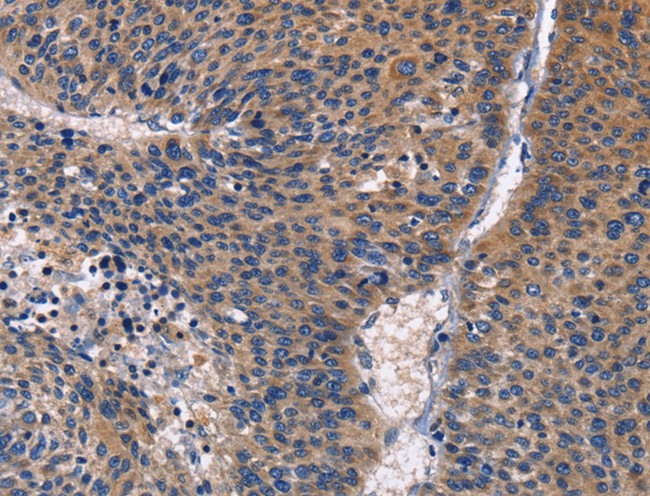
 IHC (Immunohiostchemistry)
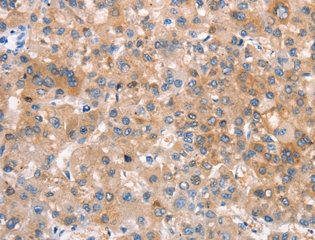
(Immunohistochemistry of paraffin-embedded Human liver cancer tissue using PHPT1 Polyclonal Antibody at dilution of 1:210(×200))
IHC (Immunohiostchemistry)
(Immunohistochemistry of paraffin-embedded Human liver cancer tissue using PHPT1 Polyclonal Antibody at dilution of 1:210(×200))
PHPT1, Polyclonal Antibody (Cat# AAA175562)
 IHC (Immunohistochemisry)
(Immunohistochemistry of paraffin-embedded Human cervical cancer tissue using RPS14 Polyclonal Antibody at dilution of 1:50(×200))
IHC (Immunohistochemisry)
(Immunohistochemistry of paraffin-embedded Human cervical cancer tissue using RPS14 Polyclonal Antibody at dilution of 1:50(×200))
RPS14, Polyclonal Antibody (Cat# AAA175576)
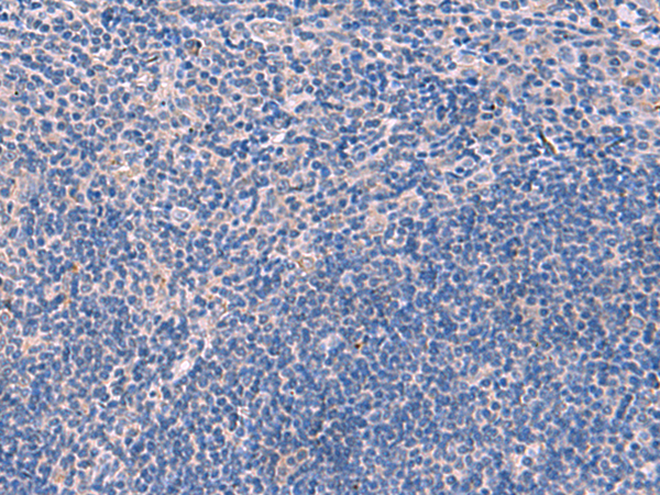
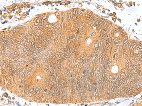
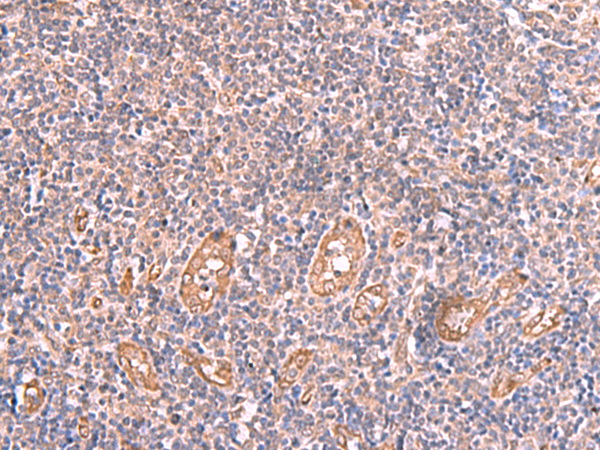
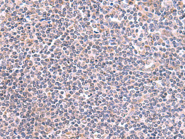
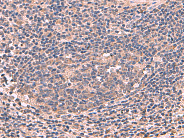
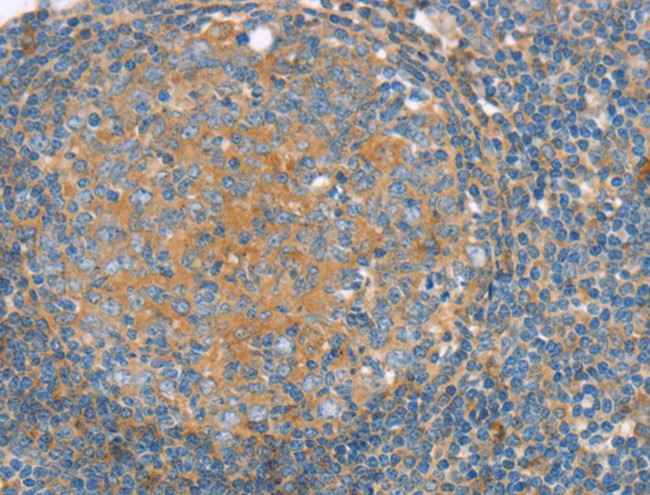
 IHC (Immunohiostchemistry)
(Immunohistochemistry of paraffin-embedded Human tonsil tissue using TNNC2 Polyclonal Antibody at dilution of 1:50(×200))
IHC (Immunohiostchemistry)
(Immunohistochemistry of paraffin-embedded Human tonsil tissue using TNNC2 Polyclonal Antibody at dilution of 1:50(×200))
TNNC2, Polyclonal Antibody (Cat# AAA175578)
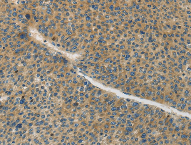
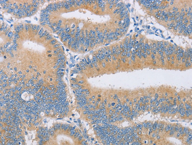
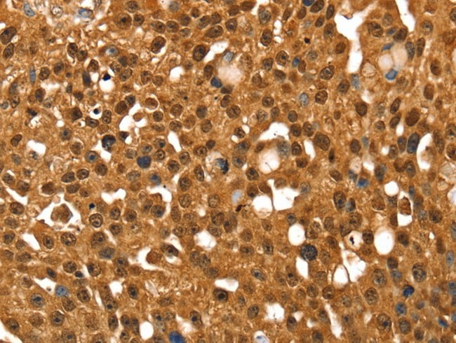
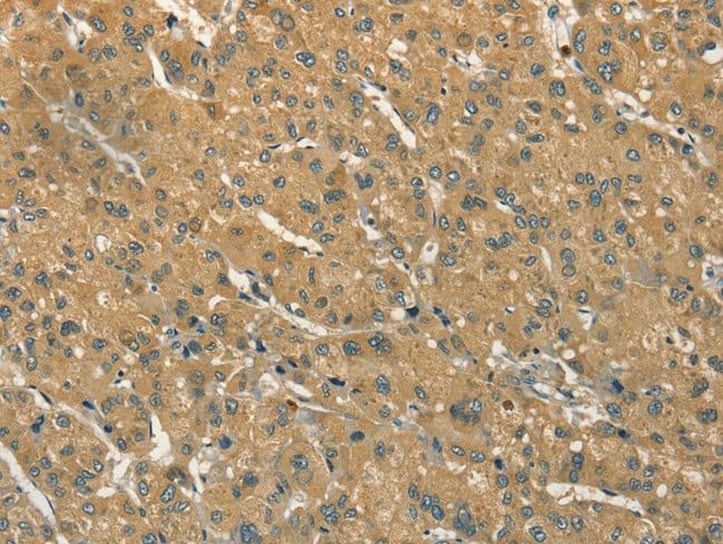
 IHC (Immunohiostchemistry)
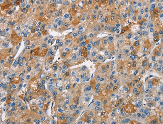
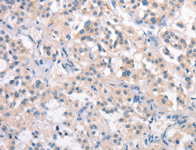
(Immunohistochemistry of paraffin-embedded Human liver cancer tissue using TXNDC12 Polyclonal Antibody at dilution of 1:70(×200))
IHC (Immunohiostchemistry)
(Immunohistochemistry of paraffin-embedded Human liver cancer tissue using TXNDC12 Polyclonal Antibody at dilution of 1:70(×200))
TXNDC12, Polyclonal Antibody (Cat# AAA175582)
 IHC (Immunohiostchemistry)
(Immunohistochemistry of paraffin-embedded Human colorectal cancer tissue using NFS1 Polyclonal Antibody at dilution of 1:50(×200))
IHC (Immunohiostchemistry)
(Immunohistochemistry of paraffin-embedded Human colorectal cancer tissue using NFS1 Polyclonal Antibody at dilution of 1:50(×200))
NFS1, Polyclonal Antibody (Cat# AAA175588)
 IHC (Immunohistochemisry)
(Immunohistochemistry of paraffin-embedded Human cervical cancer tissue using LMCD1 Polyclonal Antibody at dilution of 1:100(×200))
IHC (Immunohistochemisry)
(Immunohistochemistry of paraffin-embedded Human cervical cancer tissue using LMCD1 Polyclonal Antibody at dilution of 1:100(×200))
LMCD1, Polyclonal Antibody (Cat# AAA175491)
 IHC (Immunohiostchemistry)
(Immunohistochemistry of paraffin-embedded Human cervical cancer tissue using GMDS Polyclonal Antibody at dilution of 1:70(×200))
IHC (Immunohiostchemistry)
(Immunohistochemistry of paraffin-embedded Human cervical cancer tissue using GMDS Polyclonal Antibody at dilution of 1:70(×200))
GMDS, Polyclonal Antibody (Cat# AAA175493)
 IHC (Immunohistochemisry)
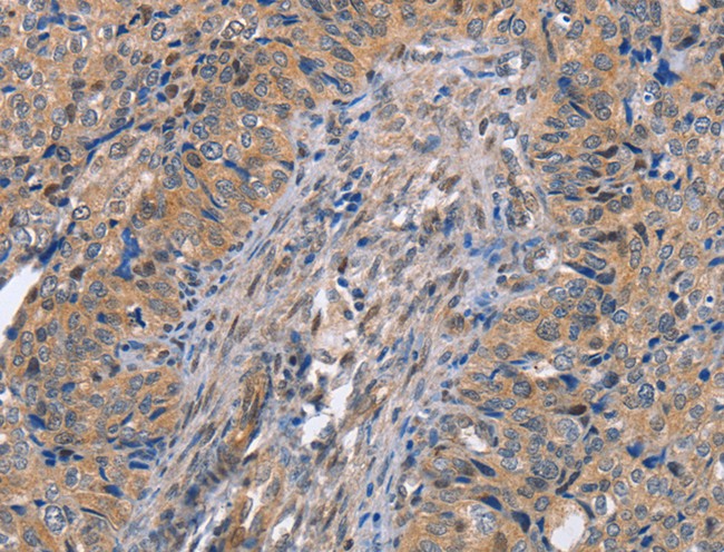
(Immunohistochemistry of paraffin-embedded Human colorectal cancer tissue using MAGEC2 Polyclonal Antibody at dilution of 1:60(×200))
IHC (Immunohistochemisry)
(Immunohistochemistry of paraffin-embedded Human colorectal cancer tissue using MAGEC2 Polyclonal Antibody at dilution of 1:60(×200))
MAGEC2, Polyclonal Antibody (Cat# AAA175496)

 IHC (Immunohiostchemistry)
(Immunohistochemistry of paraffin-embedded Human esophagus cancer tissue using GABPB1 Polyclonal Antibody at dilution of 1:70(×200))
IHC (Immunohiostchemistry)
(Immunohistochemistry of paraffin-embedded Human esophagus cancer tissue using GABPB1 Polyclonal Antibody at dilution of 1:70(×200))
GABPB1, Polyclonal Antibody (Cat# AAA175502)
 IHC (Immunohiostchemistry)
(Immunohistochemistry of paraffin-embedded Human tonsil tissue using RMDN2 Polyclonal Antibody at dilution of 1:70(×200))
IHC (Immunohiostchemistry)
(Immunohistochemistry of paraffin-embedded Human tonsil tissue using RMDN2 Polyclonal Antibody at dilution of 1:70(×200))
RMDN2, Polyclonal Antibody (Cat# AAA175505)
 IHC (Immunohistochemisry)
(Immunohistochemistry of paraffin-embedded Human gastric cancer tissue using TUBD1 Polyclonal Antibody at dilution of 1:70(×200))
IHC (Immunohistochemisry)
(Immunohistochemistry of paraffin-embedded Human gastric cancer tissue using TUBD1 Polyclonal Antibody at dilution of 1:70(×200))
TUBD1, Polyclonal Antibody (Cat# AAA175506)
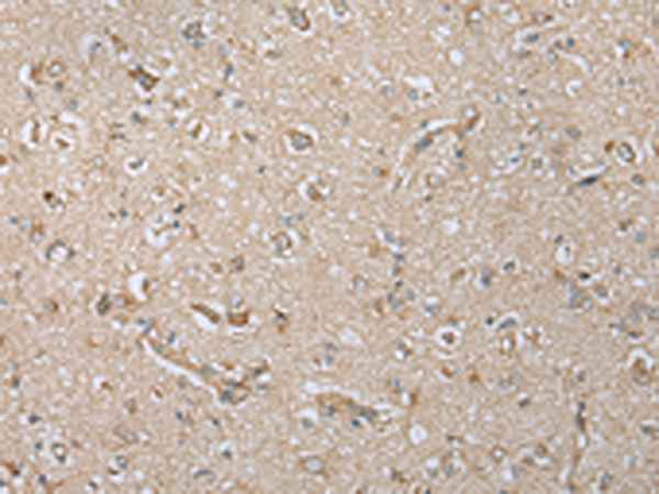
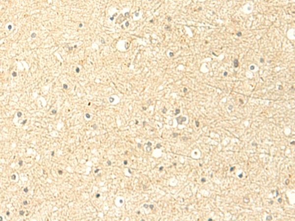 IHC (Immunohiostchemistry)
(Immunohistochemistry of paraffin-embedded Human brain tissue using MTHFS Polyclonal Antibody at dilution of 1:50(×200))
IHC (Immunohiostchemistry)
(Immunohistochemistry of paraffin-embedded Human brain tissue using MTHFS Polyclonal Antibody at dilution of 1:50(×200))
MTHFS, Polyclonal Antibody (Cat# AAA175452)
 IHC (Immunohiostchemistry)
(Immunohistochemistry of paraffin-embedded Human esophagus cancer tissue using DCAF10 Polyclonal Antibody at dilution of 1:130(×200))
IHC (Immunohiostchemistry)
(Immunohistochemistry of paraffin-embedded Human esophagus cancer tissue using DCAF10 Polyclonal Antibody at dilution of 1:130(×200))
DCAF10, Polyclonal Antibody (Cat# AAA175458)
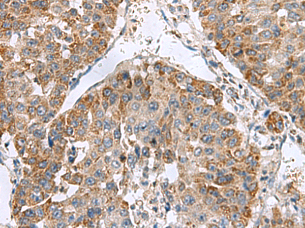
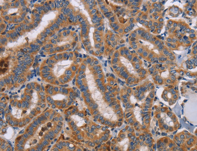
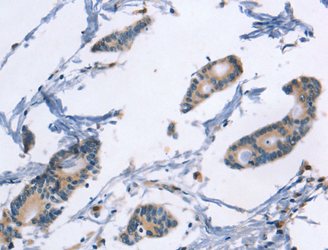
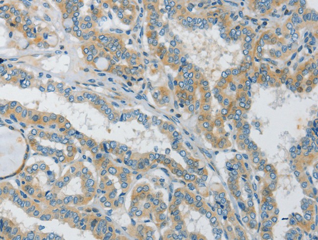
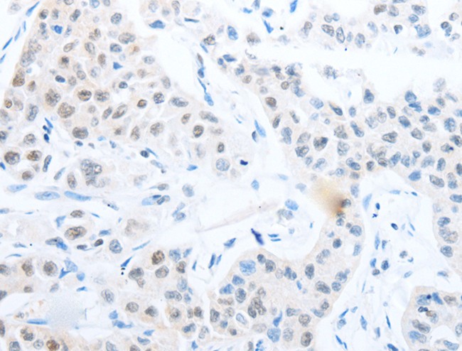
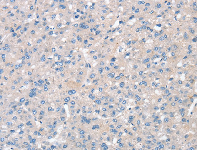
 IHC (Immunohiostchemistry)
(Immunohistochemistry of paraffin-embedded Human liver cancer tissue using RTKN2 Polyclonal Antibody at dilution of 1:75(×200))
IHC (Immunohiostchemistry)
(Immunohistochemistry of paraffin-embedded Human liver cancer tissue using RTKN2 Polyclonal Antibody at dilution of 1:75(×200))
RTKN2, Polyclonal Antibody (Cat# AAA175482)
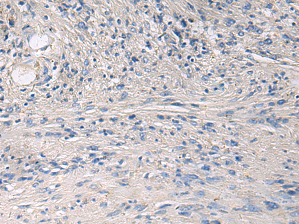
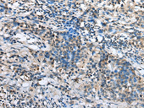
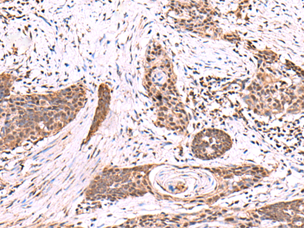
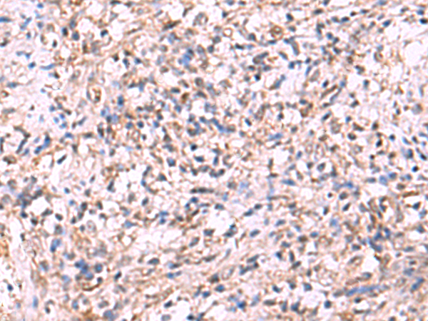
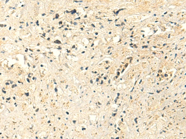
 IHC (Immunohiostchemistry)
(Immunohistochemistry of paraffin-embedded Human breast cancer tissue using ARHGEF12 Polyclonal Antibody at dilution 1:20)
IHC (Immunohiostchemistry)
(Immunohistochemistry of paraffin-embedded Human breast cancer tissue using ARHGEF12 Polyclonal Antibody at dilution 1:20)
ARHGEF12, Polyclonal Antibody (Cat# AAA168341)
 IHC (Immunohiostchemistry)
(Immunohistochemistry of paraffin-embedded Human colon cancer tissue using TLR8 Polyclonal Antibody at dilution 1:40)
IHC (Immunohiostchemistry)
(Immunohistochemistry of paraffin-embedded Human colon cancer tissue using TLR8 Polyclonal Antibody at dilution 1:40)
TLR8, Polyclonal Antibody (Cat# AAA168342)
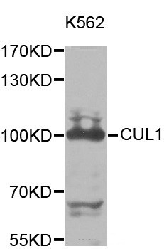
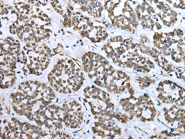
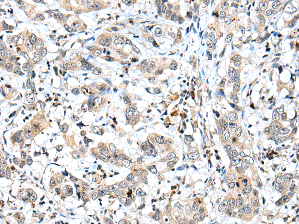
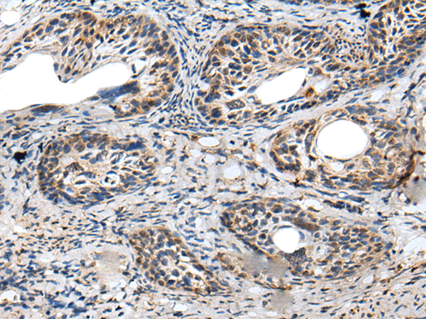
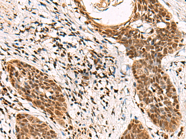
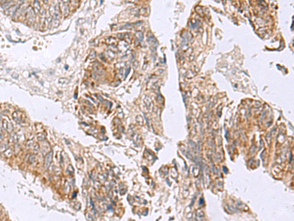
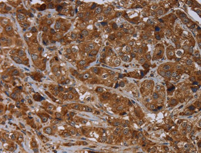
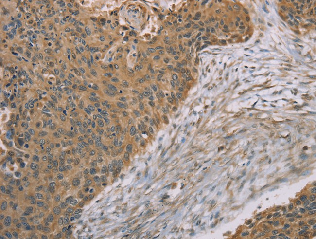
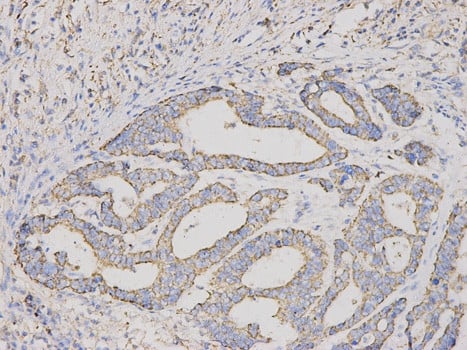 IHC (Immunohiostchemistry)
(Immunohistochemical of paraffin-embedded human stomach cancer using CUL1 antibody at dilution of 1:200 (200x lens).)
IHC (Immunohiostchemistry)
(Immunohistochemical of paraffin-embedded human stomach cancer using CUL1 antibody at dilution of 1:200 (200x lens).)
CUL1, Polyclonal Antibody (Cat# AAA168349)
 IHC (Immunohiostchemistry)
(Immunohistochemistry of paraffin-embedded Human lung cancer tissue using ACVR1C Polyclonal Antibody at dilution 1:15)
IHC (Immunohiostchemistry)
(Immunohistochemistry of paraffin-embedded Human lung cancer tissue using ACVR1C Polyclonal Antibody at dilution 1:15)
ACVR1C, Polyclonal Antibody (Cat# AAA168362)
 IHC (Immunohiostchemistry)
(Immunohistochemistry of paraffin-embedded Human thyroid cancer tissue using ANKRA2 Polyclonal Antibody at dilution 1:50)
IHC (Immunohiostchemistry)
(Immunohistochemistry of paraffin-embedded Human thyroid cancer tissue using ANKRA2 Polyclonal Antibody at dilution 1:50)
ANKRA2, Polyclonal Antibody (Cat# AAA168366)
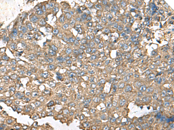
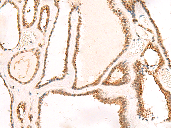
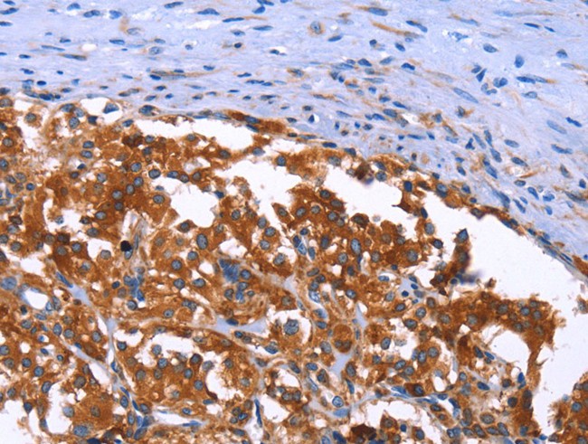
 IHC (Immunohiostchemistry)
(Immunohistochemistry of paraffin-embedded Human ovarian cancer tissue using LRP5 Polyclonal Antibody at dilution 1:55)
IHC (Immunohiostchemistry)
(Immunohistochemistry of paraffin-embedded Human ovarian cancer tissue using LRP5 Polyclonal Antibody at dilution 1:55)
LRP5, Polyclonal Antibody (Cat# AAA168367)
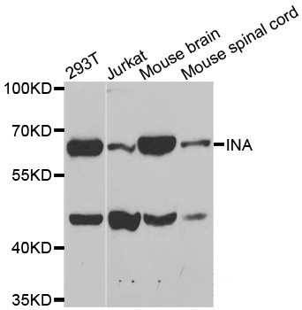
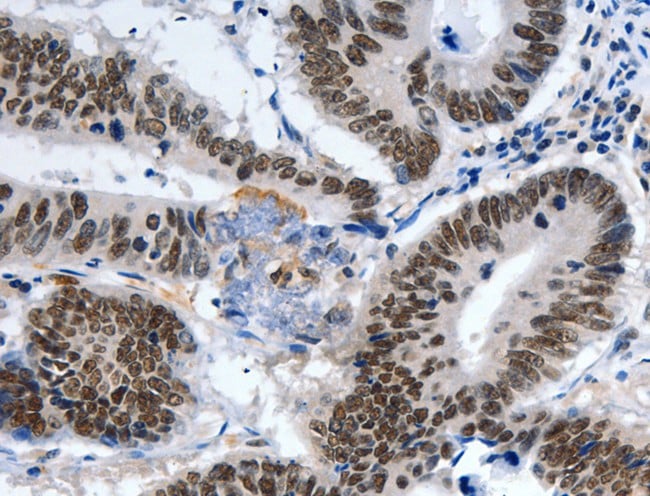
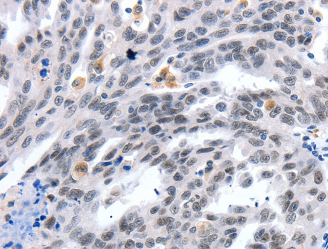
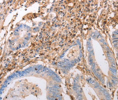 IHC (Immunohiostchemistry)
(Immunohistochemistry of paraffin-embedded human colon cancer tissue using INA antibody.)
IHC (Immunohiostchemistry)
(Immunohistochemistry of paraffin-embedded human colon cancer tissue using INA antibody.)
INA, Polyclonal Antibody (Cat# AAA168371)
 IHC (Immunohiostchemistry)
(Immunohistochemistry of paraffin-embedded Human colon cancer tissue using ACOX3 Polyclonal Antibody at dilution 1:35)
IHC (Immunohiostchemistry)
(Immunohistochemistry of paraffin-embedded Human colon cancer tissue using ACOX3 Polyclonal Antibody at dilution 1:35)
ACOX3, Polyclonal Antibody (Cat# AAA168373)
 IHC (Immunohiostchemistry)
(Immunohistochemistry of paraffin-embedded Human thyroid cancer tissue using RPS6KA6 Polyclonal Antibody at dilution 1:20)
IHC (Immunohiostchemistry)
(Immunohistochemistry of paraffin-embedded Human thyroid cancer tissue using RPS6KA6 Polyclonal Antibody at dilution 1:20)
RPS6KA6, Polyclonal Antibody (Cat# AAA168375)
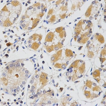
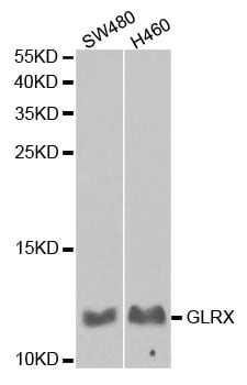
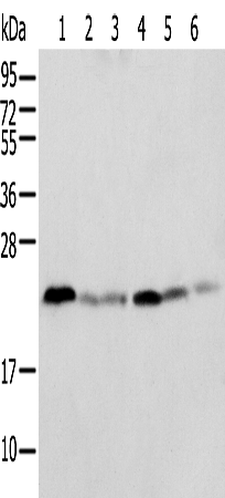
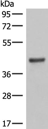
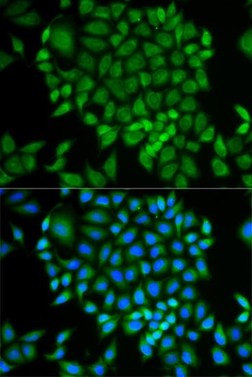 IF (Immunofluorescence)
(Immunofluorescence analysis of MCF-7 cell using GLRX antibody. Blue: DAPI for nuclear staining.)
IF (Immunofluorescence)
(Immunofluorescence analysis of MCF-7 cell using GLRX antibody. Blue: DAPI for nuclear staining.)
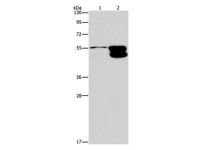
GLRX, Polyclonal Antibody (Cat# AAA168386)
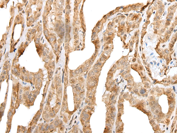
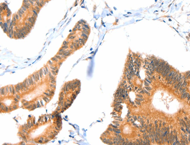
 IHC (Immunohistochemisry)
(Immunohistochemistry of paraffin-embedded Human lung cancer using RBFOX3 Polyclonal Antibody at dilution of 1:25)
IHC (Immunohistochemisry)
(Immunohistochemistry of paraffin-embedded Human lung cancer using RBFOX3 Polyclonal Antibody at dilution of 1:25)
RBFOX3, Polyclonal Antibody (Cat# AAA168391)
 IHC (Immunohiostchemistry)
(Immunohistochemistry of paraffin-embedded Human thyroid cancer tissue using GJC1 Polyclonal Antibody at dilution 1:50)
IHC (Immunohiostchemistry)
(Immunohistochemistry of paraffin-embedded Human thyroid cancer tissue using GJC1 Polyclonal Antibody at dilution 1:50)
GJC1, Polyclonal Antibody (Cat# AAA168392)
 IHC (Immunohiostchemistry)
(Immunohistochemistry of paraffin-embedded Human colon cancer tissue using MAP2K1 Polyclonal Antibody at dilution 1:20)
IHC (Immunohiostchemistry)
(Immunohistochemistry of paraffin-embedded Human colon cancer tissue using MAP2K1 Polyclonal Antibody at dilution 1:20)
MAP2K1, Polyclonal Antibody (Cat# AAA168394)
 IHC (Immunohiostchemistry)
(Immunohistochemistry of paraffin-embedded Human colon cancer tissue using CENPE Polyclonal Antibody at dilution 1:50)
IHC (Immunohiostchemistry)
(Immunohistochemistry of paraffin-embedded Human colon cancer tissue using CENPE Polyclonal Antibody at dilution 1:50)
CENPE, Polyclonal Antibody (Cat# AAA168395)

 IHC (Immunohiostchemistry)
(Immunohistochemistry of paraffin-embedded Human lung cancer using POU2F1 Polyclonal Antibody at dilution of 1:25)
IHC (Immunohiostchemistry)
(Immunohistochemistry of paraffin-embedded Human lung cancer using POU2F1 Polyclonal Antibody at dilution of 1:25)
POU2F1, Polyclonal Antibody (Cat# AAA168398)
 IHC (Immunohistochemisry)
(Immunohistochemistry of paraffin-embedded Human breast cancer using SRGAP1 Polyclonal Antibody at dilution of 1:25)
IHC (Immunohistochemisry)
(Immunohistochemistry of paraffin-embedded Human breast cancer using SRGAP1 Polyclonal Antibody at dilution of 1:25)
SRGAP1, Polyclonal Antibody (Cat# AAA168403)
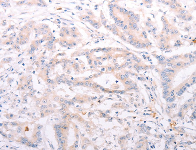
 IHC (Immunohiostchemistry)
(Immunohistochemistry of paraffin-embedded Human esophagus cancer tissue using S100A6 Polyclonal Antibody at dilution 1:35)
IHC (Immunohiostchemistry)
(Immunohistochemistry of paraffin-embedded Human esophagus cancer tissue using S100A6 Polyclonal Antibody at dilution 1:35)
S100A6, Polyclonal Antibody (Cat# AAA168404)
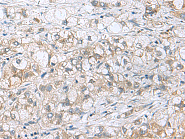
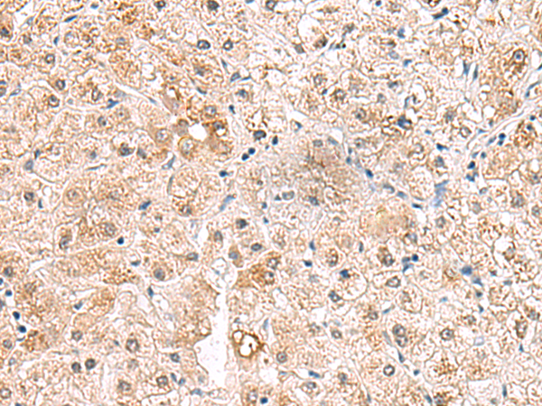
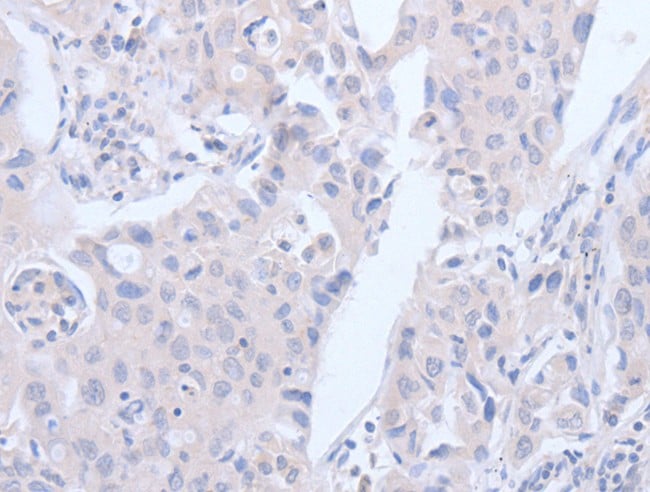
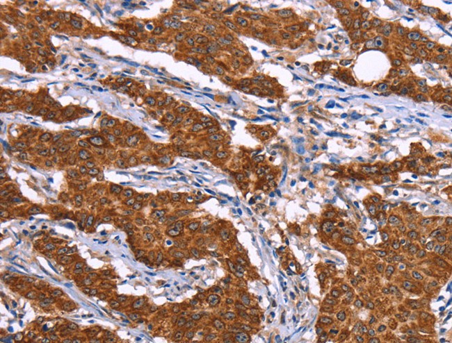
 IHC (Immunohistochemisry)
(Immunohistochemistry of paraffin-embedded Human liver cancer using BCL10 Polyclonal Antibody at dilution of 1:40)
IHC (Immunohistochemisry)
(Immunohistochemistry of paraffin-embedded Human liver cancer using BCL10 Polyclonal Antibody at dilution of 1:40)
BCL10, Polyclonal Antibody (Cat# AAA168407)
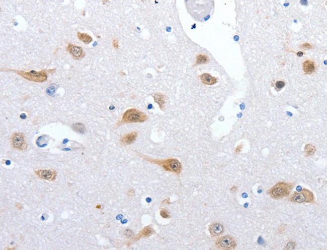
 IHC (Immunohistochemisry)
(Immunohistochemistry of paraffin-embedded Human brain using FLNA Polyclonal Antibody at dilution of 1:50)
IHC (Immunohistochemisry)
(Immunohistochemistry of paraffin-embedded Human brain using FLNA Polyclonal Antibody at dilution of 1:50)
FLNA, Polyclonal Antibody (Cat# AAA168411)
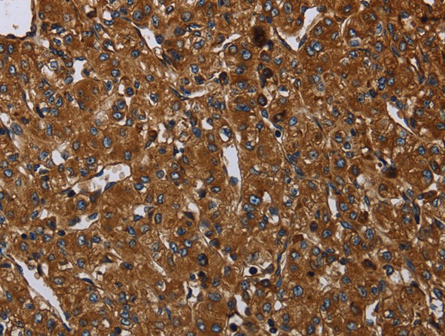
 IHC (Immunohistochemisry)
(Immunohistochemistry of paraffin-embedded Human liver cancer using SNX3 Polyclonal Antibody at dilution of 1:30)
IHC (Immunohistochemisry)
(Immunohistochemistry of paraffin-embedded Human liver cancer using SNX3 Polyclonal Antibody at dilution of 1:30)
SNX3, Polyclonal Antibody (Cat# AAA168418)
 IHC (Immunohistochemisry)
(Immunohistochemistry of paraffin-embedded Human thyroid cancer using PRKCD Polyclonal Antibody at dilution of 1:30)
IHC (Immunohistochemisry)
(Immunohistochemistry of paraffin-embedded Human thyroid cancer using PRKCD Polyclonal Antibody at dilution of 1:30)
PRKCD, Polyclonal Antibody (Cat# AAA168419)
 IHC (Immunohistochemisry)
(Immunohistochemistry of paraffin-embedded Human thyroid cancer using CRHR2 Polyclonal Antibody at dilution of 1:25)
IHC (Immunohistochemisry)
(Immunohistochemistry of paraffin-embedded Human thyroid cancer using CRHR2 Polyclonal Antibody at dilution of 1:25)
CRHR2, Polyclonal Antibody (Cat# AAA168420)
What are Polyclonal Antibodies?
Polyclonal antibodies are antibodies that come from multiple B cell clones of a host animal. The typical hosts used for the majority of polyclonal antibody production are rabbits, goats, sheep, and donkeys. These polyclonal antibodies, once having identified their target, will bind to different epitopes located at different regions or sequences on the same protein/antigen. As a result, they are ideal at locating and binding to the target, even if the target is in very low concentrations (due to many different antibodies being able to bind to the same target molecule, which allows for significant amplification of a downstream signal).
Polyclonal antibodies are typically produced by injecting an antigen into a host animal, which causes the animal’s immune system to attack the foreign antigen by mass generating antibodies against it. After a period of time, serum is collected from the animal and purified using physicochemical fractionation, class-specific affinity purification, and/or antigen-affinity purification.
Key Uses of Polyclonal Antibodies
- Western Blotting: This method is used to find specific proteins in biological samples after separating them by size.
- Immunohistochemistry: IHC helps visualize the location of proteins in tissue sections using various staining techniques.
- ELISA: (Enzyme-Linked Immunosorbent Assay) is typically used to identify specific protein quantities in a sample. ELISAs can be either “Quantitative” or “Qualitative”.
- Flow Cytometry: technique that identifies and measures the specific protein on the surface or inside the cells in a fluid suspension.
- Immunoprecipitation: IP isolates and studies a specific protein from a complex mixture using antibodies.
Why Buy Polyclonal Antibodies from AAA Biotech?
1. Ideal for Various Applications
Our antibodies are generally going to be validated for use in multiple types of assays, including ELISA, Western Blotting, Immunohistochemistry, Immunoprecipitation, amongst others. They are ideal for a wide range of research applications.
2. Rigorous Quality Control
All of the antibodies in our catalog undergo strict quality testing to ensure specificity, sensitivity, and consistent performance. We are confident in the ability of our antibodies to provide you with accurate results.
3. Wide Assortment of Antibodies
Antibodies in are catalog can be found for both common and exotic species, and these antibodies are also available in both conjugated and recombinant forms to suit many diverse experimental needs.
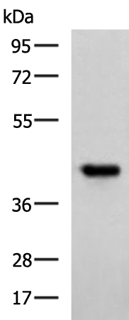
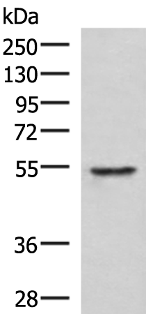
4. Highly Purified
Our antibodies are available in purified forms with over 85% purity, as confirmed by SDS-PAGE. They are also available with tags such as His, Flag, GST, or MBP. We cater to customers worldwide.
FAQ
1. How are polyclonal antibodies produced?
Traditionally, polyclonal antibodies are produced by injecting an antigen into a host animal (such as a rabbit or goat), which then triggers an immune response from the host animal. The animal’s B cells produce antibodies that will recognize different parts of the injected antigen. These antibodies are then collected from the animal’s blood and purified for use.
2. How do polyclonal antibodies differ from monoclonal antibodies?
Polyclonal antibodies are a mix of antibodies that bind to different locations (epitopes) of the same antigen, while monoclonal antibodies are identical and bind to just one specific epitope. This makes polyclonal antibodies more versatile and better at detecting proteins that may be present in low quantities or in altered/modified forms.
3. How should I store polyclonal antibodies?
Polyclonal antibodies should be stored at 4°C for short-term use (up to a few weeks) and at -20°C or -80°C for long-term storage. Avoid repeated freeze-thaw cycles by dividing them into small aliquots. Always check the datasheet for specific storage instructions.