Filters
▼Clonality
▼Type
▼Reactivity
▼Gene Name
▼Isotype
▼Host
▼Application
▼Clone
▼Polyclonal Antibodies
At AAA Biotech also known as AAA Bio or AAABio, we provide a broad range of purified polyclonal antibodies (pAbs) that are able to all be browsed online through our website. Due to their high specificity and strong binding affinity, these antibodies are ideal for wide swathes of research and experimental applications.
Our polyclonal antibodies can easily support your work, whether you use them for Western Blotting, Immunocytochemistry (with or without Immunofluorescence used in conjunction), Immunohistochemistry, Immunoprecipitation, and ELISA tests. We highly encourage you to browse our range of pAbs and choose the one that best suits your experimental model.
Viewing 2200-2250 of 96805 product results

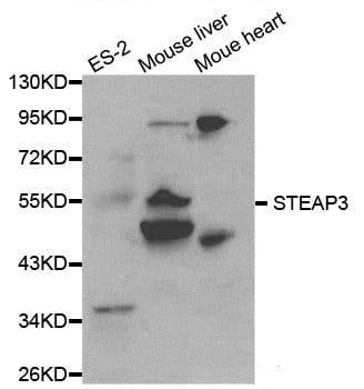 Application Data
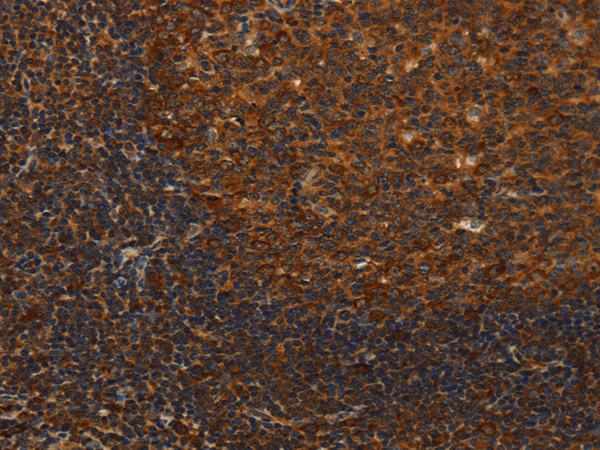
STEAP3, Polyclonal Antibody (Cat# AAA168209)

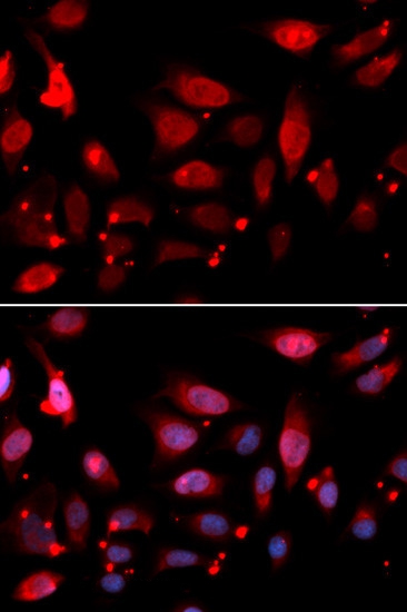 IF (Immunofluorescence)
(Immunofluorescence analysis of U20S cell using RACGAP1 antibody. Blue: DAPI for nuclear staining.)
IF (Immunofluorescence)
(Immunofluorescence analysis of U20S cell using RACGAP1 antibody. Blue: DAPI for nuclear staining.)
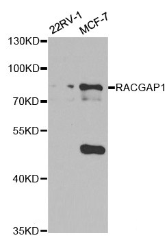
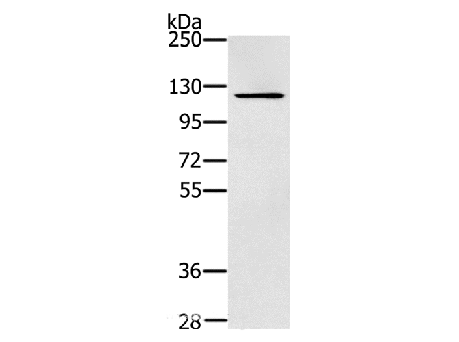
RACGAP1, Polyclonal Antibody (Cat# AAA168218)
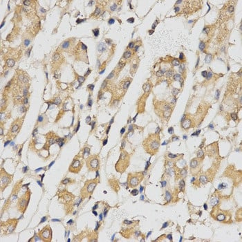
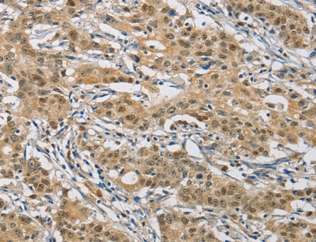
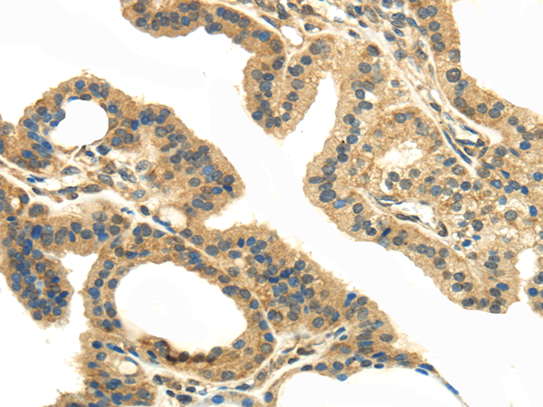
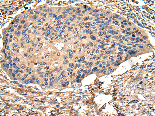
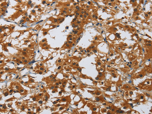
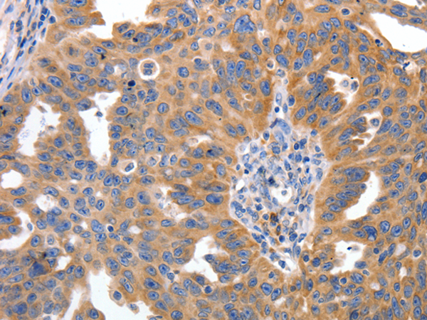
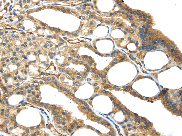
 IHC (Immunohistochemisry)
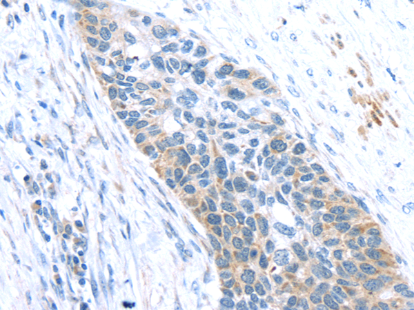
(Immunohistochemistry of paraffin-embedded Human thyroid cancer using CMTM6 Polyclonal Antibody at dilution of 1:40)
IHC (Immunohistochemisry)
(Immunohistochemistry of paraffin-embedded Human thyroid cancer using CMTM6 Polyclonal Antibody at dilution of 1:40)
CMTM6, Polyclonal Antibody (Cat# AAA168219)
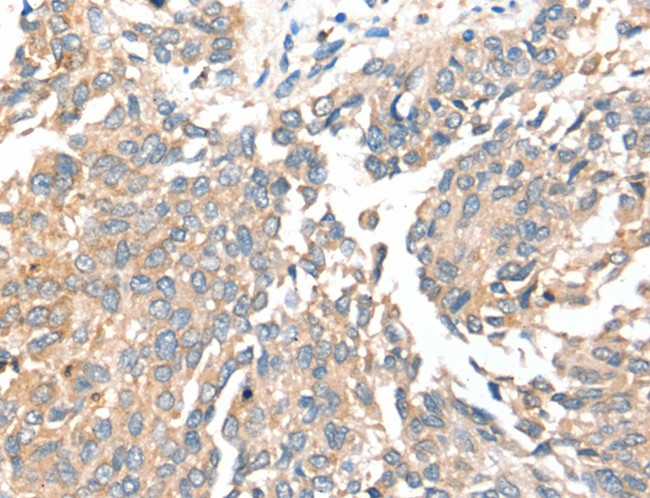
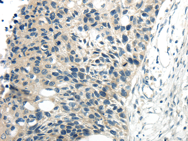
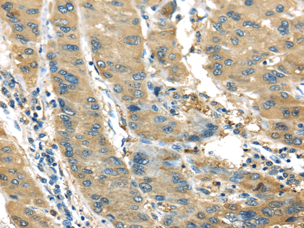
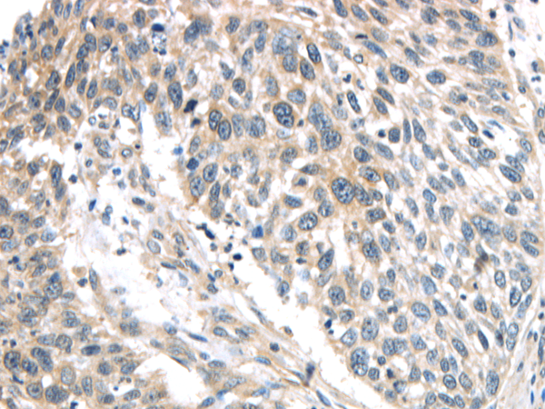
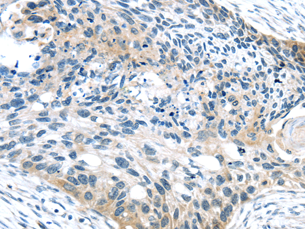
 IHC (Immunohistochemisry)
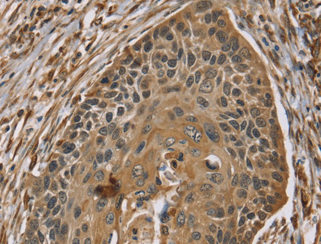
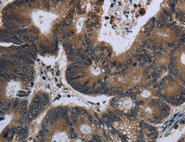
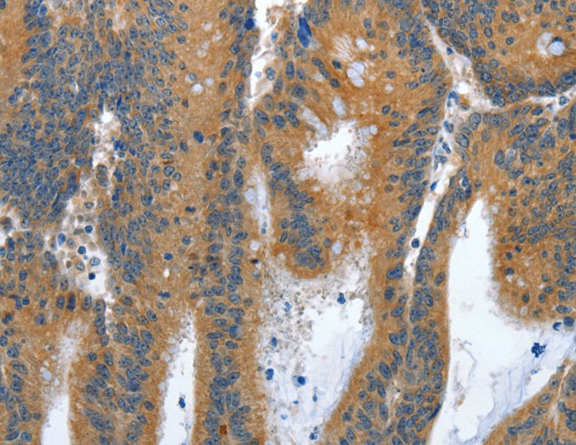
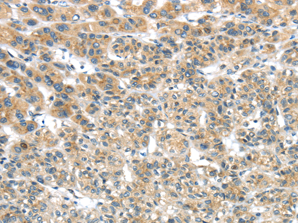
(Immunohistochemistry of paraffin-embedded Human esophagus cancer using ZMIZ1 Polyclonal Antibody at dilution of 1:40)
IHC (Immunohistochemisry)
(Immunohistochemistry of paraffin-embedded Human esophagus cancer using ZMIZ1 Polyclonal Antibody at dilution of 1:40)
ZMIZ1, Polyclonal Antibody (Cat# AAA168229)
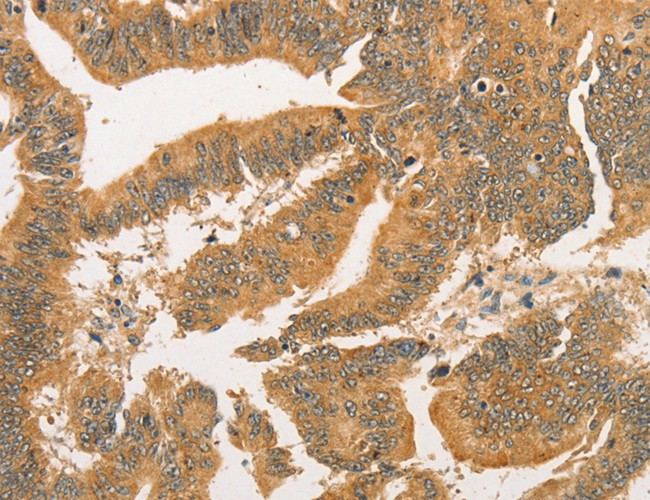
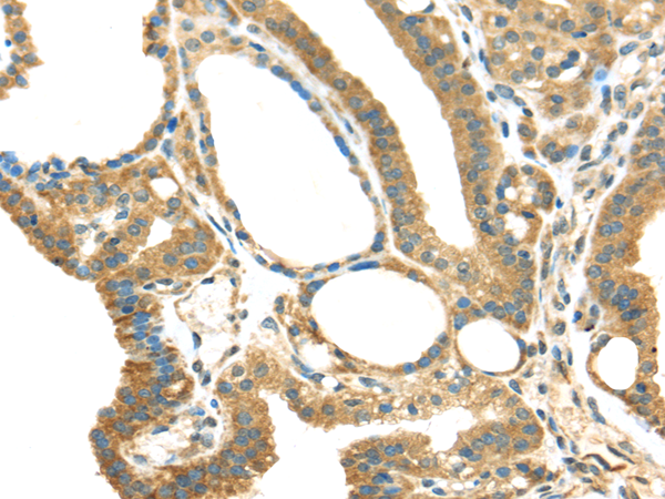
 IHC (Immunohistochemisry)
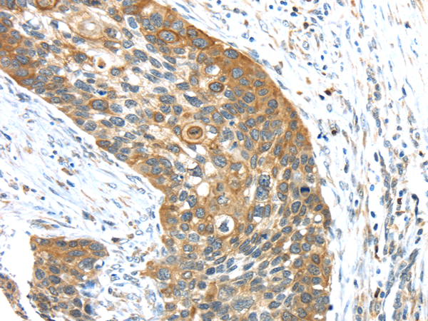
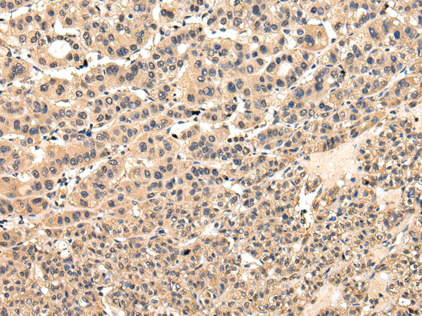
(Immunohistochemistry of paraffin-embedded Human thyroid cancer using SEC14L2 Polyclonal Antibody at dilution of 1:40)
IHC (Immunohistochemisry)
(Immunohistochemistry of paraffin-embedded Human thyroid cancer using SEC14L2 Polyclonal Antibody at dilution of 1:40)
SEC14L2, Polyclonal Antibody (Cat# AAA168230)
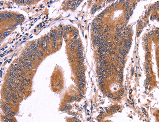
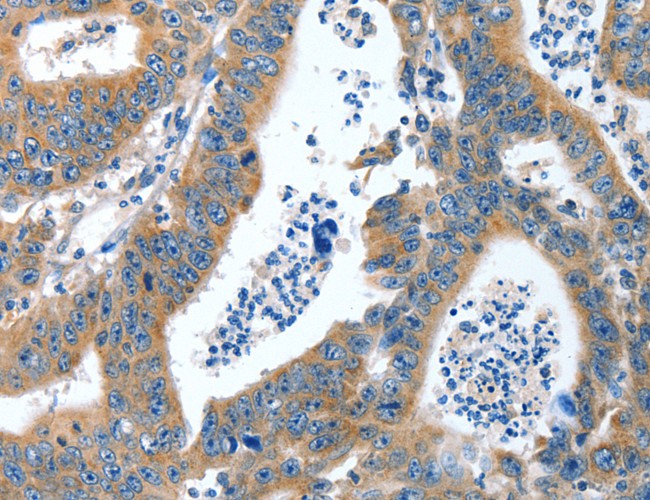
 IHC (Immunohistochemisry)
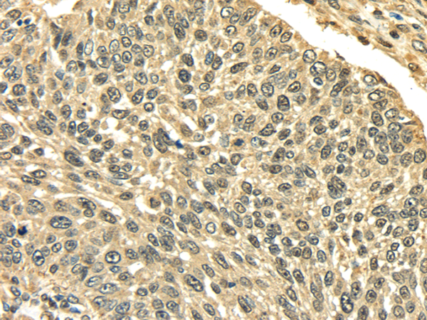
(Immunohistochemistry of paraffin-embedded Human thyroid cancer using ACOX2 Polyclonal Antibody at dilution of 1:20)
IHC (Immunohistochemisry)
(Immunohistochemistry of paraffin-embedded Human thyroid cancer using ACOX2 Polyclonal Antibody at dilution of 1:20)
ACOX2, Polyclonal Antibody (Cat# AAA168235)

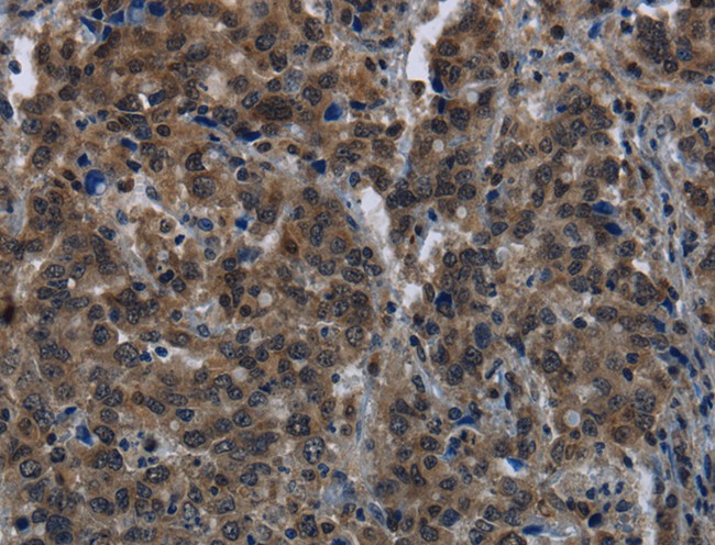
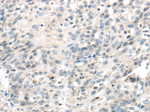
 IHC (Immunohistochemisry)
(Immunohistochemistry of paraffin-embedded Human gastric cancer using RAD50 Polyclonal Antibody at dilution of 1:40)
IHC (Immunohistochemisry)
(Immunohistochemistry of paraffin-embedded Human gastric cancer using RAD50 Polyclonal Antibody at dilution of 1:40)
RAD50, Polyclonal Antibody (Cat# AAA168240)
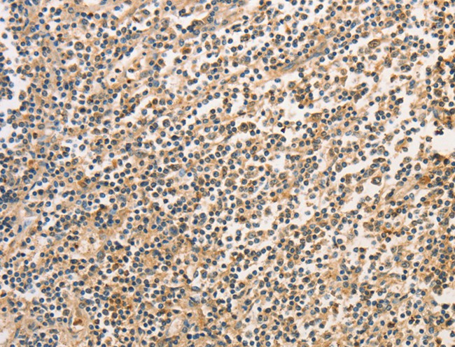
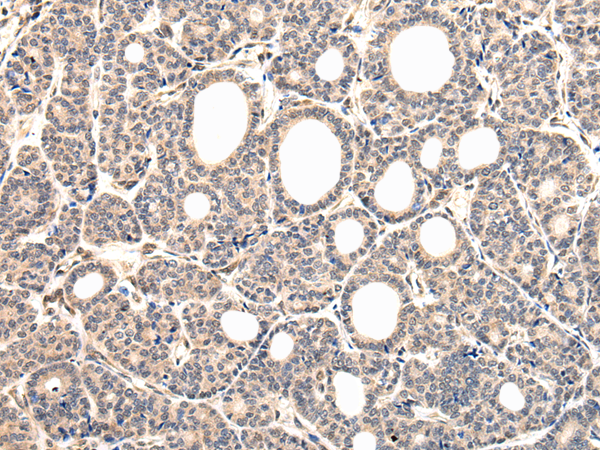
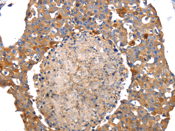
 IHC (Immunohiostchemistry)
(Immunohistochemistry of paraffin-embedded Human tonsil tissue using FPR3 Polyclonal Antibody at dilution 1:20)
IHC (Immunohiostchemistry)
(Immunohistochemistry of paraffin-embedded Human tonsil tissue using FPR3 Polyclonal Antibody at dilution 1:20)
FPR3, Polyclonal Antibody (Cat# AAA168245)
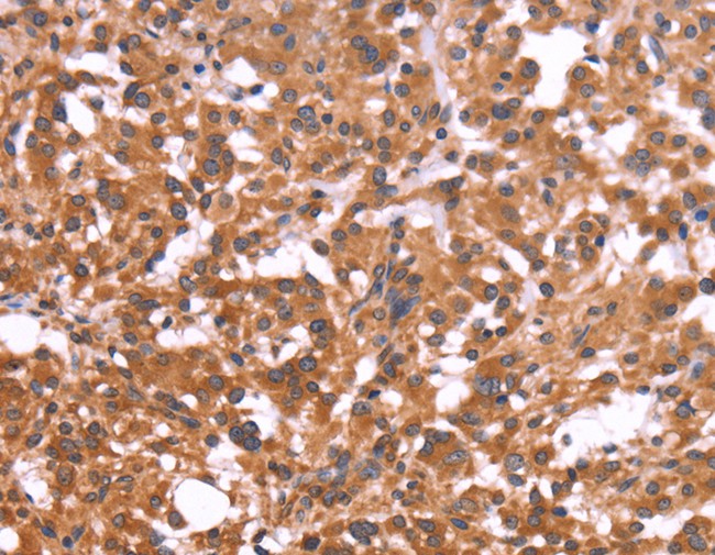
 IHC (Immunohistochemisry)
(Immunohistochemistry of paraffin-embedded Human gastric cancer using ICAM1 Polyclonal Antibody at dilution of 1:30)
IHC (Immunohistochemisry)
(Immunohistochemistry of paraffin-embedded Human gastric cancer using ICAM1 Polyclonal Antibody at dilution of 1:30)
ICAM1, Polyclonal Antibody (Cat# AAA168250)
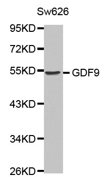
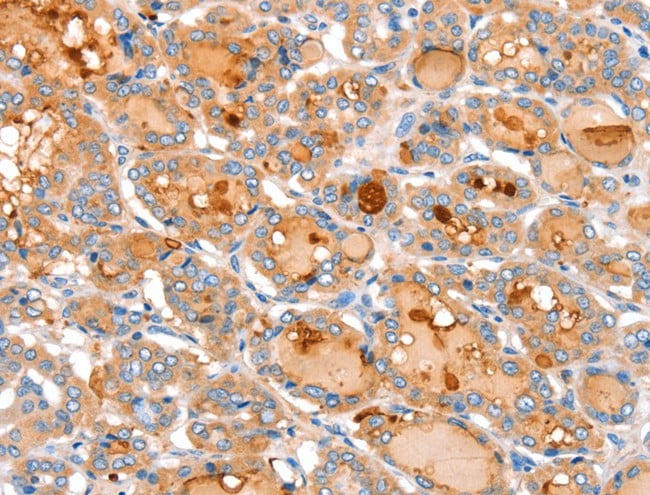
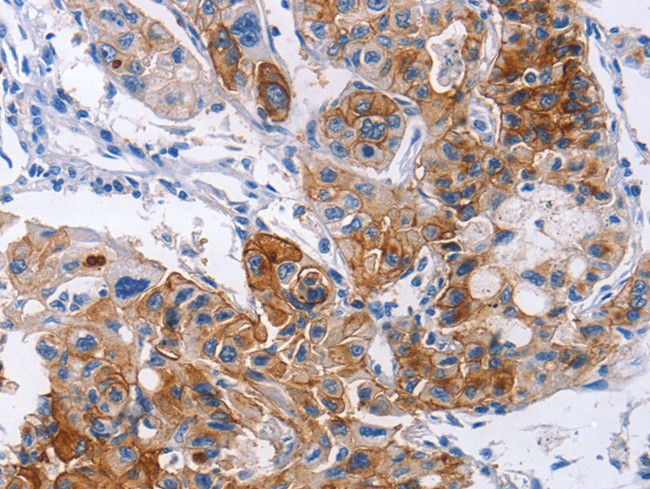
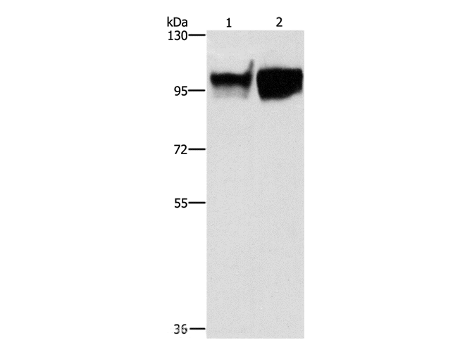
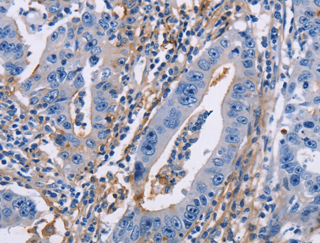
 IHC (Immunohiostchemistry)
(Immunohistochemistry of paraffin-embedded human stomach cancer tissue using GDF9 antibody.)
IHC (Immunohiostchemistry)
(Immunohistochemistry of paraffin-embedded human stomach cancer tissue using GDF9 antibody.)
GDF9, Polyclonal Antibody (Cat# AAA167870)
 IHC (Immunohiostchemistry)
(Immunohistochemistry of paraffin-embedded Human lung cancer tissue using SIRT4 Polyclonal Antibody at dilution 1:35)
IHC (Immunohiostchemistry)
(Immunohistochemistry of paraffin-embedded Human lung cancer tissue using SIRT4 Polyclonal Antibody at dilution 1:35)
SIRT4, Polyclonal Antibody (Cat# AAA167871)
 IHC (Immunohistochemisry)
(Immunohistochemistry of paraffin-embedded Human gastric cancer using NAPSA Polyclonal Antibody at dilution of 1:50)
IHC (Immunohistochemisry)
(Immunohistochemistry of paraffin-embedded Human gastric cancer using NAPSA Polyclonal Antibody at dilution of 1:50)
NAPSA, Polyclonal Antibody (Cat# AAA167875)
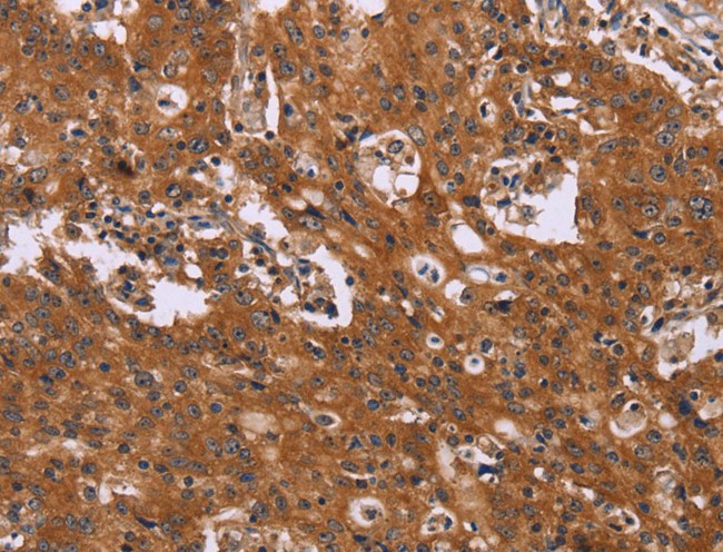
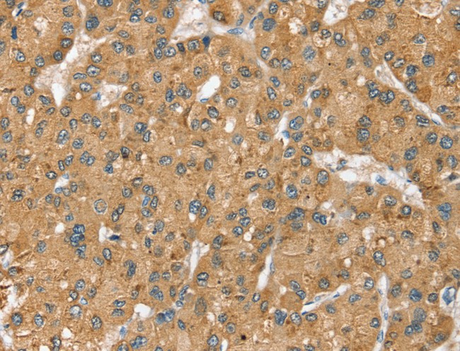
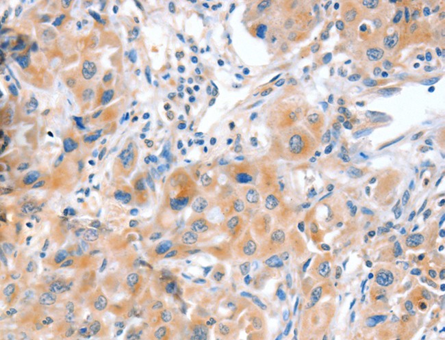
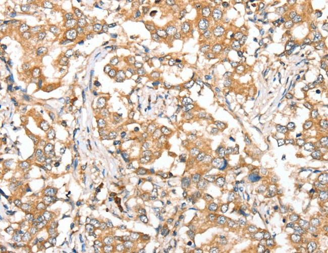
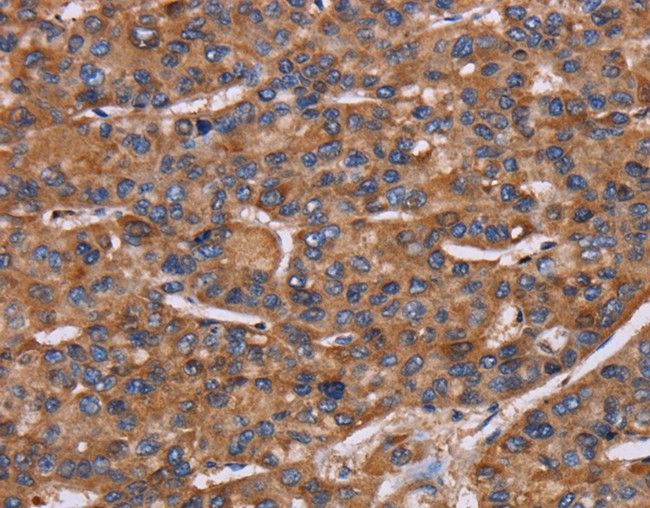
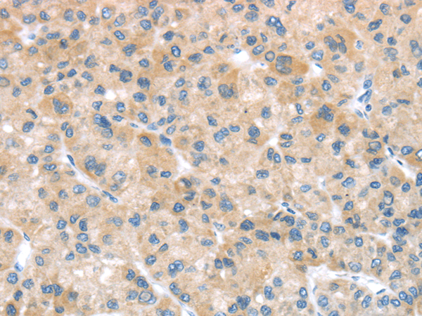
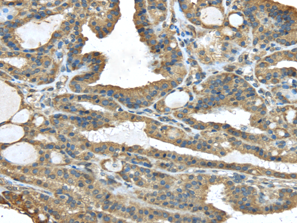
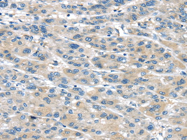
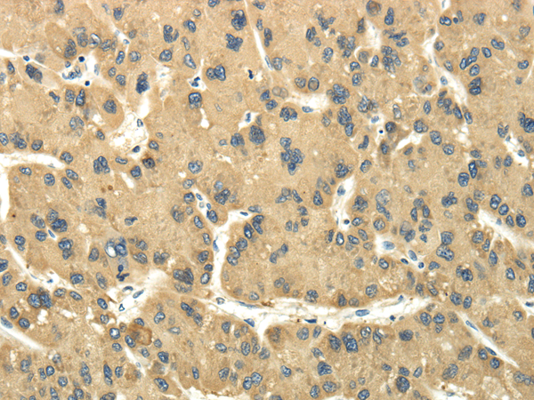
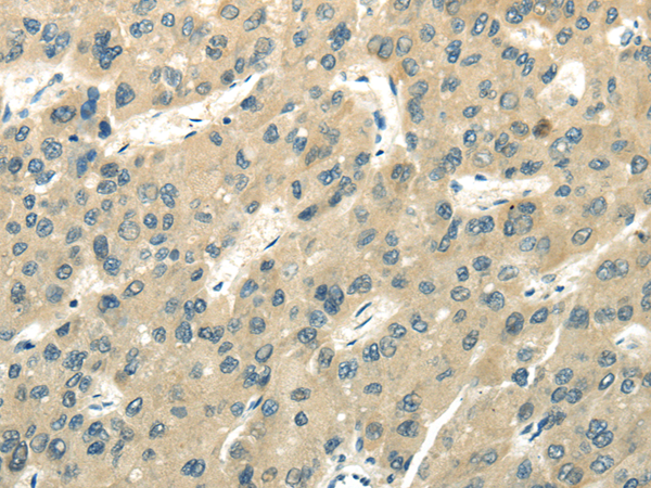
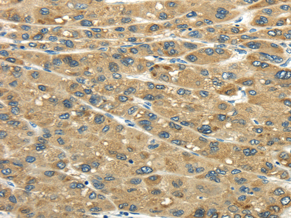
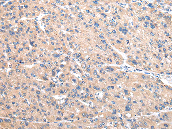
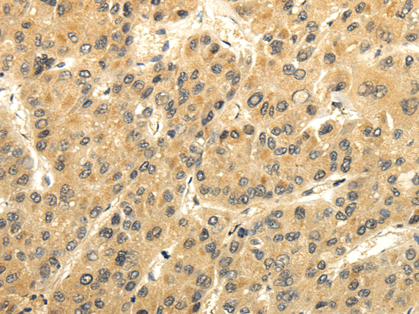
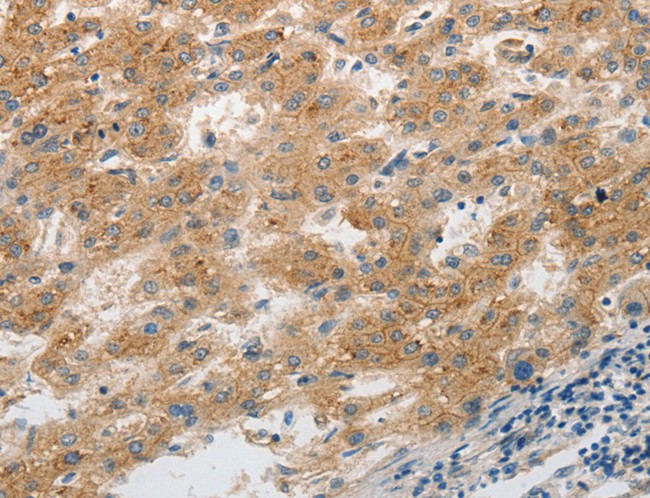 IHC (Immunohiostchemistry)
(Immunohistochemistry of paraffin-embedded Human liver cancer tissue using SLC1A2 Polyclonal Antibody at dilution 1:20)
IHC (Immunohiostchemistry)
(Immunohistochemistry of paraffin-embedded Human liver cancer tissue using SLC1A2 Polyclonal Antibody at dilution 1:20)
SLC1A2, Polyclonal Antibody (Cat# AAA167876)
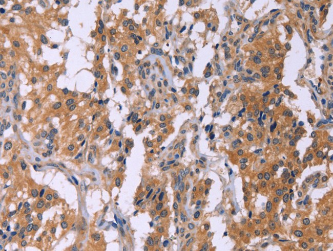
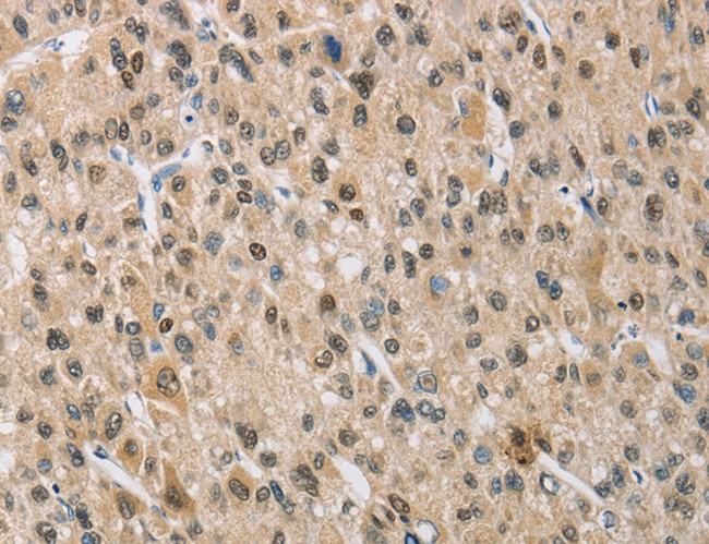
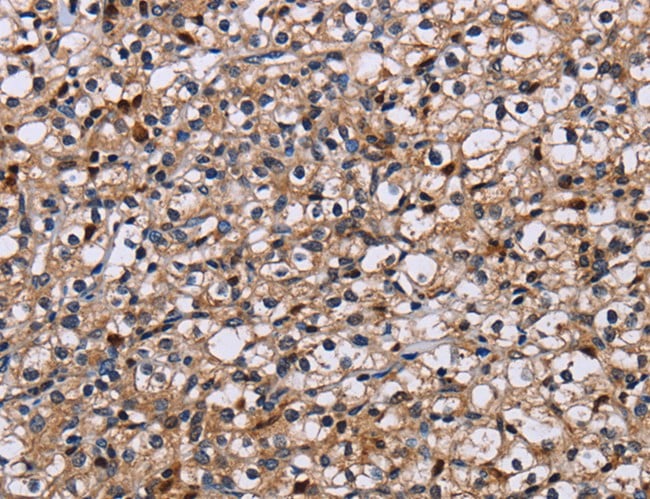
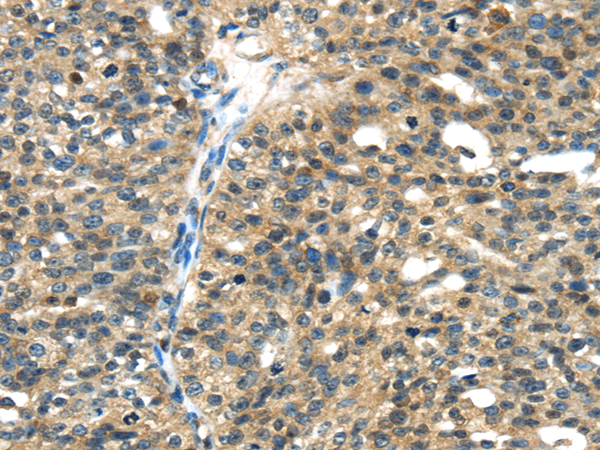
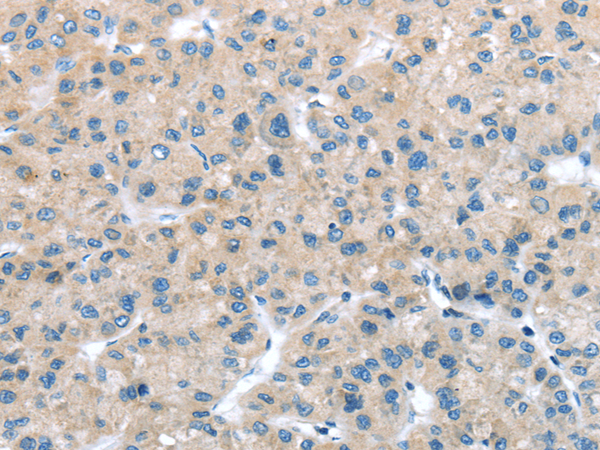
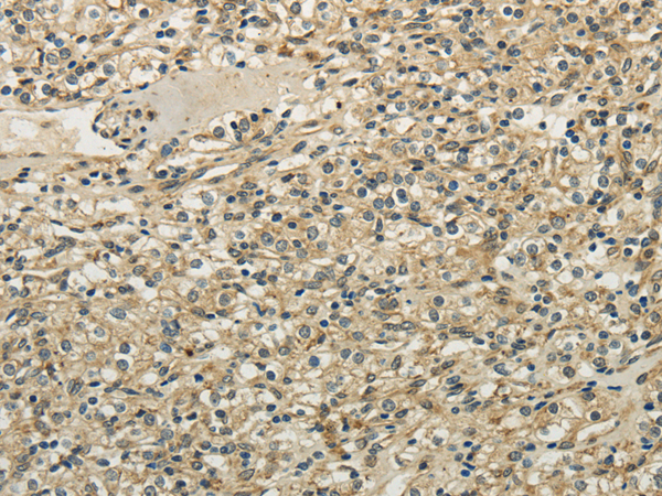
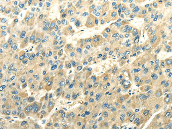
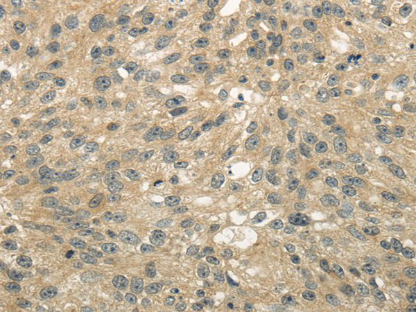
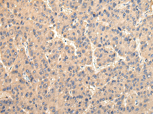
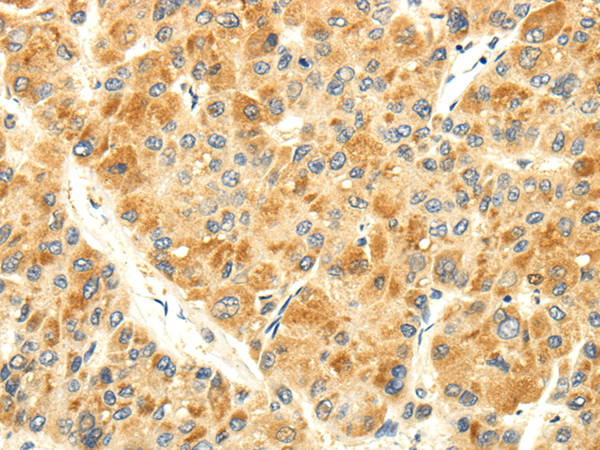
 IHC (Immunohiostchemistry)
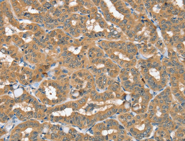
(Immunohistochemistry of paraffin-embedded Human liver cancer tissue using GK Polyclonal Antibody at dilution 1:60)
IHC (Immunohiostchemistry)
(Immunohistochemistry of paraffin-embedded Human liver cancer tissue using GK Polyclonal Antibody at dilution 1:60)
GK, Polyclonal Antibody (Cat# AAA167877)
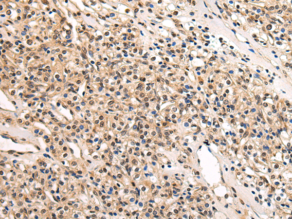
 IHC (Immunohiostchemistry)
(Immunohistochemistry of paraffin-embedded Human colon cancer tissue using CRISP3 Polyclonal Antibody at dilution 1:50)
IHC (Immunohiostchemistry)
(Immunohistochemistry of paraffin-embedded Human colon cancer tissue using CRISP3 Polyclonal Antibody at dilution 1:50)
CRISP3, Polyclonal Antibody (Cat# AAA167880)
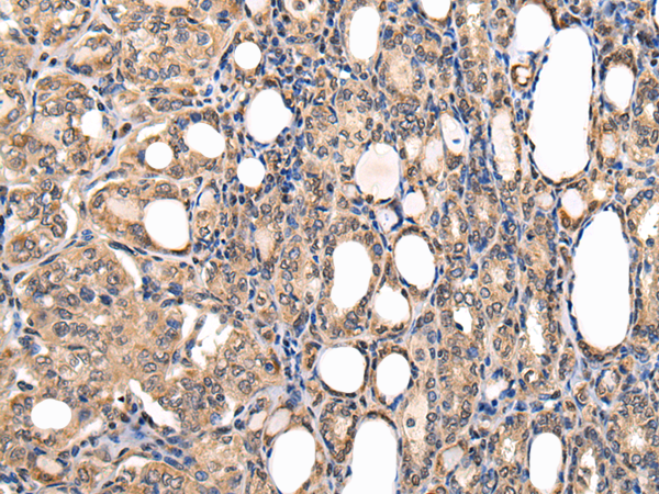
 IHC (Immunohiostchemistry)
(Immunohistochemistry of paraffin-embedded Human gastric cancer tissue using ANAPC10 Polyclonal Antibody at dilution 1:30)
IHC (Immunohiostchemistry)
(Immunohistochemistry of paraffin-embedded Human gastric cancer tissue using ANAPC10 Polyclonal Antibody at dilution 1:30)
ANAPC10, Polyclonal Antibody (Cat# AAA167897)
 IHC (Immunohiostchemistry)
(Immunohistochemistry of paraffin-embedded Human tonsil tissue using BCL2L14 Polyclonal Antibody at dilution 1:40)
IHC (Immunohiostchemistry)
(Immunohistochemistry of paraffin-embedded Human tonsil tissue using BCL2L14 Polyclonal Antibody at dilution 1:40)
BCL2L14, Polyclonal Antibody (Cat# AAA167898)
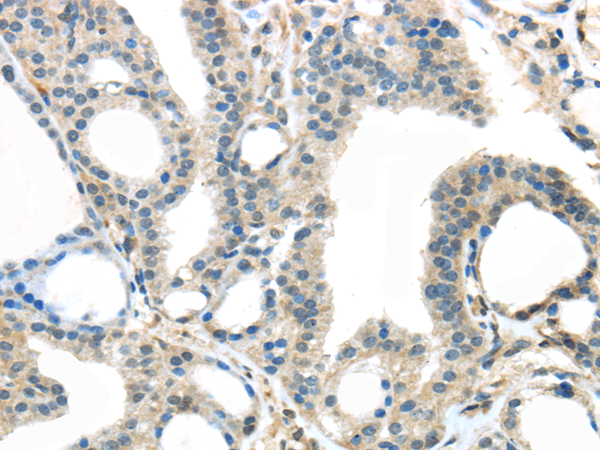
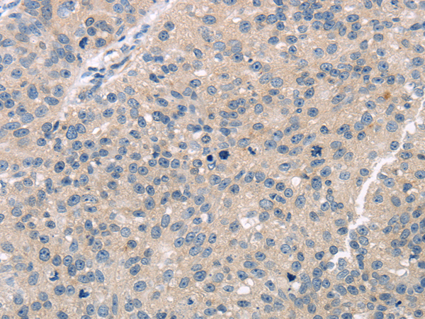
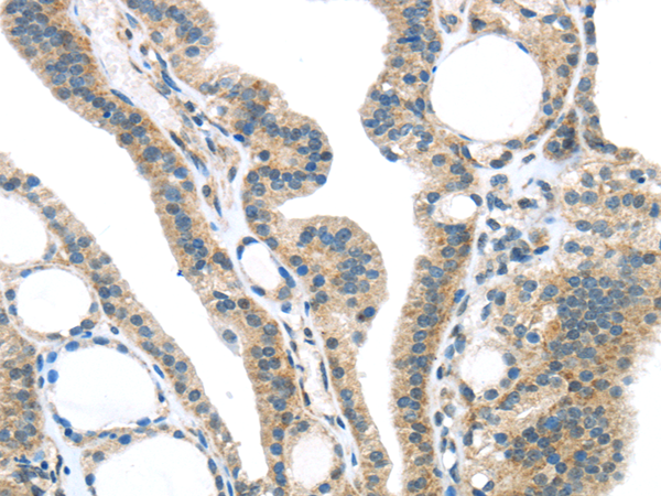
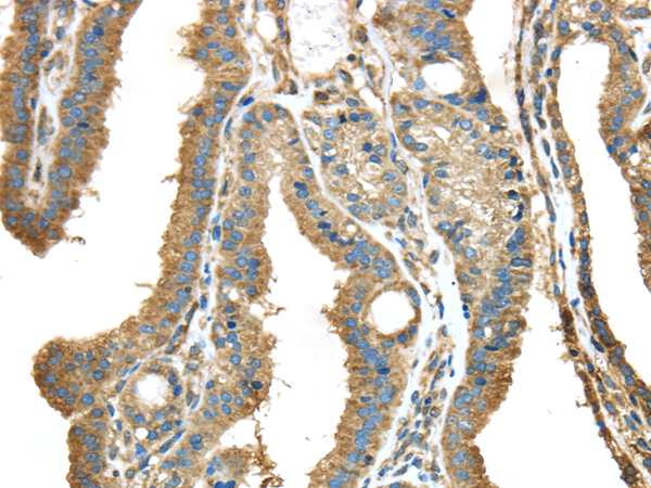
 IHC (Immunohistochemisry)
(Immunohistochemistry of paraffin-embedded Human prostate cancer using NCSTN Polyclonal Antibody at dilution of 1:50)
IHC (Immunohistochemisry)
(Immunohistochemistry of paraffin-embedded Human prostate cancer using NCSTN Polyclonal Antibody at dilution of 1:50)
NCSTN, Polyclonal Antibody (Cat# AAA167916)
 IHC (Immunohiostchemistry)
(Immunohistochemistry of paraffin-embedded Human esophagus cancer tissue using NDUFA12 Polyclonal Antibody at dilution 1:50)
IHC (Immunohiostchemistry)
(Immunohistochemistry of paraffin-embedded Human esophagus cancer tissue using NDUFA12 Polyclonal Antibody at dilution 1:50)
NDUFA12, Polyclonal Antibody (Cat# AAA167936)
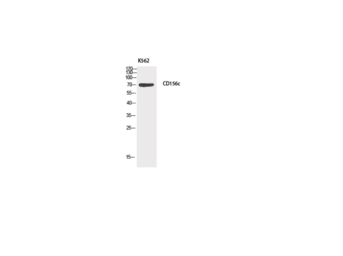
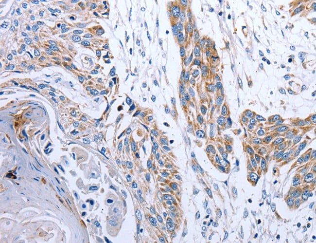
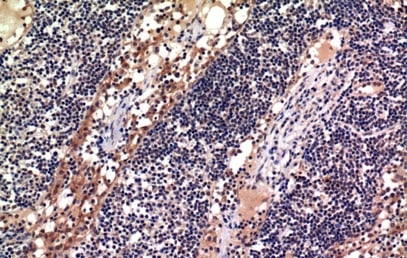 IHC (Immunohiostchemistry)
(Immunohistochemistry of paraffin-embedded Human lymph tissue using CD156c Polyclonal Antibody at dilution of 1:100.)
IHC (Immunohiostchemistry)
(Immunohistochemistry of paraffin-embedded Human lymph tissue using CD156c Polyclonal Antibody at dilution of 1:100.)
ADAM10, Polyclonal Antibody (Cat# AAA173605)
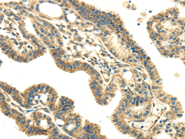
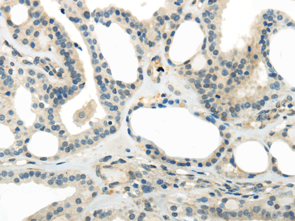
 IHC (Immunohiostchemistry)
(Immunohistochemistry of paraffin-embedded Human esophagus cancer tissue using SFTPA1 Polyclonal Antibody at dilution 1:40)
IHC (Immunohiostchemistry)
(Immunohistochemistry of paraffin-embedded Human esophagus cancer tissue using SFTPA1 Polyclonal Antibody at dilution 1:40)
SFTPA1, Polyclonal Antibody (Cat# AAA173039)
 IHC (Immunohiostchemistry)
(Immunohistochemistry of paraffin-embedded Human esophagus cancer tissue using TRIM16 Polyclonal Antibody at dilution 1:35)
IHC (Immunohiostchemistry)
(Immunohistochemistry of paraffin-embedded Human esophagus cancer tissue using TRIM16 Polyclonal Antibody at dilution 1:35)
TRIM16, Polyclonal Antibody (Cat# AAA173065)
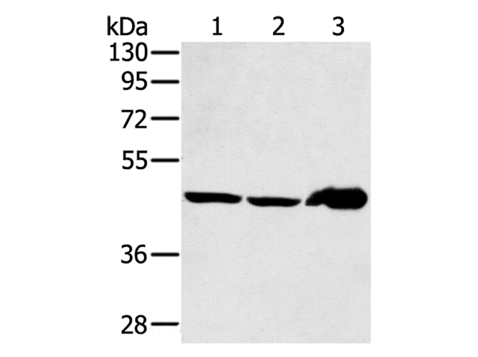
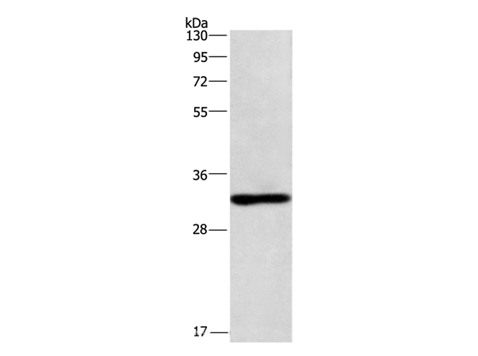
 WB (Western Blot)
(Western Blot analysis of 293T, 231 and Hela cells using PLIN3 Polyclonal Antibody at dilution of 1/800)
WB (Western Blot)
(Western Blot analysis of 293T, 231 and Hela cells using PLIN3 Polyclonal Antibody at dilution of 1/800)
PLIN3, Polyclonal Antibody (Cat# AAA173069)
 IHC (Immunohiostchemistry)
(Immunohistochemistry of paraffin-embedded Human esophagus cancer tissue using TKTL1 Polyclonal Antibody at dilution 1:40)
IHC (Immunohiostchemistry)
(Immunohistochemistry of paraffin-embedded Human esophagus cancer tissue using TKTL1 Polyclonal Antibody at dilution 1:40)
TKTL1, Polyclonal Antibody (Cat# AAA173071)
 IHC (Immunohiostchemistry)
(Immunohistochemistry of paraffin-embedded Human thyroid cancer using TMED1 Polyclonal Antibody at dilution of 1/35)
IHC (Immunohiostchemistry)
(Immunohistochemistry of paraffin-embedded Human thyroid cancer using TMED1 Polyclonal Antibody at dilution of 1/35)
TMED1, Polyclonal Antibody (Cat# AAA173072)

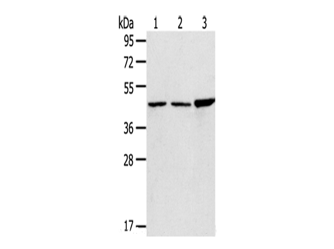
 WB (Western Blot)
(Western Blot analysis of A431 cells using TMPRSS4 Polyclonal Antibody at dilution of 1/500)
WB (Western Blot)
(Western Blot analysis of A431 cells using TMPRSS4 Polyclonal Antibody at dilution of 1/500)
TMPRSS4, Polyclonal Antibody (Cat# AAA173078)
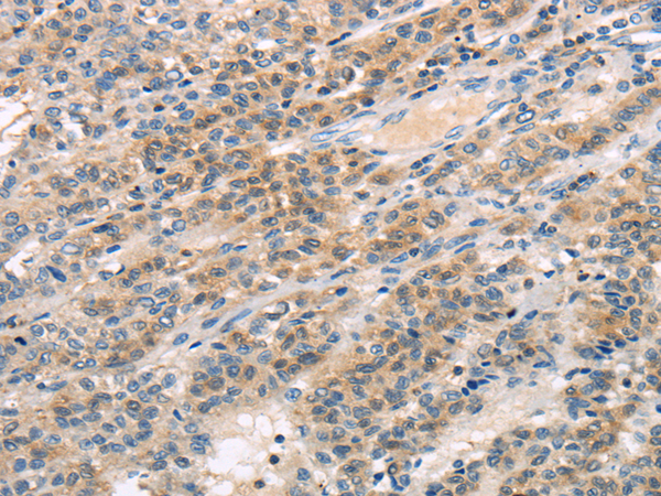
 IHC (Immunohiostchemistry)
(Immunohistochemistry of paraffin-embedded Human breast cancer using TGM5 Polyclonal Antibody at dilution of 1/30)
IHC (Immunohiostchemistry)
(Immunohistochemistry of paraffin-embedded Human breast cancer using TGM5 Polyclonal Antibody at dilution of 1/30)
TGM5, Polyclonal Antibody (Cat# AAA173096)
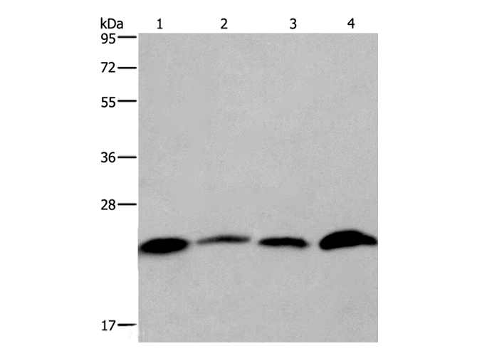
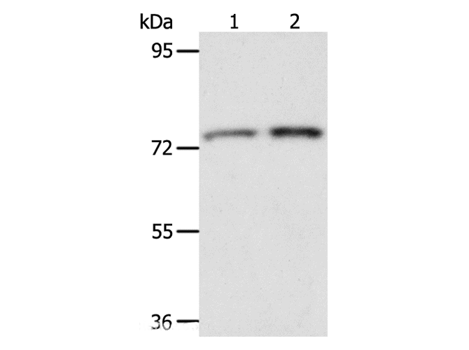
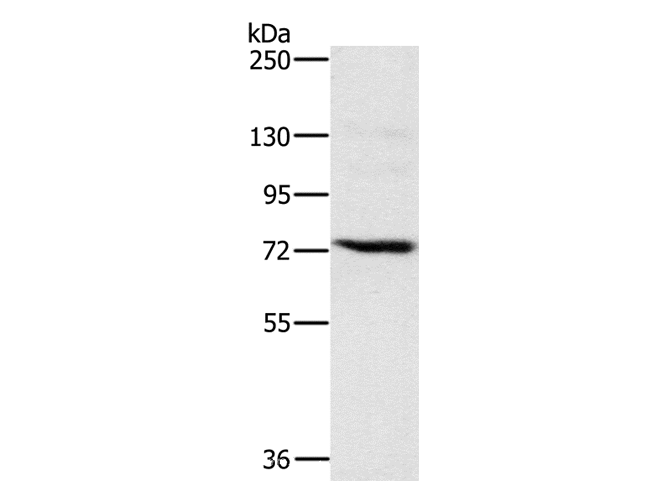
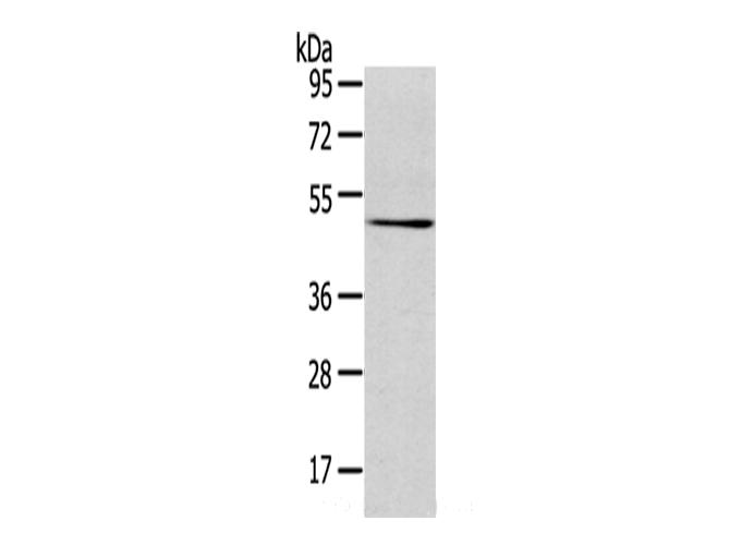
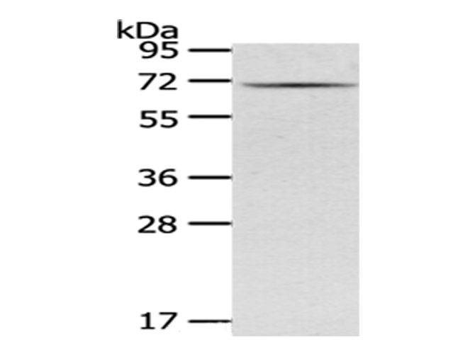
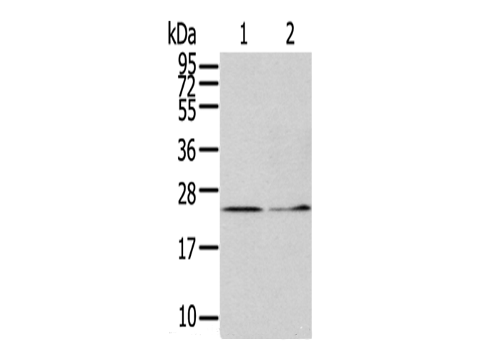 WB (Western Blot)
(Western Blot analysis of Hela cells and Human fetal liver tissue using ITPA Polyclonal Antibody at dilution of 1/400)
WB (Western Blot)
(Western Blot analysis of Hela cells and Human fetal liver tissue using ITPA Polyclonal Antibody at dilution of 1/400)
ITPA, Polyclonal Antibody (Cat# AAA173109)
 IHC (Immunohiostchemistry)
(Immunohistochemistry of paraffin-embedded Human thyroid cancer tissue using TRPM2 Polyclonal Antibody at dilution 1:30)
IHC (Immunohiostchemistry)
(Immunohistochemistry of paraffin-embedded Human thyroid cancer tissue using TRPM2 Polyclonal Antibody at dilution 1:30)
TRPM2, Polyclonal Antibody (Cat# AAA173114)
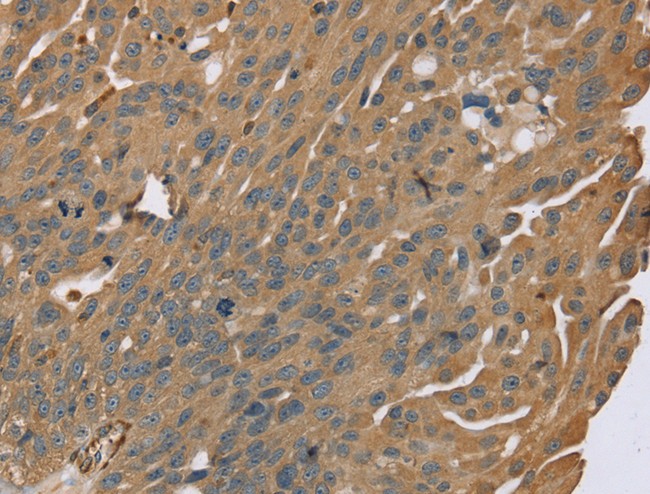
 IHC (Immunohiostchemistry)
(Immunohistochemistry of paraffin-embedded Human liver cancer using TTC23 Polyclonal Antibody at dilution of 1/30)
IHC (Immunohiostchemistry)
(Immunohistochemistry of paraffin-embedded Human liver cancer using TTC23 Polyclonal Antibody at dilution of 1/30)
TTC23, Polyclonal Antibody (Cat# AAA173116)
 IHC (Immunohiostchemistry)
(Immunohistochemistry of paraffin-embedded Human prostate cancer tissue using TULP2 Polyclonal Antibody at dilution 1:30)
IHC (Immunohiostchemistry)
(Immunohistochemistry of paraffin-embedded Human prostate cancer tissue using TULP2 Polyclonal Antibody at dilution 1:30)
TULP2, Polyclonal Antibody (Cat# AAA173120)
 IHC (Immunohiostchemistry)
(Immunohistochemistry of paraffin-embedded Human thyroid cancer tissue using UBL4A Polyclonal Antibody at dilution 1:30)
IHC (Immunohiostchemistry)
(Immunohistochemistry of paraffin-embedded Human thyroid cancer tissue using UBL4A Polyclonal Antibody at dilution 1:30)
UBL4A, Polyclonal Antibody (Cat# AAA173125)
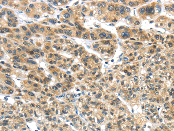
 IHC (Immunohiostchemistry)
(Immunohistochemistry of paraffin-embedded Human thyroid cancer using DLAT Polyclonal Antibody at dilution of 1/30)
IHC (Immunohiostchemistry)
(Immunohistochemistry of paraffin-embedded Human thyroid cancer using DLAT Polyclonal Antibody at dilution of 1/30)
DLAT, Polyclonal Antibody (Cat# AAA173144)
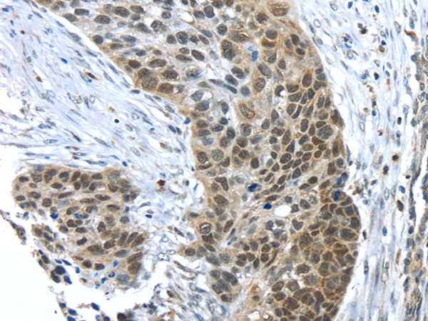
 IHC (Immunohiostchemistry)
(Immunohistochemistry of paraffin-embedded Human breast cancer tissue using UGT2B4 Polyclonal Antibody at dilution 1:45)
IHC (Immunohiostchemistry)
(Immunohistochemistry of paraffin-embedded Human breast cancer tissue using UGT2B4 Polyclonal Antibody at dilution 1:45)
UGT2B4, Polyclonal Antibody (Cat# AAA173149)
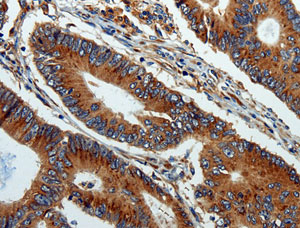
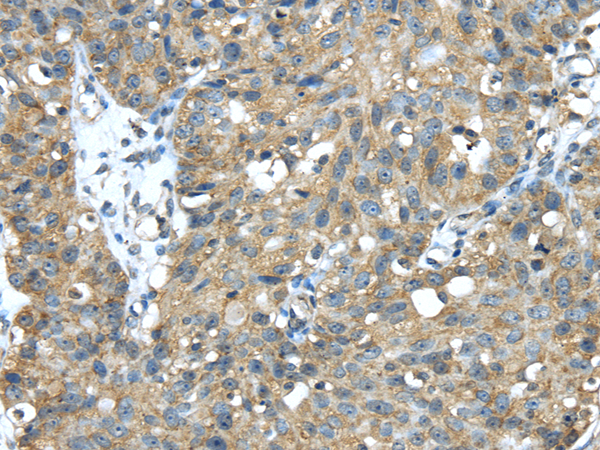
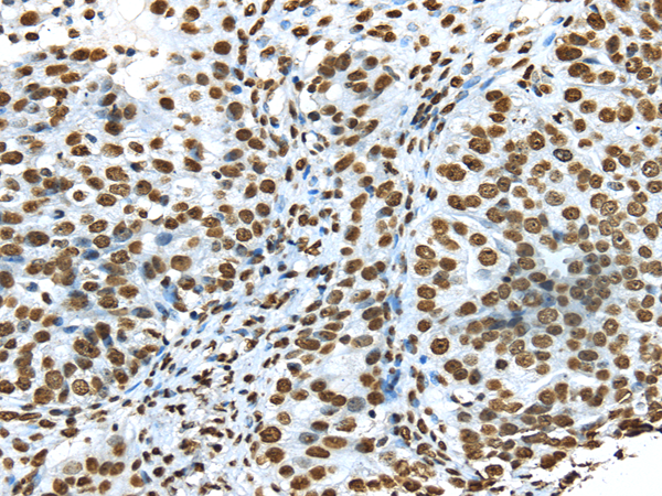
 IHC (Immunohiostchemistry)
(Immunohistochemistry of paraffin-embedded Human thyroid cancer tissue using USF1 Polyclonal Antibody at dilution 1:30)
IHC (Immunohiostchemistry)
(Immunohistochemistry of paraffin-embedded Human thyroid cancer tissue using USF1 Polyclonal Antibody at dilution 1:30)
USF1, Polyclonal Antibody (Cat# AAA173155)
 IHC (Immunohiostchemistry)
(Immunohistochemistry of paraffin-embedded Human esophagus cancer tissue using USP15 Polyclonal Antibody at dilution 1:35)
IHC (Immunohiostchemistry)
(Immunohistochemistry of paraffin-embedded Human esophagus cancer tissue using USP15 Polyclonal Antibody at dilution 1:35)
USP15, Polyclonal Antibody (Cat# AAA173158)
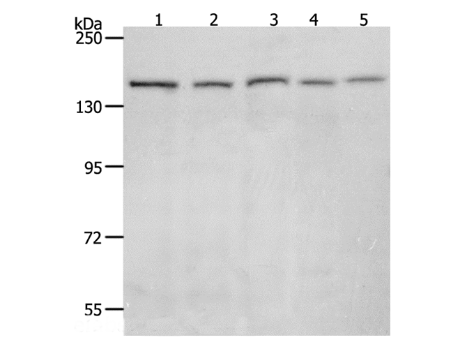
 WB (Western Blot)
(Western Blot analysis of PC3, TM4, hela and K562 cell using VAMP4 Polyclonal Antibody at dilution of 1/650)
WB (Western Blot)
(Western Blot analysis of PC3, TM4, hela and K562 cell using VAMP4 Polyclonal Antibody at dilution of 1/650)
VAMP4, Polyclonal Antibody (Cat# AAA173165)
 IHC (Immunohiostchemistry)
(Immunohistochemistry of paraffin-embedded Human thyroid cancer tissue using VPS37D Polyclonal Antibody at dilution 1:30)
IHC (Immunohiostchemistry)
(Immunohistochemistry of paraffin-embedded Human thyroid cancer tissue using VPS37D Polyclonal Antibody at dilution 1:30)
VPS37D, Polyclonal Antibody (Cat# AAA173190)
 IHC (Immunohiostchemistry)
(Immunohistochemistry of paraffin-embedded Human thyroid cancer tissue using CYP19A1 Polyclonal Antibody at dilution 1:30)
IHC (Immunohiostchemistry)
(Immunohistochemistry of paraffin-embedded Human thyroid cancer tissue using CYP19A1 Polyclonal Antibody at dilution 1:30)
CYP19A1, Polyclonal Antibody (Cat# AAA173197)
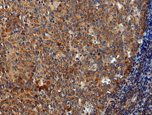
 IHC (Immunohiostchemistry)
(Immunohistochemistry of paraffin-embedded Human thyroid cancer tissue using MIP Polyclonal Antibody at dilution 1:40)
IHC (Immunohiostchemistry)
(Immunohistochemistry of paraffin-embedded Human thyroid cancer tissue using MIP Polyclonal Antibody at dilution 1:40)
MIP, Polyclonal Antibody (Cat# AAA173205)
 IHC (Immunohiostchemistry)
(Immunohistochemistry of paraffin-embedded Human thyroid cancer tissue using IL3RA Polyclonal Antibody at dilution 1:35)
IHC (Immunohiostchemistry)
(Immunohistochemistry of paraffin-embedded Human thyroid cancer tissue using IL3RA Polyclonal Antibody at dilution 1:35)
IL3RA, Polyclonal Antibody (Cat# AAA173218)
 IHC (Immunohiostchemistry)
(Immunohistochemistry of paraffin-embedded Human ovarian cancer using IQGAP2 Polyclonal Antibody at dilution of 1/40)
IHC (Immunohiostchemistry)
(Immunohistochemistry of paraffin-embedded Human ovarian cancer using IQGAP2 Polyclonal Antibody at dilution of 1/40)
IQGAP2, Polyclonal Antibody (Cat# AAA173221)
 IHC (Immunohiostchemistry)
(Immunohistochemistry of paraffin-embedded Human breast cancer tissue using IRS2 Polyclonal Antibody at dilution 1:100)
IHC (Immunohiostchemistry)
(Immunohistochemistry of paraffin-embedded Human breast cancer tissue using IRS2 Polyclonal Antibody at dilution 1:100)
IRS2, Polyclonal Antibody (Cat# AAA173223)
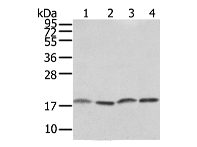
 WB (Western Blot)
(Western Blot analysis of Mouse brain tissue using GAD1 Polyclonal Antibody at dilution of 1/200)
WB (Western Blot)
(Western Blot analysis of Mouse brain tissue using GAD1 Polyclonal Antibody at dilution of 1/200)
GAD1, Polyclonal Antibody (Cat# AAA173240)
 IHC (Immunohiostchemistry)
(Immunohistochemistry of paraffin-embedded Human prostate cancer tissue using S1PR2 Polyclonal Antibody at dilution 1:30)
IHC (Immunohiostchemistry)
(Immunohistochemistry of paraffin-embedded Human prostate cancer tissue using S1PR2 Polyclonal Antibody at dilution 1:30)
S1PR2, Polyclonal Antibody (Cat# AAA173244)
 IHC (Immunohiostchemistry)
(Immunohistochemistry of paraffin-embedded Human esophagus cancer tissue using SPDL1 Polyclonal Antibody at dilution 1:35)
IHC (Immunohiostchemistry)
(Immunohistochemistry of paraffin-embedded Human esophagus cancer tissue using SPDL1 Polyclonal Antibody at dilution 1:35)
SPDL1, Polyclonal Antibody (Cat# AAA173259)
 IHC (Immunohiostchemistry)
(Immunohistochemistry of paraffin-embedded Human thyroid cancer using ISM2 Polyclonal Antibody at dilution of 1/45)
IHC (Immunohiostchemistry)
(Immunohistochemistry of paraffin-embedded Human thyroid cancer using ISM2 Polyclonal Antibody at dilution of 1/45)
ISM2, Polyclonal Antibody (Cat# AAA173265)
 IHC (Immunohiostchemistry)
(Immunohistochemistry of paraffin-embedded Human cervical cancer using NME2 Polyclonal Antibody at dilution of 1/40)
IHC (Immunohiostchemistry)
(Immunohistochemistry of paraffin-embedded Human cervical cancer using NME2 Polyclonal Antibody at dilution of 1/40)
NME2, Polyclonal Antibody (Cat# AAA173269)
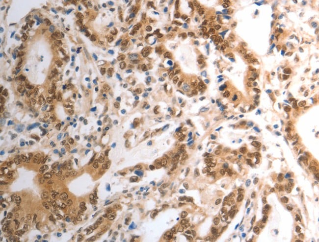
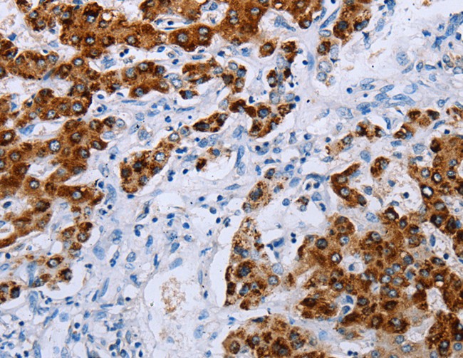
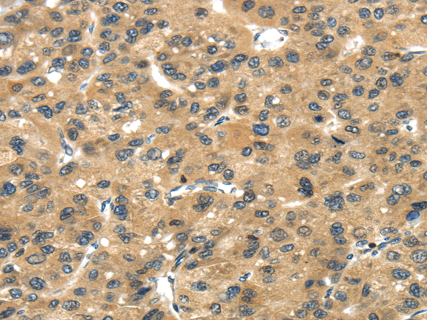
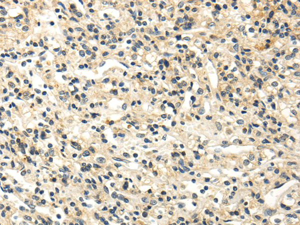
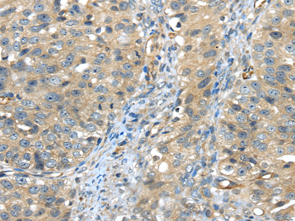
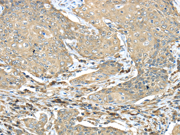
 IHC (Immunohiostchemistry)
(Immunohistochemistry of paraffin-embedded Human liver cancer tissue using TRIM27 Polyclonal Antibody at dilution 1:35)
IHC (Immunohiostchemistry)
(Immunohistochemistry of paraffin-embedded Human liver cancer tissue using TRIM27 Polyclonal Antibody at dilution 1:35)
TRIM27, Polyclonal Antibody (Cat# AAA173271)
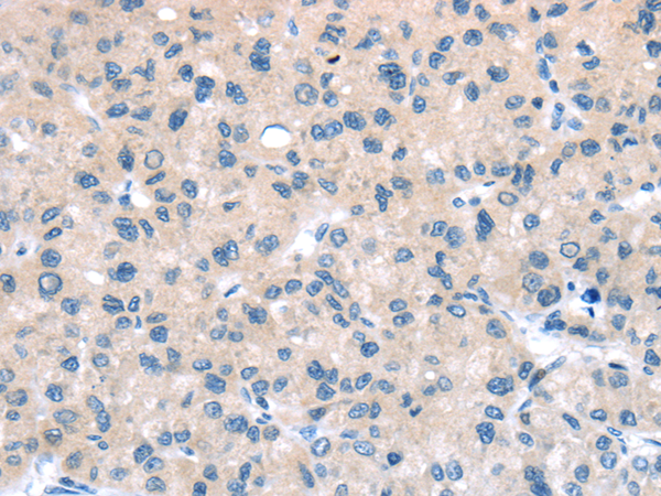
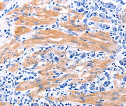
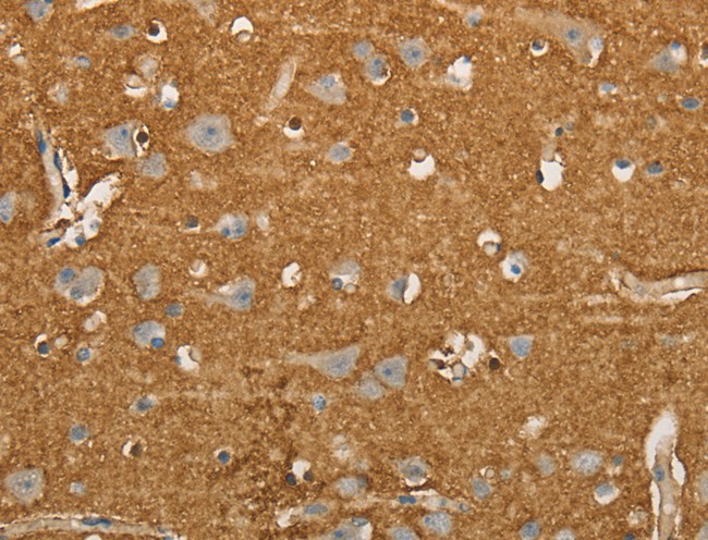
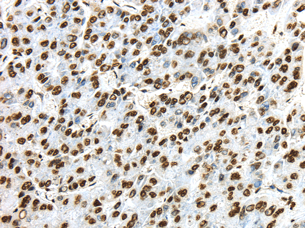
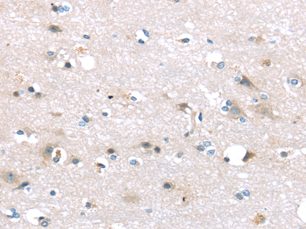 IHC (Immunohiostchemistry)
(Immunohistochemistry of paraffin-embedded Human brain tissue using TTI1 Polyclonal Antibody at dilution 1:40)
IHC (Immunohiostchemistry)
(Immunohistochemistry of paraffin-embedded Human brain tissue using TTI1 Polyclonal Antibody at dilution 1:40)
TTI1, Polyclonal Antibody (Cat# AAA173283)
What are Polyclonal Antibodies?
Polyclonal antibodies are antibodies that come from multiple B cell clones of a host animal. The typical hosts used for the majority of polyclonal antibody production are rabbits, goats, sheep, and donkeys. These polyclonal antibodies, once having identified their target, will bind to different epitopes located at different regions or sequences on the same protein/antigen. As a result, they are ideal at locating and binding to the target, even if the target is in very low concentrations (due to many different antibodies being able to bind to the same target molecule, which allows for significant amplification of a downstream signal).
Polyclonal antibodies are typically produced by injecting an antigen into a host animal, which causes the animal’s immune system to attack the foreign antigen by mass generating antibodies against it. After a period of time, serum is collected from the animal and purified using physicochemical fractionation, class-specific affinity purification, and/or antigen-affinity purification.
Key Uses of Polyclonal Antibodies
- Western Blotting: This method is used to find specific proteins in biological samples after separating them by size.
- Immunohistochemistry: IHC helps visualize the location of proteins in tissue sections using various staining techniques.
- ELISA: (Enzyme-Linked Immunosorbent Assay) is typically used to identify specific protein quantities in a sample. ELISAs can be either “Quantitative” or “Qualitative”.
- Flow Cytometry: technique that identifies and measures the specific protein on the surface or inside the cells in a fluid suspension.
- Immunoprecipitation: IP isolates and studies a specific protein from a complex mixture using antibodies.
Why Buy Polyclonal Antibodies from AAA Biotech?
1. Ideal for Various Applications
Our antibodies are generally going to be validated for use in multiple types of assays, including ELISA, Western Blotting, Immunohistochemistry, Immunoprecipitation, amongst others. They are ideal for a wide range of research applications.
2. Rigorous Quality Control
All of the antibodies in our catalog undergo strict quality testing to ensure specificity, sensitivity, and consistent performance. We are confident in the ability of our antibodies to provide you with accurate results.
3. Wide Assortment of Antibodies
Antibodies in are catalog can be found for both common and exotic species, and these antibodies are also available in both conjugated and recombinant forms to suit many diverse experimental needs.
4. Highly Purified
Our antibodies are available in purified forms with over 85% purity, as confirmed by SDS-PAGE. They are also available with tags such as His, Flag, GST, or MBP. We cater to customers worldwide.
FAQ
1. How are polyclonal antibodies produced?
Traditionally, polyclonal antibodies are produced by injecting an antigen into a host animal (such as a rabbit or goat), which then triggers an immune response from the host animal. The animal’s B cells produce antibodies that will recognize different parts of the injected antigen. These antibodies are then collected from the animal’s blood and purified for use.
2. How do polyclonal antibodies differ from monoclonal antibodies?
Polyclonal antibodies are a mix of antibodies that bind to different locations (epitopes) of the same antigen, while monoclonal antibodies are identical and bind to just one specific epitope. This makes polyclonal antibodies more versatile and better at detecting proteins that may be present in low quantities or in altered/modified forms.
3. How should I store polyclonal antibodies?
Polyclonal antibodies should be stored at 4°C for short-term use (up to a few weeks) and at -20°C or -80°C for long-term storage. Avoid repeated freeze-thaw cycles by dividing them into small aliquots. Always check the datasheet for specific storage instructions.