At AAA Biotech, we provide a broad range of purified polyclonal antibodies (pAbs) that are able to all be browsed online through our website. Due to their high specificity and strong binding affinity, these antibodies are ideal for wide swathes of research and experimental applications.
Our polyclonal antibodies can easily support your work, whether you use them for Western Blotting, Immunocytochemistry (with or without Immunofluorescence used in conjunction), Immunohistochemistry, Immunoprecipitation, and ELISA tests. We highly encourage you to browse our range of pAbs and choose the one that best suits your experimental model.
FCM (Flow Cytometry) (Figure 12. Flow Cytometry analysis of HEPA1-6 cells using anti-LSM5 antibody (AAA19332).Overlay histogram showing HEPA1-6 cells stained with AAA19332 (Blue line). The cells were blocked with 10% normal goat serum. And then incubated with rabbit anti-LSM5 Antibody (AAA19332, 1μg/1x106 cells) for 30 min at 20 degree C. DyLight®488 conjugated goat anti-rabbit IgG (5-10μg/1x106 cells) was used as secondary antibody for 30 minutes at 20 degree C. Isotype control antibody (Green line) was rabbit IgG (1μg/1x106) used under the same conditions. Unlabelled sample (Red line) was also used as a control.)
FCM (Flow Cytometry) (Figure 11. Flow Cytometry analysis of A431 cells using anti-LSM5 antibody (AAA19332).Overlay histogram showing A431 cells stained with AAA19332 (Blue line). The cells were blocked with 10% normal goat serum. And then incubated with rabbit anti-LSM5 Antibody (AAA19332, 1μg/1x106 cells) for 30 min at 20 degree C. DyLight®488 conjugated goat anti-rabbit IgG (5-10μg/1x106 cells) was used as secondary antibody for 30 minutes at 20 degree C. Isotype control antibody (Green line) was rabbit IgG (1μg/1x106) used under the same conditions. Unlabelled sample (Red line) was also used as a control.)
IF (Immunofluorescence) (Figure 10. IF analysis of LSM5 using anti- LSM5 antibody (AAA19332).LSM5 was detected in immunocytochemical section of MCF-7 cells. Enzyme antigen retrieval was performed using IHC enzyme antigen retrieval reagent for 15 mins. The cells were blocked with 10% goat serum. And then incubated with 5μg/mL rabbit anti- LSM5 Antibody (AAA19332) overnight at 4 degree C. DyLight®488 Conjugated Goat Anti-Rabbit IgG was used as secondary antibody at 1:100 dilution and incubated for 30 minutes at 37 degree C. The section was counterstained with DAPI. Visualize using a fluorescence microscope and filter sets appropriate for the label used.)
IHC (Immunohistchemistry) (Figure 9. IHC analysis of LSM5 using anti-LSM5 antibody (AAA19332).LSM5 was detected in paraffin-embedded section of human melanoma tissue. Heat mediated antigen retrieval was performed in EDTA buffer (pH8. 0, epitope retrieval solution). The tissue section was blocked with 10% goat serum. The tissue section was then incubated with 2μg/ml rabbit anti-LSM5 Antibody (AAA19332) overnight at 4 degree C. Biotinylated goat anti-rabbit IgG was used as secondary antibody and incubated for 30 minutes at 37 degree C. The tissue section was developed using Strepavidin-Biotin-Complex (SABC) (Catalog # with DAB as the chromogen.)
IHC (Immunohistochemistry) (Figure 8. IHC analysis of LSM5 using anti-LSM5 antibody (AAA19332).LSM5 was detected in paraffin-embedded section of human renal carcinoma tissue. Heat mediated antigen retrieval was performed in EDTA buffer (pH8. 0, epitope retrieval solution). The tissue section was blocked with 10% goat serum. The tissue section was then incubated with 2μg/ml rabbit anti-LSM5 Antibody (AAA19332) overnight at 4 degree C. Biotinylated goat anti-rabbit IgG was used as secondary antibody and incubated for 30 minutes at 37 degree C. The tissue section was developed using Strepavidin-Biotin-Complex (SABC) (Catalog # with DAB as the chromogen.)
IHC (Immunohistochemistry) (Figure 7. IHC analysis of LSM5 using anti-LSM5 antibody (AAA19332).LSM5 was detected in paraffin-embedded section of human ovary cancer tissue. Heat mediated antigen retrieval was performed in EDTA buffer (pH8. 0, epitope retrieval solution). The tissue section was blocked with 10% goat serum. The tissue section was then incubated with 2μg/ml rabbit anti-LSM5 Antibody (AAA19332) overnight at 4 degree C. Biotinylated goat anti-rabbit IgG was used as secondary antibody and incubated for 30 minutes at 37 degree C. The tissue section was developed using Strepavidin-Biotin-Complex (SABC) (Catalog # with DAB as the chromogen.)
IHC (Immunohistchemistry) (Figure 6. IHC analysis of LSM5 using anti-LSM5 antibody (AAA19332).LSM5 was detected in paraffin-embedded section of human lung cancer tissue. Heat mediated antigen retrieval was performed in EDTA buffer (pH8. 0, epitope retrieval solution). The tissue section was blocked with 10% goat serum. The tissue section was then incubated with 2μg/ml rabbit anti-LSM5 Antibody (AAA19332) overnight at 4 degree C. Biotinylated goat anti-rabbit IgG was used as secondary antibody and incubated for 30 minutes at 37 degree C. The tissue section was developed using Strepavidin-Biotin-Complex (SABC) (Catalog # with DAB as the chromogen.)
IHC (Immunohistochemistry) (Figure 5. IHC analysis of LSM5 using anti-LSM5 antibody (AAA19332).LSM5 was detected in paraffin-embedded section of human bladder cancer tissue. Heat mediated antigen retrieval was performed in EDTA buffer (pH8. 0, epitope retrieval solution). The tissue section was blocked with 10% goat serum. The tissue section was then incubated with 2μg/ml rabbit anti-LSM5 Antibody (AAA19332) overnight at 4 degree C. Biotinylated goat anti-rabbit IgG was used as secondary antibody and incubated for 30 minutes at 37 degree C. The tissue section was developed using Strepavidin-Biotin-Complex (SABC) (Catalog # with DAB as the chromogen.)
IHC (Immunohistochemistry) (Figure 4. IHC analysis of LSM5 using anti-LSM5 antibody (AAA19332).LSM5 was detected in paraffin-embedded section of human rectal cancer tissue. Heat mediated antigen retrieval was performed in EDTA buffer (pH8. 0, epitope retrieval solution). The tissue section was blocked with 10% goat serum. The tissue section was then incubated with 2μg/ml rabbit anti-LSM5 Antibody (AAA19332) overnight at 4 degree C. Biotinylated goat anti-rabbit IgG was used as secondary antibody and incubated for 30 minutes at 37 degree C. The tissue section was developed using Strepavidin-Biotin-Complex (SABC) (Catalog # with DAB as the chromogen.)
IHC (Immunohistochemistry) (Figure 3. IHC analysis of LSM5 using anti-LSM5 antibody (AAA19332).LSM5 was detected in paraffin-embedded section of human appendicitis tissue. Heat mediated antigen retrieval was performed in EDTA buffer (pH8. 0, epitope retrieval solution). The tissue section was blocked with 10% goat serum. The tissue section was then incubated with 2μg/ml rabbit anti-LSM5 Antibody (AAA19332) overnight at 4 degree C. Biotinylated goat anti-rabbit IgG was used as secondary antibody and incubated for 30 minutes at 37 degree C. The tissue section was developed using Strepavidin-Biotin-Complex (SABC) (Catalog # with DAB as the chromogen.)
IHC (Immunohistochemistry) (Figure 2. IHC analysis of LSM5 using anti-LSM5 antibody (AAA19332).LSM5 was detected in paraffin-embedded section of rat liver tissue. Heat mediated antigen retrieval was performed in EDTA buffer (pH8. 0, epitope retrieval solution). The tissue section was blocked with 10% goat serum. The tissue section was then incubated with 2μg/ml rabbit anti-LSM5 Antibody (AAA19332) overnight at 4 degree C. Biotinylated goat anti-rabbit IgG was used as secondary antibody and incubated for 30 minutes at 37 degree C. The tissue section was developed using Strepavidin-Biotin-Complex (SABC) (Catalog # with DAB as the chromogen.)
WB (Western Blot) (Figure 1. Western blot analysis of LSM5 using anti-LSM5 antibody (AAA19332).Electrophoresis was performed on a 5-20% SDS-PAGE gel at 70V (Stacking gel) / 90V (Resolving gel) for 2-3 hours. The sample well of each lane was loaded with 50ug of sample under reducing conditions.Lane 1: human HL-60 whole cell lysatesLane 2: human K562 whole cell lysatesLane 3: human MCF-7 whole cell lysates.After Electrophoresis, proteins were transferred to a Nitrocellulose membrane at 150mA for 50-90 minutes. Blocked the membrane with 5% Non-fat Milk/ TBS for 1. 5 hour at RT. The membrane was incubated with rabbit anti-LSM5 antigen affinity purified polyclonal antibody (Catalog # AAA19332) at 0. 5 μg/mL overnight at 4 degree C, then washed with TBS-0. 1%Tween 3 times with 5 minutes each and probed with a goat anti-rabbit IgG-HRP secondary antibody at a dilution of 1:5000 for 1. 5 hour at RT. The signal is developed using an Enhanced Chemiluminescent detection (ECL) kit (Catalog # with Tanon 5200 system. A specific band was detected for LSM5 at approximately 12KD. The expected band size for LSM5 is at 12KD.)
IHC (Immunohistochemistry) (Immunohistochemistry analysis of paraffin-embedded human liver fibrosis tissue using PELP1 antibody at dilution of 1:200 (x400 lens))
IHC (Immunohistochemistry) (Immunohistochemistry analysis of paraffin-embedded human liver cancer tissue using PELP1 antibody at dilution of 1:200 (x400 lens).)
IHC (Immunohistchemistry) (Immunohistochemistry analysis of paraffin-embedded rat heart tissue using PELP1 antibody at dilution of 1:200 (x400 lens).)
IHC (Immunohistochemistry) (Immunohistochemistry analysis of paraffin-embedded human rectal cancer tissue using PELP1 antibody at dilution of 1:200 (x400 lens).)
IHC (Immunohistochemistry) (Immunohistochemistry analysis of paraffin-embedded human lung cancer tissue using PELP1 antibody at dilution of 1:200 (x400 lens).)
IHC (Immunohistochemistry) (Immunohistochemistry analysis of paraffin-embedded rat kidney tissue using PELP1 antibody at dilution of 1:200 (x400 lens).)
IHC (Immunohistochemistry) (Immunohistochemistry analysis of paraffin-embedded rat brain tissue using PELP1 antibody at dilution of 1:200 (x400 lens).)
WB (Western Blot) (Western blot analysis of extracts of HeLa cell line, using PELP1 antibody.)
IF (Immunofluorescence) (Immunofluorescence analysis of NIH/3T3 cells using ERK1 / ERK2 Polyclonal Antibody at dilution of 1:100. Blue: DAPI for nuclear staining.)
IF (Immunofluorescence) (Immunofluorescence analysis of C6 cells using ERK1 / ERK2 Polyclonal Antibody at dilution of 1:100. Blue: DAPI for nuclear staining.)
IF (Immunofluorescence) (Immunofluorescence analysis of U-2 OS cells using ERK1 / ERK2 Polyclonal Antibody at dilution of 1:100 (40x lens). Blue: DAPI for nuclear staining.)
IF (Immunofluorescence) (Immunofluorescence analysis of NIH-3T3 cells using ERK1 / ERK2 Polyclonal Antibody at dilution of 1:100 (40x lens). Blue: DAPI for nuclear staining.)
IF (Immunofluorescence) (Immunofluorescence analysis of C6 cells using ERK1 / ERK2 Polyclonal Antibody at dilution of 1:100 (40x lens). Blue: DAPI for nuclear staining.)
IHC (Immunohistochemistry) (Immunohistochemistry of paraffin-embedded Mouse kidney using ERK1 / ERK2 Polyclonal Antibody at dilution of 1:200 (40x lens).)
IHC (Immunohistochemistry) (Immunohistochemistry of paraffin-embedded Human uterine cancer using ERK1 / ERK2 Polyclonal Antibody at dilution of 1:200 (40x lens).)
IHC (Immunohistochemistry) (Immunohistochemistry of paraffin-embedded Human lung cancer using ERK1 / ERK2 Polyclonal Antibody at dilution of 1:200 (40x lens).)
FCM (Flow Cytometry) (Figure 7. Flow Cytometry analysis of U20S cells using anti-VDAC3 antibody (AAA19301).Overlay histogram showing U20S cells stained with AAA19301 (Blue line). The cells were blocked with 10% normal goat serum. And then incubated with rabbit anti-VDAC3 Antibody (AAA19301, 1μg/1x106 cells) for 30 min at 20 degree C. DyLight®488 conjugated goat anti-rabbit IgG (5-10μg/1x106 cells) was used as secondary antibody for 30 minutes at 20 degree C. Isotype control antibody (Green line) was rabbit IgG (1μg/1x106) used under the same conditions. Unlabelled sample (Red line) was also used as a control.)
IHC (Immunohistchemistry) (Figure 6. IHC analysis of VDAC3 using anti-VDAC3 antibody (AAA19301).VDAC3 was detected in paraffin-embedded section of rat testis tissue. Heat mediated antigen retrieval was performed in EDTA buffer (pH8. 0, epitope retrieval solution). The tissue section was blocked with 10% goat serum. The tissue section was then incubated with 2μg/ml rabbit anti-VDAC3 Antibody (AAA19301) overnight at 4 degree C. Biotinylated goat anti-rabbit IgG was used as secondary antibody and incubated for 30 minutes at 37 degree C. The tissue section was developed using Strepavidin-Biotin-Complex (SABC) (Catalog # with DAB as the chromogen.)
IHC (Immunohistochemistry) (Figure 5. IHC analysis of VDAC3 using anti-VDAC3 antibody (AAA19301).VDAC3 was detected in paraffin-embedded section of mouse testis tissue. Heat mediated antigen retrieval was performed in EDTA buffer (pH8. 0, epitope retrieval solution). The tissue section was blocked with 10% goat serum. The tissue section was then incubated with 2μg/ml rabbit anti-VDAC3 Antibody (AAA19301) overnight at 4 degree C. Biotinylated goat anti-rabbit IgG was used as secondary antibody and incubated for 30 minutes at 37 degree C. The tissue section was developed using Strepavidin-Biotin-Complex (SABC) (Catalog # with DAB as the chromogen.)
IHC (Immunohistochemistry) (Figure 4. IHC analysis of VDAC3 using anti-VDAC3 antibody (AAA19301).VDAC3 was detected in paraffin-embedded section of human melanoma tissue. Heat mediated antigen retrieval was performed in EDTA buffer (pH8. 0, epitope retrieval solution). The tissue section was blocked with 10% goat serum. The tissue section was then incubated with 2μg/ml rabbit anti-VDAC3 Antibody (AAA19301) overnight at 4 degree C. Biotinylated goat anti-rabbit IgG was used as secondary antibody and incubated for 30 minutes at 37 degree C. The tissue section was developed using Strepavidin-Biotin-Complex (SABC) (Catalog # with DAB as the chromogen.)
IHC (Immunohistochemistry) (Figure 3. IHC analysis of VDAC3 using anti-VDAC3 antibody (AAA19301).VDAC3 was detected in paraffin-embedded section of human breast cancer tissue. Heat mediated antigen retrieval was performed in EDTA buffer (pH8. 0, epitope retrieval solution). The tissue section was blocked with 10% goat serum. The tissue section was then incubated with 2μg/ml rabbit anti-VDAC3 Antibody (AAA19301) overnight at 4 degree C. Biotinylated goat anti-rabbit IgG was used as secondary antibody and incubated for 30 minutes at 37 degree C. The tissue section was developed using Strepavidin-Biotin-Complex (SABC) (Catalog # with DAB as the chromogen.)
IHC (Immunohistochemistry) (Figure 2. IHC analysis of VDAC3 using anti-VDAC3 antibody (AAA19301).VDAC3 was detected in paraffin-embedded section of human testis cancer tissue. Heat mediated antigen retrieval was performed in EDTA buffer (pH8. 0, epitope retrieval solution). The tissue section was blocked with 10% goat serum. The tissue section was then incubated with 2μg/ml rabbit anti-VDAC3 Antibody (AAA19301) overnight at 4 degree C. Biotinylated goat anti-rabbit IgG was used as secondary antibody and incubated for 30 minutes at 37 degree C. The tissue section was developed using Strepavidin-Biotin-Complex (SABC) (Catalog # with DAB as the chromogen.)
WB (Western Blot) (Figure 1. Western blot analysis of VDAC3 using anti-VDAC3 antibody (AAA19301).Electrophoresis was performed on a 5-20% SDS-PAGE gel at 70V (Stacking gel) / 90V (Resolving gel) for 2-3 hours. The sample well of each lane was loaded with 50ug of sample under reducing conditions.Lane 1: human K562 whole cell lysatesLane 2: human A431 whole cell lysatesLane 3: rat heart tissue lysatesLane 4: rat kidney tissue lysatesLane 5: mouse heart tissue lysates.After Electrophoresis, proteins were transferred to a Nitrocellulose membrane at 150mA for 50-90 minutes. Blocked the membrane with 5% Non-fat Milk/ TBS for 1. 5 hour at RT. The membrane was incubated with rabbit anti-VDAC3 antigen affinity purified polyclonal antibody (Catalog # AAA19301) at 0. 5 μg/mL overnight at 4 degree C, then washed with TBS-0. 1%Tween 3 times with 5 minutes each and probed with a goat anti-rabbit IgG-HRP secondary antibody at a dilution of 1:5000 for 1. 5 hour at RT. The signal is developed using an Enhanced Chemiluminescent detection (ECL) kit (Catalog # with Tanon 5200 system. A specific band was detected for VDAC3 at approximately 31KD. The expected band size for VDAC3 is at 31KD.)
IF (Immunofluorescence) (Immunofluorescence analysis of A549 cell using BCL2 antibody. Blue: DAPI for nuclear staining.)
IHC (Immunohistchemistry) (Immunohistochemical analysis of paraffin-embedded rat lung using BCL2 antibody at dilution of 1:100 (200x lens).)
IHC (Immunohistochemistry) (Immunohistochemical analysis of paraffin-embedded rat kidney using BCL2 antibody at dilution of 1:100 (200x lens).)
IHC (Immunohistochemistry) (Immunohistochemical analysis of paraffin-embedded mouse lung using BCL2 antibody at dilution of 1:100 (200x lens).)
IHC (Immunohistochemistry) (Immunohistochemical analysis of paraffin-embedded human liver cancer using BCL2 antibody at dilution of 1:100 (200x lens).)
IHC (Immunohistochemistry) (Immunohistochemical analysis of paraffin-embedded Breast cancer using BCL2 Antibody.)
WB (Western Blot) (Western blot analysis of extracts of various cell lines using BCL2 antibody.)
FCM (Flow Cytometry) (Figure 6. Flow Cytometry analysis of HepG2 cells using anti-PFKFB1 antibody (AAA19903).Overlay histogram showing HepG2 cells stained with AAA19903 (Blue line). To facilitate intracellular staining, cells were fixed with 4% paraformaldehyde and permeabilized with permeabilization buffer. The cells were blocked with 10% normal goat serum. And then incubated with rabbit anti-PFKFB1 Antibody (AAA19903, 1ug/1x106 cells) for 30 min at 20 degree C. DyLight488 conjugated goat anti-rabbit IgG was used as secondary antibody for 30 minutes at 20 degree C. Isotype control antibody (Green line) was rabbit IgG (1ug/1x106) used under the same conditions. Unlabelled sample (Red line) was also used as a control.)
IHC (Immunohistochemistry) (Figure 5. IHC analysis of PFKFB1 using anti-PFKFB1 antibody (AAA19903).PFKFB1 was detected in a paraffin-embedded section of human tonsil tissue. Heat mediated antigen retrieval was performed in EDTA buffer (pH 8.0, epitope retrieval solution). The tissue section was blocked with 10% goat serum. The tissue section was then incubated with 2ug/ml rabbit anti-PFKFB1 Antibody (AAA19903) overnight at 4 degree C. Peroxidase Conjugated Goat Anti-rabbit IgG was used as secondary antibody and incubated for 30 minutes at 37 degree C. The tissue section was developed using HRP Conjugated Rabbit IgG Super Vision Assay Kit ( epitope retrieval solution). The tissue section was blocked with 10% goat serum. The tissue section was then incubated with 2ug/ml rabbit anti-PFKFB1 Antibody (AAA19903) overnight at 4 degree C. Peroxidase Conjugated Goat Anti-rabbit IgG was used as secondary antibody and incubated for 30 minutes at 37 degree C. The tissue section was developed using HRP Conjugated Rabbit IgG Super Vision Assay Kit ( epitope retrieval solution). The tissue section was blocked with 10% goat serum. The tissue section was then incubated with 2ug/ml rabbit anti-PFKFB1 Antibody (AAA19903) overnight at 4 degree C. Peroxidase Conjugated Goat Anti-rabbit IgG was used as secondary antibody and incubated for 30 minutes at 37 degree C. The tissue section was developed using HRP Conjugated Rabbit IgG Super Vision Assay Kit ( epitope retrieval solution). The tissue section was blocked with 10% goat serum. The tissue section was then incubated with 2ug/ml rabbit anti-PFKFB1 Antibody (AAA19903) overnight at 4 degree C. Peroxidase Conjugated Goat Anti-rabbit IgG was used as secondary antibody and incubated for 30 minutes at 37 degree C. The tissue section was developed using HRP Conjugated Rabbit IgG Super Vision Assay Kit (Lane 2: human hepatocellular carcinoma paracancerous tissue (HCCP) lysates.After electrophoresis, proteins were transferred to a nitrocellulose membrane at 150 mA for 50-90 minutes. Blocked the membrane with 5% non-fat milk/TBS for 1.5 hour at RT. The membrane was incubated with rabbit anti-PFKFB1 antigen affinity purified polyclonal antibody (#AAA19903) at 0.5ug/mL overnight at 4 degree C, then washed with TBS-0.1%Tween 3 times with 5 minutes each and probed with a goat anti-rabbit IgG-HRP secondary antibody at a dilution of 1:5000 for 1.5 hour at RT. The signal is developed using an Enhanced Chemiluminescent detection (ECL) kit (55 kDa.)
IF (Immunofluorescence) (Immunofluorescence analysis of HeLa cells using TOMM20 antibody at dilution of 1:100 (40x lens). Blue: DAPI for nuclear staining.)
IF (Immunofluorescence) (Immunofluorescence analysis of A549 cells using TOMM20 antibody. Blue: DAPI for nuclear staining.)
IHC (Immunohistochemistry) (Immunohistochemistry of paraffin-embedded mouse heart using TOMM20 antibody at dilution of 1:100 (40x lens).)
IHC (Immunohistchemistry) (Immunohistochemistry of paraffin-embedded mouse kidney using TOMM20 antibody at dilution of 1:100 (40x lens).)
IHC (Immunohistochemistry) (Immunohistochemistry of paraffin-embedded human gastric cancer using TOMM20 antibody at dilution of 1:100 (40x lens).)
IHC (Immunohistochemistry) (Immunohistochemistry of paraffin-embedded human esophageal cancer using TOMM20 antibody at dilution of 1:100 (40x lens).)
IHC (Immunohistochemistry) (Immunohistochemistry of paraffin-embedded human colon carcinoma using TOMM20 antibody at dilution of 1:100 (40x lens).)
IHC (Immunohistochemistry) (Immunohistochemistry of paraffin-embedded human liver cancer using TOMM20 antibody at dilution of 1:100 (40x lens).)
WB (Western Blot) (Western blot analysis of extracts of various cell lines, using TOMM20 antibody at 1:1000 dilution.Secondary antibody: HRP Goat Anti-Rabbit IgG (H+L) at 1:10000 dilution.Lysates/proteins: 25ug per lane.Blocking buffer: 3% nonfat dry milk in TBST.Detection: ECL Basic Kit.Exposure time: 60s.)
FCM (Flow Cytometry) (Figure 7. Flow Cytometry analysis of HeLa cells using anti-delta 1 Catenin/CAS/CTNND1 antibody (AAA19453).Overlay histogram showing HeLa cells stained with AAA19453 (Blue line). The cells were blocked with 10% normal goat serum. And then incubated with rabbit anti-delta 1 Catenin/CAS/CTNND1 Antibody (AAA19453, 1 ug/1x10^6 cells) for 30 min at 20 degree C. DyLight488 conjugated goat anti-rabbit IgG was used as secondary antibody for 30 minutes at 20 degree C. Isotype control antibody (Green line) was rabbit IgG (1 ug/1x10^6) used under the same conditions. Unlabelled sample (Red line) was also used as a control.)
IF (Immunofluorescence) (Figure 6. IF analysis of delta 1 Catenin/CAS/CTNND1 using anti-delta 1 Catenin/CAS/CTNND1 antibody (AAA19453).delta 1 Catenin/CAS/CTNND1 was detected in an immunocytochemical section of A431 cells. Enzyme antigen retrieval was performed using IHC enzyme antigen retrieval reagent (AR0022) for 15 mins. The cells were blocked with 10% goat serum. And then incubated with 5 ug/mL rabbit anti-delta 1 Catenin/CAS/CTNND1 Antibody (AAA19453) overnight at 4 degree C. DyLight488 Conjugated Goat Anti-Rabbit IgG was used as secondary antibody at 1:100 dilution and incubated for 30 minutes at 37 degree C. The section was counterstained with DAPI. Visualize using a fluorescence microscope and filter sets appropriate for the label used.)
IHC (Immunohistochemistry) (Figure 5. IHC analysis of delta 1 Catenin/CAS/CTNND1 using anti-delta 1 Catenin/CAS/CTNND1 antibody (AAA19453).delta 1 Catenin/CAS/CTNND1 was detected in a paraffin-embedded section of rat lung tissue. Heat mediated antigen retrieval was performed in EDTA buffer (pH 8.0, epitope retrieval solution). The tissue section was blocked with 10% goat serum. The tissue section was then incubated with 2 ug/ml rabbit anti-delta 1 Catenin/CAS/CTNND1 Antibody (AAA19453) overnight at 4 degree C. Biotinylated goat anti-rabbit IgG was used as secondary antibody and incubated for 30 minutes at 37 degree C. The tissue section was developed using Strepavidin-Biotin-Complex (SABC) with DAB as the chromogen.)
IHC (Immunohistochemistry) (Figure 4. IHC analysis of delta 1 Catenin/CAS/CTNND1 using anti-delta 1 Catenin/CAS/CTNND1 antibody (AAA19453).delta 1 Catenin/CAS/CTNND1 was detected in a paraffin-embedded section of mouse intestines tissue. Heat mediated antigen retrieval was performed in EDTA buffer (pH 8.0, epitope retrieval solution). The tissue section was blocked with 10% goat serum. The tissue section was then incubated with 2 ug/ml rabbit anti-delta 1 Catenin/CAS/CTNND1 Antibody (AAA19453) overnight at 4 degree C. Biotinylated goat anti-rabbit IgG was used as secondary antibody and incubated for 30 minutes at 37 degree C. The tissue section was developed using Strepavidin-Biotin-Complex (SABC) with DAB as the chromogen.)
IHC (Immunohistochemistry) (Figure 3. IHC analysis of delta 1 Catenin/CAS/CTNND1 using anti-delta 1 Catenin/CAS/CTNND1 antibody (AAA19453).delta 1 Catenin/CAS/CTNND1 was detected in a paraffin-embedded section of mouse lung tissue. Heat mediated antigen retrieval was performed in EDTA buffer (pH 8.0, epitope retrieval solution). The tissue section was blocked with 10% goat serum. The tissue section was then incubated with 2 ug/ml rabbit anti-delta 1 Catenin/CAS/CTNND1 Antibody (AAA19453) overnight at 4 degree C. Biotinylated goat anti-rabbit IgG was used as secondary antibody and incubated for 30 minutes at 37 degree C. The tissue section was developed using Strepavidin-Biotin-Complex (SABC) with DAB as the chromogen.)
IHC (Immunohistochemistry) (Figure 2. IHC analysis of delta 1 Catenin/CAS/CTNND1 using anti-delta 1 Catenin/CAS/CTNND1 antibody (AAA19453).delta 1 Catenin/CAS/CTNND1 was detected in a paraffin-embedded section of human liver cancer tissue. Heat mediated antigen retrieval was performed in EDTA buffer (pH 8.0, epitope retrieval solution). The tissue section was blocked with 10% goat serum. The tissue section was then incubated with 2 ug/ml rabbit anti-delta 1 Catenin/CAS/CTNND1 Antibody (AAA19453) overnight at 4 degree C. Biotinylated goat anti-rabbit IgG was used as secondary antibody and incubated for 30 minutes at 37 degree C. The tissue section was developed using Strepavidin-Biotin-Complex (SABC) with DAB as the chromogen.)
WB (Western Blot) (Figure 1. Western blot analysis of delta 1 Catenin/CAS/CTNND1 using anti-delta 1 Catenin/CAS/CTNND1 antibody (AAA19453).Electrophoresis was performed on a 5-20% SDS-PAGE gel at 70V (Stacking gel)/90V (Resolving gel) for 2-3 hours. The sample well of each lane was loaded with 30 ug of sample under reducing conditions.Lane 1: human Hela whole cell lysates,Lane 2: human HEK293 whole cell lysates,Lane 3: rat brain tissue lysates,Lane 4: rat PC-12 whole cell lysates,Lane 5: mouse brain tissue lysates,Lane 6: rat RAW264.7 whole cell lysates.After electrophoresis, proteins were transferred to a nitrocellulose membrane at 150 mA for 50-90 minutes. Blocked the membrane with 5% non-fat milk/TBS for 1.5 hour at RT. The membrane was incubated with rabbit anti-delta 1 Catenin/CAS/CTNND1 antigen affinity purified polyclonal antibody (#AAA19453) at 0.25 ug/mL overnight at 4 degree C, then washed with TBS-0.1%Tween 3 times with 5 minutes each and probed with a goat anti-rabbit IgG-HRP secondary antibody at a dilution of 1:5000 for 1.5 hour at RT. The signal is developed using an Enhanced Chemiluminescent detection (ECL) kit with Tanon 5200 system. A specific band was detected for delta 1 Catenin/CAS/CTNND1 at approximately 100-110 kDa. The expected band size for delta 1 Catenin/CAS/CTNND1 is at 108 kDa.)
FCM (Flow Cytometry) (Figure 9. Flow Cytometry analysis of THP-1 cells using anti-QARS1 antibody (AAA19879).Overlay histogram showing THP-1 cells stained with AAA19879 (Blue line). To facilitate intracellular staining, cells were fixed with 4% paraformaldehyde and permeabilized with permeabilization buffer. The cells were blocked with 10% normal goat serum. And then incubated with rabbit anti-QARS1 Antibody (AAA19879, 1ug/1x106 cells) for 30 min at 20 degree C. DyLight488 conjugated goat anti-rabbit IgG was used as secondary antibody for 30 minutes at 20 degree C. Isotype control antibody (Green line) was rabbit IgG (1ug/1x106) used under the same conditions. Unlabelled sample without incubation with primary antibody and secondary antibody (Red line) was used as a blank control.)
IF (Immunofluorescence) (Figure 8. IF analysis of QARS1 using anti-QARS1 antibody (AAA19879).QARS1 was detected in a paraffin-embedded section of human thyroid carcinoma tissue. Heat mediated antigen retrieval was performed in EDTA buffer (pH 8.0, epitope retrieval solution). The tissue section was blocked with 10% goat serum. The tissue section was then incubated with 5ug/mL rabbit anti-QARS1 Antibody (AAA19879) overnight at 4 degree C. Cy3 Conjugated Goat Anti-Rabbit IgG (BA1032) was used as secondary antibody at 1:500 dilution and incubated for 30 minutes at 37 degree C. The section was counterstained with DAPI. Visualize using a fluorescence microscope and filter sets appropriate for the label used.)
IF (Immunofluorescence) (Figure 7. IF analysis of QARS1 using anti-QARS1 antibody (AAA19879).QARS1 was detected in a paraffin-embedded section of human testicular cancer tissue. Heat mediated antigen retrieval was performed in EDTA buffer (pH 8.0, epitope retrieval solution). The tissue section was blocked with 10% goat serum. The tissue section was then incubated with 5ug/mL rabbit anti-QARS1 Antibody (AAA19879) overnight at 4 degree C. Cy3 Conjugated Goat Anti-Rabbit IgG (BA1032) was used as secondary antibody at 1:500 dilution and incubated for 30 minutes at 37 degree C. The section was counterstained with DAPI. Visualize using a fluorescence microscope and filter sets appropriate for the label used.)
IF (Immunofluorescence) (Figure 6. IF analysis of QARS1 using anti-QARS1 antibody (AAA19879).QARS1 was detected in an immunocytochemical section of T-47D cells. Enzyme antigen retrieval was performed using IHC enzyme antigen retrieval reagent ( epitope retrieval solution). The tissue section was blocked with 10% goat serum. The tissue section was then incubated with 2ug/ml rabbit anti-QARS1 Antibody (AAA19879) overnight at 4 degree C. Peroxidase Conjugated Goat Anti-rabbit IgG was used as secondary antibody and incubated for 30 minutes at 37 degree C. The tissue section was developed using HRP Conjugated Rabbit IgG Super Vision Assay Kit ( epitope retrieval solution). The tissue section was blocked with 10% goat serum. The tissue section was then incubated with 2ug/ml rabbit anti-QARS1 Antibody (AAA19879) overnight at 4 degree C. Peroxidase Conjugated Goat Anti-rabbit IgG was used as secondary antibody and incubated for 30 minutes at 37 degree C. The tissue section was developed using HRP Conjugated Rabbit IgG Super Vision Assay Kit ( epitope retrieval solution). The tissue section was blocked with 10% goat serum. The tissue section was then incubated with 2ug/ml rabbit anti-QARS1 Antibody (AAA19879) overnight at 4 degree C. Peroxidase Conjugated Goat Anti-rabbit IgG was used as secondary antibody and incubated for 30 minutes at 37 degree C. The tissue section was developed using HRP Conjugated Rabbit IgG Super Vision Assay Kit ( epitope retrieval solution). The tissue section was blocked with 10% goat serum. The tissue section was then incubated with 2ug/ml rabbit anti-QARS1 Antibody (AAA19879) overnight at 4 degree C. Peroxidase Conjugated Goat Anti-rabbit IgG was used as secondary antibody and incubated for 30 minutes at 37 degree C. The tissue section was developed using HRP Conjugated Rabbit IgG Super Vision Assay Kit (Lane 2: human HepG2 whole cell lysates,Lane 3: human RT4 whole cell lysates,Lane 4: rat liver tissue lysates,Lane 5: rat RH35 whole cell lysates,Lane 6: mouse liver tissue lysates,Lane 7: mouse NIH/3T3 whole cell lysates.After electrophoresis, proteins were transferred to a nitrocellulose membrane at 150 mA for 50-90 minutes. Blocked the membrane with 5% non-fat milk/TBS for 1.5 hour at RT. The membrane was incubated with rabbit anti-QARS1 antigen affinity purified polyclonal antibody (#AAA19879) at 0.5ug/mL overnight at 4 degree C, then washed with TBS-0.1%Tween 3 times with 5 minutes each and probed with a goat anti-rabbit IgG-HRP secondary antibody at a dilution of 1:5000 for 1.5 hour at RT. The signal is developed using an Enhanced Chemiluminescent detection (ECL) kit with Tanon 5200 system. A specific band was detected for QARS1 at approximately 88 kDa. The expected band size for QARS1 is at 88 kDa.)
IF (Immunofluorescence) (Immunofluorescent analysis of 4% paraformaldehyde-fixed, 0.1% Triton X-100 permeabilized MCF-7 (human breast cancer cell line) cells labeling Pdx1 with at 1:25 dilution, followed by DyLight 488-conjugated IgG goat anti-rabbit secondary antibody at 1:200 dilution (green). Immunofluorescence image showing cytoplasm staining on MCF-7 cell line. Cytoplasmic actin is detected with DyLight 554 Phalloidin (PD18466410) at 1:100 dilution (red). The nuclear counter stain is DAPI (blue).)
IHC (Immunohistochemistry) (Immunohistochemistry analysis in formalin fixed and paraffin embedded human kidney tissue using followed by peroxidase conjugation of the secondary antibody and DAB staining.This data demonstrates the use of for immunohistochemistry.)
Application Data (All lanes: at 1:1000 dilution Lane 1: human liver lysates Lane 2: MDA-MB-453 whole cell lysates Lane 3: MOLT-4 whole cell lysates Lysates/proteins at 20ug/lane. Secondary IgG, (H+L) goat anti-rabbit, Peroxidase conjugated at 1:10000 dilution. Predicted band size : 35kD Blocking/Dilution buffer: 5% NFDM/TBST.)
Application Data (039288 at 1:2000 dilution + Hela whole cell lysate Lysates/proteins at 20ug/lane. Secondary IgG, (H+L) goat anti-rabbit Peroxidase conjugated at 1:10000 dilution. Predicted band size: 35kD Blocking/Dilution buffer: 5% NFDM/TBST.)
Application Data (All lanes: at 1:2000 dilution Lane 1: Hela whole cell lysate Lane 2: HL-60 whole cell lysate Lane 3: Jurkat whole cell lysate Lane 4: MOLT-4 whole cell lysate Lysates/proteins at 20ug/lane. Secondary IgG (H+L) goat anti-rabbit, Peroxidase conjugated at 1:10000 dilution. Predicted band size: 35kD Blocking/Dilution buffer: 5% NFDM/TBST.)
WB (Western Blot) (Western Blot analysis of OPN-a/b (arrow))
WB (Western Blot) (Host: RabbitTarget Name: RBM26Sample Type: Human 293TAntibody Dilution: 1.0ug/mlThere is BioGPS gene expression data showing that RBM26 is expressed in HEK293T)
IF (Immunofluorescence) (Figure 8. IF analysis of ATP4B using anti-ATP4B antibody (AAA19909).ATP4B was detected in a paraffin-embedded section of rat stomach tissue. Heat mediated antigen retrieval was performed in EDTA buffer (pH 8.0, epitope retrieval solution). The tissue section was blocked with 10% goat serum. The tissue section was then incubated with 5ug/mL rabbit anti-ATP4B Antibody (AAA19909) overnight at 4 degree C. DyLight594 Conjugated Goat Anti-Rabbit IgG (BA1142) was used as secondary antibody at 1:500 dilution and incubated for 30 minutes at 37 degree C. The section was counterstained with DAPI. Visualize using a fluorescence microscope and filter sets appropriate for the label used.)
IF (Immunofluorescence) (Figure 7. IF analysis of ATP4B using anti-ATP4B antibody (AAA19909).ATP4B was detected in a paraffin-embedded section of mouse stomach tissue. Heat mediated antigen retrieval was performed in EDTA buffer (pH 8.0, epitope retrieval solution). The tissue section was blocked with 10% goat serum. The tissue section was then incubated with 5ug/mL rabbit anti-ATP4B Antibody (AAA19909) overnight at 4 degree C. DyLight594 Conjugated Goat Anti-Rabbit IgG (BA1142) was used as secondary antibody at 1:500 dilution and incubated for 30 minutes at 37 degree C. The section was counterstained with DAPI. Visualize using a fluorescence microscope and filter sets appropriate for the label used.)
FCM (Flow Cytometry) (Figure 6. Flow Cytometry analysis of HepG2 cells using anti-ATP4B antibody (AAA19909).Overlay histogram showing HepG2 cells stained with AAA19909 (Blue line). The cells were fixed with 4% paraformaldehyde and blocked with 10% normal goat serum. And then incubated with rabbit anti-ATP4B Antibody (AAA19909, 1ug/1x106 cells) for 30 min at 20 degree C. DyLight488 conjugated goat anti-rabbit IgG was used as secondary antibody for 30 minutes at 20 degree C. Isotype control antibody (Green line) was rabbit IgG (1ug/1x106) used under the same conditions. Unlabelled sample (Red line) was also used as a control.)
IHC (Immunohistochemistry) (Figure 5. IHC analysis of ATP4B using anti-ATP4B antibody (AAA19909).ATP4B was detected in a paraffin-embedded section of rat stomach tissue. Heat mediated antigen retrieval was performed in EDTA buffer (pH 8.0, epitope retrieval solution). The tissue section was blocked with 10% goat serum. The tissue section was then incubated with 2ug/ml rabbit anti-ATP4B Antibody (AAA19909) overnight at 4 degree C. Peroxidase Conjugated Goat Anti-rabbit IgG was used as secondary antibody and incubated for 30 minutes at 37 degree C. The tissue section was developed using HRP Conjugated Rabbit IgG Super Vision Assay Kit ( epitope retrieval solution). The tissue section was blocked with 10% goat serum. The tissue section was then incubated with 2ug/ml rabbit anti-ATP4B Antibody (AAA19909) overnight at 4 degree C. Peroxidase Conjugated Goat Anti-rabbit IgG was used as secondary antibody and incubated for 30 minutes at 37 degree C. The tissue section was developed using HRP Conjugated Rabbit IgG Super Vision Assay Kit ( epitope retrieval solution). The tissue section was blocked with 10% goat serum. The tissue section was then incubated with 2ug/ml rabbit anti-ATP4B Antibody (AAA19909) overnight at 4 degree C. Peroxidase Conjugated Goat Anti-rabbit IgG was used as secondary antibody and incubated for 30 minutes at 37 degree C. The tissue section was developed using HRP Conjugated Rabbit IgG Super Vision Assay Kit ( epitope retrieval solution). The tissue section was blocked with 10% goat serum. The tissue section was then incubated with 2ug/ml rabbit anti-ATP4B Antibody (AAA19909) overnight at 4 degree C. Peroxidase Conjugated Goat Anti-rabbit IgG was used as secondary antibody and incubated for 30 minutes at 37 degree C. The tissue section was developed using HRP Conjugated Rabbit IgG Super Vision Assay Kit (Lane 2: mouse stomach tissue lysates.After electrophoresis, proteins were transferred to a nitrocellulose membrane at 150 mA for 50-90 minutes. Blocked the membrane with 5% non-fat milk/TBS for 1.5 hour at RT. The membrane was incubated with rabbit anti-ATP4B antigen affinity purified polyclonal antibody (#AAA19909) at 0.5ug/mL overnight at 4 degree C, then washed with TBS-0.1%Tween 3 times with 5 minutes each and probed with a goat anti-rabbit IgG-HRP secondary antibody at a dilution of 1:5000 for 1.5 hour at RT. The signal is developed using an Enhanced Chemiluminescent detection (ECL) kit with Tanon 5200 system. A specific band was detected for ATP4B at approximately 75 kDa. The expected band size for ATP4B is at 33 kDa.)
IHC (Immunohistchemistry) (Immunohistochemistry of paraffin-embedded rat brain using RAB11A/RAB11B Rabbit pAb at dilution of 1:150 (40x lens).Perform high pressure antigen retrieval with 10 mM citrate buffer pH 6.0 before commencing with IHC staining protocol.)
IHC (Immunohistochemistry) (Immunohistochemistry of paraffin-embedded mouse heart using RAB11A/RAB11B Rabbit pAb at dilution of 1:150 (40x lens).Perform high pressure antigen retrieval with 10 mM citrate buffer pH 6.0 before commencing with IHC staining protocol.)
IHC (Immunohistochemistry) (Immunohistochemistry of paraffin-embedded human liver using RAB11A/RAB11B Rabbit pAb at dilution of 1:150 (40x lens).Perform high pressure antigen retrieval with 10 mM citrate buffer pH 6.0 before commencing with IHC staining protocol.)
IHC (Immunohistochemistry) (Immunohistochemistry of paraffin-embedded human brain using RAB11A/RAB11B Rabbit pAb at dilution of 1:150 (40x lens).Perform high pressure antigen retrieval with 10 mM citrate buffer pH 6.0 before commencing with IHC staining protocol.)
WB (Western Blot) (Western blot analysis of extracts of various cell lines, using RAB11A/RAB11B antibody at 1:1000 dilution.Secondary antibody: HRP Goat Anti-Rabbit IgG (H+L) (AS014) at 1:10000 dilution.Lysates/proteins: 25ug per lane.Blocking buffer: 3% nonfat dry milk in TBST.Detection: ECL Basic Kit (RM00020).Exposure time: 30s.)
WB (Western Blot) (Western blot analysis of extracts of various cell lines, using RAB11A/RAB11B antibody at 1:1000 dilution.Secondary antibody: HRP Goat Anti-Rabbit IgG (H+L) (AS014) at 1:10000 dilution.Lysates/proteins: 25ug per lane.Blocking buffer: 3% nonfat dry milk in TBST.Detection: ECL Basic Kit (RM00020).Exposure time: 1s.)
FCM (Flow Cytometry) (Figure 9. Flow Cytometry analysis of HepG2 cells using anti-MOGAT2 antibody (AAA19953).Overlay histogram showing HepG2 cells stained with AAA19953 (Blue line). To facilitate intracellular staining, cells were fixed with 4% paraformaldehyde and permeabilized with permeabilization buffer. The cells were blocked with 10% normal goat serum. And then incubated with rabbit anti-MOGAT2 Antibody (AAA19953, 1ug/1x106 cells) for 30 min at 20 degree C. DyLight488 conjugated goat anti-rabbit IgG was used as secondary antibody for 30 minutes at 20 degree C. Isotype control antibody (Green line) was rabbit IgG (1ug/1x106) used under the same conditions. Unlabelled sample (Red line) was also used as a control.)
IF (Immunofluorescence) (Figure 8. IF analysis of MOGAT2 using anti-MOGAT2 antibody (AAA19953).MOGAT2 was detected in an immunocytochemical section of HepG2 cells. Enzyme antigen retrieval was performed using IHC enzyme antigen retrieval reagent ( epitope retrieval solution). The tissue section was blocked with 10% goat serum. The tissue section was then incubated with 2ug/ml rabbit anti-MOGAT2 Antibody (AAA19953) overnight at 4 degree C. Peroxidase Conjugated Goat Anti-rabbit IgG was used as secondary antibody and incubated for 30 minutes at 37 degree C. The tissue section was developed using HRP Conjugated Rabbit IgG Super Vision Assay Kit ( epitope retrieval solution). The tissue section was blocked with 10% goat serum. The tissue section was then incubated with 2ug/ml rabbit anti-MOGAT2 Antibody (AAA19953) overnight at 4 degree C. Peroxidase Conjugated Goat Anti-rabbit IgG was used as secondary antibody and incubated for 30 minutes at 37 degree C. The tissue section was developed using HRP Conjugated Rabbit IgG Super Vision Assay Kit ( epitope retrieval solution). The tissue section was blocked with 10% goat serum. The tissue section was then incubated with 2ug/ml rabbit anti-MOGAT2 Antibody (AAA19953) overnight at 4 degree C. Peroxidase Conjugated Goat Anti-rabbit IgG was used as secondary antibody and incubated for 30 minutes at 37 degree C. The tissue section was developed using HRP Conjugated Rabbit IgG Super Vision Assay Kit ( epitope retrieval solution). The tissue section was blocked with 10% goat serum. The tissue section was then incubated with 2ug/ml rabbit anti-MOGAT2 Antibody (AAA19953) overnight at 4 degree C. Peroxidase Conjugated Goat Anti-rabbit IgG was used as secondary antibody and incubated for 30 minutes at 37 degree C. The tissue section was developed using HRP Conjugated Rabbit IgG Super Vision Assay Kit ( epitope retrieval solution). The tissue section was blocked with 10% goat serum. The tissue section was then incubated with 2ug/ml rabbit anti-MOGAT2 Antibody (AAA19953) overnight at 4 degree C. Peroxidase Conjugated Goat Anti-rabbit IgG was used as secondary antibody and incubated for 30 minutes at 37 degree C. The tissue section was developed using HRP Conjugated Rabbit IgG Super Vision Assay Kit ( epitope retrieval solution). The tissue section was blocked with 10% goat serum. The tissue section was then incubated with 2ug/ml rabbit anti-MOGAT2 Antibody (AAA19953) overnight at 4 degree C. Peroxidase Conjugated Goat Anti-rabbit IgG was used as secondary antibody and incubated for 30 minutes at 37 degree C. The tissue section was developed using HRP Conjugated Rabbit IgG Super Vision Assay Kit (Lane 2: human 293T whole cell lysates,Lane 3: rat liver tissue lysates,Lane 4: rat kidney tissue lysates,Lane 5: mouse kidney tissue lysates.After electrophoresis, proteins were transferred to a nitrocellulose membrane at 150 mA for 50-90 minutes. Blocked the membrane with 5% non-fat milk/TBS for 1.5 hour at RT. The membrane was incubated with rabbit anti-MOGAT2 antigen affinity purified polyclonal antibody (#AAA19953) at 0.5ug/mL overnight at 4 degree C, then washed with TBS-0.1%Tween 3 times with 5 minutes each and probed with a goat anti-rabbit IgG-HRP secondary antibody at a dilution of 1:5000 for 1.5 hour at RT. The signal is developed using an Enhanced Chemiluminescent detection (ECL) kit with Tanon 5200 system. A specific band was detected for MOGAT2 at approximately 43 kDa. The expected band size for MOGAT2 is at 38 kDa.)
FCM (Flow Cytometry) (Figure 9. Flow Cytometry analysis of 293T cells using anti-IDH3G antibody (AAA19943).Overlay histogram showing 293T cells stained with AAA19943 (Blue line). To facilitate intracellular staining, cells were fixed with 4% paraformaldehyde and permeabilized with permeabilization buffer. The cells were blocked with 10% normal goat serum. And then incubated with rabbit anti-IDH3G Antibody (AAA19943, 1ug/1x106 cells) for 30 min at 20 degree C. DyLight488 conjugated goat anti-rabbit IgG was used as secondary antibody for 30 minutes at 20 degree C. Isotype control antibody (Green line) was rabbit IgG (1ug/1x106) used under the same conditions. Unlabelled sample (Red line) was also used as a control.)
IF (Immunofluorescence) (Figure 8. IF analysis of IDH3G using anti-IDH3G antibody (AAA19943).IDH3G was detected in an immunocytochemical section of PC-3 cells. Enzyme antigen retrieval was performed using IHC enzyme antigen retrieval reagent ( epitope retrieval solution). The tissue section was blocked with 10% goat serum. The tissue section was then incubated with 2ug/ml rabbit anti-IDH3G Antibody (AAA19943) overnight at 4 degree C. Peroxidase Conjugated Goat Anti-rabbit IgG was used as secondary antibody and incubated for 30 minutes at 37 degree C. The tissue section was developed using HRP Conjugated Rabbit IgG Super Vision Assay Kit ( epitope retrieval solution). The tissue section was blocked with 10% goat serum. The tissue section was then incubated with 2ug/ml rabbit anti-IDH3G Antibody (AAA19943) overnight at 4 degree C. Peroxidase Conjugated Goat Anti-rabbit IgG was used as secondary antibody and incubated for 30 minutes at 37 degree C. The tissue section was developed using HRP Conjugated Rabbit IgG Super Vision Assay Kit ( epitope retrieval solution). The tissue section was blocked with 10% goat serum. The tissue section was then incubated with 2ug/ml rabbit anti-IDH3G Antibody (AAA19943) overnight at 4 degree C. Peroxidase Conjugated Goat Anti-rabbit IgG was used as secondary antibody and incubated for 30 minutes at 37 degree C. The tissue section was developed using HRP Conjugated Rabbit IgG Super Vision Assay Kit ( epitope retrieval solution). The tissue section was blocked with 10% goat serum. The tissue section was then incubated with 2ug/ml rabbit anti-IDH3G Antibody (AAA19943) overnight at 4 degree C. Peroxidase Conjugated Goat Anti-rabbit IgG was used as secondary antibody and incubated for 30 minutes at 37 degree C. The tissue section was developed using HRP Conjugated Rabbit IgG Super Vision Assay Kit ( epitope retrieval solution). The tissue section was blocked with 10% goat serum. The tissue section was then incubated with 2ug/ml rabbit anti-IDH3G Antibody (AAA19943) overnight at 4 degree C. Peroxidase Conjugated Goat Anti-rabbit IgG was used as secondary antibody and incubated for 30 minutes at 37 degree C. The tissue section was developed using HRP Conjugated Rabbit IgG Super Vision Assay Kit (Lane 2: rat kidney tissue lysates,Lane 3: rat brain tissue lysates,Lane 4: mouse heart tissue lysates,Lane 5: mouse kidney tissue lysates,Lane 6: mouse brain tissue lysates,Lane 7: rat HBZY whole cell lysates.After electrophoresis, proteins were transferred to a nitrocellulose membrane at 150 mA for 50-90 minutes. Blocked the membrane with 5% non-fat milk/TBS for 1.5 hour at RT. The membrane was incubated with rabbit anti-IDH3G antigen affinity purified polyclonal antibody (#AAA19943) at 0.5ug/mL overnight at 4 degree C, then washed with TBS-0.1%Tween 3 times with 5 minutes each and probed with a goat anti-rabbit IgG-HRP secondary antibody at a dilution of 1:5000 for 1.5 hour at RT. The signal is developed using an Enhanced Chemiluminescent detection (ECL) kit (Lane 2: human MCF-7 whole cell lysates,Lane 3: human Hela whole cell lysates,Lane 4: human 293T whole cell lysates,Lane 5: human U251 whole cell lysates,Lane 6: monkey COS-7 whole cell lysates.After electrophoresis, proteins were transferred to a nitrocellulose membrane at 150 mA for 50-90 minutes. Blocked the membrane with 5% non-fat milk/TBS for 1.5 hour at RT. The membrane was incubated with rabbit anti-IDH3G antigen affinity purified polyclonal antibody (#AAA19943) at 0.5ug/mL overnight at 4 degree C, then washed with TBS-0.1%Tween 3 times with 5 minutes each and probed with a goat anti-rabbit IgG-HRP secondary antibody at a dilution of 1:5000 for 1.5 hour at RT. The signal is developed using an Enhanced Chemiluminescent detection (ECL) kit with Tanon 5200 system. A specific band was detected for IDH3G at approximately 40 kDa. The expected band size for IDH3G is at 43 kDa.)
FCM (Flow Cytometry) (Figure 9. Flow Cytometry analysis of U87 cells using anti-PKP2 antibody (AAA19770).Overlay histogram showing U87 cells stained with AAA19770 (Blue line). To facilitate intracellular staining, cells were fixed with 4% paraformaldehyde and permeabilized with permeabilization buffer. The cells were blocked with 10% normal goat serum. And then incubated with rabbit anti-PKP2 Antibody (AAA19770, 1ug/1x106 cells) for 30 min at 20 degree C. DyLight488 conjugated goat anti-rabbit IgG was used as secondary antibody for 30 minutes at 20 degree C. Isotype control antibody (Green line) was rabbit IgG (1ug/1x106) used under the same conditions. Unlabelled sample (Red line) was also used as a control.)
IF (Immunofluorescence) (Figure 8. IF analysis of PKP2 using anti-PKP2 antibody (AAA19770) and anti-Beta Tubulin antibody (M01857-3).PKP2 was detected in immunocytochemical section of U2OS cell. Enzyme antigen retrieval was performed using IHC enzyme antigen retrieval reagent ( epitope retrieval solution). The tissue section was blocked with 10% goat serum. The tissue section was then incubated with 2ug/ml rabbit anti-PKP2 Antibody (AAA19770) overnight at 4 degree C. Peroxidase Conjugated Goat Anti-rabbit IgG was used as secondary antibody and incubated for 30 minutes at 37 degree C. The tissue section was developed using HRP Conjugated Rabbit IgG Super Vision Assay Kit ( epitope retrieval solution). The tissue section was blocked with 10% goat serum. The tissue section was then incubated with 2ug/ml rabbit anti-PKP2 Antibody (AAA19770) overnight at 4 degree C. Peroxidase Conjugated Goat Anti-rabbit IgG was used as secondary antibody and incubated for 30 minutes at 37 degree C. The tissue section was developed using HRP Conjugated Rabbit IgG Super Vision Assay Kit ( epitope retrieval solution). The tissue section was blocked with 10% goat serum. The tissue section was then incubated with 2ug/ml rabbit anti-PKP2 Antibody (AAA19770) overnight at 4 degree C. Peroxidase Conjugated Goat Anti-rabbit IgG was used as secondary antibody and incubated for 30 minutes at 37 degree C. The tissue section was developed using HRP Conjugated Rabbit IgG Super Vision Assay Kit ( epitope retrieval solution). The tissue section was blocked with 10% goat serum. The tissue section was then incubated with 2ug/ml rabbit anti-PKP2 Antibody (AAA19770) overnight at 4 degree C. Peroxidase Conjugated Goat Anti-rabbit IgG was used as secondary antibody and incubated for 30 minutes at 37 degree C. The tissue section was developed using HRP Conjugated Rabbit IgG Super Vision Assay Kit ( epitope retrieval solution). The tissue section was blocked with 10% goat serum. The tissue section was then incubated with 2ug/ml rabbit anti-PKP2 Antibody (AAA19770) overnight at 4 degree C. Peroxidase Conjugated Goat Anti-rabbit IgG was used as secondary antibody and incubated for 30 minutes at 37 degree C. The tissue section was developed using HRP Conjugated Rabbit IgG Super Vision Assay Kit ( epitope retrieval solution). The tissue section was blocked with 10% goat serum. The tissue section was then incubated with 2ug/ml rabbit anti-PKP2 Antibody (AAA19770) overnight at 4 degree C. Peroxidase Conjugated Goat Anti-rabbit IgG was used as secondary antibody and incubated for 30 minutes at 37 degree C. The tissue section was developed using HRP Conjugated Rabbit IgG Super Vision Assay Kit (Lane 2: rat heart tissue lysates,Lane 3: mouse heart tissue lysates.After electrophoresis, proteins were transferred to a nitrocellulose membrane at 150 mA for 50-90 minutes. Blocked the membrane with 5% non-fat milk/TBS for 1.5 hour at RT. The membrane was incubated with rabbit anti-PKP2 antigen affinity purified polyclonal antibody (#AAA19770) at 0.5ug/mL overnight at 4 degree C, then washed with TBS-0.1%Tween 3 times with 5 minutes each and probed with a goat anti-rabbit IgG-HRP secondary antibody at a dilution of 1:5000 for 1.5 hour at RT. The signal is developed using an Enhanced Chemiluminescent detection (ECL) kit with Tanon 5200 system. A specific band was detected for PKP2 at approximately 92 kDa. The expected band size for PKP2 is at 97 kDa.)
FCM (Flow Cytometry) (Figure 8. Flow Cytometry analysis of A431 cells using anti-PRDM15 antibody (AAA19987).Overlay histogram showing A431 cells stained with AAA19987 (Blue line). To facilitate intracellular staining, cells were fixed with 4% paraformaldehyde and permeabilized with permeabilization buffer. The cells were blocked with 10% normal goat serum. And then incubated with rabbit anti-PRDM15 Antibody (AAA19987, 1ug/1x106 cells) for 30 min at 20 degree C. DyLight488 conjugated goat anti-rabbit IgG was used as secondary antibody for 30 minutes at 20 degree C. Isotype control antibody (Green line) was rabbit IgG (1ug/1x106) used under the same conditions. Unlabelled sample (Red line) was also used as a control.)
IF (Immunofluorescence) (Figure 7. IF analysis of PRDM15 using anti-PRDM15 antibody (AAA19987).PRDM15 was detected in a paraffin-embedded section of human ovarian cancer tissue. Heat mediated antigen retrieval was performed in EDTA buffer (pH 8.0, epitope retrieval solution). The tissue section was blocked with 10% goat serum. The tissue section was then incubated with 5ug/mL rabbit anti-PRDM15 Antibody (AAA19987) overnight at 4 degree C. Cy3 Conjugated Goat Anti-Rabbit IgG (BA1032) was used as secondary antibody at 1:500 dilution and incubated for 30 minutes at 37 degree C. The section was counterstained with DAPI. Visualize using a fluorescence microscope and filter sets appropriate for the label used.)
IF (Immunofluorescence) (Figure 6. IF analysis of PRDM15 using anti-PRDM15 antibody (AAA19987).PRDM15 was detected in a paraffin-embedded section of human intestinal cancer tissue. Heat mediated antigen retrieval was performed in EDTA buffer (pH 8.0, epitope retrieval solution). The tissue section was blocked with 10% goat serum. The tissue section was then incubated with 5ug/mL rabbit anti-PRDM15 Antibody (AAA19987) overnight at 4 degree C. Cy3 Conjugated Goat Anti-Rabbit IgG (BA1032) was used as secondary antibody at 1:500 dilution and incubated for 30 minutes at 37 degree C. The section was counterstained with DAPI. Visualize using a fluorescence microscope and filter sets appropriate for the label used.)
IHC (Immunohistochemistry) (Figure 5. IHC analysis of PRDM15 using anti-PRDM15 antibody (AAA19987).PRDM15 was detected in a paraffin-embedded section of human urothelial carcinoma tissue. Heat mediated antigen retrieval was performed in EDTA buffer (pH 8.0, epitope retrieval solution). The tissue section was blocked with 10% goat serum. The tissue section was then incubated with 2ug/ml rabbit anti-PRDM15 Antibody (AAA19987) overnight at 4 degree C. Peroxidase Conjugated Goat Anti-rabbit IgG was used as secondary antibody and incubated for 30 minutes at 37 degree C. The tissue section was developed using HRP Conjugated Rabbit IgG Super Vision Assay Kit ( epitope retrieval solution). The tissue section was blocked with 10% goat serum. The tissue section was then incubated with 2ug/ml rabbit anti-PRDM15 Antibody (AAA19987) overnight at 4 degree C. Peroxidase Conjugated Goat Anti-rabbit IgG was used as secondary antibody and incubated for 30 minutes at 37 degree C. The tissue section was developed using HRP Conjugated Rabbit IgG Super Vision Assay Kit ( epitope retrieval solution). The tissue section was blocked with 10% goat serum. The tissue section was then incubated with 2ug/ml rabbit anti-PRDM15 Antibody (AAA19987) overnight at 4 degree C. Peroxidase Conjugated Goat Anti-rabbit IgG was used as secondary antibody and incubated for 30 minutes at 37 degree C. The tissue section was developed using HRP Conjugated Rabbit IgG Super Vision Assay Kit ( epitope retrieval solution). The tissue section was blocked with 10% goat serum. The tissue section was then incubated with 2ug/ml rabbit anti-PRDM15 Antibody (AAA19987) overnight at 4 degree C. Peroxidase Conjugated Goat Anti-rabbit IgG was used as secondary antibody and incubated for 30 minutes at 37 degree C. The tissue section was developed using HRP Conjugated Rabbit IgG Super Vision Assay Kit (Lane 2: human MCF-7 whole cell lysates.After electrophoresis, proteins were transferred to a nitrocellulose membrane at 150 mA for 50-90 minutes. Blocked the membrane with 5% non-fat milk/TBS for 1.5 hour at RT. The membrane was incubated with rabbit anti-PRDM15 antigen affinity purified polyclonal antibody (#AAA19987) at 0.5ug/mL overnight at 4 degree C, then washed with TBS-0.1%Tween 3 times with 5 minutes each and probed with a goat anti-rabbit IgG-HRP secondary antibody at a dilution of 1:5000 for 1.5 hour at RT. The signal is developed using an Enhanced Chemiluminescent detection (ECL) kit (62 kDa.)
IHC (Immunohistochemistry) (Dilution: Western Blot: 1/500 - 1/2000. IHC-p: 1/100-1/300. ELISA: 1/20000. Not yet tested in other applications.)
IHC (Immunohistchemistry) (Dilution: Western Blot: 1/500 - 1/2000. IHC-p: 1/100-1/300. ELISA: 1/20000. Not yet tested in other applications.)
Application Data (Dilution: Western Blot: 1/500 - 1/2000. IHC-p: 1/100-1/300. ELISA: 1/20000. Not yet tested in other applications.)
IHC (Immunohistochemistry-Paraffin) (Dilution: Western Blot: 1/500 - 1/2000. IHC-p: 1/100-1/300. ELISA: 1/20000. Not yet tested in other applications.)
IHC (Immunohistochemistry-Paraffin) (Dilution: Western Blot: 1/500 - 1/2000. IHC-p: 1/100-1/300. ELISA: 1/20000. Not yet tested in other applications.)
IHC (Immunohistochemistry-Paraffin) (Dilution: Western Blot: 1/500 - 1/2000. IHC-p: 1/100-1/300. ELISA: 1/20000. Not yet tested in other applications.)
WB (Western Blot) (Dilution: Western Blot: 1/500 - 1/2000. IHC-p: 1/100-1/300. ELISA: 1/20000. Not yet tested in other applications.)
IF (Immunofluorescence) (Immunofluorescence analysis of C6 cells using HSP90AA1 Polyclonal Antibody at dilution of 1:100. Blue: DAPI for nuclear staining.)
IF (Immunofluorescence) (Immunofluorescence analysis of NIH-3T3 cells using HSP90AA1 Polyclonal Antibody at dilution of 1:100. Blue: DAPI for nuclear staining.)
IHC (Immunohistochemistry) (Immunohistochemistry of paraffin-embedded Mouse brain using HSP90AA1 Polyclonal Antibody at dilution of 1:200 (40x lens).)
IHC (Immunohistochemistry) (Immunohistochemistry of paraffin-embedded Mouse testis using HSP90AA1 Polyclonal Antibody at dilution of 1:200 (40x lens).)
IHC (Immunohistochemistry) (Immunohistochemistry of paraffin-embedded Rat brain using HSP90AA1 Polyclonal Antibody at dilution of 1:200 (40x lens).)
IHC (Immunohistochemistry) (Immunohistochemistry of paraffin-embedded Rat testis using HSP90AA1 Polyclonal Antibody at dilution of 1:200 (40x lens).)
WB (Western Blot) (Western blot analysis of extracts of various cell lines using HSP90AA1 Polyclonal Antibody at dilution of 1:1000.)
FCM (Flow Cytometry) (Figure 8. Flow Cytometry analysis of U20S cells using anti-Dynamin 1 antibody (AAA19160).Overlay histogram showing U20S cells stained with AAA19160 (Blue line).The cells were blocked with 10% normal goat serum. And then incubated with rabbit anti-DNM1 Antibody (AAA19160,1ug/1x10^6 cells) for 30 min at 20 degree C. DyLight®488 conjugated goat anti-rabbit IgG (5-10ug/1x10^6 cells) was used as secondary antibody for 30 minutes at 20 degree C. Isotype control antibody (Green line) was rabbit IgG (1ug/1x106) used under the same conditions. Unlabelled sample (Red line) was also used as a control.)
FCM (Flow Cytometry) (Figure 7. Flow Cytometry analysis of A549 cells using anti-Dynamin 1 antibody (AAA19160).Overlay histogram showing A549 cells stained with AAA19160 (Blue line).The cells were blocked with 10% normal goat serum. And then incubated with rabbit anti-Dynamin 1 Antibody (AAA19160,1ug/1x10^6 cells) for 30 min at 20 degree C. DyLight®488 conjugated goat anti-rabbit IgG (5-10ug/1x10^6 cells) was used as secondary antibody for 30 minutes at 20 degree C. Isotype control antibody (Green line) was rabbit IgG (1ug/1x106) used under the same conditions. Unlabelled sample (Red line) was also used as a control.)
FCM (Flow Cytometry) (Figure 6. Flow Cytometry analysis of U251 cells using anti-Dynamin 1 antibody (AAA19160).Overlay histogram showing U251 cells stained with AAA19160 (Blue line).The cells were blocked with 10% normal goat serum. And then incubated with rabbit anti-Dynamin 1 Antibody (AAA19160,1ug/1x10^6 cells) for 30 min at 20 degree C. DyLight®488 conjugated goat anti-rabbit IgG (5-10ug/1x10^6 cells) was used as secondary antibody for 30 minutes at 20 degree C. Isotype control antibody (Green line) was rabbit IgG (1ug/1x106) used under the same conditions. Unlabelled sample (Red line) was also used as a control.)
IHC (Immunohistochemistry) (Figure 5. IHC analysis of Dynamin 1 using anti-Dynamin 1 antibody (AAA19160).Dynamin 1 was detected in paraffin-embedded section of rat small intestine tissue. Heat mediated antigen retrieval was performed in citrate buffer (pH6, epitope retrieval solution) for 20 mins. The tissue section was blocked with 10% goat serum. The tissue section was then incubated with 1ug/ml rabbit anti-Dynamin 1 Antibody (AAA19160) overnight at 4 degree C. Biotinylated goat anti-rabbit IgG was used as secondary antibody and incubated for 30 minutes at 37 degree C. The tissue section was developed using Strepavidin-Biotin-Complex (SABC) with DAB as the chromogen.)
IHC (Immunohistochemistry) (Figure 4. IHC analysis of Dynamin 1 using anti-Dynamin 1 antibody (AAA19160).Dynamin 1 was detected in paraffin-embedded section of mouse small intestine tissue. Heat mediated antigen retrieval was performed in citrate buffer (pH6, epitope retrieval solution) for 20 mins. The tissue section was blocked with 10% goat serum. The tissue section was then incubated with 1ug/ml rabbit anti-Dynamin 1 Antibody (AAA19160) overnight at 4 degree C. Biotinylated goat anti-rabbit IgG was used as secondary antibody and incubated for 30 minutes at 37 degree C. The tissue section was developed using Strepavidin-Biotin-Complex (SABC) with DAB as the chromogen.)
IHC (Immunohistochemistry) (Figure 3. IHC analysis of Dynamin 1 using anti-Dynamin 1 antibody (AAA19160).Dynamin 1 was detected in paraffin-embedded section of human mammary cancer tissue. Heat mediated antigen retrieval was performed in citrate buffer (pH6, epitope retrieval solution) for 20 mins. The tissue section was blocked with 10% goat serum. The tissue section was then incubated with 1ug/ml rabbit anti-Dynamin 1 Antibody (AAA19160) overnight at 4 degree C. Biotinylated goat anti-rabbit IgG was used as secondary antibody and incubated for 30 minutes at 37 degree C. The tissue section was developed using Strepavidin-Biotin-Complex (SABC) with DAB as the chromogen.)
IHC (Immunohistochemistry) (Figure 2. IHC analysis of Dynamin 1 using anti-Dynamin 1 antibody (AAA19160).Dynamin 1 was detected in paraffin-embedded section of human lung cancer tissue. Heat mediated antigen retrieval was performed in citrate buffer (pH6, epitope retrieval solution) for 20 mins. The tissue section was blocked with 10% goat serum. The tissue section was then incubated with 1ug/ml rabbit anti-Dynamin 1 Antibody (AAA19160) overnight at 4 degree C. Biotinylated goat anti-rabbit IgG was used as secondary antibody and incubated for 30 minutes at 37 degree C. The tissue section was developed using Strepavidin-Biotin-Complex (SABC) with DAB as the chromogen.)
WB (Western Blot) (Figure 1. Western blot analysis of Dynamin 1 using anti-Dynamin 1 antibody (AAA19160). Electrophoresis was performed on a 5-20% SDS-PAGE gel at 70V (Stacking gel) / 90V (Resolving gel) for 2-3 hours. The sample well of each lane was loaded with 50ug of sample under reducing conditions. Lane 1: rat brain tissue lysates,Lane 2: mouse brain tissue lysates,Lane 3: mouse NIH3T3 whole cell lysates. After Electrophoresis, proteins were transferred to a Nitrocellulose membrane at 150mA for 50-90 minutes. Blocked the membrane with 5% Non-fat Milk/ TBS for 1.5 hour at RT. The membrane was incubated with rabbit anti-Dynamin 1 antigen affinity purified polyclonal antibody at 0.5ug/mL overnight at 4 degree C, then washed with TBS-0.1%Tween 3 times with 5 minutes each and probed with a goat anti-rabbit IgG-HRP secondary antibody at a dilution of 1:10000 for 1.5 hour at RT. The signal is developed using an Enhanced Chemiluminescent detection (ECL) kit with Tanon 5200 system. A specific band was detected for Dynamin 1 at approximately 97KD. The expected band size for Dynamin 1 is at 97KD.)
FCM (Flow Cytometry) (Flow cytometric analysis of MG-63 cells with Kv4.3 antibody at 1/100 dilution (purple) compared with an unlabelled control (cells without incubation with primary antibody; yellow). Alexa Fluor 488-conjugated goat anti-rabbit IgG was used as the secondary antibody)
ICC (Immunocytochemistry) (ICC staining Kv4.3 in MG-63 cells (green). The nuclear counter stain is DAPI (blue). Cells were fixed in paraformaldehyde, permeabilised with 0.25% Triton X100/PBS.)
ICC (Immunocytochemistry) (ICC staining Kv4.3 in MCF-7 cells (green). The nuclear counter stain is DAPI (blue). Cells were fixed in paraformaldehyde, permeabilised with 0.25% Triton X100/PBS.)
ICC (Immunocytochemistry) (ICC staining Kv4.3 in LOVO cells (green). The nuclear counter stain is DAPI (blue). Cells were fixed in paraformaldehyde, permeabilised with 0.25% Triton X100/PBS.)
IHC (Immunohistochemistry) (Immunohistochemical analysis of paraffin-embedded mouse cerebellum tissue using anti-Kv4.3 antibody. Counter stained with hematoxylin.)
IHC (Immunohistochemistry) (Immunohistochemical analysis of paraffin-embedded human placenta tissue using anti-Kv4.3 antibody. Counter stained with hematoxylin.)
IHC (Immunohistochemistry) (Immunohistochemical analysis of paraffin-embedded human esophagus tissue using anti-Kv4.3 antibody. Counter stained with hematoxylin.)
WB (Western Blot) (Western blot analysis of Kv4.3 on different lysates using anti-Kv4.3 antibody at 1/1, 000 dilution. Positive control�� Lane1: MCF-7 Lane2: Mouse marrow)
IF (Immunofluorescence) (Immunofluorescence analysis of NIH/3T3 cells using Smad1 Rabbit pAb at dilution of 1:50 (40x lens). Blue: DAPI for nuclear staining.)
IHC (Immunohistochemistry) (Immunohistochemistry of paraffin-embedded rat kidney using Smad1 Rabbit pAb at dilution of 1:50 (40x lens).Perform high pressure antigen retrieval with 10 mM citrate buffer pH 6.0 before commencing with IHC staining protocol.)
IHC (Immunohistochemistry) (Immunohistochemistry of paraffin-embedded mouse lung using Smad1 Rabbit pAb at dilution of 1:50 (40x lens).Perform high pressure antigen retrieval with 10 mM citrate buffer pH 6.0 before commencing with IHC staining protocol.)
IHC (Immunohistochemistry) (Immunohistochemistry of paraffin-embedded human breast cancer using Smad1 Rabbit pAb at dilution of 1:50 (40x lens).Perform high pressure antigen retrieval with 10 mM citrate buffer pH 6.0 before commencing with IHC staining protocol.)
WB (Western Blot) (Western blot analysis of extracts from wild type (WT) and Smad1 knockout (KO) HeLa cells, using Smad1 antibody at 1:1000 dilution.Secondary antibody: HRP Goat Anti-Rabbit IgG (H+L) at 1:10000 dilution.Lysates/proteins: 25ug per lane.Blocking buffer: 3% nonfat dry milk in TBST.Detection: ECL Basic Kit.Exposure time: 10s.)
WB (Western Blot) (Western blot analysis of extracts of various cell lines, using Smad1 antibody at 1:1000 dilution.Secondary antibody: HRP Goat Anti-Rabbit IgG (H+L) at 1:10000 dilution.Lysates/proteins: 25ug per lane.Blocking buffer: 3% nonfat dry milk in TBST.Detection: ECL Basic Kit.Exposure time: 10s.)
IF (Immunofluorescence) (Immunofluorescence analysis of NIH-3T3 using Heme Oxygenase 1 (HO-1/HMOX1) antibody at dilution of 1 : 50 (40x lens). Blue: DAPI for nuclear staining.)
IF (Immunofluorescence) (Immunofluorescence analysis of HepG2 using Heme Oxygenase 1 (HO-1/HMOX1) antibody at dilution of 1 : 50 (40x lens). Blue: DAPI for nuclear staining.)
IF (Immunofluorescence) (Immunofluorescence analysis of PC-12 using Heme Oxygenase 1 (HO-1/HMOX1) antibody at dilution of 1 : 50 (40x lens). Blue: DAPI for nuclear staining.)
WB (Western Blot) (Western blot analysis of extracts from wild type (WT) and Heme Oxygenase 1 (HO-1/HMOX1) knockout (KO) HeLa cells, using Heme Oxygenase 1 (HO-1/HMOX1) antibody at 1:1000 dilution.Secondary antibody: HRP Goat Anti-Rabbit IgG (H+L) at 1:10000 dilution.Lysates/proteins: 25ug per lane.Blocking buffer: 3% nonfat dry milk in TBST.Detection: ECL Basic Kit.Exposure time: 180s.)
WB (Western Blot) (Western blot analysis of extracts of HepG2 cells, using Heme Oxygenase 1 (HO-1/HMOX1) antibody at 1:1000 dilution.Secondary antibody: HRP Goat Anti-Rabbit IgG (H+L) at 1:10000 dilution.Lysates/proteins: 25ug per lane.Blocking buffer: 3% nonfat dry milk in TBST.Detection: ECL Basic Kit.Exposure time: 180s.)
WB (Western Blot) (Western blot analysis of extracts of various cell lines, using Heme Oxygenase 1 (HO-1/HMOX1) antibody at 1:1000 dilution.Secondary antibody: HRP Goat Anti-Rabbit IgG (H+L) at 1:10000 dilution.Lysates/proteins: 25ug per lane.Blocking buffer: 3% nonfat dry milk in TBST.Detection: ECL Basic Kit.Exposure time: 90s.)
WB (Western Blot) (Western blot analysis of extracts of HeLa cells, using Heme Oxygenase 1 (HO-1/HMOX1) antibody at 1:1000 dilution.Secondary antibody: HRP Goat Anti-Rabbit IgG (H+L) at 1:10000 dilution.Lysates/proteins: 25ug per lane.Blocking buffer: 3% nonfat dry milk in TBST.Detection: ECL Basic Kit.Exposure time: 10s.)
FCM (Flow Cytometry) (Figure 9. Flow Cytometry analysis of U937 cells using anti-Syntaxin 18/STX18 antibody (AAA19967).Overlay histogram showing U937 cells stained with AAA19967 (Blue line). To facilitate intracellular staining, cells were fixed with 4% paraformaldehyde and permeabilized with permeabilization buffer. The cells were blocked with 10% normal goat serum. And then incubated with rabbit anti-Syntaxin 18/STX18 Antibody (AAA19967, 1ug/1x106 cells) for 30 min at 20 degree C. DyLight488 conjugated goat anti-rabbit IgG was used as secondary antibody for 30 minutes at 20 degree C. Isotype control antibody (Green line) was rabbit IgG (1ug/1x106) used under the same conditions. Unlabelled sample without incubation with primary antibody and secondary antibody (Red line) was used as a blank control.)
IF (Immunofluorescence) (Figure 8. IF analysis of Syntaxin 18/STX18 using anti-Syntaxin 18/STX18 antibody (AAA19967).Syntaxin 18/STX18 was detected in a paraffin-embedded section of human breast cancer tissue. Heat mediated antigen retrieval was performed in EDTA buffer (pH 8.0, epitope retrieval solution). The tissue section was blocked with 10% goat serum. The tissue section was then incubated with 5ug/mL rabbit anti-Syntaxin 18/STX18 Antibody (AAA19967) overnight at 4 degree C. Cy3 Conjugated Goat Anti-Rabbit IgG (BA1032) was used as secondary antibody at 1:500 dilution and incubated for 30 minutes at 37 degree C. The section was counterstained with DAPI. Visualize using a fluorescence microscope and filter sets appropriate for the label used.)
IF (Immunofluorescence) (Figure 7. IF analysis of Syntaxin 18/STX18 using anti-Syntaxin 18/STX18 antibody (AAA19967).Syntaxin 18/STX18 was detected in a paraffin-embedded section of human colon cancer tissue. Heat mediated antigen retrieval was performed in EDTA buffer (pH 8.0, epitope retrieval solution). The tissue section was blocked with 10% goat serum. The tissue section was then incubated with 5ug/mL rabbit anti-Syntaxin 18/STX18 Antibody (AAA19967) overnight at 4 degree C. Cy3 Conjugated Goat Anti-Rabbit IgG (BA1032) was used as secondary antibody at 1:500 dilution and incubated for 30 minutes at 37 degree C. The section was counterstained with DAPI. Visualize using a fluorescence microscope and filter sets appropriate for the label used.)
IHC (Immunohistchemistry) (Figure 6. IHC analysis of Syntaxin 18/STX18 using anti-Syntaxin 18/STX18 antibody (AAA19967).Syntaxin 18/STX18 was detected in a paraffin-embedded section of human lung adenocarcinoma tissue. Heat mediated antigen retrieval was performed in EDTA buffer (pH 8.0, epitope retrieval solution). The tissue section was blocked with 10% goat serum. The tissue section was then incubated with 2ug/ml rabbit anti-Syntaxin 18/STX18 Antibody (AAA19967) overnight at 4 degree C. Peroxidase Conjugated Goat Anti-rabbit IgG was used as secondary antibody and incubated for 30 minutes at 37 degree C. The tissue section was developed using HRP Conjugated Rabbit IgG Super Vision Assay Kit ( epitope retrieval solution). The tissue section was blocked with 10% goat serum. The tissue section was then incubated with 2ug/ml rabbit anti-Syntaxin 18/STX18 Antibody (AAA19967) overnight at 4 degree C. Peroxidase Conjugated Goat Anti-rabbit IgG was used as secondary antibody and incubated for 30 minutes at 37 degree C. The tissue section was developed using HRP Conjugated Rabbit IgG Super Vision Assay Kit ( epitope retrieval solution). The tissue section was blocked with 10% goat serum. The tissue section was then incubated with 2ug/ml rabbit anti-Syntaxin 18/STX18 Antibody (AAA19967) overnight at 4 degree C. Peroxidase Conjugated Goat Anti-rabbit IgG was used as secondary antibody and incubated for 30 minutes at 37 degree C. The tissue section was developed using HRP Conjugated Rabbit IgG Super Vision Assay Kit ( epitope retrieval solution). The tissue section was blocked with 10% goat serum. The tissue section was then incubated with 2ug/ml rabbit anti-Syntaxin 18/STX18 Antibody (AAA19967) overnight at 4 degree C. Peroxidase Conjugated Goat Anti-rabbit IgG was used as secondary antibody and incubated for 30 minutes at 37 degree C. The tissue section was developed using HRP Conjugated Rabbit IgG Super Vision Assay Kit ( epitope retrieval solution). The tissue section was blocked with 10% goat serum. The tissue section was then incubated with 2ug/ml rabbit anti-Syntaxin 18/STX18 Antibody (AAA19967) overnight at 4 degree C. Peroxidase Conjugated Goat Anti-rabbit IgG was used as secondary antibody and incubated for 30 minutes at 37 degree C. The tissue section was developed using HRP Conjugated Rabbit IgG Super Vision Assay Kit (Lane 2: human Hela whole cell lysates,Lane 3: human HepG2 whole cell lysates.After electrophoresis, proteins were transferred to a nitrocellulose membrane at 150 mA for 50-90 minutes. Blocked the membrane with 5% non-fat milk/TBS for 1.5 hour at RT. The membrane was incubated with rabbit anti-Syntaxin 18/STX18 antigen affinity purified polyclonal antibody (#AAA19967) at 0.5ug/mL overnight at 4 degree C, then washed with TBS-0.1%Tween 3 times with 5 minutes each and probed with a goat anti-rabbit IgG-HRP secondary antibody at a dilution of 1:5000 for 1.5 hour at RT. The signal is developed using an Enhanced Chemiluminescent detection (ECL) kit with Tanon 5200 system. A specific band was detected for Syntaxin 18/STX18 at approximately 39 kDa. The expected band size for Syntaxin 18/STX18 is at 39 kDa.)
FCM (Flow Cytometry) (Figure 7. Flow Cytometry analysis of Hela cells using anti- ETF/TEAD2 antibody (AAA19325).Overlay histogram showing Hela cells stained withAAA19325 (Blue line). The cells were blocked with 10% normal goat serum. And then incubated with rabbit anti- ETF/TEAD2 Antibody (AAA19325, 1μg/1x106 cells) for 30 min at 20 degree C. DyLight®488 conjugated goat anti-rabbit IgG (5-10μg/1x106 cells) was used as secondary antibody for 30 minutes at 20 degree C. Isotype control antibody (Green line) was rabbit IgG (1μg/1x106) used under the same conditions. Unlabelled sample (Red line) was also used as a control.)
IHC (Immunohistchemistry) (Figure 6. IHC analysis of ETF/TEAD2 using anti- ETF/TEAD2 antibody (AAA19325).ETF/TEAD2 was detected in paraffin-embedded section of rat intestine tissue. Heat mediated antigen retrieval was performed in EDTA buffer (pH8. 0, epitope retrieval solution). The tissue section was blocked with 10% goat serum. The tissue section was then incubated with 2μg/ml rabbit anti- ETF/TEAD2 Antibody (AAA19325) overnight at 4 degree C. Biotinylated goat anti-rabbit IgG was used as secondary antibody and incubated for 30 minutes at 37 degree C. The tissue section was developed using Strepavidin-Biotin-Complex (SABC) (Catalog # with DAB as the chromogen.)
IHC (Immunohistochemistry) (Figure 5. IHC analysis of ETF/TEAD2 using anti- ETF/TEAD2 antibody (AAA19325).ETF/TEAD2 was detected in paraffin-embedded section of rat intestine tissue. Heat mediated antigen retrieval was performed in EDTA buffer (pH8. 0, epitope retrieval solution). The tissue section was blocked with 10% goat serum. The tissue section was then incubated with 2μg/ml rabbit anti- ETF/TEAD2 Antibody (AAA19325) overnight at 4 degree C. Biotinylated goat anti-rabbit IgG was used as secondary antibody and incubated for 30 minutes at 37 degree C. The tissue section was developed using Strepavidin-Biotin-Complex (SABC) (Catalog # with DAB as the chromogen.)
IHC (Immunohistochemistry) (Figure 4. IHC analysis of ETF/TEAD2 using anti- ETF/TEAD2 antibody (AAA19325).ETF/TEAD2 was detected in paraffin-embedded section of mouse intestine (lymph node) tissue. Heat mediated antigen retrieval was performed in EDTA buffer (pH8. 0, epitope retrieval solution). The tissue section was blocked with 10% goat serum. The tissue section was then incubated with 2μg/ml rabbit anti- ETF/TEAD2 Antibody (AAA19325) overnight at 4 degree C. Biotinylated goat anti-rabbit IgG was used as secondary antibody and incubated for 30 minutes at 37 degree C. The tissue section was developed using Strepavidin-Biotin-Complex (SABC) (Catalog # with DAB as the chromogen.)
IHC (Immunohistochemistry) (Figure 3. IHC analysis of ETF/TEAD2 using anti- ETF/TEAD2 antibody (AAA19325).ETF/TEAD2 was detected in paraffin-embedded section of human mammary cancer tissue. Heat mediated antigen retrieval was performed in EDTA buffer (pH8. 0, epitope retrieval solution). The tissue section was blocked with 10% goat serum. The tissue section was then incubated with 2μg/ml rabbit anti- ETF/TEAD2 Antibody (AAA19325) overnight at 4 degree C. Biotinylated goat anti-rabbit IgG was used as secondary antibody and incubated for 30 minutes at 37 degree C. The tissue section was developed using Strepavidin-Biotin-Complex (SABC) (Catalog # with DAB as the chromogen.)
IHC (Immunohistochemistry) (Figure 2. IHC analysis of ETF/TEAD2 using anti- ETF/TEAD2 antibody (AAA19325).ETF/TEAD2 was detected in paraffin-embedded section of human mammary cancer tissue. Heat mediated antigen retrieval was performed in EDTA buffer (pH8. 0, epitope retrieval solution). The tissue section was blocked with 10% goat serum. The tissue section was then incubated with 2μg/ml rabbit anti- ETF/TEAD2 Antibody (AAA19325) overnight at 4 degree C. Biotinylated goat anti-rabbit IgG was used as secondary antibody and incubated for 30 minutes at 37 degree C. The tissue section was developed using Strepavidin-Biotin-Complex (SABC) (Catalog # with DAB as the chromogen.)
WB (Western Blot) (Figure 1. Western blot analysis of ETF/TEAD2 using anti-ETF/TEAD2 antibody (AAA19325).Electrophoresis was performed on a 5-20% SDS-PAGE gel at 70V (Stacking gel) / 90V (Resolving gel) for 2-3 hours. The sample well of each lane was loaded with 50ug of sample under reducing conditions.Lane 1: human Hela whole cell lysates.After Electrophoresis, proteins were transferred to a Nitrocellulose membrane at 150mA for 50-90 minutes. Blocked the membrane with 5% Non-fat Milk/ TBS for 1. 5 hour at RT. The membrane was incubated with rabbit anti-ETF/TEAD2 antigen affinity purified polyclonal antibody (Catalog # AAA19325) at 0. 5 μg/mL overnight at 4 degree C, then washed with TBS-0. 1%Tween 3 times with 5 minutes each and probed with a goat anti-rabbit IgG-HRP secondary antibody at a dilution of 1:5000 for 1. 5 hour at RT. The signal is developed using an Enhanced Chemiluminescent detection (ECL) kit (Catalog # with Tanon 5200 system. A specific band was detected for ETF/TEAD2 at approximately 49KD. The expected band size for ETF/TEAD2 is at 49KD.)
FCM (Flow Cytometry) (Figure 6. Flow Cytometry analysis of A431 cells using anti-NDUFB5 antibody (AAA19333).Overlay histogram showing A431 cells stained with AAA19333 (Blue line). The cells were blocked with 10% normal goat serum. And then incubated with rabbit anti-NDUFB5 Antibody (AAA19333, 1μg/1x106 cells) for 30 min at 20 degree C. DyLight®488 conjugated goat anti-rabbit IgG (5-10μg/1x106 cells) was used as secondary antibody for 30 minutes at 20 degree C. Isotype control antibody (Green line) was rabbit IgG (1μg/1x106) used under the same conditions. Unlabelled sample (Red line) was also used as a control.)
IF (Immunofluorescence) (Figure 5. IF analysis of NDUFB5 using anti- NDUFB5 antibody (AAA19333).NDUFB5 was detected in immunocytochemical section of MCF-7 cells. Enzyme antigen retrieval was performed using IHC enzyme antigen retrieval reagent for 15 mins. The cells were blocked with 10% goat serum. And then incubated with 5μg/mL rabbit anti- NDUFB5 Antibody (AAA19333) overnight at 4 degree C. DyLight®594 Conjugated Goat Anti-Rabbit IgG (BA1142) was used as secondary antibody at 1:100 dilution and incubated for 30 minutes at 37 degree C. The section was counterstained with DAPI. Visualize using a fluorescence microscope and filter sets appropriate for the label used.)
IHC (Immunohistochemistry) (Figure 4. IHC analysis of NDUFB5 using anti-NDUFB5 antibody (AAA19333).NDUFB5 was detected in paraffin-embedded section of human prostate cancer tissue. Heat mediated antigen retrieval was performed in EDTA buffer (pH8. 0, epitope retrieval solution). The tissue section was blocked with 10% goat serum. The tissue section was then incubated with 2μg/ml rabbit anti-NDUFB5 Antibody (AAA19333) overnight at 4 degree C. Biotinylated goat anti-rabbit IgG was used as secondary antibody and incubated for 30 minutes at 37 degree C. The tissue section was developed using Strepavidin-Biotin-Complex (SABC) (Catalog # with DAB as the chromogen.)
IHC (Immunohistochemistry) (Figure 3. IHC analysis of NDUFB5 using anti-NDUFB5 antibody (AAA19333).NDUFB5 was detected in paraffin-embedded section of rat cardiac muscle tissue. Heat mediated antigen retrieval was performed in EDTA buffer (pH8. 0, epitope retrieval solution). The tissue section was blocked with 10% goat serum. The tissue section was then incubated with 2μg/ml rabbit anti-NDUFB5 Antibody (AAA19333) overnight at 4 degree C. Biotinylated goat anti-rabbit IgG was used as secondary antibody and incubated for 30 minutes at 37 degree C. The tissue section was developed using Strepavidin-Biotin-Complex (SABC) (Catalog # with DAB as the chromogen.)
IHC (Immunohistochemistry) (Figure 2. IHC analysis of NDUFB5 using anti-NDUFB5 antibody (AAA19333).NDUFB5 was detected in paraffin-embedded section of mouse cardiac muscle tissue. Heat mediated antigen retrieval was performed in EDTA buffer (pH8. 0, epitope retrieval solution). The tissue section was blocked with 10% goat serum. The tissue section was then incubated with 2μg/ml rabbit anti-NDUFB5 Antibody (AAA19333) overnight at 4 degree C. Biotinylated goat anti-rabbit IgG was used as secondary antibody and incubated for 30 minutes at 37 degree C. The tissue section was developed using Strepavidin-Biotin-Complex (SABC) (Catalog # with DAB as the chromogen.)
WB (Western Blot) (Figure 1. Western blot analysis of NDUFB5 using anti-NDUFB5 antibody (AAA19333).Electrophoresis was performed on a 5-20% SDS-PAGE gel at 70V (Stacking gel) / 90V (Resolving gel) for 2-3 hours. The sample well of each lane was loaded with 50ug of sample under reducing conditions.Lane 1: human HELA whole cell lysatesLane 2: human MCF-7 whole cell lysatesLane 3: human THP-1 whole cell lysatesLane 4: human A431 whole cell lysatesLane 5: rat heart tissue lysatesLane 6: rat kidney tissue lysatesLane 7: rat liver tissue lysatesLane 8: mouse heart tissue lysatesLane 9: mouse kidney tissue lysatesLane 10: mouse liver tissue lysates.After Electrophoresis, proteins were transferred to a Nitrocellulose membrane at 150mA for 50-90 minutes. Blocked the membrane with 5% Non-fat Milk/ TBS for 1. 5 hour at RT. The membrane was incubated with rabbit anti-NDUFB5 antigen affinity purified polyclonal antibody (Catalog # AAA19333) at 0. 5 μg/mL overnight at 4 degree C, then washed with TBS-0. 1%Tween 3 times with 5 minutes each and probed with a goat anti-rabbit IgG-HRP secondary antibody at a dilution of 1:5000 for 1. 5 hour at RT. The signal is developed using an Enhanced Chemiluminescent detection (ECL) kit (Catalog # with Tanon 5200 system. A specific band was detected for NDUFB5 at approximately 22KD. The expected band size for NDUFB5 is at 16KD.)
FCM (Flow Cytometry) (Figure 12. Flow Cytometry analysis of 293T cells using anti-ATP5F1B antibody (AAA19652).Overlay histogram showing 293T cells stained with AAA19652 (Blue line). The cells were blocked with 10% normal goat serum. And then incubated with rabbit anti-ATP5F1B Antibody (AAA19652, 1 ug/1x106 cells) for 30 min at 20 degree C. DyLight488 conjugated goat anti-rabbit IgG (BA1127, 5-10 ug/1x106 cells) was used as secondary antibody for 30 minutes at 20 degree C. Isotype control antibody (Green line) was rabbit IgG (1 ug/1x106) used under the same conditions. Unlabelled sample (Red line) was also used as a control.)
IF (Immunofluorescence) (Figure 11. IF analysis of ATP5F1B using anti-ATP5F1B antibody (AAA19652).ATP5F1B was detected in an immunocytochemical section of PC-3 cells. Enzyme antigen retrieval was performed using IHC enzyme antigen retrieval reagent (AR0022) for 15 mins. The cells were blocked with 10% goat serum. And then incubated with 5 ug/mL rabbit anti-ATP5F1B Antibody (AAA19652) overnight at 4 degree C. DyLight488 Conjugated Goat Anti-Rabbit IgG (BA1127) was used as secondary antibody at 1:100 dilution and incubated for 30 minutes at 37 degree C. The section was counterstained with DAPI. Visualize using a fluorescence microscope and filter sets appropriate for the label used.)
IHC (Immunohistochemistry) (Figure 10. IHC analysis of ATP5F1B using anti-ATP5F1B antibody (AAA19652).ATP5F1B was detected in a paraffin-embedded section of rat heart tissue. Heat mediated antigen retrieval was performed in EDTA buffer (pH 8.0, epitope retrieval solution). The tissue section was blocked with 10% goat serum. The tissue section was then incubated with 2 ug/ml rabbit anti-ATP5F1B Antibody (AAA19652) overnight at 4 degree C. Peroxidase Conjugated Goat Anti-rabbit IgG was used as secondary antibody and incubated for 30 minutes at 37 degree C. The tissue section was developed using HRP Conjugated Rabbit IgG Super Vision Assay Kit with DAB as the chromogen.)
IHC (Immunohistchemistry) (Figure 9. IHC analysis of ATP5F1B using anti-ATP5F1B antibody (AAA19652).ATP5F1B was detected in a paraffin-embedded section of mouse heart tissue. Heat mediated antigen retrieval was performed in EDTA buffer (pH 8.0, epitope retrieval solution). The tissue section was blocked with 10% goat serum. The tissue section was then incubated with 2 ug/ml rabbit anti-ATP5F1B Antibody (AAA19652) overnight at 4 degree C. Peroxidase Conjugated Goat Anti-rabbit IgG was used as secondary antibody and incubated for 30 minutes at 37 degree C. The tissue section was developed using HRP Conjugated Rabbit IgG Super Vision Assay Kit with DAB as the chromogen.)
IHC (Immunohistochemistry) (Figure 8. IHC analysis of ATP5F1B using anti-ATP5F1B antibody (AAA19652).ATP5F1B was detected in a paraffin-embedded section of human esophageal squamous carcinoma tissue. Heat mediated antigen retrieval was performed in EDTA buffer (pH 8.0, epitope retrieval solution). The tissue section was blocked with 10% goat serum. The tissue section was then incubated with 2 ug/ml rabbit anti-ATP5F1B Antibody (AAA19652) overnight at 4 degree C. Peroxidase Conjugated Goat Anti-rabbit IgG was used as secondary antibody and incubated for 30 minutes at 37 degree C. The tissue section was developed using HRP Conjugated Rabbit IgG Super Vision Assay Kit with DAB as the chromogen.)
IHC (Immunohistochemistry) (Figure 7. IHC analysis of ATP5F1B using anti-ATP5F1B antibody (AAA19652).ATP5F1B was detected in a paraffin-embedded section of human breast cancer tissue. Heat mediated antigen retrieval was performed in EDTA buffer (pH 8.0, epitope retrieval solution). The tissue section was blocked with 10% goat serum. The tissue section was then incubated with 2 ug/ml rabbit anti-ATP5F1B Antibody (AAA19652) overnight at 4 degree C. Peroxidase Conjugated Goat Anti-rabbit IgG was used as secondary antibody and incubated for 30 minutes at 37 degree C. The tissue section was developed using HRP Conjugated Rabbit IgG Super Vision Assay Kit with DAB as the chromogen.)
IHC (Immunohistchemistry) (Figure 6. IHC analysis of ATP5F1B using anti-ATP5F1B antibody (AAA19652).ATP5F1B was detected in a paraffin-embedded section of human rectal cancer tissue. Heat mediated antigen retrieval was performed in EDTA buffer (pH 8.0, epitope retrieval solution). The tissue section was blocked with 10% goat serum. The tissue section was then incubated with 2 ug/ml rabbit anti-ATP5F1B Antibody (AAA19652) overnight at 4 degree C. Peroxidase Conjugated Goat Anti-rabbit IgG was used as secondary antibody and incubated for 30 minutes at 37 degree C. The tissue section was developed using HRP Conjugated Rabbit IgG Super Vision Assay Kit with DAB as the chromogen.)
IHC (Immunohistochemistry) (Figure 5. IHC analysis of ATP5F1B using anti-ATP5F1B antibody (AAA19652).ATP5F1B was detected in a paraffin-embedded section of human placenta tissue. Heat mediated antigen retrieval was performed in EDTA buffer (pH 8.0, epitope retrieval solution). The tissue section was blocked with 10% goat serum. The tissue section was then incubated with 2 ug/ml rabbit anti-ATP5F1B Antibody (AAA19652) overnight at 4 degree C. Peroxidase Conjugated Goat Anti-rabbit IgG was used as secondary antibody and incubated for 30 minutes at 37 degree C. The tissue section was developed using HRP Conjugated Rabbit IgG Super Vision Assay Kit with DAB as the chromogen.)
IHC (Immunohistochemistry) (Figure 4. IHC analysis of ATP5F1B using anti-ATP5F1B antibody (AAA19652).ATP5F1B was detected in a paraffin-embedded section of human ovarian cancer tissue. Heat mediated antigen retrieval was performed in EDTA buffer (pH 8.0, epitope retrieval solution). The tissue section was blocked with 10% goat serum. The tissue section was then incubated with 2 ug/ml rabbit anti-ATP5F1B Antibody (AAA19652) overnight at 4 degree C. Peroxidase Conjugated Goat Anti-rabbit IgG was used as secondary antibody and incubated for 30 minutes at 37 degree C. The tissue section was developed using HRP Conjugated Rabbit IgG Super Vision Assay Kit with DAB as the chromogen.)
IHC (Immunohistochemistry) (Figure 3. IHC analysis of ATP5F1B using anti-ATP5F1B antibody (AAA19652).ATP5F1B was detected in a paraffin-embedded section of human endometrial adenocarcinoma tissue. Heat mediated antigen retrieval was performed in EDTA buffer (pH 8.0, epitope retrieval solution). The tissue section was blocked with 10% goat serum. The tissue section was then incubated with 2 ug/ml rabbit anti-ATP5F1B Antibody (AAA19652) overnight at 4 degree C. Peroxidase Conjugated Goat Anti-rabbit IgG was used as secondary antibody and incubated for 30 minutes at 37 degree C. The tissue section was developed using HRP Conjugated Rabbit IgG Super Vision Assay Kit with DAB as the chromogen.)
WB (Western Blot) (Figure 2. Western blot analysis of ATP5F1B using anti-ATP5F1B antibody (AAA19652).Electrophoresis was performed on a 5-20% SDS-PAGE gel at 70V (Stacking gel)/90V (Resolving gel) for 2-3 hours. The sample well of each lane was loaded with 30 ug of sample under reducing conditions.Lane 1: rat heart tissue lysates,Lane 2: rat liver tissue lysates,Lane 3: rat lung tissue lysates,Lane 4: rat RH35 whole cell lysates,Lane 5: mouse heart tissue lysates,Lane 6: mouse liver tissue lysates,Lane 7: mouse lung tissue lysates,Lane 8: mosue RAW264.7 whole cell lysates.After electrophoresis, proteins were transferred to a nitrocellulose membrane at 150 mA for 50-90 minutes. Blocked the membrane with 5% non-fat milk/TBS for 1.5 hour at RT. The membrane was incubated with rabbit anti-ATP5F1B antigen affinity purified polyclonal antibody (#AAA19652) at 0.5 ug/mL overnight at 4 degree C, then washed with TBS-0.1%Tween 3 times with 5 minutes each and probed with a goat anti-rabbit IgG-HRP secondary antibody at a dilution of 1:5000 for 1.5 hour at RT. The signal is developed using an Enhanced Chemiluminescent detection (ECL) kit with Tanon 5200 system. A specific band was detected for ATP5F1B at approximately 54 kDa. The expected band size for ATP5F1B is at 57 kDa.)
WB (Western Blot) (Figure 1. Western blot analysis of ATP5F1B using anti-ATP5F1B antibody (AAA19652).Electrophoresis was performed on a 5-20% SDS-PAGE gel at 70V (Stacking gel)/90V (Resolving gel) for 2-3 hours. The sample well of each lane was loaded with 30 ug of sample under reducing conditions.Lane 1: human HepG2 whole cell lysates,Lane 2: human Hela whole cell lysates,Lane 3: human PC-3 whole cell lysates,Lane 4: human HL-60 whole cell lysates,Lane 5: human T-47D whole cell lysates,Lane 6: human CACO-2 whole cell lysates,Lane 7: human HEL whole cell lysates,Lane 8: human K562 whole cell lysates.After electrophoresis, proteins were transferred to a nitrocellulose membrane at 150 mA for 50-90 minutes. Blocked the membrane with 5% non-fat milk/TBS for 1.5 hour at RT. The membrane was incubated with rabbit anti-ATP5F1B antigen affinity purified polyclonal antibody (#AAA19652) at 0.5 ug/mL overnight at 4 degree C, then washed with TBS-0.1%Tween 3 times with 5 minutes each and probed with a goat anti-rabbit IgG-HRP secondary antibody at a dilution of 1:5000 for 1.5 hour at RT. The signal is developed using an Enhanced Chemiluminescent detection (ECL) kit with Tanon 5200 system. A specific band was detected for ATP5F1B at approximately 54 kDa. The expected band size for ATP5F1B is at 57 kDa.)
FCM (Flow Cytometry) (Figure 8. Flow Cytometry analysis of Caco-2 cells using anti-TOMM70 antibody (AAA19606).Overlay histogram showing Caco-2 cells stained with AAA19606 (Blue line). The cells were blocked with 10% normal goat serum. And then incubated with rabbit anti-TOMM70 Antibody (AAA19606, 1 ug/1x10^6 cells) for 30 min at 20 degree C. DyLight488 conjugated goat anti-rabbit IgG was used as secondary antibody for 30 minutes at 20 degree C. Isotype control antibody (Green line) was rabbit IgG (1 ug/1x10^6) used under the same conditions. Unlabelled sample (Red line) was also used as a control.)
IF (Immunofluorescence) (Figure 7. IF analysis of TOMM70 using anti-TOMM70 antibody (AAA19606).TOMM70 was detected in an immunocytochemical section of A431 cells. Enzyme antigen retrieval was performed using IHC enzyme antigen retrieval reagent (AR0022) for 15 mins. The cells were blocked with 10% goat serum. And then incubated with 5 ug/mL rabbit anti-TOMM70 Antibody (AAA19606) overnight at 4 degree C. DyLight594 Conjugated Goat Anti-Rabbit IgG (BA1142) was used as secondary antibody at 1:100 dilution and incubated for 30 minutes at 37 degree C. The section was counterstained with DAPI. Visualize using a fluorescence microscope and filter sets appropriate for the label used.)
IHC (Immunohistchemistry) (Figure 6. IHC analysis of TOMM70 using anti-TOMM70 antibody (AAA19606).TOMM70 was detected in a paraffin-embedded section of human a differentiated adenocarcinoma of the rectum tissue. Heat mediated antigen retrieval was performed in EDTA buffer (pH 8.0, epitope retrieval solution). The tissue section was blocked with 10% goat serum. The tissue section was then incubated with 2 ug/ml rabbit anti-TOMM70 Antibody (AAA19606) overnight at 4 degree C. Biotinylated goat anti-rabbit IgG was used as secondary antibody and incubated for 30 minutes at 37 degree C. The tissue section was developed using Strepavidin-Biotin-Complex (SABC) with DAB as the chromogen.)
IHC (Immunohistochemistry) (Figure 5. IHC analysis of TOMM70 using anti-TOMM70 antibody (AAA19606).TOMM70 was detected in a paraffin-embedded section of human renal cancer tissue. Heat mediated antigen retrieval was performed in EDTA buffer (pH 8.0, epitope retrieval solution). The tissue section was blocked with 10% goat serum. The tissue section was then incubated with 2 ug/ml rabbit anti-TOMM70 Antibody (AAA19606) overnight at 4 degree C. Biotinylated goat anti-rabbit IgG was used as secondary antibody and incubated for 30 minutes at 37 degree C. The tissue section was developed using Strepavidin-Biotin-Complex (SABC) with DAB as the chromogen.)
IHC (Immunohistochemistry) (Figure 4. IHC analysis of TOMM70 using anti-TOMM70 antibody (AAA19606).TOMM70 was detected in a paraffin-embedded section of human bladder epithelial carcinoma tissue. Heat mediated antigen retrieval was performed in EDTA buffer (pH 8.0, epitope retrieval solution). The tissue section was blocked with 10% goat serum. The tissue section was then incubated with 2 ug/ml rabbit anti-TOMM70 Antibody (AAA19606) overnight at 4 degree C. Biotinylated goat anti-rabbit IgG was used as secondary antibody and incubated for 30 minutes at 37 degree C. The tissue section was developed using Strepavidin-Biotin-Complex (SABC) with DAB as the chromogen.)
IHC (Immunohistochemistry) (Figure 3. IHC analysis of TOMM70 using anti-TOMM70 antibody (AAA19606).TOMM70 was detected in a paraffin-embedded section of human ovarian serous adenocarcinoma tissue. Heat mediated antigen retrieval was performed in EDTA buffer (pH 8.0, epitope retrieval solution). The tissue section was blocked with 10% goat serum. The tissue section was then incubated with 2 ug/ml rabbit anti-TOMM70 Antibody (AAA19606) overnight at 4 degree C. Biotinylated goat anti-rabbit IgG was used as secondary antibody and incubated for 30 minutes at 37 degree C. The tissue section was developed using Strepavidin-Biotin-Complex (SABC) with DAB as the chromogen.)
IHC (Immunohistochemistry) (Figure 2. IHC analysis of TOMM70 using anti-TOMM70 antibody (AAA19606).TOMM70 was detected in a paraffin-embedded section of human papillary carcinoma of the left breast tissue. Heat mediated antigen retrieval was performed in EDTA buffer (pH 8.0, epitope retrieval solution). The tissue section was blocked with 10% goat serum. The tissue section was then incubated with 2 ug/ml rabbit anti-TOMM70 Antibody (AAA19606) overnight at 4 degree C. Biotinylated goat anti-rabbit IgG was used as secondary antibody and incubated for 30 minutes at 37 degree C. The tissue section was developed using Strepavidin-Biotin-Complex (SABC) with DAB as the chromogen.)
WB (Western Blot) (Figure 1. Western blot analysis of TOMM70 using anti-TOMM70 antibody (AAA19606).Electrophoresis was performed on a 5-20% SDS-PAGE gel at 70V (Stacking gel)/90V (Resolving gel) for 2-3 hours. The sample well of each lane was loaded with 30 ug of sample under reducing conditions.Lane 1: human HL-60 whole cell lysates,Lane 2: human Caco-2 whole cell lysates,Lane 3: human RT4 whole cell lysates,Lane 4: human U251 whole cell lysates.After electrophoresis, proteins were transferred to a nitrocellulose membrane at 150 mA for 50-90 minutes. Blocked the membrane with 5% non-fat milk/TBS for 1.5 hour at RT. The membrane was incubated with rabbit anti-TOMM70 antigen affinity purified polyclonal antibody (#AAA19606) at 0.5 ug/mL overnight at 4 degree C, then washed with TBS-0.1%Tween 3 times with 5 minutes each and probed with a goat anti-rabbit IgG-HRP secondary antibody at a dilution of 1:5000 for 1.5 hour at RT. The signal is developed using an Enhanced Chemiluminescent detection (ECL) kit with Tanon 5200 system. A specific band was detected for TOMM70 at approximately 67 kDa. The expected band size for TOMM70 is at 67 kDa.)
IHC (Immunohistochemistry) (Immunohistochemical analysis of Laminin alpha 5 staining in human lung cancer formalin fixed paraffin embedded tissue section. The section was pre-treated using heat mediated antigen retrieval with sodium citrate buffer (pH 6.0). The section was then incubated with the antibody at room temperature and detected using an HRP conjugated compact polymer system. DAB was used as the chromogen. The section was then counterstained with haematoxylin and mounted with DPX.)
WB (Western Blot) (Western blot analysis of Laminin alpha 5 expression in JAR (A), A431 (B), RAW264.7 (C) whole cell lysates.(Predicted band size: 399 kD; Observed band size: 400 kD))
ChIP (Chromatin Immunoprecipitation) (Chromatin Immunoprecipitation 293T(1.6^106)were cross-linked with formaldehyde, sonicated, and immunoprecipitated with 4ug anti-MCM3 or a control normal rabbit IgG . The resulting ChIP DNA was quantified using real-time PCR with primers against the IRF-1 promoter.)
IHC (Immunohistochemistry) (Immunohistochemistry of paraffin-embedded human ovarian cancer using AAA18870 at dilution 1:100)
IHC (Immunohistochemistry) (Immunohistochemistry of paraffin-embedded human colon cancer using AAA18870 at dilution 1:100)
IHC (Immunohistochemistry) (Immunohistochemistry of paraffin-embedded human liver using AAA18870 at dilution 1:100)
IHC (Immunohistochemistry) (Immunohistochemistry of paraffin-embedded human tonsil using AAA18870 at dilution 1:100)
WB (Western Blot) (All lanes: MCM3 antibody at 4 ug/mlLane 1: Hela whole cell lysateLane 2: 293T whole cell lysateLane 3: K562 whole cell lysateLane 4:HepG2 whole cell lysateLane 5:Jurkats whole cell lysateLane 6:U917 whole cell lysateSecondaryGoat polyclonal to rabbit at 1/10000 dilutionPredicted band size: 91,96 kDaObserved band size: 91 kDa)
WB (Western Blot) (Western Blot analysis of FKBP4 expression in transfected 293T cell line by FKBP4 polyclonal antibody. Lane 1: FKBP4 transfected lysate (51.8kD). Lane 2: Non-transfected lysate.)
WB (Western Blot) (FKBP4 rabbit polyclonal antibody. Western Blot analysis of FKBP4 expression in K-562.)
WB (Western Blot) (FKBP4 rabbit polyclonal antibody. Western Blot analysis of FKBP4 expression in Jurkat.)
WB (Western Blot) (FKBP4 rabbit polyclonal antibody. Western Blot analysis of FKBP4 expression in mouse testis.)
WB (Western Blot) (FKBP4 rabbit polyclonal antibody. Western Blot analysis of FKBP4 expression in mouse spleen.)
WB (Western Blot) (FKBP4 rabbit polyclonal antibody. Western Blot analysis of FKBP4 expression in human pancreas.)
WB (Western Blot) (FKBP4 rabbit polyclonal antibody. Western Blot analysis of FKBP4 expression in human liver.)
IHC (Immunohistochemistry) (Figure 8. IHC analysis of COX IV/COX4I1 using anti-COX IV/COX4I1 antibody (AAA19549).COX IV/COX4I1 was detected in a paraffin-embedded section of rat colon tissue. Heat mediated antigen retrieval was performed in EDTA buffer (pH 8.0, epitope retrieval solution). The tissue section was blocked with 10% goat serum. The tissue section was then incubated with 2 ug/ml rabbit anti-COX IV/COX4I1 Antibody (AAA19549) overnight at 4 degree C. Peroxidase Conjugated Goat Anti-rabbit IgG was used as secondary antibody and incubated for 30 minutes at 37 degree C. The tissue section was developed using HRP Conjugated Rabbit IgG Super Vision Assay Kit with DAB as the chromogen.)
IHC (Immunohistochemistry) (Figure 7. IHC analysis of COX IV/COX4I1 using anti-COX IV/COX4I1 antibody (AAA19549).COX IV/COX4I1 was detected in a paraffin-embedded section of mouse colon tissue. Heat mediated antigen retrieval was performed in EDTA buffer (pH 8.0, epitope retrieval solution). The tissue section was blocked with 10% goat serum. The tissue section was then incubated with 2 ug/ml rabbit anti-COX IV/COX4I1 Antibody (AAA19549) overnight at 4 degree C. Peroxidase Conjugated Goat Anti-rabbit IgG was used as secondary antibody and incubated for 30 minutes at 37 degree C. The tissue section was developed using HRP Conjugated Rabbit IgG Super Vision Assay Kit with DAB as the chromogen.)
IHC (Immunohistchemistry) (Figure 6. IHC analysis of COX IV/COX4I1 using anti-COX IV/COX4I1 antibody (AAA19549).COX IV/COX4I1 was detected in a paraffin-embedded section of human liver cancer tissue. Heat mediated antigen retrieval was performed in EDTA buffer (pH 8.0, epitope retrieval solution). The tissue section was blocked with 10% goat serum. The tissue section was then incubated with 2 ug/ml rabbit anti-COX IV/COX4I1 Antibody (AAA19549) overnight at 4 degree C. Peroxidase Conjugated Goat Anti-rabbit IgG was used as secondary antibody and incubated for 30 minutes at 37 degree C. The tissue section was developed using HRP Conjugated Rabbit IgG Super Vision Assay Kit with DAB as the chromogen.)
IHC (Immunohistochemistry) (Figure 5. IHC analysis of COX IV/COX4I1 using anti-COX IV/COX4I1 antibody (AAA19549).COX IV/COX4I1 was detected in a paraffin-embedded section of human laryngeal squamous cell carcinoma tissue. Heat mediated antigen retrieval was performed in EDTA buffer (pH 8.0, epitope retrieval solution). The tissue section was blocked with 10% goat serum. The tissue section was then incubated with 2 ug/ml rabbit anti-COX IV/COX4I1 Antibody (AAA19549) overnight at 4 degree C. Peroxidase Conjugated Goat Anti-rabbit IgG was used as secondary antibody and incubated for 30 minutes at 37 degree C. The tissue section was developed using HRP Conjugated Rabbit IgG Super Vision Assay Kit with DAB as the chromogen.)
IHC (Immunohistochemistry) (Figure 4. IHC analysis of COX IV/COX4I1 using anti-COX IV/COX4I1 antibody (AAA19549).COX IV/COX4I1 was detected in a paraffin-embedded section of human endometrial cancer tissue. Heat mediated antigen retrieval was performed in EDTA buffer (pH 8.0, epitope retrieval solution). The tissue section was blocked with 10% goat serum. The tissue section was then incubated with 2 ug/ml rabbit anti-COX IV/COX4I1 Antibody (AAA19549) overnight at 4 degree C. Peroxidase Conjugated Goat Anti-rabbit IgG was used as secondary antibody and incubated for 30 minutes at 37 degree C. The tissue section was developed using HRP Conjugated Rabbit IgG Super Vision Assay Kit with DAB as the chromogen.)
IHC (Immunohistochemistry) (Figure 3. IHC analysis of COX IV/COX4I1 using anti-COX IV/COX4I1 antibody (AAA19549).COX IV/COX4I1 was detected in a paraffin-embedded section of human bladder epithelial carcinoma tissue. Heat mediated antigen retrieval was performed in EDTA buffer (pH 8.0, epitope retrieval solution). The tissue section was blocked with 10% goat serum. The tissue section was then incubated with 2 ug/ml rabbit anti-COX IV/COX4I1 Antibody (AAA19549) overnight at 4 degree C. Peroxidase Conjugated Goat Anti-rabbit IgG was used as secondary antibody and incubated for 30 minutes at 37 degree C. The tissue section was developed using HRP Conjugated Rabbit IgG Super Vision Assay Kit with DAB as the chromogen.)
WB (Western Blot) (Figure 2. Western blot analysis of COX IV/COX4I1 using anti-COX IV/COX4I1 antibody (AAA19549).Electrophoresis was performed on a 5-20% SDS-PAGE gel at 70V (Stacking gel)/90V (Resolving gel) for 2-3 hours. The sample well of each lane was loaded with 30 ug of sample under reducing conditions.Lane 1: rat skeletal muscle tissue lysates,Lane 2: rat brain tissue lysates,Lane 3: rat stomach tissue lysates,Lane 4: rat small intestine tissue lysates,Lane 5: mouse skeletal muscle tissue lysates,Lane 6: mouse brain tissue lysates,Lane 7: mouse stomach tissue lysates,Lane 8: mouse small intestine tissue lysates.After electrophoresis, proteins were transferred to a nitrocellulose membrane at 150 mA for 50-90 minutes. Blocked the membrane with 5% non-fat milk/TBS for 1.5 hour at RT. The membrane was incubated with rabbit anti-COX IV/COX4I1 antigen affinity purified polyclonal antibody (#AAA19549) at 0.5 ug/mL overnight at 4 degree C, then washed with TBS-0.1%Tween 3 times with 5 minutes each and probed with a goat anti-rabbit IgG-HRP secondary antibody at a dilution of 1:5000 for 1.5 hour at RT. The signal is developed using an Enhanced Chemiluminescent detection (ECL) kit with Tanon 5200 system. A specific band was detected for COX IV/COX4I1 at approximately 17 kDa. The expected band size for COX IV/COX4I1 is at 17 kDa.)
WB (Western Blot) (Figure 1. Western blot analysis of COX IV/COX4I1 using anti-COX IV/COX4I1 antibody (AAA19549).Electrophoresis was performed on a 5-20% SDS-PAGE gel at 70V (Stacking gel)/90V (Resolving gel) for 2-3 hours. The sample well of each lane was loaded with 30 ug of sample under reducing conditions.Lane 1: human A431 whole cell lysates,Lane 2: human MOLT4 whole cell lysates,Lane 3: human Raji whole cell lysates,Lane 4: human MCF-7 whole cell lysates,Lane 5: human Hela whole cell lysates,Lane 6: human Caco-2 whole cell lysates,Lane 7: human HepG2 whole cell lysates.After electrophoresis, proteins were transferred to a nitrocellulose membrane at 150 mA for 50-90 minutes. Blocked the membrane with 5% non-fat milk/TBS for 1.5 hour at RT. The membrane was incubated with rabbit anti-COX IV/COX4I1 antigen affinity purified polyclonal antibody (#AAA19549) at 0.5 ug/mL overnight at 4 degree C, then washed with TBS-0.1%Tween 3 times with 5 minutes each and probed with a goat anti-rabbit IgG-HRP secondary antibody at a dilution of 1:5000 for 1.5 hour at RT. The signal is developed using an Enhanced Chemiluminescent detection (ECL) kit with Tanon 5200 system. A specific band was detected for COX IV/COX4I1 at approximately 17 kDa. The expected band size for COX IV/COX4I1 is at 17 kDa.)
FCM (Flow Cytometry) (Figure 11. Flow Cytometry analysis of THP-1 cells using anti-CDC123 antibody (AAA19323).Overlay histogram showing THP-1 cells stained with AAA19323 (Blue line). The cells were blocked with 10% normal goat serum. And then incubated with rabbit anti-CDC123 Antibody (AAA19323, 1μg/1x106 cells) for 30 min at 20 degree C. DyLight®488 conjugated goat anti-rabbit IgG (5-10μg/1x106 cells) was used as secondary antibody for 30 minutes at 20 degree C. Isotype control antibody (Green line) was rabbit IgG (1μg/1x106) used under the same conditions. Unlabelled sample (Red line) was also used as a control.)
IF (Immunofluorescence) (Figure 10. IF analysis of CDC123 using anti- CDC123 antibody (AAA19323).CDC123 was detected in immunocytochemical section of A431 cells. Enzyme antigen retrieval was performed using IHC enzyme antigen retrieval reagent for 15 mins. The cells were blocked with 10% goat serum. And then incubated with 5μg/mL rabbit anti- CDC123 Antibody (AAA19323) overnight at 4 degree C. DyLight®488 Conjugated Goat Anti-Rabbit IgG was used as secondary antibody at 1:100 dilution and incubated for 30 minutes at 37 degree C. The section was counterstained with DAPI. Visualize using a fluorescence microscope and filter sets appropriate for the label used.)
IHC (Immunohistchemistry) (Figure 9. IHC analysis of CDC123 using anti-CDC123 antibody (AAA19323).CDC123 was detected in paraffin-embedded section of human pancreatic cancer tissue. Heat mediated antigen retrieval was performed in EDTA buffer (pH8. 0, epitope retrieval solution). The tissue section was blocked with 10% goat serum. The tissue section was then incubated with 2μg/ml rabbit anti-CDC123 Antibody (AAA19323) overnight at 4 degree C. Biotinylated goat anti-rabbit IgG was used as secondary antibody and incubated for 30 minutes at 37 degree C. The tissue section was developed using Strepavidin-Biotin-Complex (SABC) (Catalog # with DAB as the chromogen.)
IHC (Immunohistochemistry) (Figure 8. IHC analysis of CDC123 using anti-CDC123 antibody (AAA19323).CDC123 was detected in paraffin-embedded section of human liver cancer tissue. Heat mediated antigen retrieval was performed in EDTA buffer (pH8. 0, epitope retrieval solution). The tissue section was blocked with 10% goat serum. The tissue section was then incubated with 2μg/ml rabbit anti-CDC123 Antibody (AAA19323) overnight at 4 degree C. Biotinylated goat anti-rabbit IgG was used as secondary antibody and incubated for 30 minutes at 37 degree C. The tissue section was developed using Strepavidin-Biotin-Complex (SABC) (Catalog # with DAB as the chromogen.)
IHC (Immunohistochemistry) (Figure 7. IHC analysis of CDC123 using anti-CDC123 antibody (AAA19323).CDC123 was detected in paraffin-embedded section of human gastric cancer tissue. Heat mediated antigen retrieval was performed in EDTA buffer (pH8. 0, epitope retrieval solution). The tissue section was blocked with 10% goat serum. The tissue section was then incubated with 2μg/ml rabbit anti-CDC123 Antibody (AAA19323) overnight at 4 degree C. Biotinylated goat anti-rabbit IgG was used as secondary antibody and incubated for 30 minutes at 37 degree C. The tissue section was developed using Strepavidin-Biotin-Complex (SABC) (Catalog # with DAB as the chromogen.)
IHC (Immunohistchemistry) (Figure 6. IHC analysis of CDC123 using anti-CDC123 antibody (AAA19323).CDC123 was detected in paraffin-embedded section of human bladder cancer tissue. Heat mediated antigen retrieval was performed in EDTA buffer (pH8. 0, epitope retrieval solution). The tissue section was blocked with 10% goat serum. The tissue section was then incubated with 2μg/ml rabbit anti-CDC123 Antibody (AAA19323) overnight at 4 degree C. Biotinylated goat anti-rabbit IgG was used as secondary antibody and incubated for 30 minutes at 37 degree C. The tissue section was developed using Strepavidin-Biotin-Complex (SABC) (Catalog # with DAB as the chromogen.)
IHC (Immunohistochemistry) (Figure 5. IHC analysis of CDC123 using anti-CDC123 antibody (AAA19323).CDC123 was detected in paraffin-embedded section of human bladder cancer tissue. Heat mediated antigen retrieval was performed in EDTA buffer (pH8. 0, epitope retrieval solution). The tissue section was blocked with 10% goat serum. The tissue section was then incubated with 2μg/ml rabbit anti-CDC123 Antibody (AAA19323) overnight at 4 degree C. Biotinylated goat anti-rabbit IgG was used as secondary antibody and incubated for 30 minutes at 37 degree C. The tissue section was developed using Strepavidin-Biotin-Complex (SABC) (Catalog # with DAB as the chromogen.)
IHC (Immunohistochemistry) (Figure 4. IHC analysis of CDC123 using anti-CDC123 antibody (AAA19323).CDC123 was detected in paraffin-embedded section of human appendicitis tissue. Heat mediated antigen retrieval was performed in EDTA buffer (pH8. 0, epitope retrieval solution). The tissue section was blocked with 10% goat serum. The tissue section was then incubated with 2μg/ml rabbit anti-CDC123 Antibody (AAA19323) overnight at 4 degree C. Biotinylated goat anti-rabbit IgG was used as secondary antibody and incubated for 30 minutes at 37 degree C. The tissue section was developed using Strepavidin-Biotin-Complex (SABC) (Catalog # with DAB as the chromogen.)
IHC (Immunohistochemistry) (Figure 3. IHC analysis of CDC123 using anti-CDC123 antibody (AAA19323).CDC123 was detected in paraffin-embedded section of human placenta tissue. Heat mediated antigen retrieval was performed in EDTA buffer (pH8. 0, epitope retrieval solution). The tissue section was blocked with 10% goat serum. The tissue section was then incubated with 2μg/ml rabbit anti-CDC123 Antibody (AAA19323) overnight at 4 degree C. Biotinylated goat anti-rabbit IgG was used as secondary antibody and incubated for 30 minutes at 37 degree C. The tissue section was developed using Strepavidin-Biotin-Complex (SABC) (Catalog # with DAB as the chromogen.)
IHC (Immunohistochemistry) (Figure 2. IHC analysis of CDC123 using anti-CDC123 antibody (AAA19323).CDC123 was detected in paraffin-embedded section of human ovarian cancer tissue. Heat mediated antigen retrieval was performed in EDTA buffer (pH8. 0, epitope retrieval solution). The tissue section was blocked with 10% goat serum. The tissue section was then incubated with 2μg/ml rabbit anti-CDC123 Antibody (AAA19323) overnight at 4 degree C. Biotinylated goat anti-rabbit IgG was used as secondary antibody and incubated for 30 minutes at 37 degree C. The tissue section was developed using Strepavidin-Biotin-Complex (SABC) (Catalog # with DAB as the chromogen.)
WB (Western Blot) (Figure 1. Western blot analysis of CDC123 using anti-CDC123 antibody (AAA19323).Electrophoresis was performed on a 5-20% SDS-PAGE gel at 70V (Stacking gel) / 90V (Resolving gel) for 2-3 hours. The sample well of each lane was loaded with 50ug of sample under reducing conditions.Lane 1: human Jurkat whole cell lysatesLane 2: human HEK293 whole cell lysatesLane 3: human SW620 whole cell lysatesLane 4: human A549 whole cell lysates.After Electrophoresis, proteins were transferred to a Nitrocellulose membrane at 150mA for 50-90 minutes. Blocked the membrane with 5% Non-fat Milk/ TBS for 1. 5 hour at RT. The membrane was incubated with rabbit anti-CDC123 antigen affinity purified polyclonal antibody (Catalog # AAA19323) at 0. 5 μg/mL overnight at 4 degree C, then washed with TBS-0. 1%Tween 3 times with 5 minutes each and probed with a goat anti-rabbit IgG-HRP secondary antibody at a dilution of 1:5000 for 1. 5 hour at RT. The signal is developed using an Enhanced Chemiluminescent detection (ECL) kit (Catalog # with Tanon 5200 system. A specific band was detected for CDC123 at approximately 45KD. The expected band size for CDC123 is at 45KD.)
IHC (Immunohistochemistry) (Figure 7. IHC analysis of VEGF Receptor 3 using anti-VEGF Receptor 3 antibody (AAA19147).VEGF Receptor 3 was detected in paraffin-embedded section of mouse liver tissue. Heat mediated antigen retrieval was performed in citrate buffer (pH6, epitope retrieval solution) for 20 mins. The tissue section was blocked with 10% goat serum. The tissue section was then incubated with 1ug/ml rabbit anti-VEGF Receptor 3 Antibody (AAA19147) overnight at 4 degree C. Biotinylated goat anti-rabbit IgG was used as secondary antibody and incubated for 30 minutes at 37 degree C. The tissue section was developed using Strepavidin-Biotin-Complex (SABC) with DAB as the chromogen.)
IHC (Immunohistchemistry) (Figure 6. IHC analysis of VEGF Receptor 3 using anti-VEGF Receptor 3 antibody (AAA19147).VEGF Receptor 3 was detected in paraffin-embedded section of human rectal cancer tissue. Heat mediated antigen retrieval was performed in citrate buffer (pH6, epitope retrieval solution) for 20 mins. The tissue section was blocked with 10% goat serum. The tissue section was then incubated with 1ug/ml rabbit anti-VEGF Receptor 3 Antibody (AAA19147) overnight at 4 degree C. Biotinylated goat anti-rabbit IgG was used as secondary antibody and incubated for 30 minutes at 37 degree C. The tissue section was developed using Strepavidin-Biotin-Complex (SABC) with DAB as the chromogen.)
IHC (Immunohistochemistry) (Figure 5. IHC analysis of VEGF Receptor 3 using anti-VEGF Receptor 3 antibody (AAA19147).VEGF Receptor 3 was detected in paraffin-embedded section of rat liver tissue. Heat mediated antigen retrieval was performed in citrate buffer (pH6, epitope retrieval solution) for 20 mins. The tissue section was blocked with 10% goat serum. The tissue section was then incubated with 1ug/ml rabbit anti-VEGF Receptor 3 Antibody (AAA19147) overnight at 4 degree C. Biotinylated goat anti-rabbit IgG was used as secondary antibody and incubated for 30 minutes at 37 degree C. The tissue section was developed using Strepavidin-Biotin-Complex (SABC) with DAB as the chromogen.)
IHC (Immunohistochemistry) (Figure 4. IHC analysis of VEGF Receptor 3 using anti-VEGF Receptor 3 antibody (AAA19147).VEGF Receptor 3 was detected in paraffin-embedded section of human prostatic cancer tissue. Heat mediated antigen retrieval was performed in citrate buffer (pH6, epitope retrieval solution) for 20 mins. The tissue section was blocked with 10% goat serum. The tissue section was then incubated with 1ug/ml rabbit anti-VEGF Receptor 3 Antibody (AAA19147) overnight at 4 degree C. Biotinylated goat anti-rabbit IgG was used as secondary antibody and incubated for 30 minutes at 37 degree C. The tissue section was developed using Strepavidin-Biotin-Complex (SABC) with DAB as the chromogen. )
IHC (Immunohistochemistry) (Figure 3. IHC analysis of VEGF Receptor 3 using anti-VEGF Receptor 3 antibody (AAA19147).VEGF Receptor 3 was detected in paraffin-embedded section of human placenta tissue. Heat mediated antigen retrieval was performed in citrate buffer (pH6, epitope retrieval solution) for 20 mins. The tissue section was blocked with 10% goat serum. The tissue section was then incubated with 1ug/ml rabbit anti-VEGF Receptor 3 Antibody (AAA19147) overnight at 4 degree C. Biotinylated goat anti-rabbit IgG was used as secondary antibody and incubated for 30 minutes at 37 degree C. The tissue section was developed using Strepavidin-Biotin-Complex (SABC) with DAB as the chromogen.)
IHC (Immunohistochemistry) (Figure 2. IHC analysis of VEGF Receptor 3 using anti-VEGF Receptor 3 antibody (AAA19147).VEGF Receptor 3 was detected in paraffin-embedded section of human lung cancer tissue. Heat mediated antigen retrieval was performed in citrate buffer (pH6, epitope retrieval solution) for 20 mins. The tissue section was blocked with 10% goat serum. The tissue section was then incubated with 1ug/ml rabbit anti-VEGF Receptor 3 Antibody (AAA19147) overnight at 4 degree C. Biotinylated goat anti-rabbit IgG was used as secondary antibody and incubated for 30 minutes at 37 degree C. The tissue section was developed using Strepavidin-Biotin-Complex (SABC) with DAB as the chromogen.)
WB (Western Blot) (Figure 1. Western blot analysis of VEGF Receptor 3 using anti-VEGF Receptor 3 antibody (AAA19147).Electrophoresis was performed on a 5-20% SDS-PAGE gel at 70V (Stacking gel) / 90V (Resolving gel) for 2-3 hours. The sample well of each lane was loaded with 50ug of sample under reducing conditions.Lane 1: rat liver tissue lysates.After Electrophoresis, proteins were transferred to a Nitrocellulose membrane at 150mA for 50-90 minutes. Blocked the membrane with 5% Non-fat Milk/ TBS for 1.5 hour at RT. The membrane was incubated with rabbit anti-VEGF Receptor 3 antigen affinity purified polyclonal antibody at 0.5ug/mL overnight at 4 degree C, then washed with TBS-0.1%Tween 3 times with 5 minutes each and probed with a goat anti-rabbit IgG-HRP secondary antibody at a dilution of 1:10000 for 1.5 hour at RT. The signal is developed using an Enhanced Chemiluminescent detection (ECL) kit with Tanon 5200 system. A specific band was detected for VEGF Receptor 3 at approximately 153KD. The expected band size for VEGF Receptor 3 is at 153KD.)
ICC (Immunocytochemistry) (ICC staining MSH6 in Hela cells (green). Cells were fixed in paraformaldehyde, permeabilised with 0.25% Triton X100/PBS.)
IHC (Immunohistochemistry) (Immunohistochemical analysis of paraffin-embedded mouse testicle tissue using anti-MSH6 antibody. Counter stained with hematoxylin.)
IHC (Immunohistochemistry) (Immunohistochemical analysis of paraffin-embedded human colon cancer tissue using anti-MSH6 antibody. Counter stained with hematoxylin.)
IHC (Immunohistochemistry) (Immunohistochemical analysis of paraffin-embedded rat testicle tissue using anti-MSH6 antibody. Counter stained with hematoxylin.)
IHC (Immunohistochemistry) (Immunohistochemical analysis of paraffin-embedded rat liver tissue using anti-MSH6 antibody. Counter stained with hematoxylin.)
WB (Western Blot) (Western blot analysis of MSH6 on A431 cell lysates using anti-MSH6 antibody at 1/1000 dilution.)
IP (Immunoprecipitation) (Immunoprecipitation analysis of 300ug extracts of K-562 cells usingug hnRNP C Polyclonal Antibody. Western blot was performed from the immunoprecipitate using hnRNP C Polyclonal Antibody at a dilution of 1:1000.)
IF (Immunofluorescence) (Immunofluorescence analysis of U2OS cells using hnRNP C Polyclonal Antibody at dilution of 1:100 (40x lens). Blue: DAPI for nuclear staining.)
IHC (Immunohistochemistry) (Immunohistochemistry of paraffin-embedded mouse spinal cord using hnRNP C Polyclonal Antibody at dilution of 1:100 (40x lens).Perform microwave antigen retrieval with 10 mM Tris/EDTA buffer pH 9.0 before commencing with IHC staining protocol.)
IHC (Immunohistochemistry) (Immunohistochemistry of paraffin-embedded human brain using hnRNP C Polyclonal Antibody at dilution of 1:100 (40x lens).Perform microwave antigen retrieval with 10 mM Tris/EDTA buffer pH 9.0 before commencing with IHC staining protocol.)
WB (Western Blot) (Western blot analysis of extracts of Rat testis using hnRNP C Polyclonal Antibody at 1:1000 dilution.)
WB (Western Blot) (Western blot analysis of extracts of various cell lines using hnRNP C Polyclonal Antibody at 1:1000 dilution.)
IF (Immunofluorescence) (Immunofluorescence analysis of C6 cells using AIF Polyclonal Antibody at dilution of 1:100 (40x lens). Blue: DAPI for nuclear staining.)
IF (Immunofluorescence) (Immunofluorescence analysis of Mouse skeletal muscle cells using AIF Polyclonal Antibody at dilution of 1:100 (40x lens). Blue: DAPI for nuclear staining.)
IF (Immunofluorescence) (Immunofluorescence analysis of L929 cells using AIF Polyclonal Antibody at dilution of 1:100 (40x lens). Blue: DAPI for nuclear staining.)
IHC (Immunohistochemistry) (Immunohistochemistry of paraffin-embedded mouse stomach using [KO Validated] AIF Polyclonal Antibody at dilution of 1:50 (40x lens).Perform high pressure antigen retrieval with 10 mM citrate buffer pH 6.0 before commencing with IHC staining protocol.)
IHC (Immunohistochemistry) (Immunohistochemistry of paraffin-embedded human thyroid cancer using [KO Validated] AIF Polyclonal Antibody at dilution of 1:50 (40x lens).Perform high pressure antigen retrieval with 10 mM citrate buffer pH 6.0 before commencing with IHC staining protocol.)
IHC (Immunohistochemistry) (Immunohistochemistry of paraffin-embedded rat kidney using [KO Validated] AIF Polyclonal Antibody at dilution of 1:50 (40x lens).Perform high pressure antigen retrieval with 10 mM citrate buffer pH 6.0 before commencing with IHC staining protocol.)
WB (Western Blot) (Western blot analysis of extracts of HepG2 cells using AIF Polyclonal Antibody)
IF (Immunofluorescence) (Figure 8. IF analysis of TTC11/FIS1 using anti-TTC11/FIS1 antibody (AAA19439).TTC11/FIS1 was detected in an immunocytochemical section of MCF-7 cells. Enzyme antigen retrieval was performed using IHC enzyme antigen retrieval reagent (AR0022) for 15 mins. The cells were blocked with 10% goat serum. And then incubated with 5 ug/mL rabbit anti-TTC11/FIS1 Antibody (AAA19439) overnight at 4 degree C. DyLight488 Conjugated Goat Anti-Rabbit IgG was used as secondary antibody at 1:100 dilution and incubated for 30 minutes at 37 degree C. The section was counterstained with DAPI. Visualize using a fluorescence microscope and filter sets appropriate for the label used.)
IHC (Immunohistochemistry) (Figure 7. IHC analysis of TTC11/FIS1 using anti-TTC11/FIS1 antibody (AAA19439).TTC11/FIS1 was detected in a paraffin-embedded section of human poison impregnated thyroid gland tissue. Heat mediated antigen retrieval was performed in EDTA buffer (pH 8.0, epitope retrieval solution). The tissue section was blocked with 10% goat serum. The tissue section was then incubated with 2 ug/ml rabbit anti-TTC11/FIS1 Antibody (AAA19439) overnight at 4 degree C. Peroxidase Conjugated Goat Anti-rabbit IgG was used as secondary antibody and incubated for 30 minutes at 37 degree C. The tissue section was developed using HRP Conjugated Rabbit IgG Super Vision Assay Kit with DAB as the chromogen.)
IHC (Immunohistchemistry) (Figure 6. IHC analysis of TTC11/FIS1 using anti-TTC11/FIS1 antibody (AAA19439).TTC11/FIS1 was detected in a paraffin-embedded section of rat gaster tissue. Heat mediated antigen retrieval was performed in EDTA buffer (pH 8.0, epitope retrieval solution). The tissue section was blocked with 10% goat serum. The tissue section was then incubated with 2 ug/ml rabbit anti-TTC11/FIS1 Antibody (AAA19439) overnight at 4 degree C. Peroxidase Conjugated Goat Anti-rabbit IgG was used as secondary antibody and incubated for 30 minutes at 37 degree C. The tissue section was developed using HRP Conjugated Rabbit IgG Super Vision Assay Kit with DAB as the chromogen.)
IHC (Immunohistochemistry) (Figure 5. IHC analysis of TTC11/FIS1 using anti-TTC11/FIS1 antibody (AAA19439).TTC11/FIS1 was detected in a paraffin-embedded section of human placenta tissue. Heat mediated antigen retrieval was performed in EDTA buffer (pH 8.0, epitope retrieval solution). The tissue section was blocked with 10% goat serum. The tissue section was then incubated with 2 ug/ml rabbit anti-TTC11/FIS1 Antibody (AAA19439) overnight at 4 degree C. Peroxidase Conjugated Goat Anti-rabbit IgG was used as secondary antibody and incubated for 30 minutes at 37 degree C. The tissue section was developed using HRP Conjugated Rabbit IgG Super Vision Assay Kit with DAB as the chromogen.)
IHC (Immunohistochemistry) (Figure 4. IHC analysis of TTC11/FIS1 using anti-TTC11/FIS1 antibody (AAA19439).TTC11/FIS1 was detected in a paraffin-embedded section of human liver cancer tissue. Heat mediated antigen retrieval was performed in EDTA buffer (pH 8.0, epitope retrieval solution). The tissue section was blocked with 10% goat serum. The tissue section was then incubated with 2 ug/ml rabbit anti-TTC11/FIS1 Antibody (AAA19439) overnight at 4 degree C. Peroxidase Conjugated Goat Anti-rabbit IgG was used as secondary antibody and incubated for 30 minutes at 37 degree C. The tissue section was developed using HRP Conjugated Rabbit IgG Super Vision Assay Kit with DAB as the chromogen.)
IHC (Immunohistochemistry) (Figure 3. IHC analysis of TTC11/FIS1 using anti-TTC11/FIS1 antibody (AAA19439).TTC11/FIS1 was detected in a paraffin-embedded section of human laryngeal squamous cell carcinomas tissue. Heat mediated antigen retrieval was performed in EDTA buffer (pH 8.0, epitope retrieval solution). The tissue section was blocked with 10% goat serum. The tissue section was then incubated with 2 ug/ml rabbit anti-TTC11/FIS1 Antibody (AAA19439) overnight at 4 degree C. Peroxidase Conjugated Goat Anti-rabbit IgG was used as secondary antibody and incubated for 30 minutes at 37 degree C. The tissue section was developed using HRP Conjugated Rabbit IgG Super Vision Assay Kit with DAB as the chromogen.)
IHC (Immunohistochemistry) (Figure 2. IHC analysis of TTC11/FIS1 using anti-TTC11/FIS1 antibody (AAA19439).TTC11/FIS1 was detected in a paraffin-embedded section of human chronic tonsillitis tissue. Heat mediated antigen retrieval was performed in EDTA buffer (pH 8.0, epitope retrieval solution). The tissue section was blocked with 10% goat serum. The tissue section was then incubated with 2 ug/ml rabbit anti-TTC11/FIS1 Antibody (AAA19439) overnight at 4 degree C. Peroxidase Conjugated Goat Anti-rabbit IgG was used as secondary antibody and incubated for 30 minutes at 37 degree C. The tissue section was developed using HRP Conjugated Rabbit IgG Super Vision Assay Kit with DAB as the chromogen.)
WB (Western Blot) (Figure 1. Western blot analysis of TTC11/FIS1 using anti-TTC11/FIS1 antibody (AAA19439).Electrophoresis was performed on a 5-20% SDS-PAGE gel at 70V (Stacking gel)/90V (Resolving gel) for 2-3 hours. The sample well of each lane was loaded with 30 ug of sample under reducing conditions.Lane 1: human Hela whole cell lysates,Lane 2: human Raji whole cell lysates,Lane 3: human Caco-2 whole cell lysates.After electrophoresis, proteins were transferred to a nitrocellulose membrane at 150 mA for 50-90 minutes. Blocked the membrane with 5% non-fat milk/TBS for 1.5 hour at RT. The membrane was incubated with rabbit anti-TTC11/FIS1 antigen affinity purified polyclonal antibody (#AAA19439) at 0.5 ug/mL overnight at 4 degree C, then washed with TBS-0.1%Tween 3 times with 5 minutes each and probed with a goat anti-rabbit IgG-HRP secondary antibody at a dilution of 1:5000 for 1.5 hour at RT. The signal is developed using an Enhanced Chemiluminescent detection (ECL) kit with Tanon 5200 system. A specific band was detected for TTC11/FIS1 at approximately 17 kDa. The expected band size for TTC11/FIS1 is at 17 kDa.)
FCM (Flow Cytometry) (Figure 6. Flow Cytometry analysis of HeLa cells using anti-SLU7 antibody (AAA19583).Overlay histogram showing HeLa cells stained with AAA19583 (Blue line). The cells were blocked with 10% normal goat serum. And then incubated with rabbit anti-SLU7 Antibody (AAA19583, 1 ug/1x10^6 cells) for 30 min at 20 degree C. DyLight488 conjugated goat anti-rabbit IgG was used as secondary antibody for 30 minutes at 20 degree C. Isotype control antibody (Green line) was rabbit IgG (1 ug/1x10^6) used under the same conditions. Unlabelled sample (Red line) was also used as a control.)
IF (Immunofluorescence) (Figure 5. IF analysis of SLU7 using anti-SLU7 antibody (AAA19583).SLU7 was detected in an immunocytochemical section of U87 cells. Enzyme antigen retrieval was performed using IHC enzyme antigen retrieval reagent (AR0022) for 15 mins. The cells were blocked with 10% goat serum. And then incubated with 5 ug/mL rabbit anti-SLU7 Antibody (AAA19583) overnight at 4 degree C. DyLight488 Conjugated Goat Anti-Rabbit IgG was used as secondary antibody at 1:100 dilution and incubated for 30 minutes at 37 degree C. The section was counterstained with DAPI. Visualize using a fluorescence microscope and filter sets appropriate for the label used.)
IHC (Immunohistochemistry) (Figure 4. IHC analysis of SLU7 using anti-SLU7 antibody (AAA19583).SLU7 was detected in a paraffin-embedded section of human liver cancer tissue. Heat mediated antigen retrieval was performed in EDTA buffer (pH 8.0, epitope retrieval solution). The tissue section was blocked with 10% goat serum. The tissue section was then incubated with 2 ug/ml rabbit anti-SLU7 Antibody (AAA19583) overnight at 4 degree C. Peroxidase Conjugated Goat Anti-rabbit IgG was used as secondary antibody and incubated for 30 minutes at 37 degree C. The tissue section was developed using HRP Conjugated Rabbit IgG Super Vision Assay Kit with DAB as the chromogen.)
IHC (Immunohistochemistry) (Figure 3. IHC analysis of SLU7 using anti-SLU7 antibody (AAA19583).SLU7 was detected in a paraffin-embedded section of human colorectal adenocarcinoma tissue. Heat mediated antigen retrieval was performed in EDTA buffer (pH 8.0, epitope retrieval solution). The tissue section was blocked with 10% goat serum. The tissue section was then incubated with 2 ug/ml rabbit anti-SLU7 Antibody (AAA19583) overnight at 4 degree C. Peroxidase Conjugated Goat Anti-rabbit IgG was used as secondary antibody and incubated for 30 minutes at 37 degree C. The tissue section was developed using HRP Conjugated Rabbit IgG Super Vision Assay Kit with DAB as the chromogen.)
IHC (Immunohistochemistry) (Figure 2. IHC analysis of SLU7 using anti-SLU7 antibody (AAA19583).SLU7 was detected in a paraffin-embedded section of human thyroid cancer tissue. Heat mediated antigen retrieval was performed in EDTA buffer (pH 8.0, epitope retrieval solution). The tissue section was blocked with 10% goat serum. The tissue section was then incubated with 2 ug/ml rabbit anti-SLU7 Antibody (AAA19583) overnight at 4 degree C. Peroxidase Conjugated Goat Anti-rabbit IgG was used as secondary antibody and incubated for 30 minutes at 37 degree C. The tissue section was developed using HRP Conjugated Rabbit IgG Super Vision Assay Kit with DAB as the chromogen.)
WB (Western Blot) (Figure 1. Western blot analysis of SLU7 using anti-SLU7 antibody (AAA19583).Electrophoresis was performed on a 5-20% SDS-PAGE gel at 70V (Stacking gel)/90V (Resolving gel) for 2-3 hours. The sample well of each lane was loaded with 30 ug of sample under reducing conditions.Lane 1: human Hela whole cell lysates,Lane 2: human 293T whole cell lysates,Lane 3: human K562 whole cell lysates,Lane 4: human U-87MG whole cell lysates,Lane 5: human T-47D whole cell lysates,Lane 6: human SH-SY5Y whole cell lysates.After electrophoresis, proteins were transferred to a nitrocellulose membrane at 150 mA for 50-90 minutes. Blocked the membrane with 5% non-fat milk/TBS for 1.5 hour at RT. The membrane was incubated with rabbit anti-SLU7 antigen affinity purified polyclonal antibody (#AAA19583) at 0.5 ug/mL overnight at 4 degree C, then washed with TBS-0.1%Tween 3 times with 5 minutes each and probed with a goat anti-rabbit IgG-HRP secondary antibody at a dilution of 1:5000 for 1.5 hour at RT. The signal is developed using an Enhanced Chemiluminescent detection (ECL) kit with Tanon 5200 system. A specific band was detected for SLU7 at approximately 68 kDa. The expected band size for SLU7 is at 68 kDa.)
FCM (Flow Cytometry) (Figure 7. Flow Cytometry analysis of HepG2 cells using anti-IFT52 antibody (AAA19977).Overlay histogram showing HepG2 cells stained with AAA19977 (Blue line). The cells were fixed with 4% paraformaldehyde and blocked with 10% normal goat serum. And then incubated with rabbit anti-IFT52 Antibody (AAA19977, 1ug/1x106 cells) for 30 min at 20 degree C. DyLight488 conjugated goat anti-rabbit IgG was used as secondary antibody for 30 minutes at 20 degree C. Isotype control antibody (Green line) was rabbit IgG (1ug/1x106) used under the same conditions. Unlabelled sample (Red line) was also used as a control.)
IF (Immunofluorescence) (Figure 6. IF analysis of IFT52 using anti-IFT52 antibody (AAA19977).IFT52 was detected in a paraffin-embedded section of human intestinal cancer tissue. Heat mediated antigen retrieval was performed in EDTA buffer (pH 8.0, epitope retrieval solution). The tissue section was blocked with 10% goat serum. The tissue section was then incubated with 5ug/mL rabbit anti-IFT52 Antibody (AAA19977) overnight at 4 degree C. Cy3 Conjugated Goat Anti-Rabbit IgG (BA1032) was used as secondary antibody at 1:500 dilution and incubated for 30 minutes at 37 degree C. Visualize using a fluorescence microscope and filter sets appropriate for the label used.)
IHC (Immunohistochemistry) (Figure 5. IHC analysis of IFT52 using anti-IFT52 antibody (AAA19977).IFT52 was detected in a paraffin-embedded section of rat testis tissue. Heat mediated antigen retrieval was performed in EDTA buffer (pH 8.0, epitope retrieval solution). The tissue section was blocked with 10% goat serum. The tissue section was then incubated with 2ug/ml rabbit anti-IFT52 Antibody (AAA19977) overnight at 4 degree C. Peroxidase Conjugated Goat Anti-rabbit IgG was used as secondary antibody and incubated for 30 minutes at 37 degree C. The tissue section was developed using HRP Conjugated Rabbit IgG Super Vision Assay Kit ( epitope retrieval solution). The tissue section was blocked with 10% goat serum. The tissue section was then incubated with 2ug/ml rabbit anti-IFT52 Antibody (AAA19977) overnight at 4 degree C. Peroxidase Conjugated Goat Anti-rabbit IgG was used as secondary antibody and incubated for 30 minutes at 37 degree C. The tissue section was developed using HRP Conjugated Rabbit IgG Super Vision Assay Kit ( epitope retrieval solution). The tissue section was blocked with 10% goat serum. The tissue section was then incubated with 2ug/ml rabbit anti-IFT52 Antibody (AAA19977) overnight at 4 degree C. Peroxidase Conjugated Goat Anti-rabbit IgG was used as secondary antibody and incubated for 30 minutes at 37 degree C. The tissue section was developed using HRP Conjugated Rabbit IgG Super Vision Assay Kit ( epitope retrieval solution). The tissue section was blocked with 10% goat serum. The tissue section was then incubated with 2ug/ml rabbit anti-IFT52 Antibody (AAA19977) overnight at 4 degree C. Peroxidase Conjugated Goat Anti-rabbit IgG was used as secondary antibody and incubated for 30 minutes at 37 degree C. The tissue section was developed using HRP Conjugated Rabbit IgG Super Vision Assay Kit (Lane 2: rat testis tissue lysates,Lane 3: mouse testis tissue lysates.After electrophoresis, proteins were transferred to a nitrocellulose membrane at 150 mA for 50-90 minutes. Blocked the membrane with 5% non-fat milk/TBS for 1.5 hour at RT. The membrane was incubated with rabbit anti-IFT52 antigen affinity purified polyclonal antibody (#AAA19977) at 0.5ug/mL overnight at 4 degree C, then washed with TBS-0.1%Tween 3 times with 5 minutes each and probed with a goat anti-rabbit IgG-HRP secondary antibody at a dilution of 1:5000 for 1.5 hour at RT. The signal is developed using an Enhanced Chemiluminescent detection (ECL) kit with Tanon 5200 system. A specific band was detected for IFT52 at approximately 45 kDa. The expected band size for IFT52 is at 50 kDa.)
IP (Immunoprecipitation) (Immunoprecipitation analysis of 300ug extracts of HeLa cells using 3ug DGCR8 antibody . Western blot was performed from the immunoprecipitate using DGCR8 antibody at a dilition of 1:1000.)
IHC (Immunohistochemistry) (Immunohistochemistry of paraffin-embedded mouse testis using DGCR8 Rabbit pAb at dilution of 1:200 (40x lens).)
IHC (Immunohistochemistry) (Immunohistochemistry of paraffin-embedded human colon carcinoma using DGCR8 Rabbit pAb at dilution of 1:200 (40x lens).)
IHC (Immunohistochemistry) (Immunohistochemistry of paraffin-embedded rat spleen using DGCR8 Rabbit pAb at dilution of 1:200 (40x lens).)
IHC (Immunohistochemistry) (Immunohistochemistry of paraffin-embedded rat ovary using DGCR8 Rabbit pAb at dilution of 1:200 (40x lens).)
WB (Western Blot) (Western blot analysis of extracts of various cell lines, using DGCR8 antibody at 1:1000 dilution.Secondary antibody: HRP Goat Anti-Rabbit IgG (H+L) (AS014) at 1:10000 dilution.Lysates/proteins: 25ug per lane.Blocking buffer: 3% nonfat dry milk in TBST.Detection: ECL Basic Kit (RM00020).Exposure time: 90s.)
IF (Immunofluorescence) (Immunofluorescence analysis of C6 cells using LLGL1 antibody at dilution of 1:100. Blue: DAPI for nuclear staining.)
IF (Immunofluorescence) (Immunofluorescence analysis of A-549 cells using LLGL1 antibody at dilution of 1:100. Blue: DAPI for nuclear staining.)
IHC (Immunohistochemistry) (Immunohistochemistry of paraffin-embedded human breast cancer using LLGL1 antibody at dilution of 1:50 (40x lens).Perform high pressure antigen retrieval with 10 mM citrate buffer pH 6.0 before commencing with IHC staining protocol.)
IHC (Immunohistochemistry) (Immunohistochemistry of paraffin-embedded human liver cancer using LLGL1 antibody at dilution of 1:50 (40x lens).Perform high pressure antigen retrieval with 10 mM citrate buffer pH 6.0 before commencing with IHC staining protocol.)
WB (Western Blot) (Western blot analysis of extracts of Rat testis, using LLGL1 antibody at 1:1000 dilution.Secondary antibody: HRP Goat Anti-Rabbit IgG (H+L) (AS014) at 1:10000 dilution.Lysates/proteins: 25ug per lane.Blocking buffer: 3% nonfat dry milk in TBST.Detection: ECL Basic Kit (RM00020).Exposure time: 60s.)
WB (Western Blot) (Western blot analysis of extracts of various cell lines, using LLGL1 antibody at 1:1000 dilution.Secondary antibody: HRP Goat Anti-Rabbit IgG (H+L) (AS014) at 1:10000 dilution.Lysates/proteins: 25ug per lane.Blocking buffer: 3% nonfat dry milk in TBST.Detection: ECL Basic Kit (RM00020).Exposure time: 10s.)
Polyclonal antibodies are antibodies that come from multiple B cell clones of a host animal. The typical hosts used for the majority of polyclonal antibody production are rabbits, goats, sheep, and donkeys. These polyclonal antibodies, once having identified their target, will bind to different epitopes located at different regions or sequences on the same protein/antigen. As a result, they are ideal at locating and binding to the target, even if the target is in very low concentrations (due to many different antibodies being able to bind to the same target molecule, which allows for significant amplification of a downstream signal).
Polyclonal antibodies are typically produced by injecting an antigen into a host animal, which causes the animal’s immune system to attack the foreign antigen by mass generating antibodies against it. After a period of time, serum is collected from the animal and purified using physicochemical fractionation, class-specific affinity purification, and/or antigen-affinity purification.
Key Uses of Polyclonal Antibodies
Western Blotting: This method is used to find specific proteins in biological samples after separating them by size.
Immunohistochemistry: IHC helps visualize the location of proteins in tissue sections using various staining techniques.
ELISA: (Enzyme-Linked Immunosorbent Assay) is typically used to identify specific protein quantities in a sample. ELISAs can be either “Quantitative” or “Qualitative”.
Flow Cytometry: technique that identifies and measures the specific protein on the surface or inside the cells in a fluid suspension.
Immunoprecipitation: IP isolates and studies a specific protein from a complex mixture using antibodies.
Why Buy Polyclonal Antibodies from AAA Biotech?
1. Ideal for Various Applications
Our antibodies are generally going to be validated for use in multiple types of assays, including ELISA, Western Blotting, Immunohistochemistry, Immunoprecipitation, amongst others. They are ideal for a wide range of research applications.
2. Rigorous Quality Control
All of the antibodies in our catalog undergo strict quality testing to ensure specificity, sensitivity, and consistent performance. We are confident in the ability of our antibodies to provide you with accurate results.
3. Wide Assortment of Antibodies
Antibodies in are catalog can be found for both common and exotic species, and these antibodies are also available in both conjugated and recombinant forms to suit many diverse experimental needs.
4. Highly Purified
Our antibodies are available in purified forms with over 85% purity, as confirmed by SDS-PAGE. They are also available with tags such as His, Flag, GST, or MBP. We cater to customers worldwide.
FAQ
1. How are polyclonal antibodies produced?
Traditionally, polyclonal antibodies are produced by injecting an antigen into a host animal (such as a rabbit or goat), which then triggers an immune response from the host animal. The animal’s B cells produce antibodies that will recognize different parts of the injected antigen. These antibodies are then collected from the animal’s blood and purified for use.
2. How do polyclonal antibodies differ from monoclonal antibodies?
Polyclonal antibodies are a mix of antibodies that bind to different locations (epitopes) of the same antigen, while monoclonal antibodies are identical and bind to just one specific epitope. This makes polyclonal antibodies more versatile and better at detecting proteins that may be present in low quantities or in altered/modified forms.
3. How should I store polyclonal antibodies?
Polyclonal antibodies should be stored at 4°C for short-term use (up to a few weeks) and at -20°C or -80°C for long-term storage. Avoid repeated freeze-thaw cycles by dividing them into small aliquots. Always check the datasheet for specific storage instructions.
Submit a Question
Please complete the form below and a representative will contact you as soon as possible.
Request more Information
Please complete the form below and a representative will contact you as soon as possible.
Request a Manual
Please complete the form below and a representative will contact you as soon as possible.
Request a Quote
Please complete the form below and a representative will contact you as soon as possible.


 FCM (Flow Cytometry)
(Figure 12. Flow Cytometry analysis of HEPA1-6 cells using anti-LSM5 antibody (AAA19332).Overlay histogram showing HEPA1-6 cells stained with AAA19332 (Blue line). The cells were blocked with 10% normal goat serum. And then incubated with rabbit anti-LSM5 Antibody (AAA19332, 1μg/1x106 cells) for 30 min at 20 degree C. DyLight®488 conjugated goat anti-rabbit IgG (5-10μg/1x106 cells) was used as secondary antibody for 30 minutes at 20 degree C. Isotype control antibody (Green line) was rabbit IgG (1μg/1x106) used under the same conditions. Unlabelled sample (Red line) was also used as a control.)
FCM (Flow Cytometry)
(Figure 12. Flow Cytometry analysis of HEPA1-6 cells using anti-LSM5 antibody (AAA19332).Overlay histogram showing HEPA1-6 cells stained with AAA19332 (Blue line). The cells were blocked with 10% normal goat serum. And then incubated with rabbit anti-LSM5 Antibody (AAA19332, 1μg/1x106 cells) for 30 min at 20 degree C. DyLight®488 conjugated goat anti-rabbit IgG (5-10μg/1x106 cells) was used as secondary antibody for 30 minutes at 20 degree C. Isotype control antibody (Green line) was rabbit IgG (1μg/1x106) used under the same conditions. Unlabelled sample (Red line) was also used as a control.)


 IHC (Immunohistochemistry)
(DAB staining on IHC-P; Samples: Rat Lung Tissue)
IHC (Immunohistochemistry)
(DAB staining on IHC-P; Samples: Rat Lung Tissue)


 IHC (Immunohistochemistry)
(Immunohistochemistry analysis of paraffin-embedded human liver fibrosis tissue using PELP1 antibody at dilution of 1:200 (x400 lens))
IHC (Immunohistochemistry)
(Immunohistochemistry analysis of paraffin-embedded human liver fibrosis tissue using PELP1 antibody at dilution of 1:200 (x400 lens))


 IF (Immunofluorescence)
(Immunofluorescence analysis of NIH/3T3 cells using ERK1 / ERK2 Polyclonal Antibody at dilution of 1:100. Blue: DAPI for nuclear staining.)
IF (Immunofluorescence)
(Immunofluorescence analysis of NIH/3T3 cells using ERK1 / ERK2 Polyclonal Antibody at dilution of 1:100. Blue: DAPI for nuclear staining.)


 FCM (Flow Cytometry)
(Figure 7. Flow Cytometry analysis of U20S cells using anti-VDAC3 antibody (AAA19301).Overlay histogram showing U20S cells stained with AAA19301 (Blue line). The cells were blocked with 10% normal goat serum. And then incubated with rabbit anti-VDAC3 Antibody (AAA19301, 1μg/1x106 cells) for 30 min at 20 degree C. DyLight®488 conjugated goat anti-rabbit IgG (5-10μg/1x106 cells) was used as secondary antibody for 30 minutes at 20 degree C. Isotype control antibody (Green line) was rabbit IgG (1μg/1x106) used under the same conditions. Unlabelled sample (Red line) was also used as a control.)
FCM (Flow Cytometry)
(Figure 7. Flow Cytometry analysis of U20S cells using anti-VDAC3 antibody (AAA19301).Overlay histogram showing U20S cells stained with AAA19301 (Blue line). The cells were blocked with 10% normal goat serum. And then incubated with rabbit anti-VDAC3 Antibody (AAA19301, 1μg/1x106 cells) for 30 min at 20 degree C. DyLight®488 conjugated goat anti-rabbit IgG (5-10μg/1x106 cells) was used as secondary antibody for 30 minutes at 20 degree C. Isotype control antibody (Green line) was rabbit IgG (1μg/1x106) used under the same conditions. Unlabelled sample (Red line) was also used as a control.)

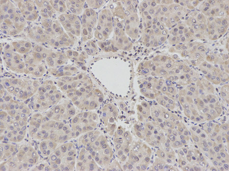


 IF (Immunofluorescence)
(Immunofluorescence analysis of A549 cell using BCL2 antibody. Blue: DAPI for nuclear staining.)
IF (Immunofluorescence)
(Immunofluorescence analysis of A549 cell using BCL2 antibody. Blue: DAPI for nuclear staining.)

 FCM (Flow Cytometry)
(Figure 6. Flow Cytometry analysis of HepG2 cells using anti-PFKFB1 antibody (AAA19903).Overlay histogram showing HepG2 cells stained with AAA19903 (Blue line). To facilitate intracellular staining, cells were fixed with 4% paraformaldehyde and permeabilized with permeabilization buffer. The cells were blocked with 10% normal goat serum. And then incubated with rabbit anti-PFKFB1 Antibody (AAA19903, 1ug/1x106 cells) for 30 min at 20 degree C. DyLight488 conjugated goat anti-rabbit IgG was used as secondary antibody for 30 minutes at 20 degree C. Isotype control antibody (Green line) was rabbit IgG (1ug/1x106) used under the same conditions. Unlabelled sample (Red line) was also used as a control.)
FCM (Flow Cytometry)
(Figure 6. Flow Cytometry analysis of HepG2 cells using anti-PFKFB1 antibody (AAA19903).Overlay histogram showing HepG2 cells stained with AAA19903 (Blue line). To facilitate intracellular staining, cells were fixed with 4% paraformaldehyde and permeabilized with permeabilization buffer. The cells were blocked with 10% normal goat serum. And then incubated with rabbit anti-PFKFB1 Antibody (AAA19903, 1ug/1x106 cells) for 30 min at 20 degree C. DyLight488 conjugated goat anti-rabbit IgG was used as secondary antibody for 30 minutes at 20 degree C. Isotype control antibody (Green line) was rabbit IgG (1ug/1x106) used under the same conditions. Unlabelled sample (Red line) was also used as a control.)


 IF (Immunofluorescence)
(Immunofluorescence analysis of HeLa cells using TOMM20 antibody at dilution of 1:100 (40x lens). Blue: DAPI for nuclear staining.)
IF (Immunofluorescence)
(Immunofluorescence analysis of HeLa cells using TOMM20 antibody at dilution of 1:100 (40x lens). Blue: DAPI for nuclear staining.)


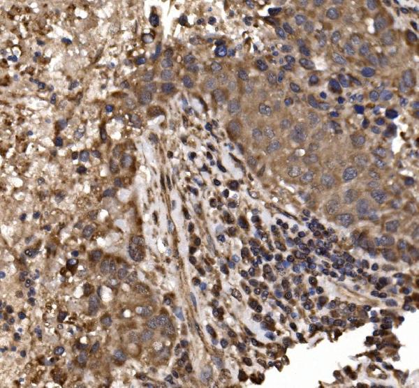

 FCM (Flow Cytometry)
(Figure 7. Flow Cytometry analysis of HeLa cells using anti-delta 1 Catenin/CAS/CTNND1 antibody (AAA19453).Overlay histogram showing HeLa cells stained with AAA19453 (Blue line). The cells were blocked with 10% normal goat serum. And then incubated with rabbit anti-delta 1 Catenin/CAS/CTNND1 Antibody (AAA19453, 1 ug/1x10^6 cells) for 30 min at 20 degree C. DyLight488 conjugated goat anti-rabbit IgG was used as secondary antibody for 30 minutes at 20 degree C. Isotype control antibody (Green line) was rabbit IgG (1 ug/1x10^6) used under the same conditions. Unlabelled sample (Red line) was also used as a control.)
FCM (Flow Cytometry)
(Figure 7. Flow Cytometry analysis of HeLa cells using anti-delta 1 Catenin/CAS/CTNND1 antibody (AAA19453).Overlay histogram showing HeLa cells stained with AAA19453 (Blue line). The cells were blocked with 10% normal goat serum. And then incubated with rabbit anti-delta 1 Catenin/CAS/CTNND1 Antibody (AAA19453, 1 ug/1x10^6 cells) for 30 min at 20 degree C. DyLight488 conjugated goat anti-rabbit IgG was used as secondary antibody for 30 minutes at 20 degree C. Isotype control antibody (Green line) was rabbit IgG (1 ug/1x10^6) used under the same conditions. Unlabelled sample (Red line) was also used as a control.)


 FCM (Flow Cytometry)
(Figure 9. Flow Cytometry analysis of THP-1 cells using anti-QARS1 antibody (AAA19879).Overlay histogram showing THP-1 cells stained with AAA19879 (Blue line). To facilitate intracellular staining, cells were fixed with 4% paraformaldehyde and permeabilized with permeabilization buffer. The cells were blocked with 10% normal goat serum. And then incubated with rabbit anti-QARS1 Antibody (AAA19879, 1ug/1x106 cells) for 30 min at 20 degree C. DyLight488 conjugated goat anti-rabbit IgG was used as secondary antibody for 30 minutes at 20 degree C. Isotype control antibody (Green line) was rabbit IgG (1ug/1x106) used under the same conditions. Unlabelled sample without incubation with primary antibody and secondary antibody (Red line) was used as a blank control.)
FCM (Flow Cytometry)
(Figure 9. Flow Cytometry analysis of THP-1 cells using anti-QARS1 antibody (AAA19879).Overlay histogram showing THP-1 cells stained with AAA19879 (Blue line). To facilitate intracellular staining, cells were fixed with 4% paraformaldehyde and permeabilized with permeabilization buffer. The cells were blocked with 10% normal goat serum. And then incubated with rabbit anti-QARS1 Antibody (AAA19879, 1ug/1x106 cells) for 30 min at 20 degree C. DyLight488 conjugated goat anti-rabbit IgG was used as secondary antibody for 30 minutes at 20 degree C. Isotype control antibody (Green line) was rabbit IgG (1ug/1x106) used under the same conditions. Unlabelled sample without incubation with primary antibody and secondary antibody (Red line) was used as a blank control.)


 IF (Immunofluorescence)
(Immunofluorescent analysis of 4% paraformaldehyde-fixed, 0.1% Triton X-100 permeabilized MCF-7 (human breast cancer cell line) cells labeling Pdx1 with at 1:25 dilution, followed by DyLight 488-conjugated IgG goat anti-rabbit secondary antibody at 1:200 dilution (green). Immunofluorescence image showing cytoplasm staining on MCF-7 cell line. Cytoplasmic actin is detected with DyLight 554 Phalloidin (PD18466410) at 1:100 dilution (red). The nuclear counter stain is DAPI (blue).)
IF (Immunofluorescence)
(Immunofluorescent analysis of 4% paraformaldehyde-fixed, 0.1% Triton X-100 permeabilized MCF-7 (human breast cancer cell line) cells labeling Pdx1 with at 1:25 dilution, followed by DyLight 488-conjugated IgG goat anti-rabbit secondary antibody at 1:200 dilution (green). Immunofluorescence image showing cytoplasm staining on MCF-7 cell line. Cytoplasmic actin is detected with DyLight 554 Phalloidin (PD18466410) at 1:100 dilution (red). The nuclear counter stain is DAPI (blue).)

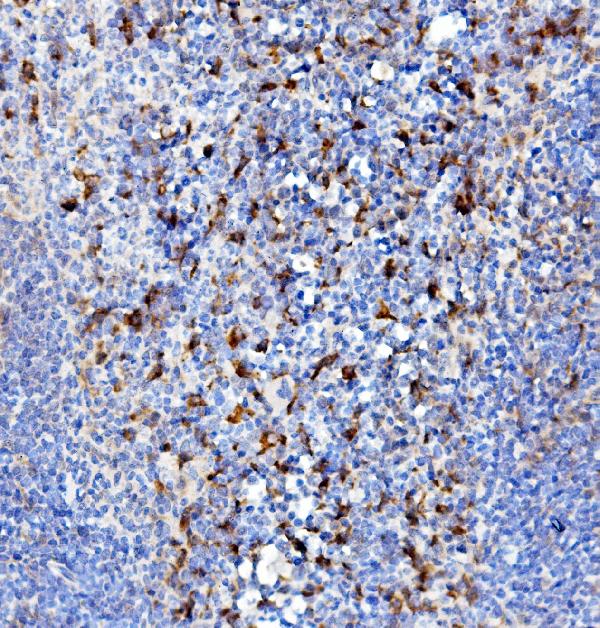

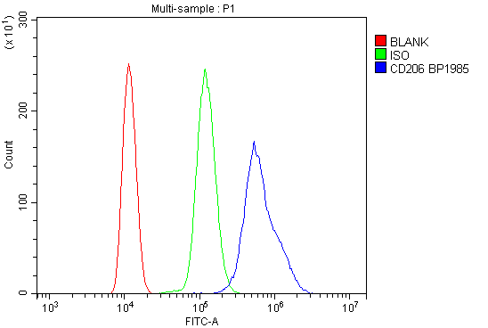


 WB (Western Blot)
(Figure 1. Western blot analysis of MRC1 using anti-MRC1 antibody.)
WB (Western Blot)
(Figure 1. Western blot analysis of MRC1 using anti-MRC1 antibody.)


 WB (Western Blot)
(WB Suggested Anti-RBM26 Antibody Titration: 0.2-1 ug/mlELISA Titer: 1:62500Positive Control: HepG2 cell lysateThere is BioGPS gene expression data showing that RBM26 is expressed in HepG2)
WB (Western Blot)
(WB Suggested Anti-RBM26 Antibody Titration: 0.2-1 ug/mlELISA Titer: 1:62500Positive Control: HepG2 cell lysateThere is BioGPS gene expression data showing that RBM26 is expressed in HepG2)
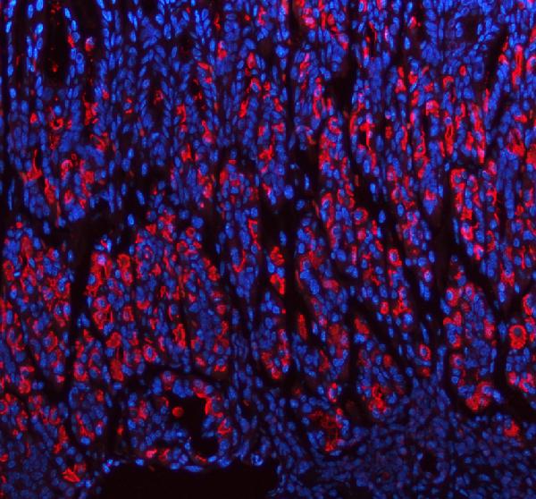


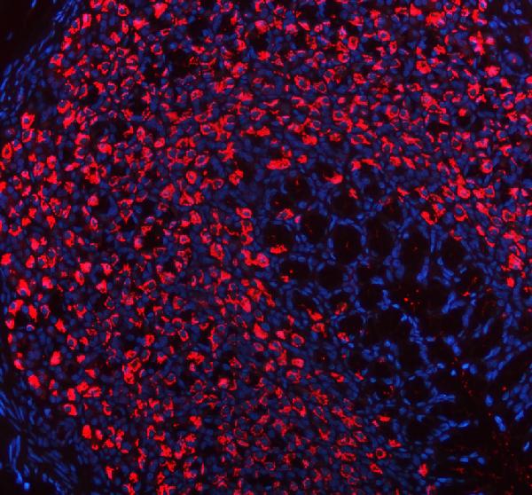 IF (Immunofluorescence)
(Figure 8. IF analysis of ATP4B using anti-ATP4B antibody (AAA19909).ATP4B was detected in a paraffin-embedded section of rat stomach tissue. Heat mediated antigen retrieval was performed in EDTA buffer (pH 8.0, epitope retrieval solution). The tissue section was blocked with 10% goat serum. The tissue section was then incubated with 5ug/mL rabbit anti-ATP4B Antibody (AAA19909) overnight at 4 degree C. DyLight594 Conjugated Goat Anti-Rabbit IgG (BA1142) was used as secondary antibody at 1:500 dilution and incubated for 30 minutes at 37 degree C. The section was counterstained with DAPI. Visualize using a fluorescence microscope and filter sets appropriate for the label used.)
IF (Immunofluorescence)
(Figure 8. IF analysis of ATP4B using anti-ATP4B antibody (AAA19909).ATP4B was detected in a paraffin-embedded section of rat stomach tissue. Heat mediated antigen retrieval was performed in EDTA buffer (pH 8.0, epitope retrieval solution). The tissue section was blocked with 10% goat serum. The tissue section was then incubated with 5ug/mL rabbit anti-ATP4B Antibody (AAA19909) overnight at 4 degree C. DyLight594 Conjugated Goat Anti-Rabbit IgG (BA1142) was used as secondary antibody at 1:500 dilution and incubated for 30 minutes at 37 degree C. The section was counterstained with DAPI. Visualize using a fluorescence microscope and filter sets appropriate for the label used.)


 IHC (Immunohistchemistry)
(Immunohistochemistry of paraffin-embedded rat brain using RAB11A/RAB11B Rabbit pAb at dilution of 1:150 (40x lens).Perform high pressure antigen retrieval with 10 mM citrate buffer pH 6.0 before commencing with IHC staining protocol.)
IHC (Immunohistchemistry)
(Immunohistochemistry of paraffin-embedded rat brain using RAB11A/RAB11B Rabbit pAb at dilution of 1:150 (40x lens).Perform high pressure antigen retrieval with 10 mM citrate buffer pH 6.0 before commencing with IHC staining protocol.)

 FCM (Flow Cytometry)
(Figure 9. Flow Cytometry analysis of HepG2 cells using anti-MOGAT2 antibody (AAA19953).Overlay histogram showing HepG2 cells stained with AAA19953 (Blue line). To facilitate intracellular staining, cells were fixed with 4% paraformaldehyde and permeabilized with permeabilization buffer. The cells were blocked with 10% normal goat serum. And then incubated with rabbit anti-MOGAT2 Antibody (AAA19953, 1ug/1x106 cells) for 30 min at 20 degree C. DyLight488 conjugated goat anti-rabbit IgG was used as secondary antibody for 30 minutes at 20 degree C. Isotype control antibody (Green line) was rabbit IgG (1ug/1x106) used under the same conditions. Unlabelled sample (Red line) was also used as a control.)
FCM (Flow Cytometry)
(Figure 9. Flow Cytometry analysis of HepG2 cells using anti-MOGAT2 antibody (AAA19953).Overlay histogram showing HepG2 cells stained with AAA19953 (Blue line). To facilitate intracellular staining, cells were fixed with 4% paraformaldehyde and permeabilized with permeabilization buffer. The cells were blocked with 10% normal goat serum. And then incubated with rabbit anti-MOGAT2 Antibody (AAA19953, 1ug/1x106 cells) for 30 min at 20 degree C. DyLight488 conjugated goat anti-rabbit IgG was used as secondary antibody for 30 minutes at 20 degree C. Isotype control antibody (Green line) was rabbit IgG (1ug/1x106) used under the same conditions. Unlabelled sample (Red line) was also used as a control.)


 IHC (Immunohistochemistry)
(Dilution: Western Blot: 1/500 - 1/2000. Immunohistochemistry: 1/100 - 1/300. Immunofluorescence: 1/200 - 1/1000. ELISA: 1/20000. Not yet tested in other applications.)
IHC (Immunohistochemistry)
(Dilution: Western Blot: 1/500 - 1/2000. Immunohistochemistry: 1/100 - 1/300. Immunofluorescence: 1/200 - 1/1000. ELISA: 1/20000. Not yet tested in other applications.)


 IHC (Immunohistchemistry)
(DAB staining on IHC-P; Samples: Mouse Ovary Tissue.)
IHC (Immunohistchemistry)
(DAB staining on IHC-P; Samples: Mouse Ovary Tissue.)
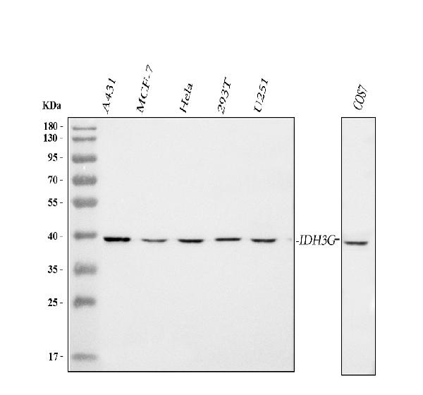
 FCM (Flow Cytometry)
(Figure 9. Flow Cytometry analysis of 293T cells using anti-IDH3G antibody (AAA19943).Overlay histogram showing 293T cells stained with AAA19943 (Blue line). To facilitate intracellular staining, cells were fixed with 4% paraformaldehyde and permeabilized with permeabilization buffer. The cells were blocked with 10% normal goat serum. And then incubated with rabbit anti-IDH3G Antibody (AAA19943, 1ug/1x106 cells) for 30 min at 20 degree C. DyLight488 conjugated goat anti-rabbit IgG was used as secondary antibody for 30 minutes at 20 degree C. Isotype control antibody (Green line) was rabbit IgG (1ug/1x106) used under the same conditions. Unlabelled sample (Red line) was also used as a control.)
FCM (Flow Cytometry)
(Figure 9. Flow Cytometry analysis of 293T cells using anti-IDH3G antibody (AAA19943).Overlay histogram showing 293T cells stained with AAA19943 (Blue line). To facilitate intracellular staining, cells were fixed with 4% paraformaldehyde and permeabilized with permeabilization buffer. The cells were blocked with 10% normal goat serum. And then incubated with rabbit anti-IDH3G Antibody (AAA19943, 1ug/1x106 cells) for 30 min at 20 degree C. DyLight488 conjugated goat anti-rabbit IgG was used as secondary antibody for 30 minutes at 20 degree C. Isotype control antibody (Green line) was rabbit IgG (1ug/1x106) used under the same conditions. Unlabelled sample (Red line) was also used as a control.)

 FCM (Flow Cytometry)
(Figure 9. Flow Cytometry analysis of U87 cells using anti-PKP2 antibody (AAA19770).Overlay histogram showing U87 cells stained with AAA19770 (Blue line). To facilitate intracellular staining, cells were fixed with 4% paraformaldehyde and permeabilized with permeabilization buffer. The cells were blocked with 10% normal goat serum. And then incubated with rabbit anti-PKP2 Antibody (AAA19770, 1ug/1x106 cells) for 30 min at 20 degree C. DyLight488 conjugated goat anti-rabbit IgG was used as secondary antibody for 30 minutes at 20 degree C. Isotype control antibody (Green line) was rabbit IgG (1ug/1x106) used under the same conditions. Unlabelled sample (Red line) was also used as a control.)
FCM (Flow Cytometry)
(Figure 9. Flow Cytometry analysis of U87 cells using anti-PKP2 antibody (AAA19770).Overlay histogram showing U87 cells stained with AAA19770 (Blue line). To facilitate intracellular staining, cells were fixed with 4% paraformaldehyde and permeabilized with permeabilization buffer. The cells were blocked with 10% normal goat serum. And then incubated with rabbit anti-PKP2 Antibody (AAA19770, 1ug/1x106 cells) for 30 min at 20 degree C. DyLight488 conjugated goat anti-rabbit IgG was used as secondary antibody for 30 minutes at 20 degree C. Isotype control antibody (Green line) was rabbit IgG (1ug/1x106) used under the same conditions. Unlabelled sample (Red line) was also used as a control.)


 FCM (Flow Cytometry)
(Figure 8. Flow Cytometry analysis of A431 cells using anti-PRDM15 antibody (AAA19987).Overlay histogram showing A431 cells stained with AAA19987 (Blue line). To facilitate intracellular staining, cells were fixed with 4% paraformaldehyde and permeabilized with permeabilization buffer. The cells were blocked with 10% normal goat serum. And then incubated with rabbit anti-PRDM15 Antibody (AAA19987, 1ug/1x106 cells) for 30 min at 20 degree C. DyLight488 conjugated goat anti-rabbit IgG was used as secondary antibody for 30 minutes at 20 degree C. Isotype control antibody (Green line) was rabbit IgG (1ug/1x106) used under the same conditions. Unlabelled sample (Red line) was also used as a control.)
FCM (Flow Cytometry)
(Figure 8. Flow Cytometry analysis of A431 cells using anti-PRDM15 antibody (AAA19987).Overlay histogram showing A431 cells stained with AAA19987 (Blue line). To facilitate intracellular staining, cells were fixed with 4% paraformaldehyde and permeabilized with permeabilization buffer. The cells were blocked with 10% normal goat serum. And then incubated with rabbit anti-PRDM15 Antibody (AAA19987, 1ug/1x106 cells) for 30 min at 20 degree C. DyLight488 conjugated goat anti-rabbit IgG was used as secondary antibody for 30 minutes at 20 degree C. Isotype control antibody (Green line) was rabbit IgG (1ug/1x106) used under the same conditions. Unlabelled sample (Red line) was also used as a control.)


 IHC (Immunohistochemistry)
(Dilution: Western Blot: 1/500 - 1/2000. IHC-p: 1/100-1/300. ELISA: 1/20000. Not yet tested in other applications.)
IHC (Immunohistochemistry)
(Dilution: Western Blot: 1/500 - 1/2000. IHC-p: 1/100-1/300. ELISA: 1/20000. Not yet tested in other applications.)


 IF (Immunofluorescence)
(Immunofluorescence analysis of C6 cells using HSP90AA1 Polyclonal Antibody at dilution of 1:100. Blue: DAPI for nuclear staining.)
IF (Immunofluorescence)
(Immunofluorescence analysis of C6 cells using HSP90AA1 Polyclonal Antibody at dilution of 1:100. Blue: DAPI for nuclear staining.)


 FCM (Flow Cytometry)
(Figure 8. Flow Cytometry analysis of U20S cells using anti-Dynamin 1 antibody (AAA19160).Overlay histogram showing U20S cells stained with AAA19160 (Blue line).The cells were blocked with 10% normal goat serum. And then incubated with rabbit anti-DNM1 Antibody (AAA19160,1ug/1x10^6 cells) for 30 min at 20 degree C. DyLight®488 conjugated goat anti-rabbit IgG (5-10ug/1x10^6 cells) was used as secondary antibody for 30 minutes at 20 degree C. Isotype control antibody (Green line) was rabbit IgG (1ug/1x106) used under the same conditions. Unlabelled sample (Red line) was also used as a control.)
FCM (Flow Cytometry)
(Figure 8. Flow Cytometry analysis of U20S cells using anti-Dynamin 1 antibody (AAA19160).Overlay histogram showing U20S cells stained with AAA19160 (Blue line).The cells were blocked with 10% normal goat serum. And then incubated with rabbit anti-DNM1 Antibody (AAA19160,1ug/1x10^6 cells) for 30 min at 20 degree C. DyLight®488 conjugated goat anti-rabbit IgG (5-10ug/1x10^6 cells) was used as secondary antibody for 30 minutes at 20 degree C. Isotype control antibody (Green line) was rabbit IgG (1ug/1x106) used under the same conditions. Unlabelled sample (Red line) was also used as a control.)

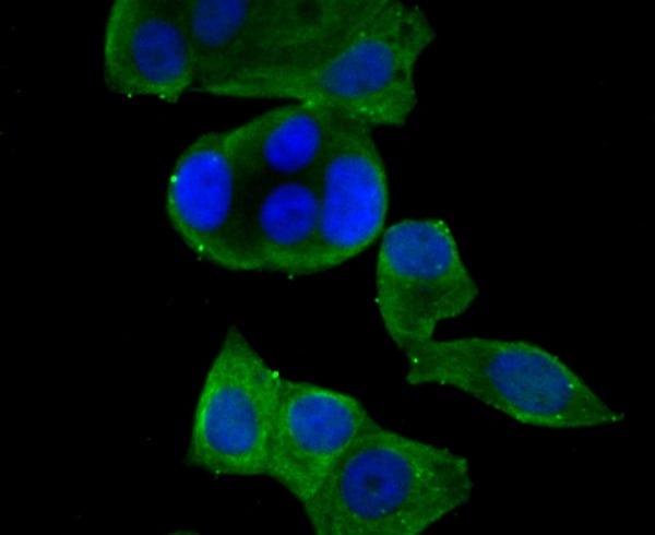


 FCM (Flow Cytometry)
(Flow cytometric analysis of MG-63 cells with Kv4.3 antibody at 1/100 dilution (purple) compared with an unlabelled control (cells without incubation with primary antibody; yellow). Alexa Fluor 488-conjugated goat anti-rabbit IgG was used as the secondary antibody)
FCM (Flow Cytometry)
(Flow cytometric analysis of MG-63 cells with Kv4.3 antibody at 1/100 dilution (purple) compared with an unlabelled control (cells without incubation with primary antibody; yellow). Alexa Fluor 488-conjugated goat anti-rabbit IgG was used as the secondary antibody)


 IF (Immunofluorescence)
(Immunofluorescence analysis of NIH/3T3 cells using Smad1 Rabbit pAb at dilution of 1:50 (40x lens). Blue: DAPI for nuclear staining.)
IF (Immunofluorescence)
(Immunofluorescence analysis of NIH/3T3 cells using Smad1 Rabbit pAb at dilution of 1:50 (40x lens). Blue: DAPI for nuclear staining.)


 IF (Immunofluorescence)
(Immunofluorescence analysis of NIH-3T3 using Heme Oxygenase 1 (HO-1/HMOX1) antibody at dilution of 1 : 50 (40x lens). Blue: DAPI for nuclear staining.)
IF (Immunofluorescence)
(Immunofluorescence analysis of NIH-3T3 using Heme Oxygenase 1 (HO-1/HMOX1) antibody at dilution of 1 : 50 (40x lens). Blue: DAPI for nuclear staining.)


 FCM (Flow Cytometry)
(Figure 9. Flow Cytometry analysis of U937 cells using anti-Syntaxin 18/STX18 antibody (AAA19967).Overlay histogram showing U937 cells stained with AAA19967 (Blue line). To facilitate intracellular staining, cells were fixed with 4% paraformaldehyde and permeabilized with permeabilization buffer. The cells were blocked with 10% normal goat serum. And then incubated with rabbit anti-Syntaxin 18/STX18 Antibody (AAA19967, 1ug/1x106 cells) for 30 min at 20 degree C. DyLight488 conjugated goat anti-rabbit IgG was used as secondary antibody for 30 minutes at 20 degree C. Isotype control antibody (Green line) was rabbit IgG (1ug/1x106) used under the same conditions. Unlabelled sample without incubation with primary antibody and secondary antibody (Red line) was used as a blank control.)
FCM (Flow Cytometry)
(Figure 9. Flow Cytometry analysis of U937 cells using anti-Syntaxin 18/STX18 antibody (AAA19967).Overlay histogram showing U937 cells stained with AAA19967 (Blue line). To facilitate intracellular staining, cells were fixed with 4% paraformaldehyde and permeabilized with permeabilization buffer. The cells were blocked with 10% normal goat serum. And then incubated with rabbit anti-Syntaxin 18/STX18 Antibody (AAA19967, 1ug/1x106 cells) for 30 min at 20 degree C. DyLight488 conjugated goat anti-rabbit IgG was used as secondary antibody for 30 minutes at 20 degree C. Isotype control antibody (Green line) was rabbit IgG (1ug/1x106) used under the same conditions. Unlabelled sample without incubation with primary antibody and secondary antibody (Red line) was used as a blank control.)


 IHC (Immunohistochemistry)
(DAB staining on IHC-P;Samples: Mouse Uterine Fibroids Tissue)
IHC (Immunohistochemistry)
(DAB staining on IHC-P;Samples: Mouse Uterine Fibroids Tissue)


 FCM (Flow Cytometry)
(Figure 7. Flow Cytometry analysis of Hela cells using anti- ETF/TEAD2 antibody (AAA19325).Overlay histogram showing Hela cells stained withAAA19325 (Blue line). The cells were blocked with 10% normal goat serum. And then incubated with rabbit anti- ETF/TEAD2 Antibody (AAA19325, 1μg/1x106 cells) for 30 min at 20 degree C. DyLight®488 conjugated goat anti-rabbit IgG (5-10μg/1x106 cells) was used as secondary antibody for 30 minutes at 20 degree C. Isotype control antibody (Green line) was rabbit IgG (1μg/1x106) used under the same conditions. Unlabelled sample (Red line) was also used as a control.)
FCM (Flow Cytometry)
(Figure 7. Flow Cytometry analysis of Hela cells using anti- ETF/TEAD2 antibody (AAA19325).Overlay histogram showing Hela cells stained withAAA19325 (Blue line). The cells were blocked with 10% normal goat serum. And then incubated with rabbit anti- ETF/TEAD2 Antibody (AAA19325, 1μg/1x106 cells) for 30 min at 20 degree C. DyLight®488 conjugated goat anti-rabbit IgG (5-10μg/1x106 cells) was used as secondary antibody for 30 minutes at 20 degree C. Isotype control antibody (Green line) was rabbit IgG (1μg/1x106) used under the same conditions. Unlabelled sample (Red line) was also used as a control.)


 FCM (Flow Cytometry)
(Figure 6. Flow Cytometry analysis of A431 cells using anti-NDUFB5 antibody (AAA19333).Overlay histogram showing A431 cells stained with AAA19333 (Blue line). The cells were blocked with 10% normal goat serum. And then incubated with rabbit anti-NDUFB5 Antibody (AAA19333, 1μg/1x106 cells) for 30 min at 20 degree C. DyLight®488 conjugated goat anti-rabbit IgG (5-10μg/1x106 cells) was used as secondary antibody for 30 minutes at 20 degree C. Isotype control antibody (Green line) was rabbit IgG (1μg/1x106) used under the same conditions. Unlabelled sample (Red line) was also used as a control.)
FCM (Flow Cytometry)
(Figure 6. Flow Cytometry analysis of A431 cells using anti-NDUFB5 antibody (AAA19333).Overlay histogram showing A431 cells stained with AAA19333 (Blue line). The cells were blocked with 10% normal goat serum. And then incubated with rabbit anti-NDUFB5 Antibody (AAA19333, 1μg/1x106 cells) for 30 min at 20 degree C. DyLight®488 conjugated goat anti-rabbit IgG (5-10μg/1x106 cells) was used as secondary antibody for 30 minutes at 20 degree C. Isotype control antibody (Green line) was rabbit IgG (1μg/1x106) used under the same conditions. Unlabelled sample (Red line) was also used as a control.)


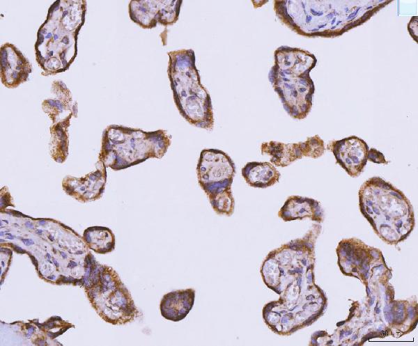

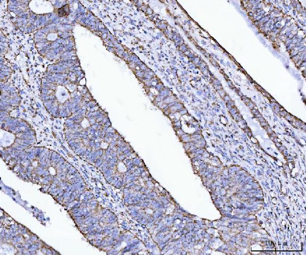


 FCM (Flow Cytometry)
(Figure 12. Flow Cytometry analysis of 293T cells using anti-ATP5F1B antibody (AAA19652).Overlay histogram showing 293T cells stained with AAA19652 (Blue line). The cells were blocked with 10% normal goat serum. And then incubated with rabbit anti-ATP5F1B Antibody (AAA19652, 1 ug/1x106 cells) for 30 min at 20 degree C. DyLight488 conjugated goat anti-rabbit IgG (BA1127, 5-10 ug/1x106 cells) was used as secondary antibody for 30 minutes at 20 degree C. Isotype control antibody (Green line) was rabbit IgG (1 ug/1x106) used under the same conditions. Unlabelled sample (Red line) was also used as a control.)
FCM (Flow Cytometry)
(Figure 12. Flow Cytometry analysis of 293T cells using anti-ATP5F1B antibody (AAA19652).Overlay histogram showing 293T cells stained with AAA19652 (Blue line). The cells were blocked with 10% normal goat serum. And then incubated with rabbit anti-ATP5F1B Antibody (AAA19652, 1 ug/1x106 cells) for 30 min at 20 degree C. DyLight488 conjugated goat anti-rabbit IgG (BA1127, 5-10 ug/1x106 cells) was used as secondary antibody for 30 minutes at 20 degree C. Isotype control antibody (Green line) was rabbit IgG (1 ug/1x106) used under the same conditions. Unlabelled sample (Red line) was also used as a control.)


 FCM (Flow Cytometry)
(Figure 8. Flow Cytometry analysis of Caco-2 cells using anti-TOMM70 antibody (AAA19606).Overlay histogram showing Caco-2 cells stained with AAA19606 (Blue line). The cells were blocked with 10% normal goat serum. And then incubated with rabbit anti-TOMM70 Antibody (AAA19606, 1 ug/1x10^6 cells) for 30 min at 20 degree C. DyLight488 conjugated goat anti-rabbit IgG was used as secondary antibody for 30 minutes at 20 degree C. Isotype control antibody (Green line) was rabbit IgG (1 ug/1x10^6) used under the same conditions. Unlabelled sample (Red line) was also used as a control.)
FCM (Flow Cytometry)
(Figure 8. Flow Cytometry analysis of Caco-2 cells using anti-TOMM70 antibody (AAA19606).Overlay histogram showing Caco-2 cells stained with AAA19606 (Blue line). The cells were blocked with 10% normal goat serum. And then incubated with rabbit anti-TOMM70 Antibody (AAA19606, 1 ug/1x10^6 cells) for 30 min at 20 degree C. DyLight488 conjugated goat anti-rabbit IgG was used as secondary antibody for 30 minutes at 20 degree C. Isotype control antibody (Green line) was rabbit IgG (1 ug/1x10^6) used under the same conditions. Unlabelled sample (Red line) was also used as a control.)

 IHC (Immunohistochemistry)
(Immunohistochemical analysis of Laminin alpha 5 staining in human lung cancer formalin fixed paraffin embedded tissue section. The section was pre-treated using heat mediated antigen retrieval with sodium citrate buffer (pH 6.0). The section was then incubated with the antibody at room temperature and detected using an HRP conjugated compact polymer system. DAB was used as the chromogen. The section was then counterstained with haematoxylin and mounted with DPX.)
IHC (Immunohistochemistry)
(Immunohistochemical analysis of Laminin alpha 5 staining in human lung cancer formalin fixed paraffin embedded tissue section. The section was pre-treated using heat mediated antigen retrieval with sodium citrate buffer (pH 6.0). The section was then incubated with the antibody at room temperature and detected using an HRP conjugated compact polymer system. DAB was used as the chromogen. The section was then counterstained with haematoxylin and mounted with DPX.)


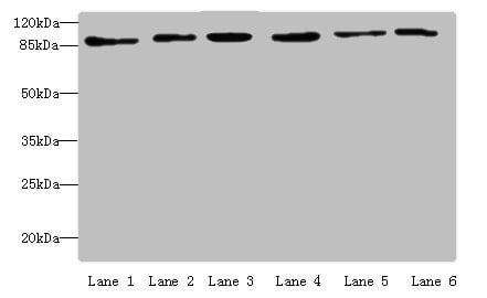
 ChIP (Chromatin Immunoprecipitation)
(Chromatin Immunoprecipitation 293T(1.6^106)were cross-linked with formaldehyde, sonicated, and immunoprecipitated with 4ug anti-MCM3 or a control normal rabbit IgG . The resulting ChIP DNA was quantified using real-time PCR with primers against the IRF-1 promoter.)
ChIP (Chromatin Immunoprecipitation)
(Chromatin Immunoprecipitation 293T(1.6^106)were cross-linked with formaldehyde, sonicated, and immunoprecipitated with 4ug anti-MCM3 or a control normal rabbit IgG . The resulting ChIP DNA was quantified using real-time PCR with primers against the IRF-1 promoter.)


 WB (Western Blot)
(Western Blot analysis of FKBP4 expression in transfected 293T cell line by FKBP4 polyclonal antibody. Lane 1: FKBP4 transfected lysate (51.8kD). Lane 2: Non-transfected lysate.)
WB (Western Blot)
(Western Blot analysis of FKBP4 expression in transfected 293T cell line by FKBP4 polyclonal antibody. Lane 1: FKBP4 transfected lysate (51.8kD). Lane 2: Non-transfected lysate.)


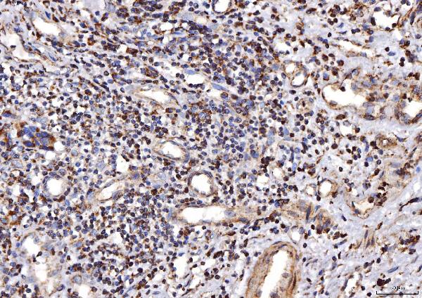


 IHC (Immunohistochemistry)
(Figure 8. IHC analysis of COX IV/COX4I1 using anti-COX IV/COX4I1 antibody (AAA19549).COX IV/COX4I1 was detected in a paraffin-embedded section of rat colon tissue. Heat mediated antigen retrieval was performed in EDTA buffer (pH 8.0, epitope retrieval solution). The tissue section was blocked with 10% goat serum. The tissue section was then incubated with 2 ug/ml rabbit anti-COX IV/COX4I1 Antibody (AAA19549) overnight at 4 degree C. Peroxidase Conjugated Goat Anti-rabbit IgG was used as secondary antibody and incubated for 30 minutes at 37 degree C. The tissue section was developed using HRP Conjugated Rabbit IgG Super Vision Assay Kit with DAB as the chromogen.)
IHC (Immunohistochemistry)
(Figure 8. IHC analysis of COX IV/COX4I1 using anti-COX IV/COX4I1 antibody (AAA19549).COX IV/COX4I1 was detected in a paraffin-embedded section of rat colon tissue. Heat mediated antigen retrieval was performed in EDTA buffer (pH 8.0, epitope retrieval solution). The tissue section was blocked with 10% goat serum. The tissue section was then incubated with 2 ug/ml rabbit anti-COX IV/COX4I1 Antibody (AAA19549) overnight at 4 degree C. Peroxidase Conjugated Goat Anti-rabbit IgG was used as secondary antibody and incubated for 30 minutes at 37 degree C. The tissue section was developed using HRP Conjugated Rabbit IgG Super Vision Assay Kit with DAB as the chromogen.)


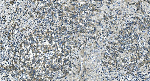


 FCM (Flow Cytometry)
(Figure 11. Flow Cytometry analysis of THP-1 cells using anti-CDC123 antibody (AAA19323).Overlay histogram showing THP-1 cells stained with AAA19323 (Blue line). The cells were blocked with 10% normal goat serum. And then incubated with rabbit anti-CDC123 Antibody (AAA19323, 1μg/1x106 cells) for 30 min at 20 degree C. DyLight®488 conjugated goat anti-rabbit IgG (5-10μg/1x106 cells) was used as secondary antibody for 30 minutes at 20 degree C. Isotype control antibody (Green line) was rabbit IgG (1μg/1x106) used under the same conditions. Unlabelled sample (Red line) was also used as a control.)
FCM (Flow Cytometry)
(Figure 11. Flow Cytometry analysis of THP-1 cells using anti-CDC123 antibody (AAA19323).Overlay histogram showing THP-1 cells stained with AAA19323 (Blue line). The cells were blocked with 10% normal goat serum. And then incubated with rabbit anti-CDC123 Antibody (AAA19323, 1μg/1x106 cells) for 30 min at 20 degree C. DyLight®488 conjugated goat anti-rabbit IgG (5-10μg/1x106 cells) was used as secondary antibody for 30 minutes at 20 degree C. Isotype control antibody (Green line) was rabbit IgG (1μg/1x106) used under the same conditions. Unlabelled sample (Red line) was also used as a control.)


 IHC (Immunohistochemistry)
(Figure 7. IHC analysis of VEGF Receptor 3 using anti-VEGF Receptor 3 antibody (AAA19147).VEGF Receptor 3 was detected in paraffin-embedded section of mouse liver tissue. Heat mediated antigen retrieval was performed in citrate buffer (pH6, epitope retrieval solution) for 20 mins. The tissue section was blocked with 10% goat serum. The tissue section was then incubated with 1ug/ml rabbit anti-VEGF Receptor 3 Antibody (AAA19147) overnight at 4 degree C. Biotinylated goat anti-rabbit IgG was used as secondary antibody and incubated for 30 minutes at 37 degree C. The tissue section was developed using Strepavidin-Biotin-Complex (SABC) with DAB as the chromogen.)
IHC (Immunohistochemistry)
(Figure 7. IHC analysis of VEGF Receptor 3 using anti-VEGF Receptor 3 antibody (AAA19147).VEGF Receptor 3 was detected in paraffin-embedded section of mouse liver tissue. Heat mediated antigen retrieval was performed in citrate buffer (pH6, epitope retrieval solution) for 20 mins. The tissue section was blocked with 10% goat serum. The tissue section was then incubated with 1ug/ml rabbit anti-VEGF Receptor 3 Antibody (AAA19147) overnight at 4 degree C. Biotinylated goat anti-rabbit IgG was used as secondary antibody and incubated for 30 minutes at 37 degree C. The tissue section was developed using Strepavidin-Biotin-Complex (SABC) with DAB as the chromogen.)


 ICC (Immunocytochemistry)
(ICC staining MSH6 in Hela cells (green). Cells were fixed in paraformaldehyde, permeabilised with 0.25% Triton X100/PBS.)
ICC (Immunocytochemistry)
(ICC staining MSH6 in Hela cells (green). Cells were fixed in paraformaldehyde, permeabilised with 0.25% Triton X100/PBS.)


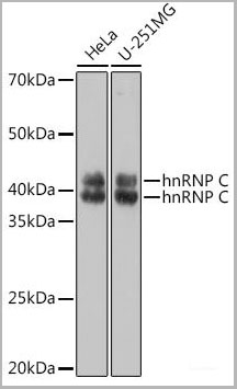
 IP (Immunoprecipitation)
(Immunoprecipitation analysis of 300ug extracts of K-562 cells usingug hnRNP C Polyclonal Antibody. Western blot was performed from the immunoprecipitate using hnRNP C Polyclonal Antibody at a dilution of 1:1000.)
IP (Immunoprecipitation)
(Immunoprecipitation analysis of 300ug extracts of K-562 cells usingug hnRNP C Polyclonal Antibody. Western blot was performed from the immunoprecipitate using hnRNP C Polyclonal Antibody at a dilution of 1:1000.)


 IF (Immunofluorescence)
(Immunofluorescence analysis of C6 cells using AIF Polyclonal Antibody at dilution of 1:100 (40x lens). Blue: DAPI for nuclear staining.)
IF (Immunofluorescence)
(Immunofluorescence analysis of C6 cells using AIF Polyclonal Antibody at dilution of 1:100 (40x lens). Blue: DAPI for nuclear staining.)


 IF (Immunofluorescence)
(Figure 8. IF analysis of TTC11/FIS1 using anti-TTC11/FIS1 antibody (AAA19439).TTC11/FIS1 was detected in an immunocytochemical section of MCF-7 cells. Enzyme antigen retrieval was performed using IHC enzyme antigen retrieval reagent (AR0022) for 15 mins. The cells were blocked with 10% goat serum. And then incubated with 5 ug/mL rabbit anti-TTC11/FIS1 Antibody (AAA19439) overnight at 4 degree C. DyLight488 Conjugated Goat Anti-Rabbit IgG was used as secondary antibody at 1:100 dilution and incubated for 30 minutes at 37 degree C. The section was counterstained with DAPI. Visualize using a fluorescence microscope and filter sets appropriate for the label used.)
IF (Immunofluorescence)
(Figure 8. IF analysis of TTC11/FIS1 using anti-TTC11/FIS1 antibody (AAA19439).TTC11/FIS1 was detected in an immunocytochemical section of MCF-7 cells. Enzyme antigen retrieval was performed using IHC enzyme antigen retrieval reagent (AR0022) for 15 mins. The cells were blocked with 10% goat serum. And then incubated with 5 ug/mL rabbit anti-TTC11/FIS1 Antibody (AAA19439) overnight at 4 degree C. DyLight488 Conjugated Goat Anti-Rabbit IgG was used as secondary antibody at 1:100 dilution and incubated for 30 minutes at 37 degree C. The section was counterstained with DAPI. Visualize using a fluorescence microscope and filter sets appropriate for the label used.)


 FCM (Flow Cytometry)
(Figure 6. Flow Cytometry analysis of HeLa cells using anti-SLU7 antibody (AAA19583).Overlay histogram showing HeLa cells stained with AAA19583 (Blue line). The cells were blocked with 10% normal goat serum. And then incubated with rabbit anti-SLU7 Antibody (AAA19583, 1 ug/1x10^6 cells) for 30 min at 20 degree C. DyLight488 conjugated goat anti-rabbit IgG was used as secondary antibody for 30 minutes at 20 degree C. Isotype control antibody (Green line) was rabbit IgG (1 ug/1x10^6) used under the same conditions. Unlabelled sample (Red line) was also used as a control.)
FCM (Flow Cytometry)
(Figure 6. Flow Cytometry analysis of HeLa cells using anti-SLU7 antibody (AAA19583).Overlay histogram showing HeLa cells stained with AAA19583 (Blue line). The cells were blocked with 10% normal goat serum. And then incubated with rabbit anti-SLU7 Antibody (AAA19583, 1 ug/1x10^6 cells) for 30 min at 20 degree C. DyLight488 conjugated goat anti-rabbit IgG was used as secondary antibody for 30 minutes at 20 degree C. Isotype control antibody (Green line) was rabbit IgG (1 ug/1x10^6) used under the same conditions. Unlabelled sample (Red line) was also used as a control.)


 FCM (Flow Cytometry)
(Figure 7. Flow Cytometry analysis of HepG2 cells using anti-IFT52 antibody (AAA19977).Overlay histogram showing HepG2 cells stained with AAA19977 (Blue line). The cells were fixed with 4% paraformaldehyde and blocked with 10% normal goat serum. And then incubated with rabbit anti-IFT52 Antibody (AAA19977, 1ug/1x106 cells) for 30 min at 20 degree C. DyLight488 conjugated goat anti-rabbit IgG was used as secondary antibody for 30 minutes at 20 degree C. Isotype control antibody (Green line) was rabbit IgG (1ug/1x106) used under the same conditions. Unlabelled sample (Red line) was also used as a control.)
FCM (Flow Cytometry)
(Figure 7. Flow Cytometry analysis of HepG2 cells using anti-IFT52 antibody (AAA19977).Overlay histogram showing HepG2 cells stained with AAA19977 (Blue line). The cells were fixed with 4% paraformaldehyde and blocked with 10% normal goat serum. And then incubated with rabbit anti-IFT52 Antibody (AAA19977, 1ug/1x106 cells) for 30 min at 20 degree C. DyLight488 conjugated goat anti-rabbit IgG was used as secondary antibody for 30 minutes at 20 degree C. Isotype control antibody (Green line) was rabbit IgG (1ug/1x106) used under the same conditions. Unlabelled sample (Red line) was also used as a control.)
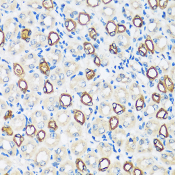


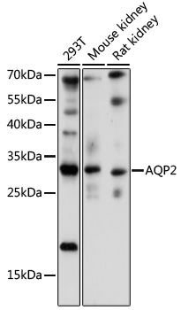 WB (Western Blot)
(Western blot analysis of extracts of various cell lines, using AQP2 at 1:1000 dilution.)
WB (Western Blot)
(Western blot analysis of extracts of various cell lines, using AQP2 at 1:1000 dilution.)


 IHC (Immunohistochemistry)
(Immunohistochemistry of paraffin-embedded mouse lung using ZNF148 at dilution of 1:100 (40x lens).)
IHC (Immunohistochemistry)
(Immunohistochemistry of paraffin-embedded mouse lung using ZNF148 at dilution of 1:100 (40x lens).)


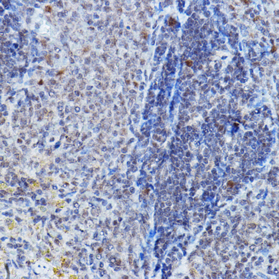


 IP (Immunoprecipitation)
(Immunoprecipitation analysis of 300ug extracts of HeLa cells using 3ug DGCR8 antibody . Western blot was performed from the immunoprecipitate using DGCR8 antibody at a dilition of 1:1000.)
IP (Immunoprecipitation)
(Immunoprecipitation analysis of 300ug extracts of HeLa cells using 3ug DGCR8 antibody . Western blot was performed from the immunoprecipitate using DGCR8 antibody at a dilition of 1:1000.)


 IF (Immunofluorescence)
(Immunofluorescence analysis of C6 cells using LLGL1 antibody at dilution of 1:100. Blue: DAPI for nuclear staining.)
IF (Immunofluorescence)
(Immunofluorescence analysis of C6 cells using LLGL1 antibody at dilution of 1:100. Blue: DAPI for nuclear staining.)


 IHC (Immunohistchemistry)
(Dilution: Western Blot: 1/500 - 1/2000. IHC-p: 1:100-300 ELISA: 1/20000. Not yet tested in other applications.)
IHC (Immunohistchemistry)
(Dilution: Western Blot: 1/500 - 1/2000. IHC-p: 1:100-300 ELISA: 1/20000. Not yet tested in other applications.)