Filters
▼Clonality
▼Type
▼Reactivity
▼Gene Name
▼Isotype
▼Host
▼Application
▼Clone
▼Polyclonal Antibodies
At AAA Biotech also known as AAA Bio or AAABio, we provide a broad range of purified polyclonal antibodies (pAbs) that are able to all be browsed online through our website. Due to their high specificity and strong binding affinity, these antibodies are ideal for wide swathes of research and experimental applications.
Our polyclonal antibodies can easily support your work, whether you use them for Western Blotting, Immunocytochemistry (with or without Immunofluorescence used in conjunction), Immunohistochemistry, Immunoprecipitation, and ELISA tests. We highly encourage you to browse our range of pAbs and choose the one that best suits your experimental model.
Viewing 2300-2350 of 96805 product results
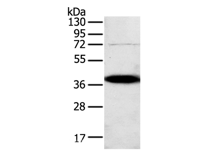
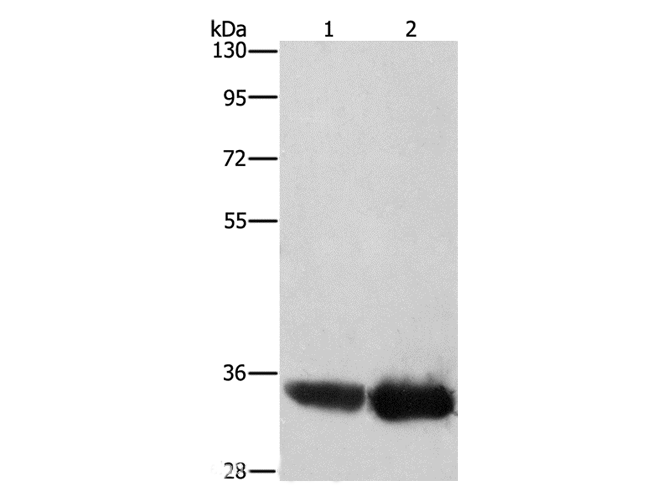
 WB (Western Blot)
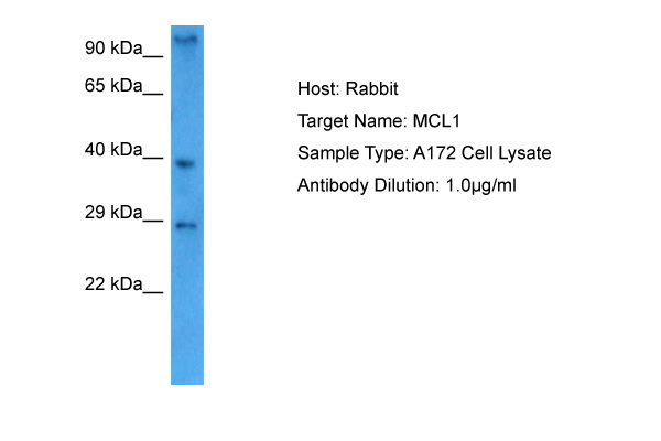
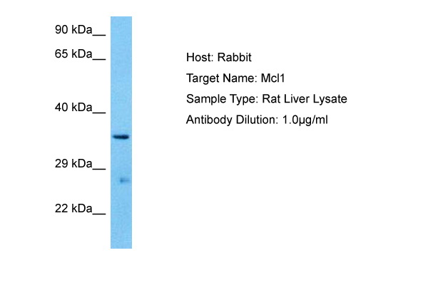
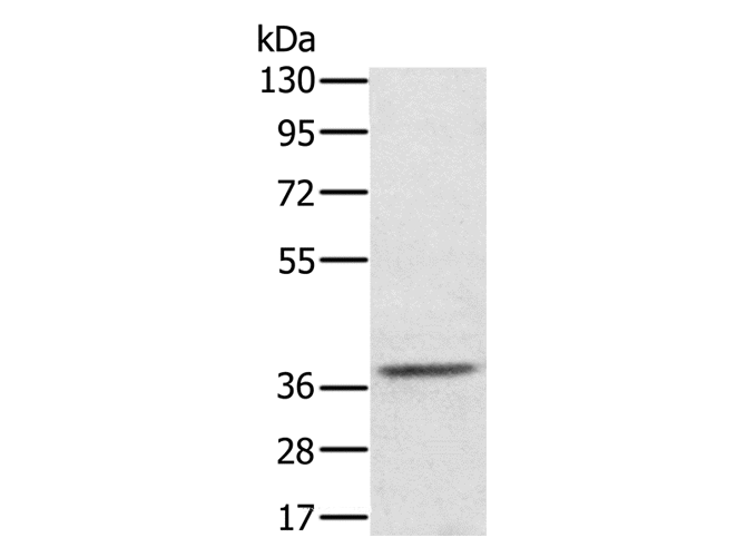
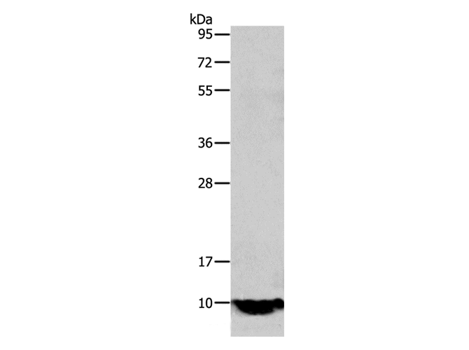
(Host: RatTarget Name: MCL1Sample Tissue: Rat LiverAntibody Dilution: 1ug/ml)
WB (Western Blot)
(Host: RatTarget Name: MCL1Sample Tissue: Rat LiverAntibody Dilution: 1ug/ml)
MCL1, Polyclonal Antibody (Cat# AAA201565)
 WB (Western Blot)
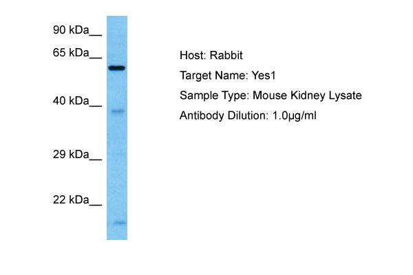
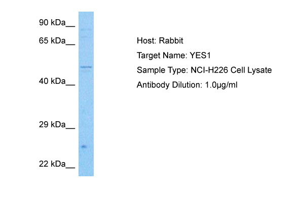
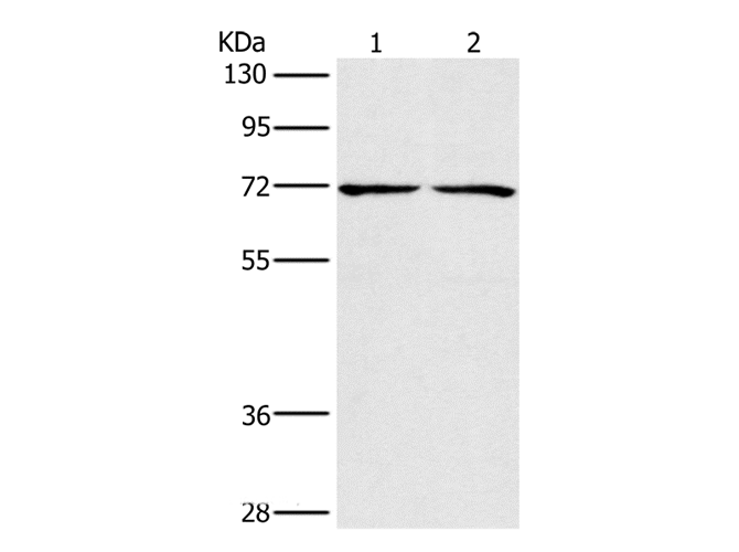
(Host: RabbitTarget Name: YES1Sample Tissue: Human NCI-H226 Whole Cell lysatesAntibody Dilution: 1ug/ml)
WB (Western Blot)
(Host: RabbitTarget Name: YES1Sample Tissue: Human NCI-H226 Whole Cell lysatesAntibody Dilution: 1ug/ml)
YES1, Polyclonal Antibody (Cat# AAA201570)
 WB (Western Blot)
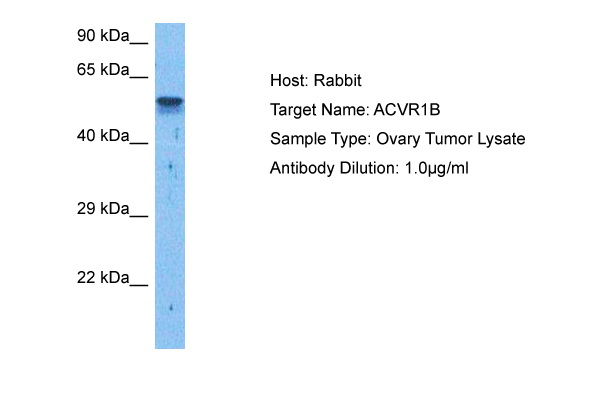
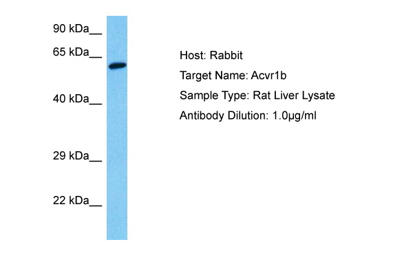
(Host: RatTarget Name: ACVR1BSample Tissue: Rat LiverAntibody Dilution: 1ug/ml)
WB (Western Blot)
(Host: RatTarget Name: ACVR1BSample Tissue: Rat LiverAntibody Dilution: 1ug/ml)
ACVR1B, Polyclonal Antibody (Cat# AAA201571)
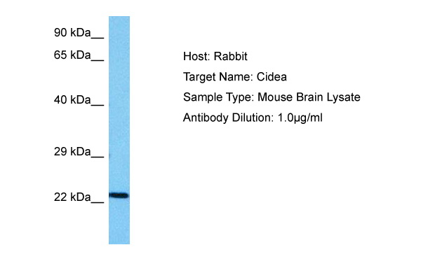
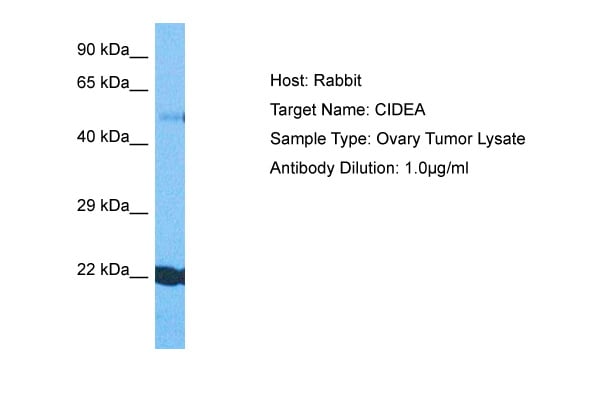 WB (Western Blot)
(Host: RabbitTarget Name: CIDEASample Tissue: Human Ovary TumorAntibody Dilution: 1.0ug/ml)
WB (Western Blot)
(Host: RabbitTarget Name: CIDEASample Tissue: Human Ovary TumorAntibody Dilution: 1.0ug/ml)
CIDEA, Polyclonal Antibody (Cat# AAA201572)
 WB (Western Blot)
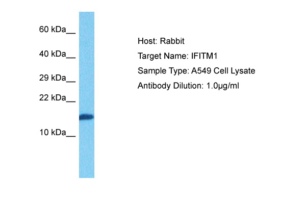
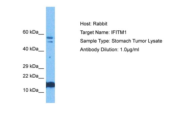
(Host: RabbitTarget Name: IFITM1Sample Tissue: Human Stomach TumorAntibody Dilution: 1.0ug/ml)
WB (Western Blot)
(Host: RabbitTarget Name: IFITM1Sample Tissue: Human Stomach TumorAntibody Dilution: 1.0ug/ml)
IFITM1, Polyclonal Antibody (Cat# AAA201573)
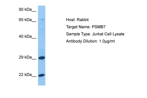 WB (Western Blot)
(Host: RabbitTarget Name: PSMB7Sample Tissue: Jurkat Whole Cell lysatesAntibody Dilution: 1.0ug/ml)
WB (Western Blot)
(Host: RabbitTarget Name: PSMB7Sample Tissue: Jurkat Whole Cell lysatesAntibody Dilution: 1.0ug/ml)
PSMB7, Polyclonal Antibody (Cat# AAA201577)
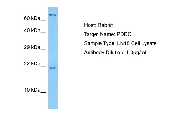 WB (Western Blot)
(Host: RabbitTarget Name: PDDC1Sample Tissue: Human LN18 Whole Cell lysatesAntibody Dilution: 1ug/ml)
WB (Western Blot)
(Host: RabbitTarget Name: PDDC1Sample Tissue: Human LN18 Whole Cell lysatesAntibody Dilution: 1ug/ml)
GATD1, Polyclonal Antibody (Cat# AAA201579)
 WB (Western Blot)
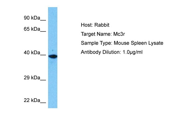
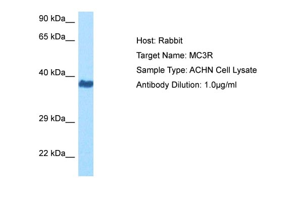
(Host: RabbitTarget Name: MC3RSample Tissue: Human ACHN Whole Cell lysatesAntibody Dilution: 1ug/ml)
WB (Western Blot)
(Host: RabbitTarget Name: MC3RSample Tissue: Human ACHN Whole Cell lysatesAntibody Dilution: 1ug/ml)
MC3R, Polyclonal Antibody (Cat# AAA201581)
 WB (Western Blot)
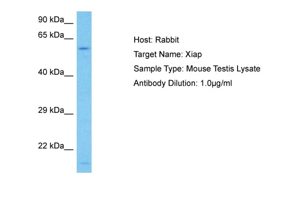
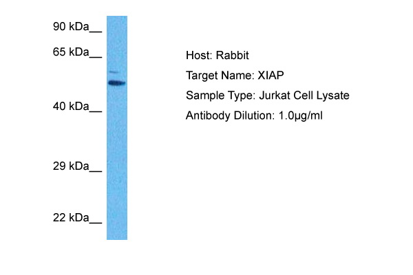
(Host: RabbitTarget Name: XIAPSample Tissue: Human Jurkat Whole Cell lysatesAntibody Dilution: 1ug/ml)
WB (Western Blot)
(Host: RabbitTarget Name: XIAPSample Tissue: Human Jurkat Whole Cell lysatesAntibody Dilution: 1ug/ml)
XIAP, Polyclonal Antibody (Cat# AAA201583)
 WB (Western Blot)
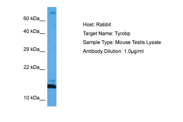
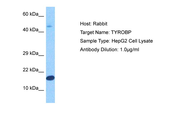
(Host: RabbitTarget Name: TYROBPSample Tissue: Human HepG2 Whole Cell lysatesAntibody Dilution: 1ug/ml)
WB (Western Blot)
(Host: RabbitTarget Name: TYROBPSample Tissue: Human HepG2 Whole Cell lysatesAntibody Dilution: 1ug/ml)
TYROBP, Polyclonal Antibody (Cat# AAA201584)
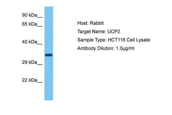 WB (Western Blot)
(Host: RabbitTarget Name: UCP2Sample Tissue: Human HCT116 Whole Cell lysatesAntibody Dilution: 1ug/ml)
WB (Western Blot)
(Host: RabbitTarget Name: UCP2Sample Tissue: Human HCT116 Whole Cell lysatesAntibody Dilution: 1ug/ml)
UCP2, Polyclonal Antibody (Cat# AAA201587)
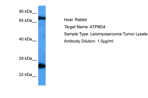 WB (Western Blot)
(Host: RabbitTarget Name: ATPBD4Sample Tissue: Human Leiomyosarcoma lysatesAntibody Dilution: 1ug/ml)
WB (Western Blot)
(Host: RabbitTarget Name: ATPBD4Sample Tissue: Human Leiomyosarcoma lysatesAntibody Dilution: 1ug/ml)
DPH6, Polyclonal Antibody (Cat# AAA201593)
 WB (Western Blot)
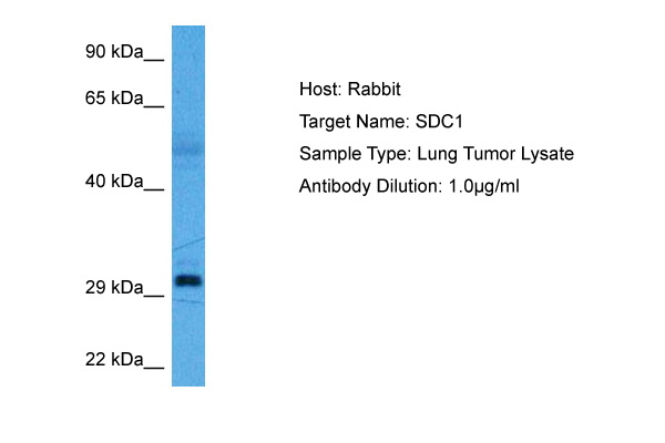
(Host: RabbitTarget Name: SDC1Sample Tissue: Human Lung Tumor lysatesAntibody Dilution: 1ug/ml)
WB (Western Blot)
(Host: RabbitTarget Name: SDC1Sample Tissue: Human Lung Tumor lysatesAntibody Dilution: 1ug/ml)
SDC1, Polyclonal Antibody (Cat# AAA201594)
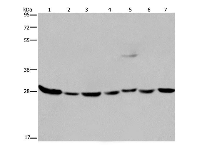
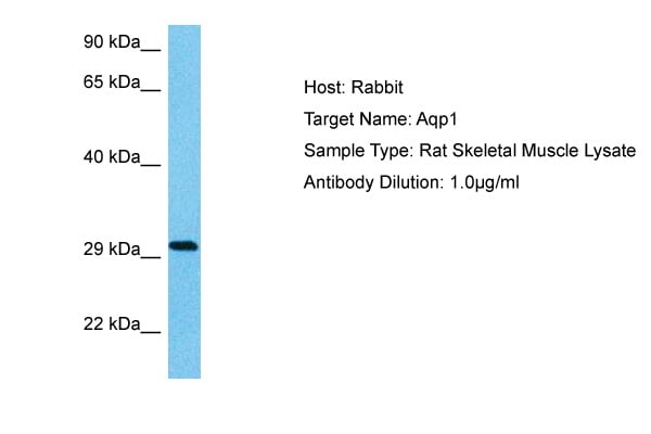 WB (Western Blot)
(Host: RatTarget Name: AQP1Sample Tissue: Rat Skeletal MuscleAntibody Dilution: 1ug/ml)
WB (Western Blot)
(Host: RatTarget Name: AQP1Sample Tissue: Rat Skeletal MuscleAntibody Dilution: 1ug/ml)
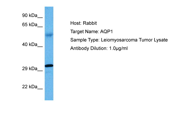
AQP1, Polyclonal Antibody (Cat# AAA201599)
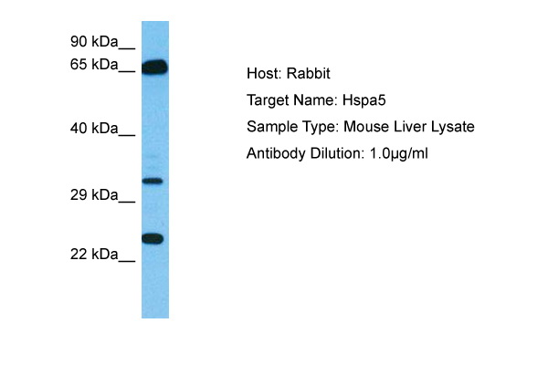
 WB (Western Blot)
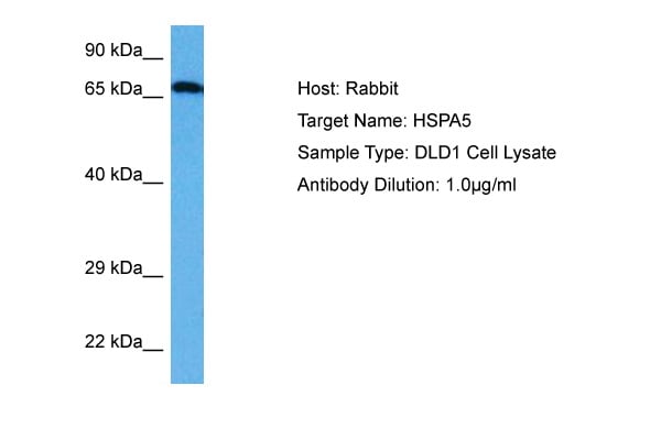
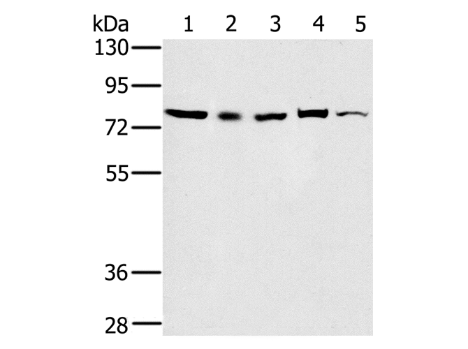
(Host: RabbitTarget Name: HSPA5Sample Tissue: Human DLD1 Whole Cell lysatesAntibody Dilution: 1ug/ml)
WB (Western Blot)
(Host: RabbitTarget Name: HSPA5Sample Tissue: Human DLD1 Whole Cell lysatesAntibody Dilution: 1ug/ml)
HSPA5, Polyclonal Antibody (Cat# AAA201600)
 WB (Western Blot)
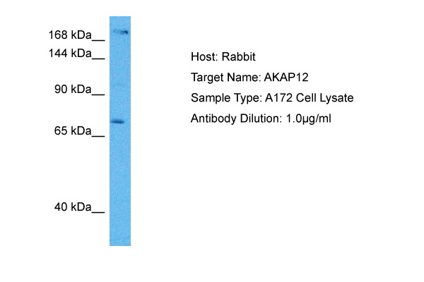
(Host: RabbitTarget Name: AKAP12Sample Tissue: Human A172 Whole Cell lysatesAntibody Dilution: 1ug/ml)
WB (Western Blot)
(Host: RabbitTarget Name: AKAP12Sample Tissue: Human A172 Whole Cell lysatesAntibody Dilution: 1ug/ml)
AKAP12, Polyclonal Antibody (Cat# AAA201605)

 WB (Western Blot)
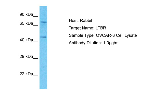
(Host: RabbitTarget Name: LTBRSample Tissue: Human OVCAR-3 Whole CellAntibody Dilution: 1ug/ml)
WB (Western Blot)
(Host: RabbitTarget Name: LTBRSample Tissue: Human OVCAR-3 Whole CellAntibody Dilution: 1ug/ml)
LTBR, Polyclonal Antibody (Cat# AAA201606)
 WB (Western Blot)
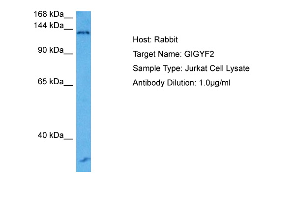
(Host: RabbitTarget Name: GIGYF2Sample Tissue: Human Jurkat Whole Cell lysatesAntibody Dilution: 1ug/ml)
WB (Western Blot)
(Host: RabbitTarget Name: GIGYF2Sample Tissue: Human Jurkat Whole Cell lysatesAntibody Dilution: 1ug/ml)
GIGYF2, Polyclonal Antibody (Cat# AAA201608)
 WB (Western Blot)
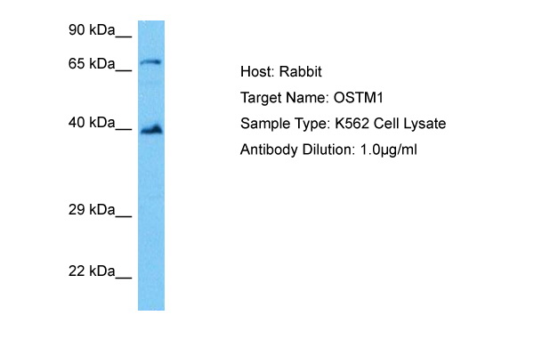
(Host: RabbitTarget Name: OSTM1Sample Tissue: Human K562 Whole Cell lysatesAntibody Dilution: 1.0ug/ml)
WB (Western Blot)
(Host: RabbitTarget Name: OSTM1Sample Tissue: Human K562 Whole Cell lysatesAntibody Dilution: 1.0ug/ml)
OSTM1, Polyclonal Antibody (Cat# AAA201612)
 WB (Western Blot)
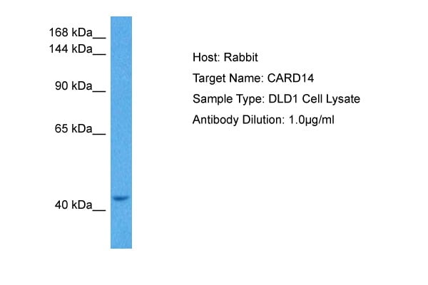
(Host: RabbitTarget Name: CARD14Sample Tissue: Human DLD1 Whole Cell lysatesAntibody Dilution: 1ug/ml)
WB (Western Blot)
(Host: RabbitTarget Name: CARD14Sample Tissue: Human DLD1 Whole Cell lysatesAntibody Dilution: 1ug/ml)
CARD14, Polyclonal Antibody (Cat# AAA201613)
 WB (Western Blot)
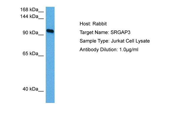
(Host: RabbitTarget Name: SRGAP3Sample Tissue: Human Jurkat Whole Cell lysatesAntibody Dilution: 1ug/ml)
WB (Western Blot)
(Host: RabbitTarget Name: SRGAP3Sample Tissue: Human Jurkat Whole Cell lysatesAntibody Dilution: 1ug/ml)
SRGAP3, Polyclonal Antibody (Cat# AAA201614)
Predicted Reactivity: Human
 WB (Western Blot)
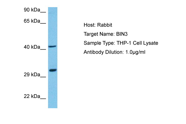
(Host: RabbitTarget Name: BIN3Sample Tissue: Human THP-1 Whole Cell lysatesAntibody Dilution: 1ug/ml)
WB (Western Blot)
(Host: RabbitTarget Name: BIN3Sample Tissue: Human THP-1 Whole Cell lysatesAntibody Dilution: 1ug/ml)
BIN3, Polyclonal Antibody (Cat# AAA201615)
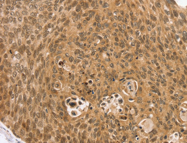
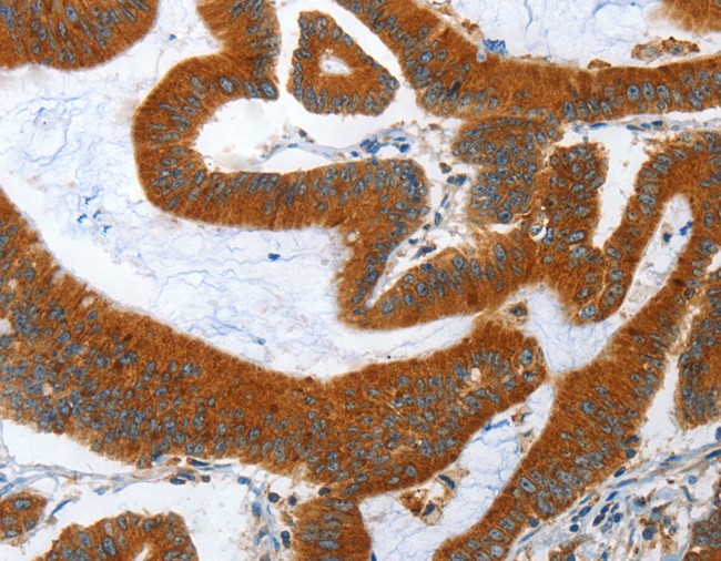
 IHC (Immunohiostchemistry)
(Immunohistochemistry of paraffin-embedded Human thyroid cancer tissue using ARHGEF11 Polyclonal Antibody at dilution 1:25)
IHC (Immunohiostchemistry)
(Immunohistochemistry of paraffin-embedded Human thyroid cancer tissue using ARHGEF11 Polyclonal Antibody at dilution 1:25)
ARHGEF11, Polyclonal Antibody (Cat# AAA167278)
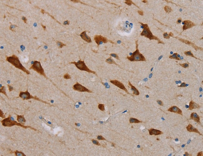
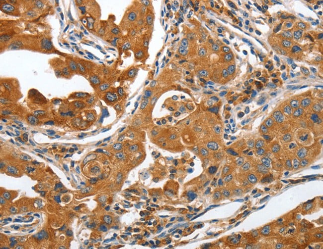
 IHC (Immunohistochemisry)
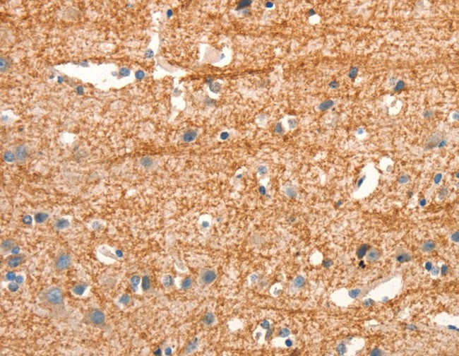
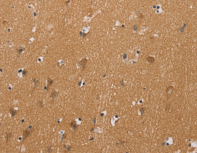
(Immunohistochemistry of paraffin-embedded Human brain using CHRNA4 Polyclonal Antibody at dilution of 1:20)
IHC (Immunohistochemisry)
(Immunohistochemistry of paraffin-embedded Human brain using CHRNA4 Polyclonal Antibody at dilution of 1:20)
CHRNA4, Polyclonal Antibody (Cat# AAA167279)
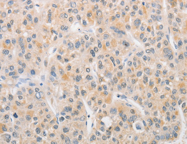
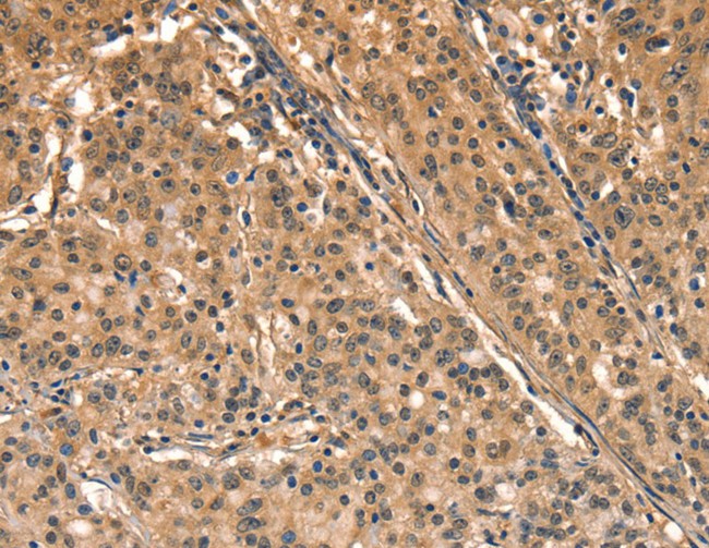
 IHC (Immunohiostchemistry)
(Immunohistochemistry of paraffin-embedded Human gastric cancer tissue using APLF Polyclonal Antibody at dilution 1:20)
IHC (Immunohiostchemistry)
(Immunohistochemistry of paraffin-embedded Human gastric cancer tissue using APLF Polyclonal Antibody at dilution 1:20)
APLF, Polyclonal Antibody (Cat# AAA167298)
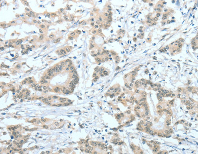
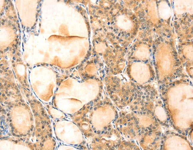
 IHC (Immunohiostchemistry)
(Immunohistochemistry of paraffin-embedded Human thyroid cancer tissue using ASH2L Polyclonal Antibody at dilution 1:60)
IHC (Immunohiostchemistry)
(Immunohistochemistry of paraffin-embedded Human thyroid cancer tissue using ASH2L Polyclonal Antibody at dilution 1:60)
ASH2L, Polyclonal Antibody (Cat# AAA167395)
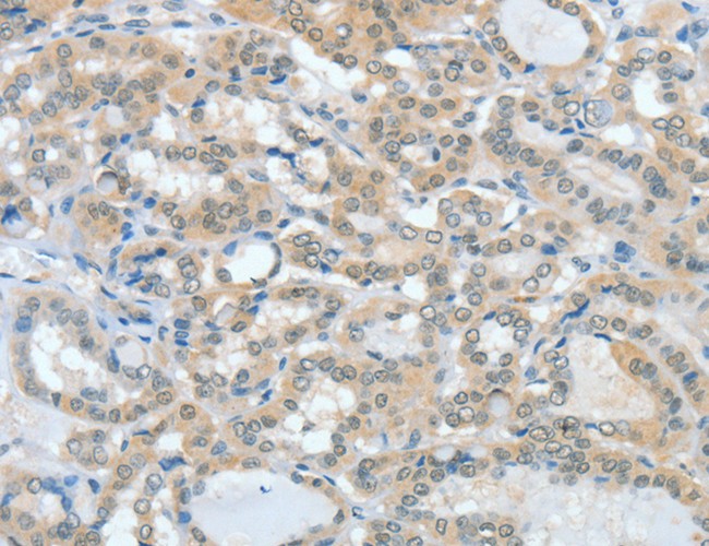
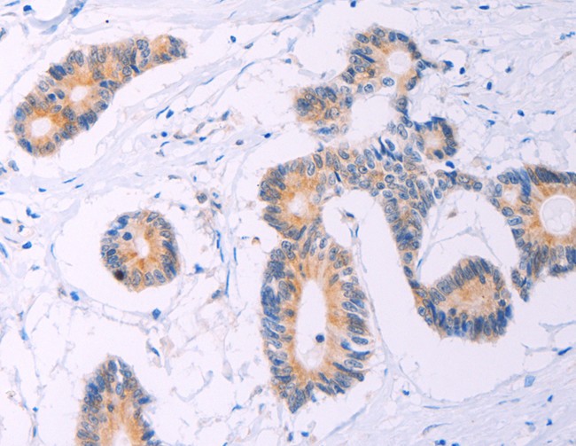
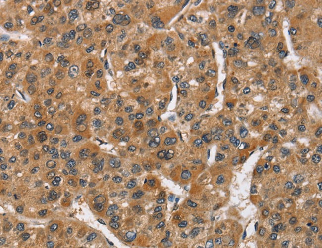
 IHC (Immunohiostchemistry)
(Immunohistochemistry of paraffin-embedded Human thyroid cancer tissue using VMP1 Polyclonal Antibody at dilution 1:35)
IHC (Immunohiostchemistry)
(Immunohistochemistry of paraffin-embedded Human thyroid cancer tissue using VMP1 Polyclonal Antibody at dilution 1:35)
VMP1, Polyclonal Antibody (Cat# AAA167403)
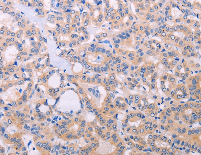 IHC (Immunohiostchemistry)
(Immunohistochemistry of paraffin-embedded Human thyroid cancer tissue using ADCY5 Polyclonal Antibody at dilution 1:30)
IHC (Immunohiostchemistry)
(Immunohistochemistry of paraffin-embedded Human thyroid cancer tissue using ADCY5 Polyclonal Antibody at dilution 1:30)
ADCY5, Polyclonal Antibody (Cat# AAA167405)

 IHC (Immunohistochemisry)
(Immunohistochemistry of paraffin-embedded Human brain using RPSA Polyclonal Antibody at dilution of 1:60)
IHC (Immunohistochemisry)
(Immunohistochemistry of paraffin-embedded Human brain using RPSA Polyclonal Antibody at dilution of 1:60)
RPSA, Polyclonal Antibody (Cat# AAA167413)
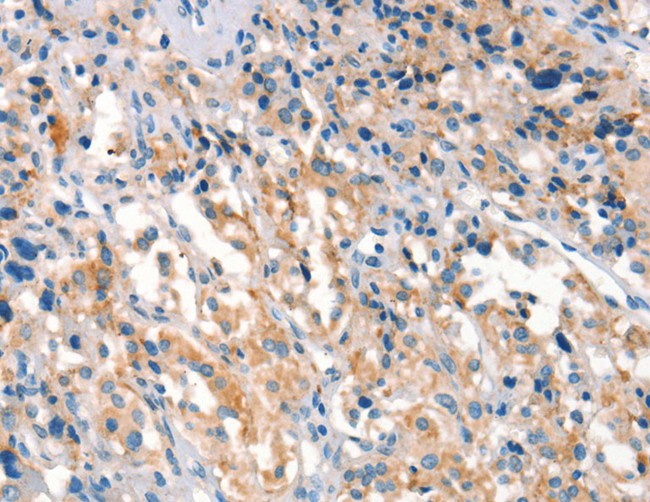
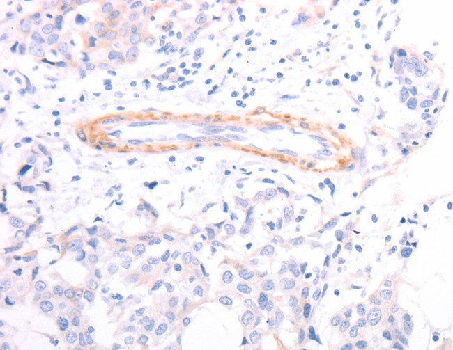
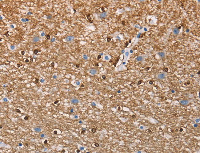
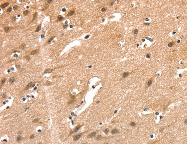
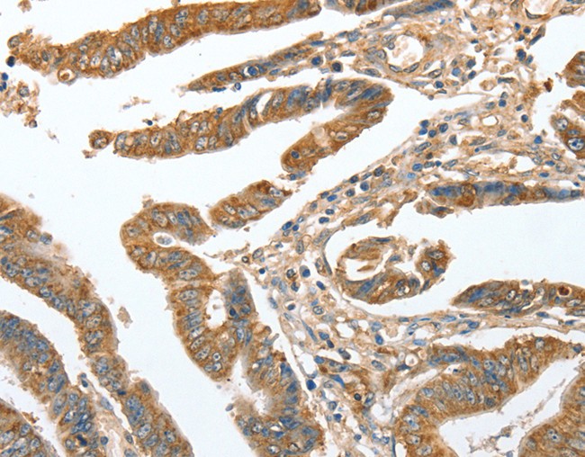
 IHC (Immunohiostchemistry)
(Immunohistochemistry of paraffin-embedded Human brain tissue using PROM1 Polyclonal Antibody at dilution 1:70)
IHC (Immunohiostchemistry)
(Immunohistochemistry of paraffin-embedded Human brain tissue using PROM1 Polyclonal Antibody at dilution 1:70)
PROM1, Polyclonal Antibody (Cat# AAA167415)

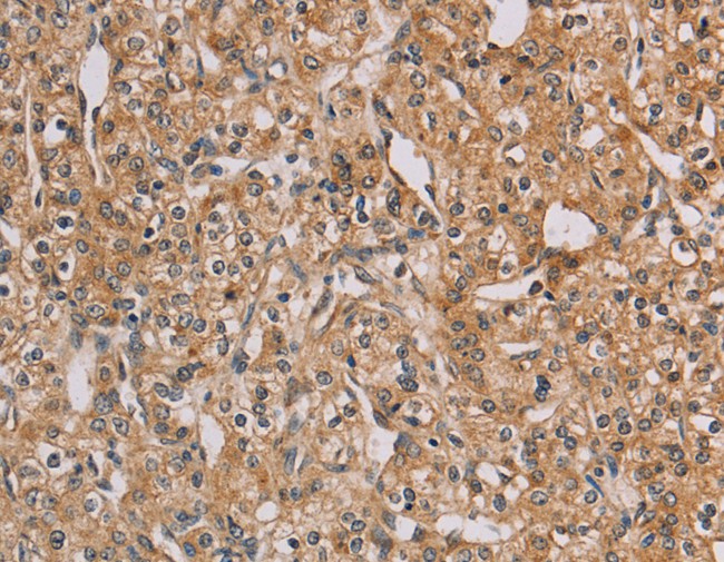
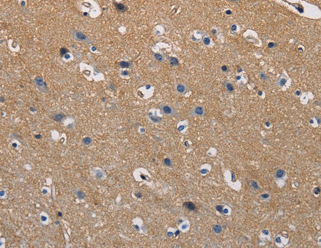
 IHC (Immunohistochemisry)
(Immunohistochemistry of paraffin-embedded Human brain using TAB3 Polyclonal Antibody at dilution of 1:25)
IHC (Immunohistochemisry)
(Immunohistochemistry of paraffin-embedded Human brain using TAB3 Polyclonal Antibody at dilution of 1:25)
TAB3, Polyclonal Antibody (Cat# AAA167416)
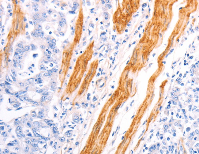
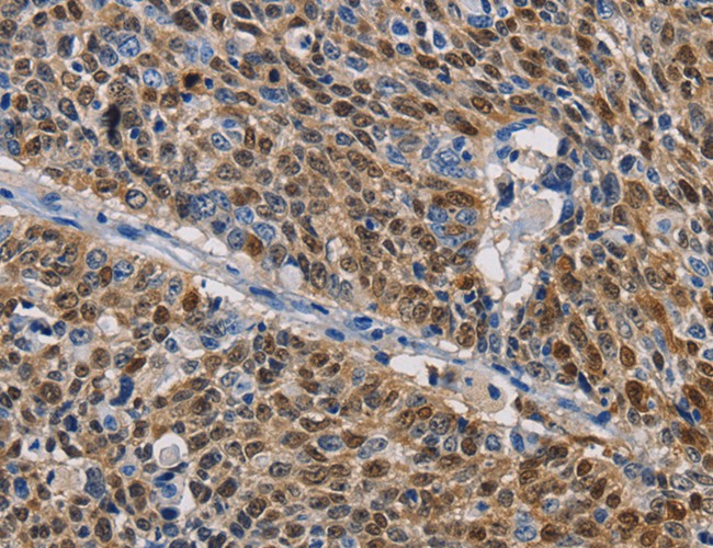
 IHC (Immunohistochemisry)
(Immunohistochemistry of paraffin-embedded Human breast cancer using TPM1 Polyclonal Antibody at dilution of 1:40)
IHC (Immunohistochemisry)
(Immunohistochemistry of paraffin-embedded Human breast cancer using TPM1 Polyclonal Antibody at dilution of 1:40)
TPM1, Polyclonal Antibody (Cat# AAA167423)
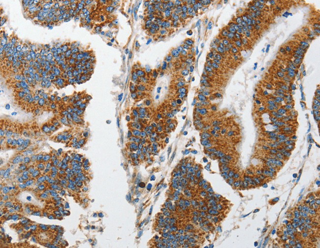
 IHC (Immunohiostchemistry)
(Immunohistochemistry of paraffin-embedded Human lung cancer tissue using GSTA2 Polyclonal Antibody at dilution 1:50)
IHC (Immunohiostchemistry)
(Immunohistochemistry of paraffin-embedded Human lung cancer tissue using GSTA2 Polyclonal Antibody at dilution 1:50)
GSTA2, Polyclonal Antibody (Cat# AAA167427)
 IHC (Immunohiostchemistry)
(Immunohistochemistry of paraffin-embedded Human cervical cancer tissue using MARK2 Polyclonal Antibody at dilution 1:15)
IHC (Immunohiostchemistry)
(Immunohistochemistry of paraffin-embedded Human cervical cancer tissue using MARK2 Polyclonal Antibody at dilution 1:15)
MARK2, Polyclonal Antibody (Cat# AAA167454)
 IHC (Immunohiostchemistry)
(Immunohistochemistry of paraffin-embedded Human esophagus cancer tissue using OPTN Polyclonal Antibody at dilution 1:25)
IHC (Immunohiostchemistry)
(Immunohistochemistry of paraffin-embedded Human esophagus cancer tissue using OPTN Polyclonal Antibody at dilution 1:25)
OPTN, Polyclonal Antibody (Cat# AAA167455)
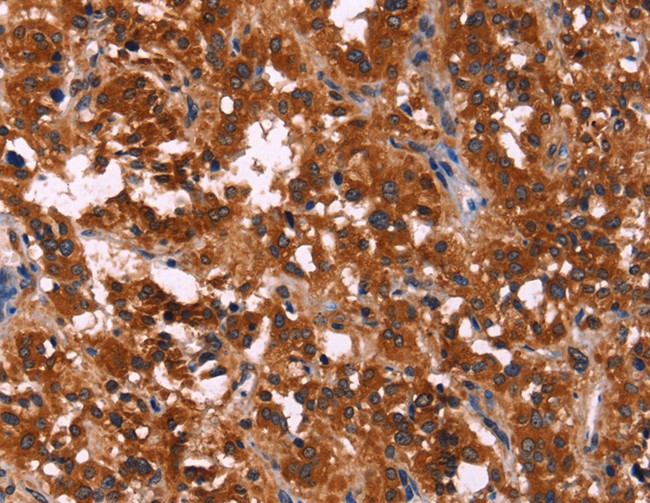
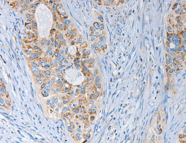
 IHC (Immunohiostchemistry)
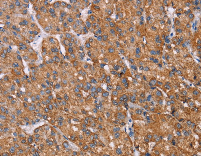
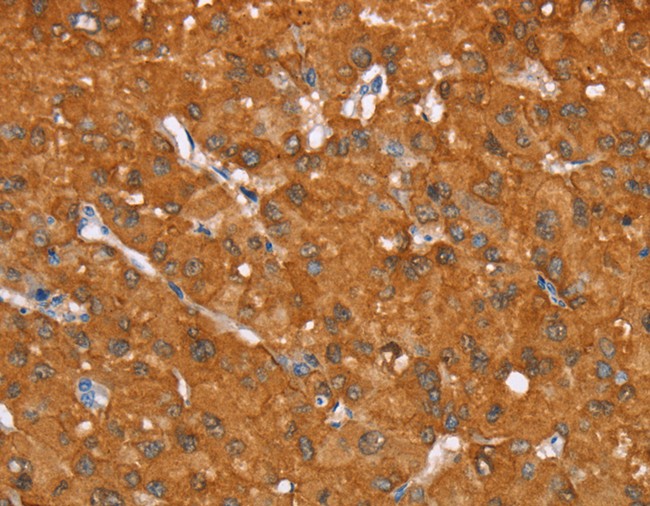
(Immunohistochemistry of paraffin-embedded Human liver cancer tissue using ATF4 Polyclonal Antibody at dilution 1:50)
IHC (Immunohiostchemistry)
(Immunohistochemistry of paraffin-embedded Human liver cancer tissue using ATF4 Polyclonal Antibody at dilution 1:50)
ATF4, Polyclonal Antibody (Cat# AAA167465)
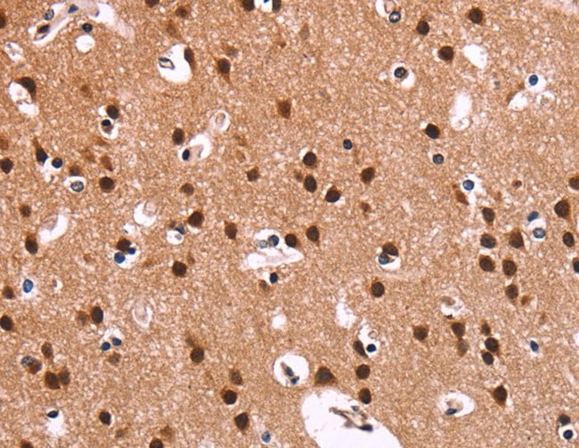
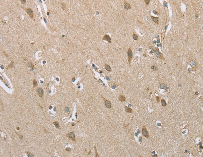
 IHC (Immunohiostchemistry)
(Immunohistochemistry of paraffin-embedded Human brain tissue using CEACAM6 Polyclonal Antibody at dilution 1:70)
IHC (Immunohiostchemistry)
(Immunohistochemistry of paraffin-embedded Human brain tissue using CEACAM6 Polyclonal Antibody at dilution 1:70)
CEACAM6, Polyclonal Antibody (Cat# AAA167470)
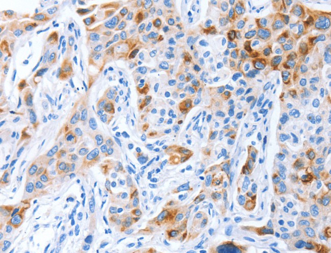
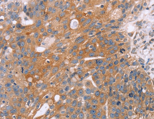
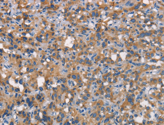
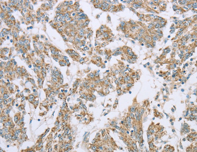
 IHC (Immunohiostchemistry)
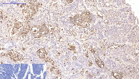
(Immunohistochemistry of paraffin-embedded Human liver cancer using SPARC Polyclonal Antibody at dilution of 1:35)
IHC (Immunohiostchemistry)
(Immunohistochemistry of paraffin-embedded Human liver cancer using SPARC Polyclonal Antibody at dilution of 1:35)
SPARC, Polyclonal Antibody (Cat# AAA167474)
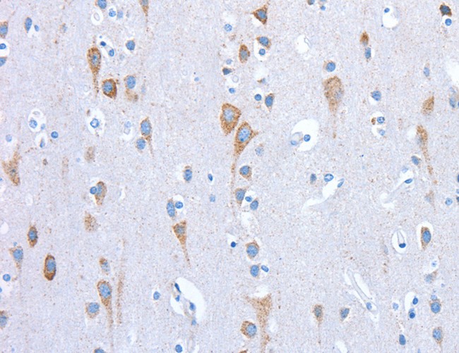
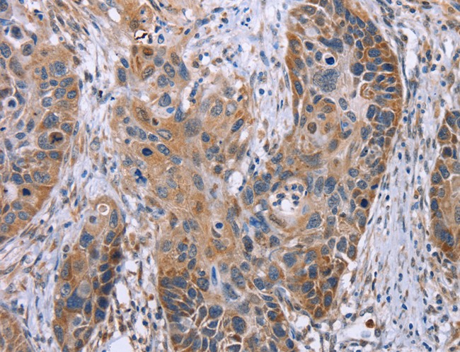
 IHC (Immunohiostchemistry)
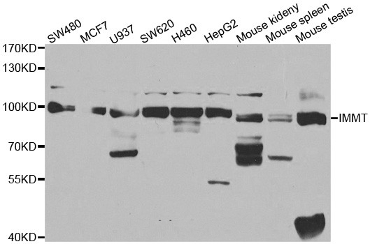
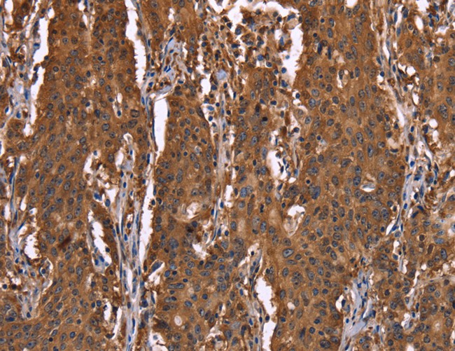
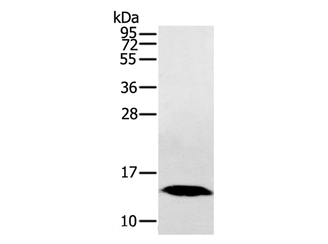
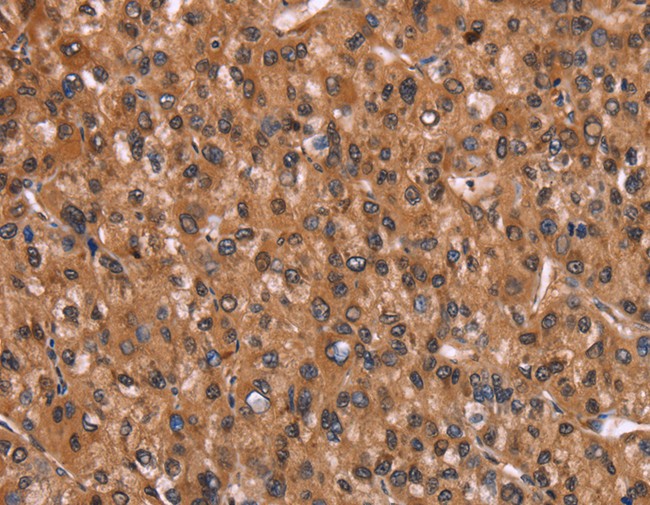
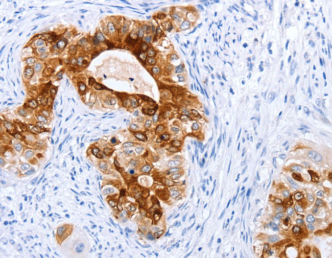
(Immunohistochemistry of paraffin-embedded human breast cancer tissue using IMMT antibody.)
IHC (Immunohiostchemistry)
(Immunohistochemistry of paraffin-embedded human breast cancer tissue using IMMT antibody.)
IMMT, Polyclonal Antibody (Cat# AAA167479)
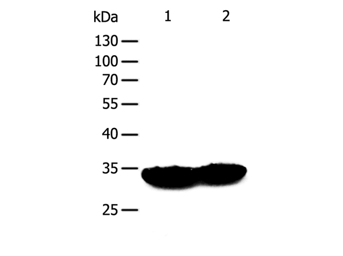
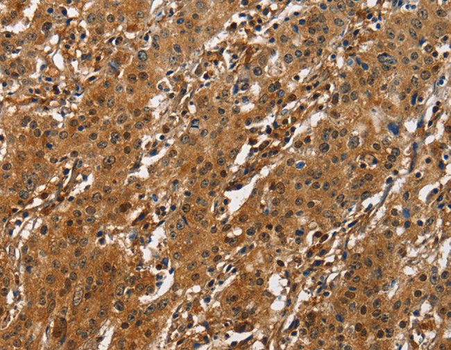
 IHC (Immunohistochemisry)
(Immunohistochemistry of paraffin-embedded Human liver cancer using HINT1 Polyclonal Antibody at dilution of 1:30)
IHC (Immunohistochemisry)
(Immunohistochemistry of paraffin-embedded Human liver cancer using HINT1 Polyclonal Antibody at dilution of 1:30)
HINT1, Polyclonal Antibody (Cat# AAA167320)
 IHC (Immunohiostchemistry)
(Immunohistochemistry of paraffin-embedded Human breast cancer tissue using RMDN3 Polyclonal Antibody at dilution 1:25)
IHC (Immunohiostchemistry)
(Immunohistochemistry of paraffin-embedded Human breast cancer tissue using RMDN3 Polyclonal Antibody at dilution 1:25)
RMDN3, Polyclonal Antibody (Cat# AAA167325)
 IF (Immunofluorescence)
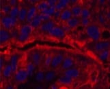
(Immunofluorescence analysis of Human liver cancer tissue using CD15 Monoclonal Antibody at dilution of 1:200.)
IF (Immunofluorescence)
(Immunofluorescence analysis of Human liver cancer tissue using CD15 Monoclonal Antibody at dilution of 1:200.)
FUT4, Polyclonal Antibody (Cat# AAA167327)
 IHC (Immunohiostchemistry)
(Immunohistochemistry of paraffin-embedded Human liver cancer using AKR1C4 Polyclonal Antibody at dilution of 1:25)
IHC (Immunohiostchemistry)
(Immunohistochemistry of paraffin-embedded Human liver cancer using AKR1C4 Polyclonal Antibody at dilution of 1:25)
AKR1C4, Polyclonal Antibody (Cat# AAA167332)
 IHC (Immunohistochemisry)
(Immunohistochemistry of paraffin-embedded Human liver cancer using TFF1 Polyclonal Antibody at dilution of 1:20)
IHC (Immunohistochemisry)
(Immunohistochemistry of paraffin-embedded Human liver cancer using TFF1 Polyclonal Antibody at dilution of 1:20)
TFF1, Polyclonal Antibody (Cat# AAA167339)
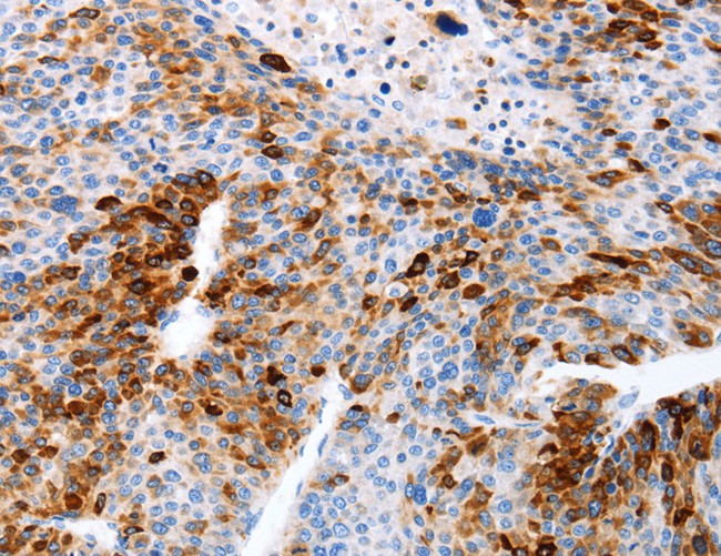
 IHC (Immunohiostchemistry)
(Immunohistochemistry of paraffin-embedded Human prostate cancer tissue using ROR1 Polyclonal Antibody at dilution 1:45)
IHC (Immunohiostchemistry)
(Immunohistochemistry of paraffin-embedded Human prostate cancer tissue using ROR1 Polyclonal Antibody at dilution 1:45)
ROR1, Polyclonal Antibody (Cat# AAA167341)

 IHC (Immunohistochemisry)
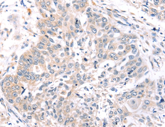
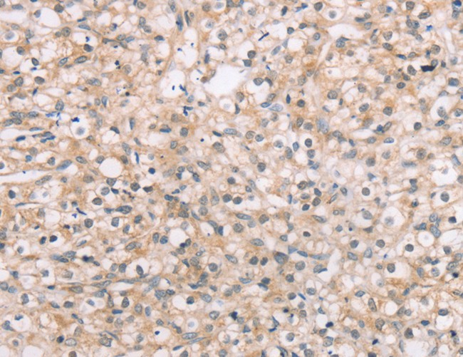
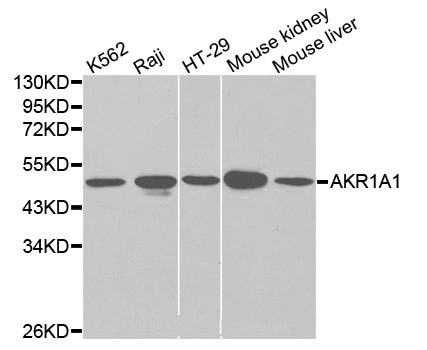
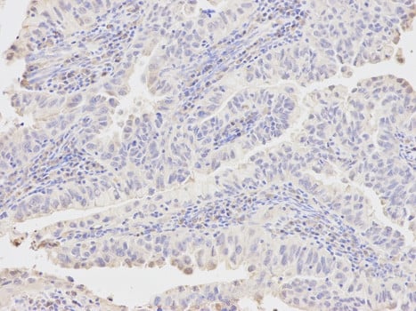
(Immunohistochemistry of paraffin-embedded human endometrial cancer using AKR1A1 antibody at dilution of 1:400 (200x lens).)
IHC (Immunohistochemisry)
(Immunohistochemistry of paraffin-embedded human endometrial cancer using AKR1A1 antibody at dilution of 1:400 (200x lens).)
AKR1A1, Polyclonal Antibody (Cat# AAA167342)
 IHC (Immunohiostchemistry)
(Immunohistochemistry of paraffin-embedded Human lung cancer tissue using ANAPC10 Polyclonal Antibody at dilution 1:40)
IHC (Immunohiostchemistry)
(Immunohistochemistry of paraffin-embedded Human lung cancer tissue using ANAPC10 Polyclonal Antibody at dilution 1:40)
ANAPC10, Polyclonal Antibody (Cat# AAA167349)
 IHC (Immunohistochemisry)
(Immunohistochemistry of paraffin-embedded Human brain using AIMP1 Polyclonal Antibody at dilution of 1:40)
IHC (Immunohistochemisry)
(Immunohistochemistry of paraffin-embedded Human brain using AIMP1 Polyclonal Antibody at dilution of 1:40)
AIMP1, Polyclonal Antibody (Cat# AAA167353)
 IHC (Immunohistochemisry)
(Immunohistochemistry of paraffin-embedded Human brain using ECHS1 Polyclonal Antibody at dilution of 1:20)
IHC (Immunohistochemisry)
(Immunohistochemistry of paraffin-embedded Human brain using ECHS1 Polyclonal Antibody at dilution of 1:20)
ECHS1, Polyclonal Antibody (Cat# AAA167355)
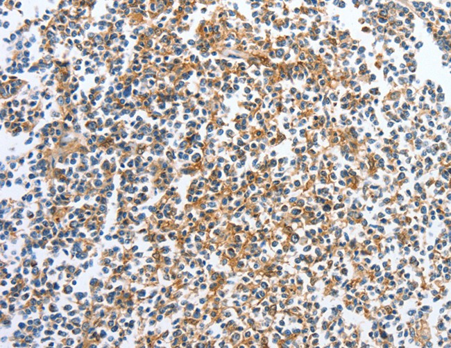 IHC (Immunohiostchemistry)
(Immunohistochemistry of paraffin-embedded Human tonsil tissue using CKAP2 Polyclonal Antibody at dilution 1:70)
IHC (Immunohiostchemistry)
(Immunohistochemistry of paraffin-embedded Human tonsil tissue using CKAP2 Polyclonal Antibody at dilution 1:70)
CKAP2, Polyclonal Antibody (Cat# AAA167356)
What are Polyclonal Antibodies?
Polyclonal antibodies are antibodies that come from multiple B cell clones of a host animal. The typical hosts used for the majority of polyclonal antibody production are rabbits, goats, sheep, and donkeys. These polyclonal antibodies, once having identified their target, will bind to different epitopes located at different regions or sequences on the same protein/antigen. As a result, they are ideal at locating and binding to the target, even if the target is in very low concentrations (due to many different antibodies being able to bind to the same target molecule, which allows for significant amplification of a downstream signal).
Polyclonal antibodies are typically produced by injecting an antigen into a host animal, which causes the animal’s immune system to attack the foreign antigen by mass generating antibodies against it. After a period of time, serum is collected from the animal and purified using physicochemical fractionation, class-specific affinity purification, and/or antigen-affinity purification.
Key Uses of Polyclonal Antibodies
- Western Blotting: This method is used to find specific proteins in biological samples after separating them by size.
- Immunohistochemistry: IHC helps visualize the location of proteins in tissue sections using various staining techniques.
- ELISA: (Enzyme-Linked Immunosorbent Assay) is typically used to identify specific protein quantities in a sample. ELISAs can be either “Quantitative” or “Qualitative”.
- Flow Cytometry: technique that identifies and measures the specific protein on the surface or inside the cells in a fluid suspension.
- Immunoprecipitation: IP isolates and studies a specific protein from a complex mixture using antibodies.
Why Buy Polyclonal Antibodies from AAA Biotech?
1. Ideal for Various Applications
Our antibodies are generally going to be validated for use in multiple types of assays, including ELISA, Western Blotting, Immunohistochemistry, Immunoprecipitation, amongst others. They are ideal for a wide range of research applications.
2. Rigorous Quality Control
All of the antibodies in our catalog undergo strict quality testing to ensure specificity, sensitivity, and consistent performance. We are confident in the ability of our antibodies to provide you with accurate results.
3. Wide Assortment of Antibodies
Antibodies in are catalog can be found for both common and exotic species, and these antibodies are also available in both conjugated and recombinant forms to suit many diverse experimental needs.
4. Highly Purified
Our antibodies are available in purified forms with over 85% purity, as confirmed by SDS-PAGE. They are also available with tags such as His, Flag, GST, or MBP. We cater to customers worldwide.
FAQ
1. How are polyclonal antibodies produced?
Traditionally, polyclonal antibodies are produced by injecting an antigen into a host animal (such as a rabbit or goat), which then triggers an immune response from the host animal. The animal’s B cells produce antibodies that will recognize different parts of the injected antigen. These antibodies are then collected from the animal’s blood and purified for use.
2. How do polyclonal antibodies differ from monoclonal antibodies?
Polyclonal antibodies are a mix of antibodies that bind to different locations (epitopes) of the same antigen, while monoclonal antibodies are identical and bind to just one specific epitope. This makes polyclonal antibodies more versatile and better at detecting proteins that may be present in low quantities or in altered/modified forms.
3. How should I store polyclonal antibodies?
Polyclonal antibodies should be stored at 4°C for short-term use (up to a few weeks) and at -20°C or -80°C for long-term storage. Avoid repeated freeze-thaw cycles by dividing them into small aliquots. Always check the datasheet for specific storage instructions.