Filters
▼Clonality
▼Type
▼Reactivity
▼Gene Name
▼Isotype
▼Host
▼Application
▼Clone
▼Polyclonal Antibodies
At AAA Biotech also known as AAA Bio or AAABio, we provide a broad range of purified polyclonal antibodies (pAbs) that are able to all be browsed online through our website. Due to their high specificity and strong binding affinity, these antibodies are ideal for wide swathes of research and experimental applications.
Our polyclonal antibodies can easily support your work, whether you use them for Western Blotting, Immunocytochemistry (with or without Immunofluorescence used in conjunction), Immunohistochemistry, Immunoprecipitation, and ELISA tests. We highly encourage you to browse our range of pAbs and choose the one that best suits your experimental model.
Viewing 2250-2300 of 96805 product results
 IHC (Immunohiostchemistry)
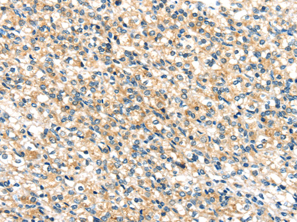
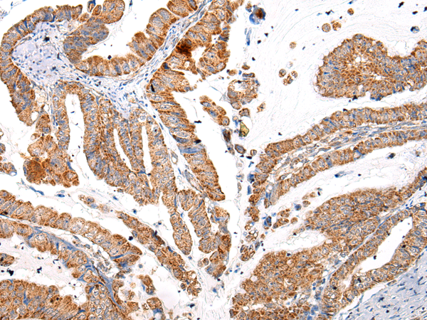
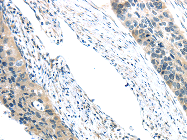
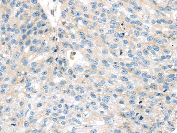
(Immunohistochemistry of paraffin-embedded Human esophagus cancer tissue using TTI1 Polyclonal Antibody at dilution 1:40)
IHC (Immunohiostchemistry)
(Immunohistochemistry of paraffin-embedded Human esophagus cancer tissue using TTI1 Polyclonal Antibody at dilution 1:40)
TTI1, Polyclonal Antibody (Cat# AAA173284)
 IHC (Immunohiostchemistry)
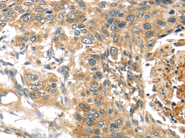
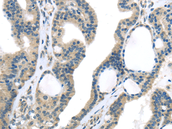
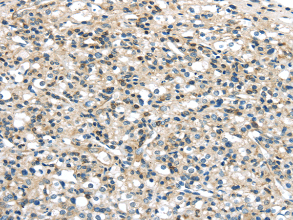
(Immunohistochemistry of paraffin-embedded Human thyroid cancer tissue using TSC2 Polyclonal Antibody at dilution 1:35)
IHC (Immunohiostchemistry)
(Immunohistochemistry of paraffin-embedded Human thyroid cancer tissue using TSC2 Polyclonal Antibody at dilution 1:35)
TSC2, Polyclonal Antibody (Cat# AAA173285)
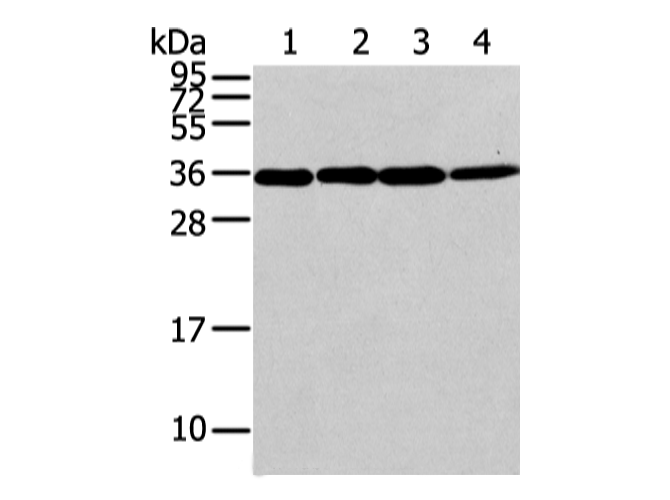
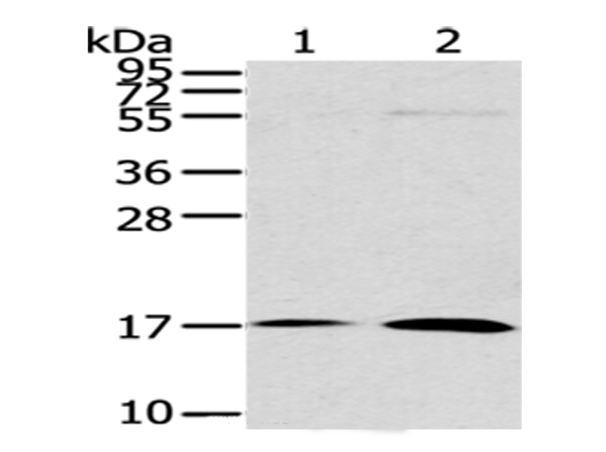
 WB (Western Blot)
(Western Blot analysis of Mouse brain tissue using WWOX Polyclonal Antibody at dilution of 1/200)
WB (Western Blot)
(Western Blot analysis of Mouse brain tissue using WWOX Polyclonal Antibody at dilution of 1/200)
WWOX, Polyclonal Antibody (Cat# AAA173375)
 IHC (Immunohiostchemistry)
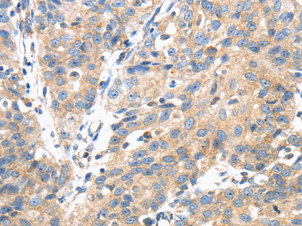
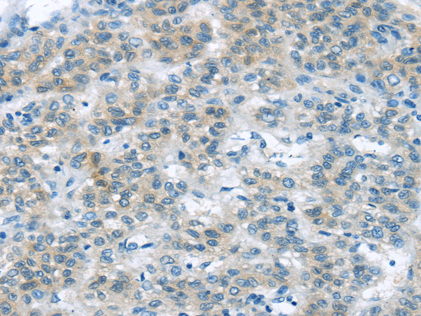
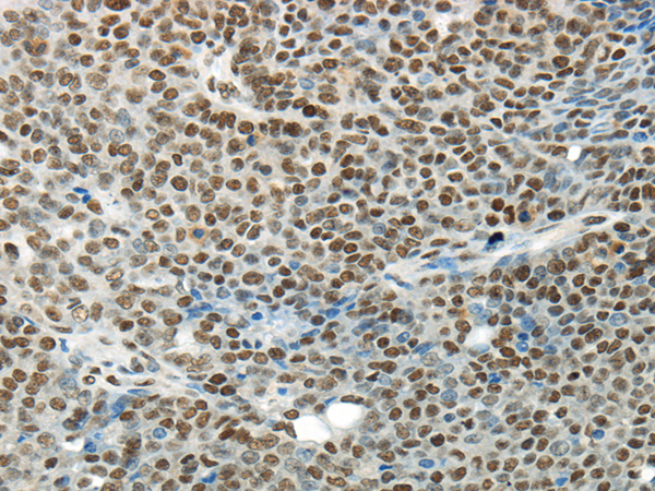
(Immunohistochemistry of paraffin-embedded Human breast cancer using XKRX Polyclonal Antibody at dilution of 1/30)
IHC (Immunohiostchemistry)
(Immunohistochemistry of paraffin-embedded Human breast cancer using XKRX Polyclonal Antibody at dilution of 1/30)
XKRX, Polyclonal Antibody (Cat# AAA173382)
 IHC (Immunohiostchemistry)
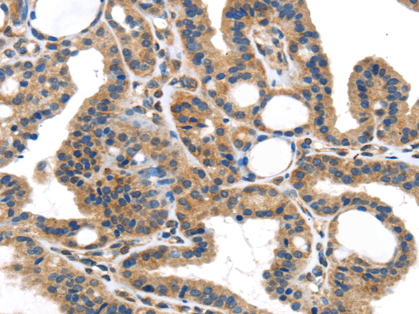
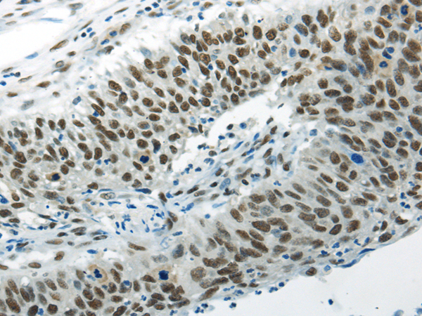
(Immunohistochemistry of paraffin-embedded Human esophagus cancer tissue using ZFAND2A Polyclonal Antibody at dilution 1:30)
IHC (Immunohiostchemistry)
(Immunohistochemistry of paraffin-embedded Human esophagus cancer tissue using ZFAND2A Polyclonal Antibody at dilution 1:30)
ZFAND2A, Polyclonal Antibody (Cat# AAA173385)
 IHC (Immunohiostchemistry)
(Immunohistochemistry of paraffin-embedded Human colorectal cancer tissue using ZNF281 Polyclonal Antibody at dilution 1:35)
IHC (Immunohiostchemistry)
(Immunohistochemistry of paraffin-embedded Human colorectal cancer tissue using ZNF281 Polyclonal Antibody at dilution 1:35)
ZNF281, Polyclonal Antibody (Cat# AAA173387)
 IHC (Immunohiostchemistry)
(Immunohistochemistry of paraffin-embedded Human colorectal cancer tissue using ZNF473 Polyclonal Antibody at dilution 1:30)
IHC (Immunohiostchemistry)
(Immunohistochemistry of paraffin-embedded Human colorectal cancer tissue using ZNF473 Polyclonal Antibody at dilution 1:30)
ZNF473, Polyclonal Antibody (Cat# AAA173388)
 WB (Western Blot)
(Western Blot analysis of A431 and hela cell using ARFGAP2 Polyclonal Antibody at dilution of 1/200)
WB (Western Blot)
(Western Blot analysis of A431 and hela cell using ARFGAP2 Polyclonal Antibody at dilution of 1/200)
ARFGAP2, Polyclonal Antibody (Cat# AAA173390)
 WB (Western Blot)
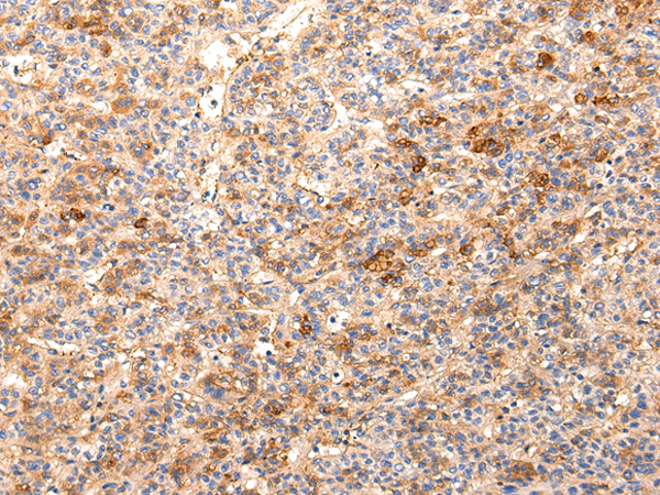
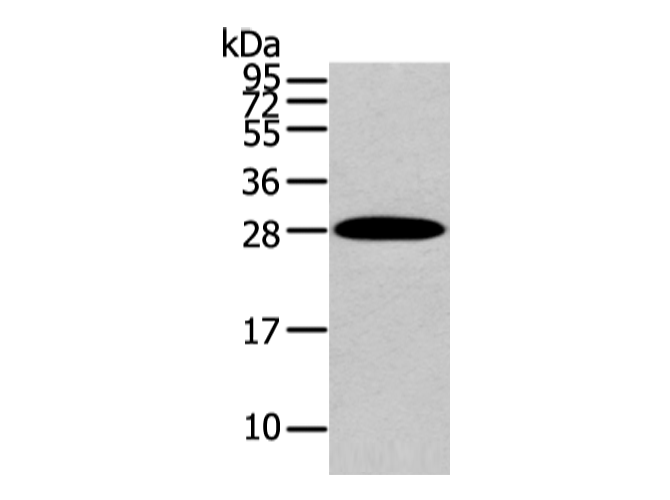
(Western Blot analysis of Human liver cancer tissue using ZNF365 Polyclonal Antibody at dilution of 1/200)
WB (Western Blot)
(Western Blot analysis of Human liver cancer tissue using ZNF365 Polyclonal Antibody at dilution of 1/200)
ZNF365, Polyclonal Antibody (Cat# AAA173392)
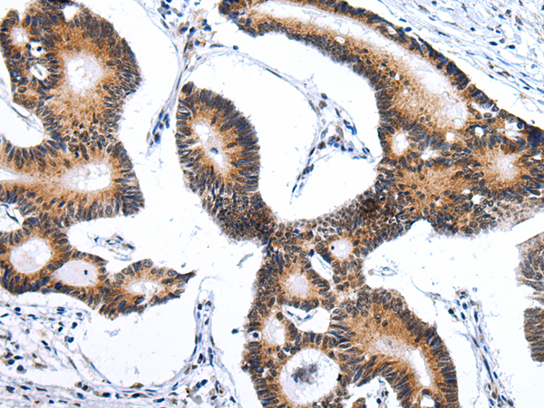
 IHC (Immunohiostchemistry)
(Immunohistochemistry of paraffin-embedded Human esophagus cancer tissue using KRT20 Polyclonal Antibody at dilution 1:35)
IHC (Immunohiostchemistry)
(Immunohistochemistry of paraffin-embedded Human esophagus cancer tissue using KRT20 Polyclonal Antibody at dilution 1:35)
KRT20, Polyclonal Antibody (Cat# AAA173403)
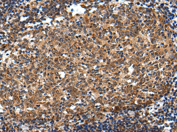
 IHC (Immunohiostchemistry)
(Immunohistochemistry of paraffin-embedded Human prostate cancer tissue using ADAMTS1 Polyclonal Antibody at dilution 1:30)
IHC (Immunohiostchemistry)
(Immunohistochemistry of paraffin-embedded Human prostate cancer tissue using ADAMTS1 Polyclonal Antibody at dilution 1:30)
ADAMTS1, Polyclonal Antibody (Cat# AAA173411)
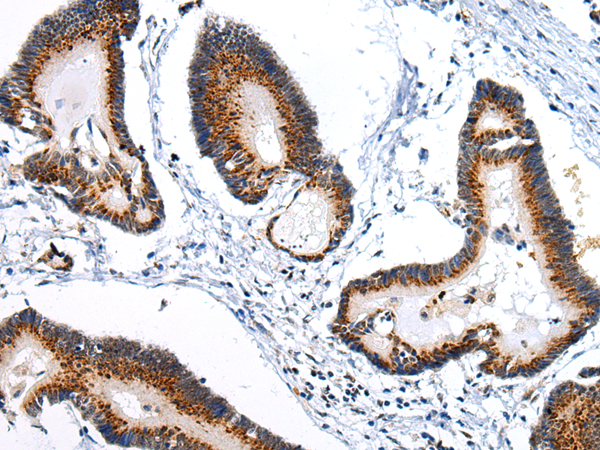
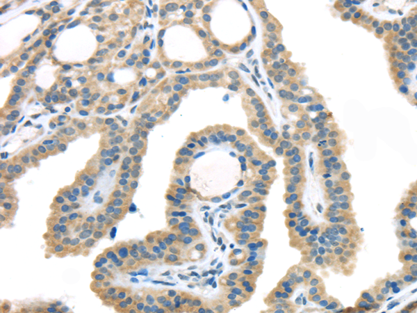
 IHC (Immunohiostchemistry)
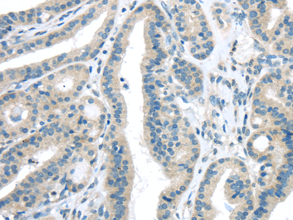
(Immunohistochemistry of paraffin-embedded Human thyroid cancer tissue using AQP7 Polyclonal Antibody at dilution 1:40)
IHC (Immunohiostchemistry)
(Immunohistochemistry of paraffin-embedded Human thyroid cancer tissue using AQP7 Polyclonal Antibody at dilution 1:40)
AQP7, Polyclonal Antibody (Cat# AAA173413)

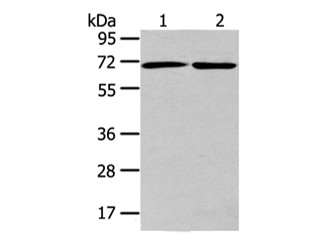
 WB (Western Blot)
(Western Blot analysis of Lovo, 293T, Jurkat and hela cell using CBX7 Polyclonal Antibody at dilution of 1/200)
WB (Western Blot)
(Western Blot analysis of Lovo, 293T, Jurkat and hela cell using CBX7 Polyclonal Antibody at dilution of 1/200)
CBX7, Polyclonal Antibody (Cat# AAA173423)
 IHC (Immunohiostchemistry)
(Immunohistochemistry of paraffin-embedded Human esophagus cancer tissue using COLEC12 Polyclonal Antibody at dilution 1:40)
IHC (Immunohiostchemistry)
(Immunohistochemistry of paraffin-embedded Human esophagus cancer tissue using COLEC12 Polyclonal Antibody at dilution 1:40)
COLEC12, Polyclonal Antibody (Cat# AAA173434)
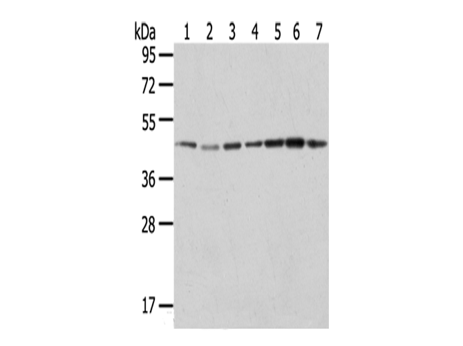 WB (Western Blot)
(Western Blot analysis of Raji, A549, Hela, HepG2, 231, K562 and A431 cells using ERP44 Polyclonal Antibody at dilution of 1/200)
WB (Western Blot)
(Western Blot analysis of Raji, A549, Hela, HepG2, 231, K562 and A431 cells using ERP44 Polyclonal Antibody at dilution of 1/200)
ERP44, Polyclonal Antibody (Cat# AAA173291)
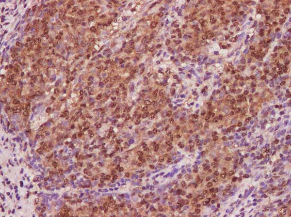
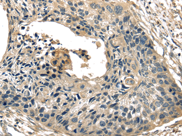 IHC (Immunohistochemistry)
(Immunohistochemistry of paraffin-embedded Human esophagus cancer tissue using TXNIP Polyclonal Antibody at dilution 1:40)
IHC (Immunohistochemistry)
(Immunohistochemistry of paraffin-embedded Human esophagus cancer tissue using TXNIP Polyclonal Antibody at dilution 1:40)
TXNIP, Polyclonal Antibody (Cat# AAA173293)
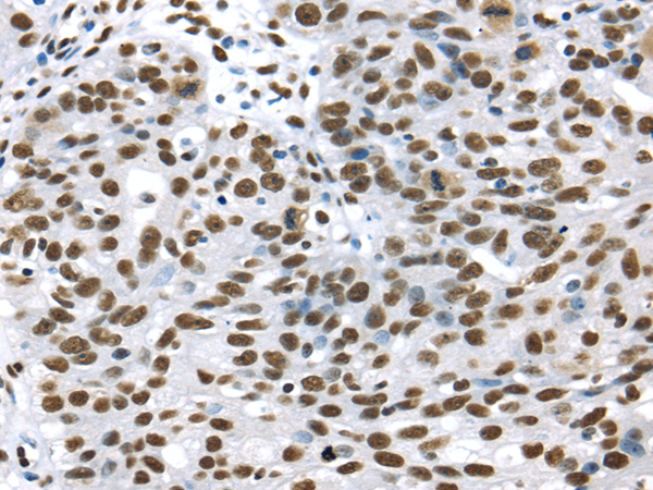
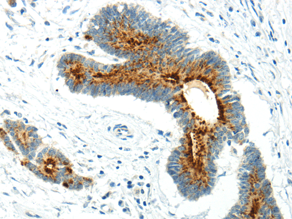
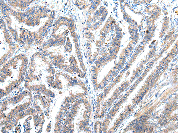
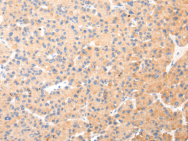
 IHC (Immunohiostchemistry)
(Immunohistochemistry of paraffin-embedded Human liver cancer using DDX39B Polyclonal Antibody at dilution of 1/35)
IHC (Immunohiostchemistry)
(Immunohistochemistry of paraffin-embedded Human liver cancer using DDX39B Polyclonal Antibody at dilution of 1/35)
DDX39B, Polyclonal Antibody (Cat# AAA173294)
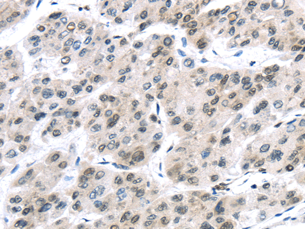
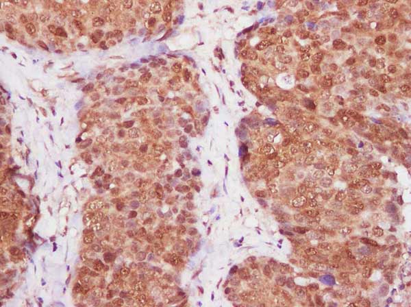
 IHC (Immunohiostchemistry)
(Immunohistochemistry of paraffin-embedded Human cervical cancer using MSI1 Polyclonal Antibody at dilution of 1/30)
IHC (Immunohiostchemistry)
(Immunohistochemistry of paraffin-embedded Human cervical cancer using MSI1 Polyclonal Antibody at dilution of 1/30)
MSI1, Polyclonal Antibody (Cat# AAA173306)
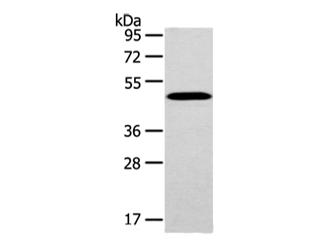
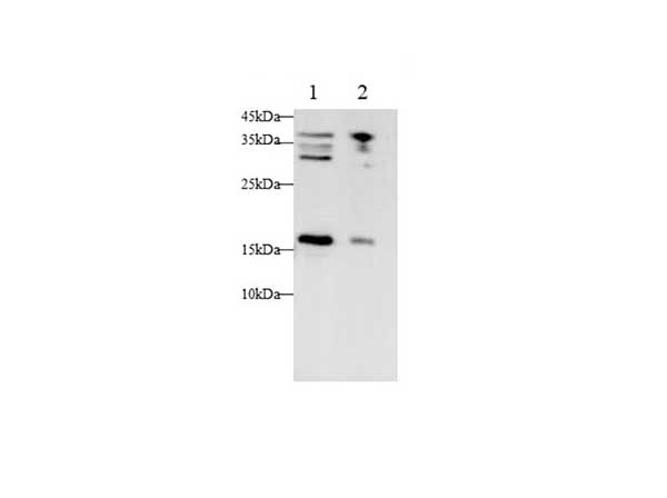
 WB (Western Blot)
(Western Blot analysis of 293T cells and Human normal kidney tissue using CHRDL1 Polyclonal Antibody at dilution of 1/400)
WB (Western Blot)
(Western Blot analysis of 293T cells and Human normal kidney tissue using CHRDL1 Polyclonal Antibody at dilution of 1/400)
CHRDL1, Polyclonal Antibody (Cat# AAA173313)
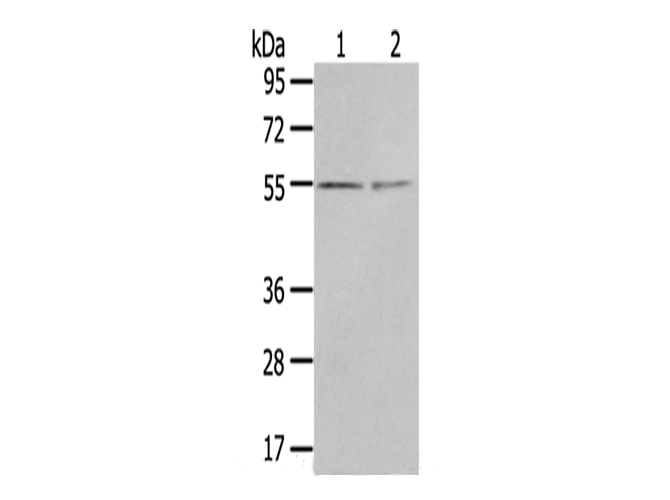
 WB (Western Blot)
(Western Blot analysis of Lovo cells and Human liver cancer tissue using UTS2B Polyclonal Antibody at dilution of 1/200)
WB (Western Blot)
(Western Blot analysis of Lovo cells and Human liver cancer tissue using UTS2B Polyclonal Antibody at dilution of 1/200)
UTS2B, Polyclonal Antibody (Cat# AAA173316)
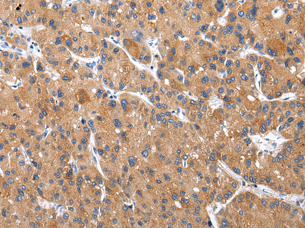
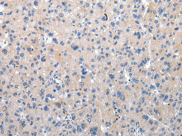
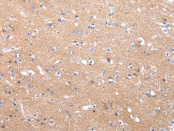
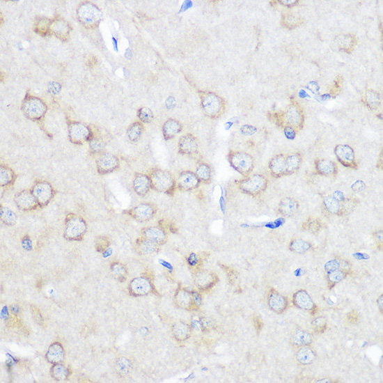
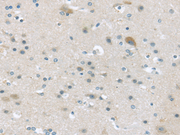 IHC (Immunohiostchemistry)
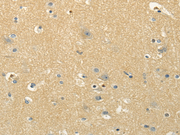
(Immunohistochemistry of paraffin-embedded Human brain using VGF Polyclonal Antibody at dilution of 1/45)
IHC (Immunohiostchemistry)
(Immunohistochemistry of paraffin-embedded Human brain using VGF Polyclonal Antibody at dilution of 1/45)
VGF, Polyclonal Antibody (Cat# AAA173324)
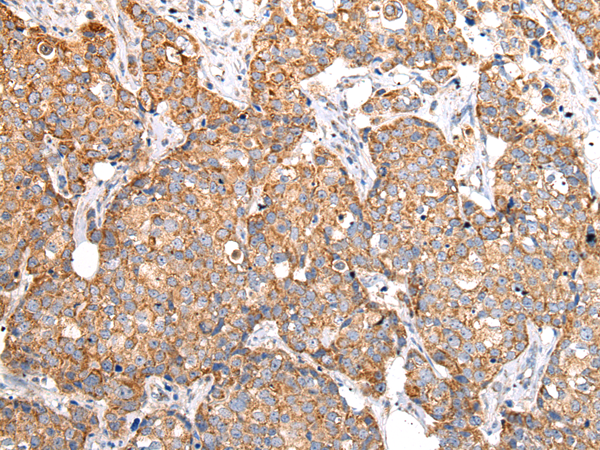
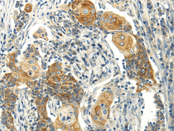
 IHC (Immunohiostchemistry)
(Immunohistochemistry of paraffin-embedded Human esophagus cancer tissue using VGF Polyclonal Antibody at dilution 1:35)
IHC (Immunohiostchemistry)
(Immunohistochemistry of paraffin-embedded Human esophagus cancer tissue using VGF Polyclonal Antibody at dilution 1:35)
VGF, Polyclonal Antibody (Cat# AAA173325)
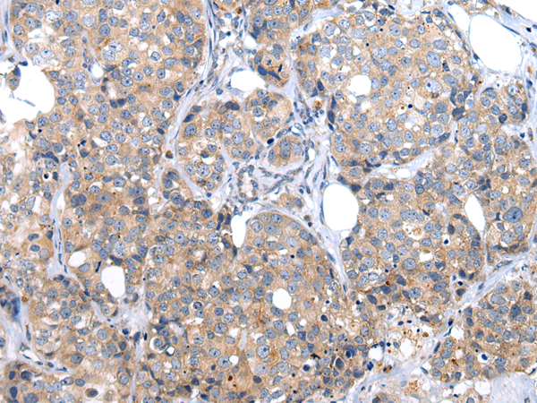
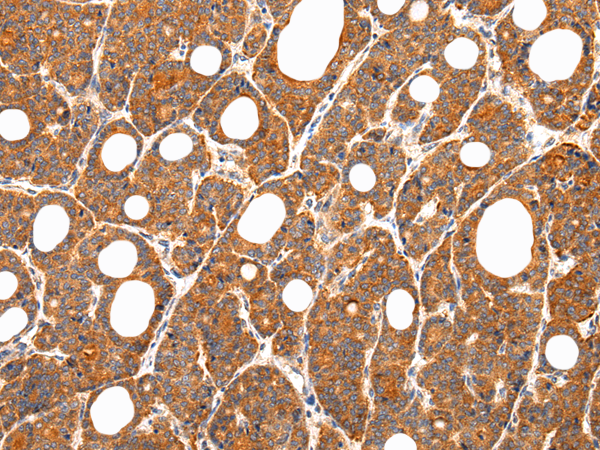
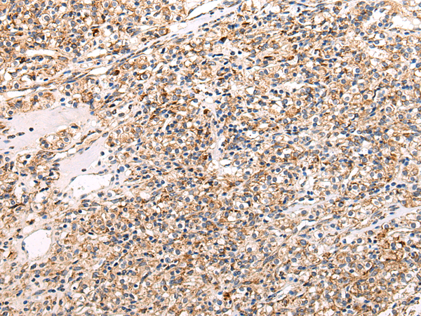
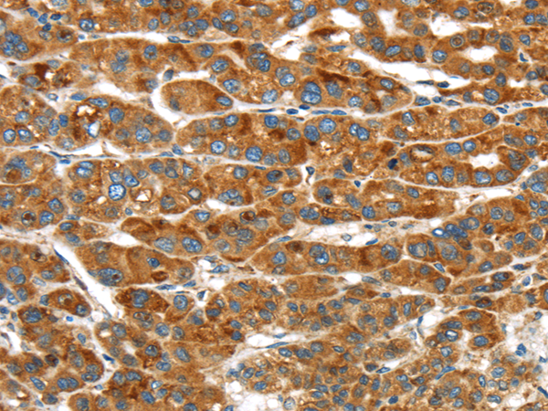
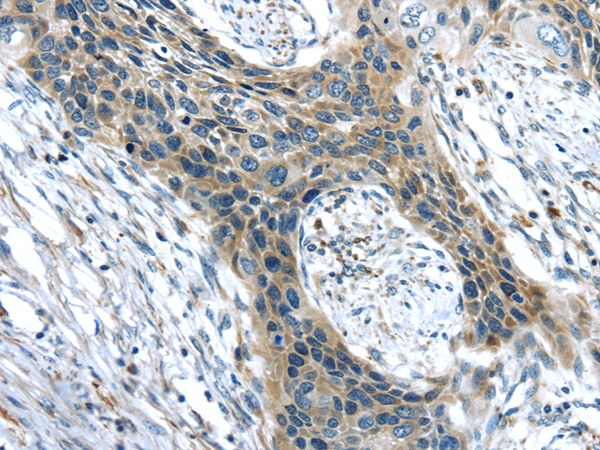
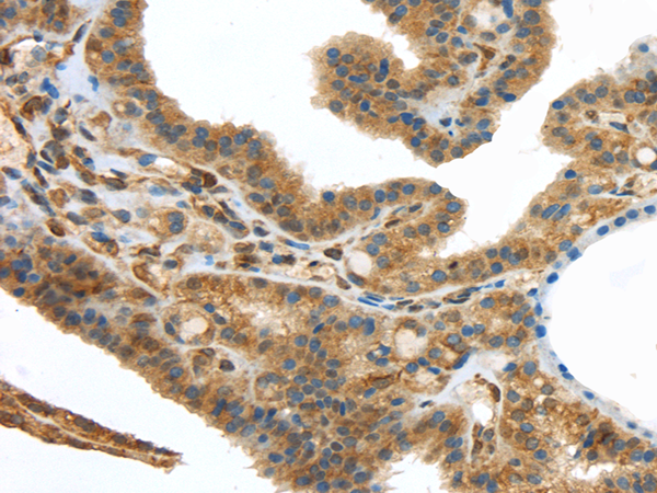
 IHC (Immunohiostchemistry)
(Immunohistochemistry of paraffin-embedded Human thyroid cancer tissue using VLDLR Polyclonal Antibody at dilution 1:35)
IHC (Immunohiostchemistry)
(Immunohistochemistry of paraffin-embedded Human thyroid cancer tissue using VLDLR Polyclonal Antibody at dilution 1:35)
VLDLR, Polyclonal Antibody (Cat# AAA173329)
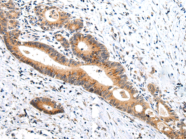
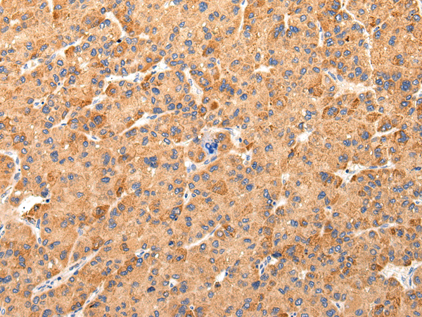
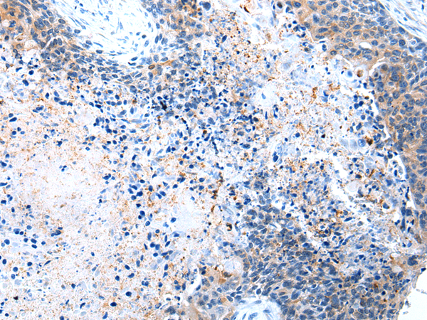
 IHC (Immunohiostchemistry)
(Immunohistochemistry of paraffin-embedded Human prostate cancer using VPS26A Polyclonal Antibody at dilution of 1/35)
IHC (Immunohiostchemistry)
(Immunohistochemistry of paraffin-embedded Human prostate cancer using VPS26A Polyclonal Antibody at dilution of 1/35)
VPS26A, Polyclonal Antibody (Cat# AAA173335)
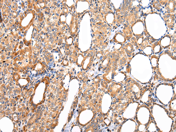
 IHC (Immunohiostchemistry)
(Immunohistochemistry of paraffin-embedded Human lung cancer using HNRNPM Polyclonal Antibody at dilution of 1/35)
IHC (Immunohiostchemistry)
(Immunohistochemistry of paraffin-embedded Human lung cancer using HNRNPM Polyclonal Antibody at dilution of 1/35)
HNRNPM, Polyclonal Antibody (Cat# AAA173353)
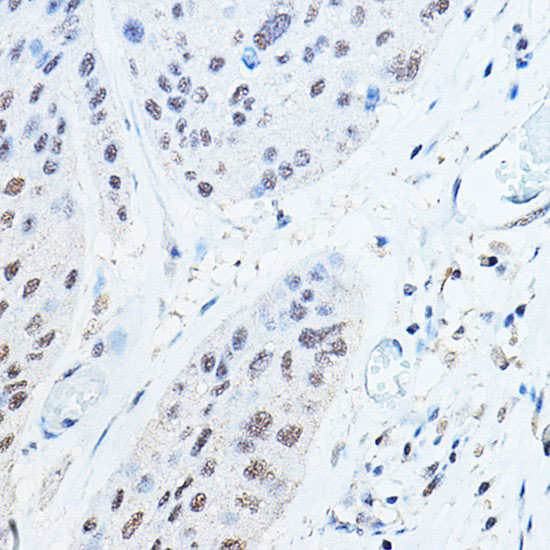
 IHC (Immunohistochemisry)
(Immunohistochemistry of paraffin-embedded Human endometrium cancer using CDKN2A Polyclonal Antibody at dilution of 1:600.)
IHC (Immunohistochemisry)
(Immunohistochemistry of paraffin-embedded Human endometrium cancer using CDKN2A Polyclonal Antibody at dilution of 1:600.)
CDKN2A, Polyclonal Antibody (Cat# AAA179743)
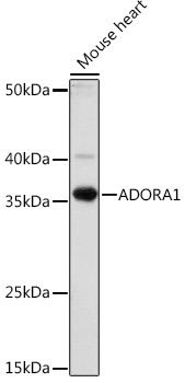
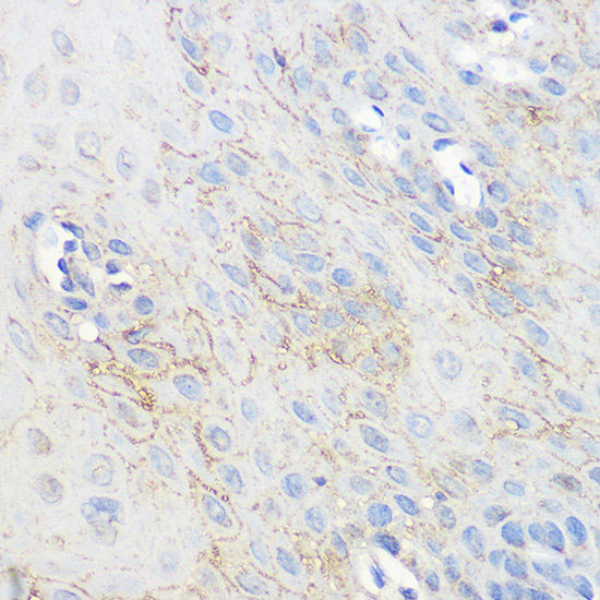 IHC (Immunohistochemisry)
(Immunohistochemistry of paraffin-embedded human esophageal using ADORA1 Polyclonal Antibody at dilution of 1:100 (40x lens).Perform microwave antigen retrieval with 10 mM PBS buffer pH 7.2 before commencing with IHC staining protocol.)
IHC (Immunohistochemisry)
(Immunohistochemistry of paraffin-embedded human esophageal using ADORA1 Polyclonal Antibody at dilution of 1:100 (40x lens).Perform microwave antigen retrieval with 10 mM PBS buffer pH 7.2 before commencing with IHC staining protocol.)
ADORA1, Polyclonal Antibody (Cat# AAA179181)
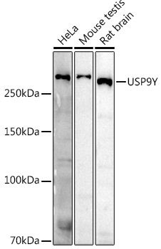
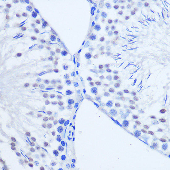
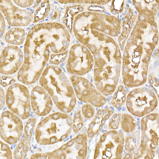 IHC (Immunohistochemistry)
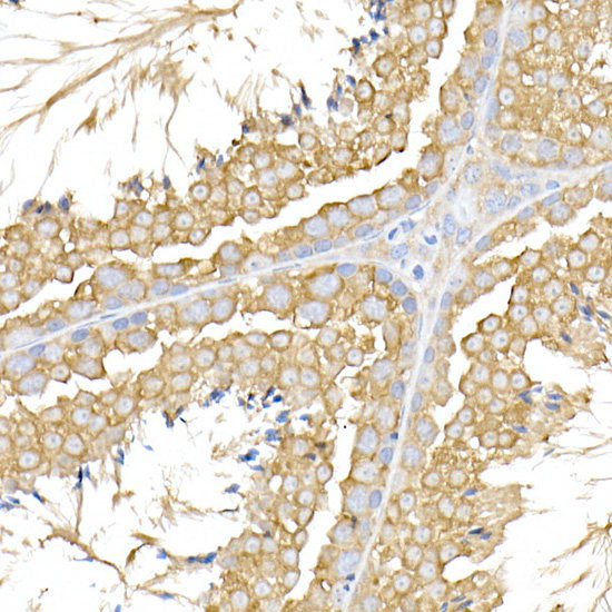
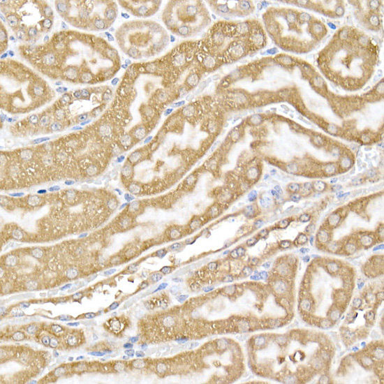
(Immunohistochemistry of paraffin-embedded rat kidney using USP9Y Polyclonal Antibody at dilution of 1:100 (40x lens).Perform high pressure antigen retrieval with 10 mM citrate buffer pH 6.0 before commencing with IHC staining protocol.)
IHC (Immunohistochemistry)
(Immunohistochemistry of paraffin-embedded rat kidney using USP9Y Polyclonal Antibody at dilution of 1:100 (40x lens).Perform high pressure antigen retrieval with 10 mM citrate buffer pH 6.0 before commencing with IHC staining protocol.)
USP9Y, Polyclonal Antibody (Cat# AAA179306)
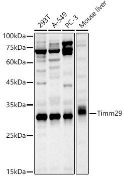
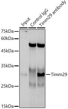 IP (Immunoprecipitation)
(Immunoprecipitation analysis of 300ug extracts of A-549 cells usingug Timm29 Polyclonal Antibody.Western blot was performed from the immunoprecipitate using Timm29 Polyclonal Antibody at a dilution of 1:1000.)
IP (Immunoprecipitation)
(Immunoprecipitation analysis of 300ug extracts of A-549 cells usingug Timm29 Polyclonal Antibody.Western blot was performed from the immunoprecipitate using Timm29 Polyclonal Antibody at a dilution of 1:1000.)
Timm29, Polyclonal Antibody (Cat# AAA179309)
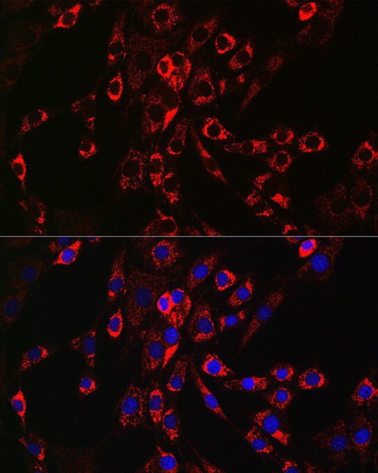
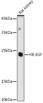
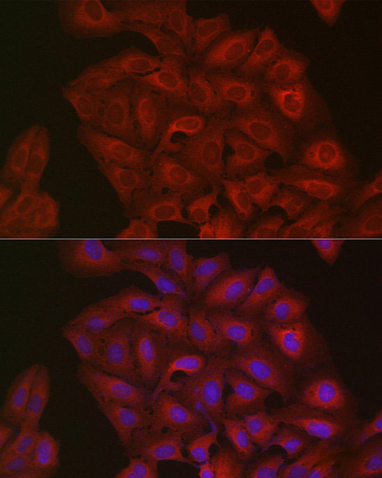
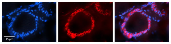
 IF (Immunofluorescence)
(Immunofluorescence analysis of PC-12 cells using HB-EGF Polyclonal Antibody at dilution of 1:100 (40x lens). Blue: DAPI for nuclear staining.)
IF (Immunofluorescence)
(Immunofluorescence analysis of PC-12 cells using HB-EGF Polyclonal Antibody at dilution of 1:100 (40x lens). Blue: DAPI for nuclear staining.)
HB-EGF, Polyclonal Antibody (Cat# AAA179082)
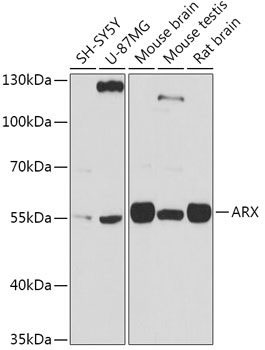
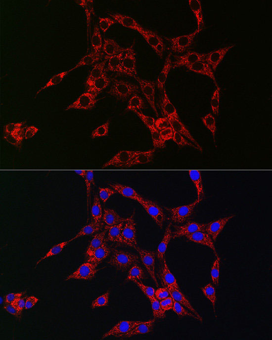
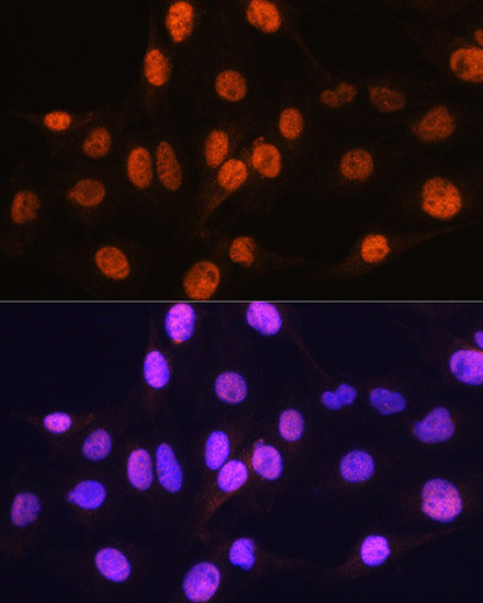 IF (Immunofluorescence)
(Immunofluorescence analysis of C6 cells using ARX Polyclonal Antibody at dilution of 1:100. Blue: DAPI for nuclear staining.)
IF (Immunofluorescence)
(Immunofluorescence analysis of C6 cells using ARX Polyclonal Antibody at dilution of 1:100. Blue: DAPI for nuclear staining.)
ARX, Polyclonal Antibody (Cat# AAA179104)
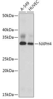
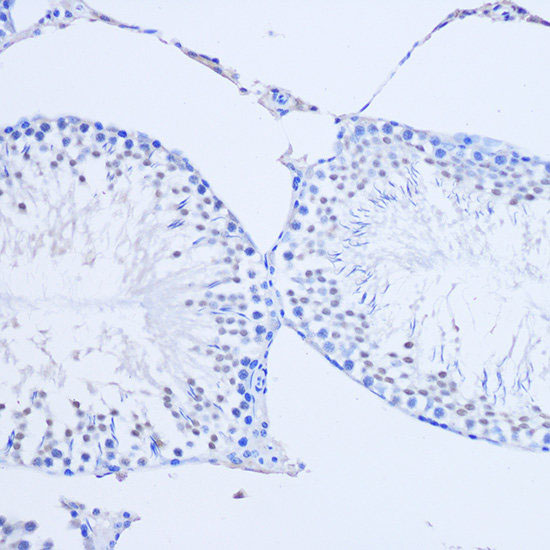
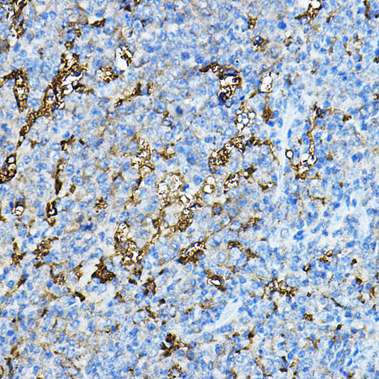 IHC (Immunohiostchemistry)
(Immunohistochemistry of paraffin-embedded mouse spleen using NXPH4 Polyclonal Antibody at dilution of 1:100 (40x lens).Perform microwave antigen retrieval with 10 mM PBS buffer pH 7.2 before commencing with IHC staining protocol.)
IHC (Immunohiostchemistry)
(Immunohistochemistry of paraffin-embedded mouse spleen using NXPH4 Polyclonal Antibody at dilution of 1:100 (40x lens).Perform microwave antigen retrieval with 10 mM PBS buffer pH 7.2 before commencing with IHC staining protocol.)
NXPH4, Polyclonal Antibody (Cat# AAA179148)
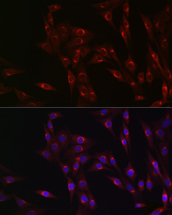
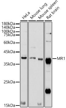
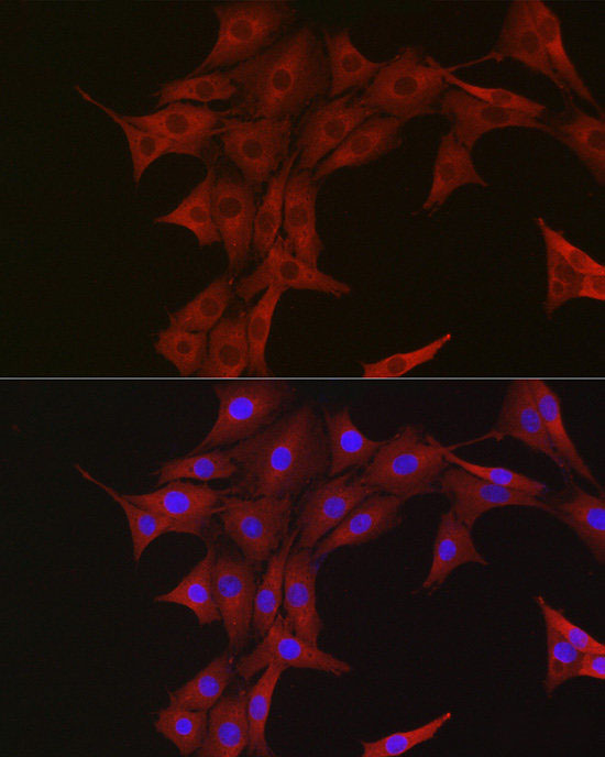
 IF (Immunofluorescence)
(Immunofluorescence analysis of PC-12 cells using MR1 Polyclonal Antibody at dilution of 1:100 (40x lens). Blue: DAPI for nuclear staining.)
IF (Immunofluorescence)
(Immunofluorescence analysis of PC-12 cells using MR1 Polyclonal Antibody at dilution of 1:100 (40x lens). Blue: DAPI for nuclear staining.)
MR1, Polyclonal Antibody (Cat# AAA179447)
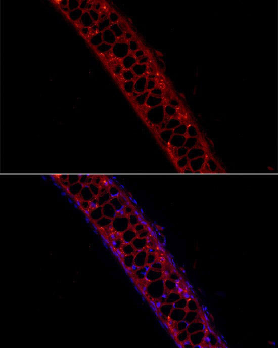
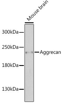
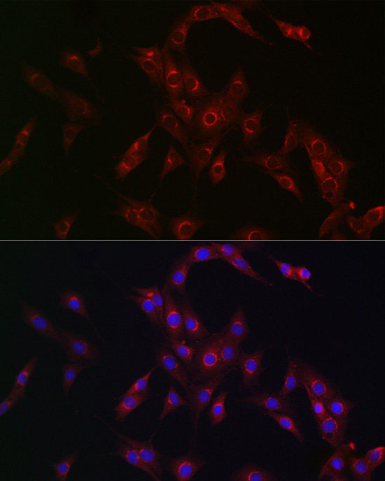
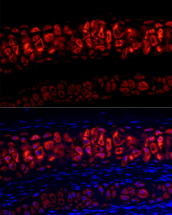 IF (Immunofluorescence)
(Immunofluorescence analysis of Rat cartilage cells using Aggrecan Polyclonal Antibody at dilution of 1:100 (40x lens).Blue: DAPI for nuclear staining.)
IF (Immunofluorescence)
(Immunofluorescence analysis of Rat cartilage cells using Aggrecan Polyclonal Antibody at dilution of 1:100 (40x lens).Blue: DAPI for nuclear staining.)
Aggrecan, Polyclonal Antibody (Cat# AAA179481)
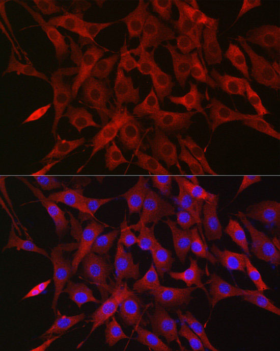
 IF (Immunofluorescence)
(Immunofluorescence analysis of U2OS cells using Ubiquitin Polyclonal Antibody at dilution of 1:50 (40x lens). Blue: DAPI for nuclear staining.)
IF (Immunofluorescence)
(Immunofluorescence analysis of U2OS cells using Ubiquitin Polyclonal Antibody at dilution of 1:50 (40x lens). Blue: DAPI for nuclear staining.)
Ubiquitin, Polyclonal Antibody (Cat# AAA179537)
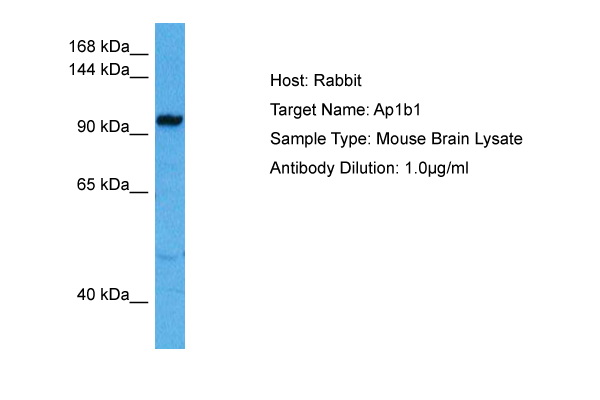
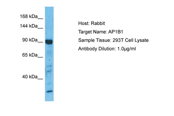 WB (Western Blot)
(Host: RabbitTarget Name: AP1B1Sample Type: 293T Whole Cell lysatesAntibody Dilution: 1.0ug/ml)
WB (Western Blot)
(Host: RabbitTarget Name: AP1B1Sample Type: 293T Whole Cell lysatesAntibody Dilution: 1.0ug/ml)
AP1B1, Polyclonal Antibody (Cat# AAA201536)
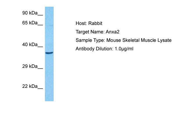
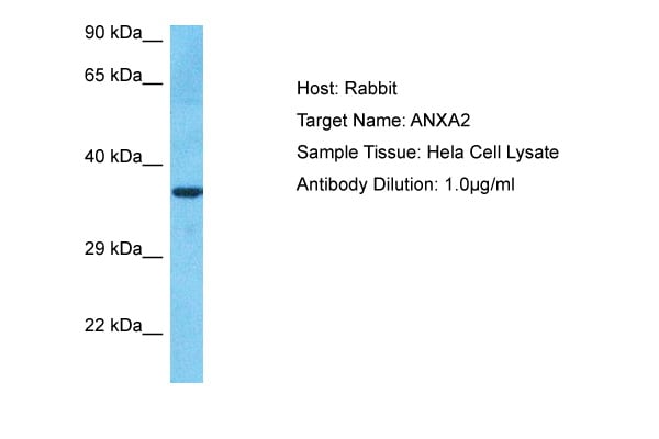 WB (Western Blot)
(Host: RabbitTarget Name: ANXA2Sample Type: Hela Whole Cell lysatesAntibody Dilution: 1.0ug/ml)
WB (Western Blot)
(Host: RabbitTarget Name: ANXA2Sample Type: Hela Whole Cell lysatesAntibody Dilution: 1.0ug/ml)
ANXA2, Polyclonal Antibody (Cat# AAA201537)
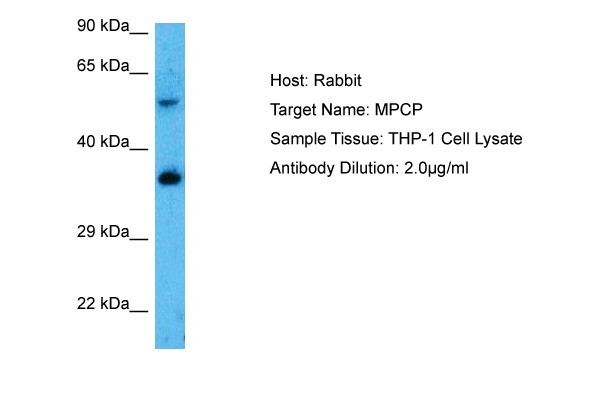
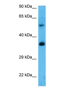 WB (Western Blot)
(Host: RabbitTarget Name: SLC25A3Sample Tissue: Human THP-1 Whole CellAntibody Dilution: 2.0ug/ml)
WB (Western Blot)
(Host: RabbitTarget Name: SLC25A3Sample Tissue: Human THP-1 Whole CellAntibody Dilution: 2.0ug/ml)
SLC25A3, Polyclonal Antibody (Cat# AAA201542)
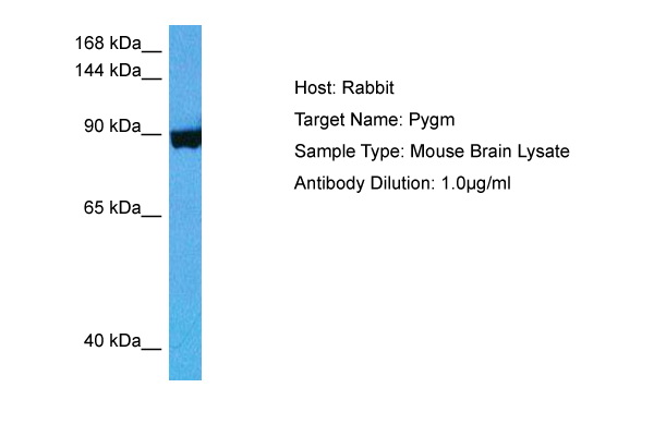
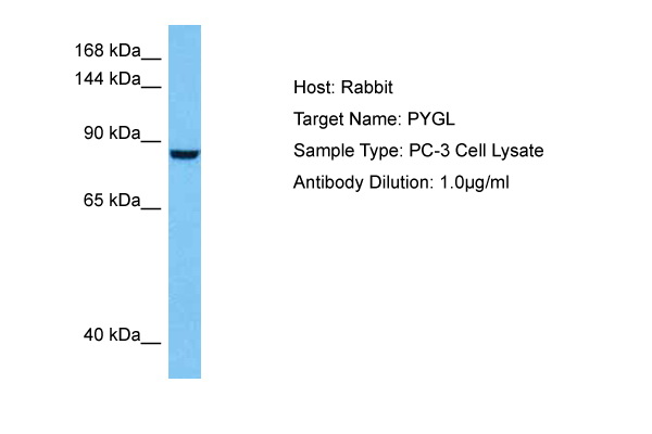 WB (Western Blot)
(Host: RabbitTarget Name: PYGLSample Tissue: Human PC-3 Whole Cell lysatesAntibody Dilution: 1ug/ml)
WB (Western Blot)
(Host: RabbitTarget Name: PYGLSample Tissue: Human PC-3 Whole Cell lysatesAntibody Dilution: 1ug/ml)
PYGM, Polyclonal Antibody (Cat# AAA201545)
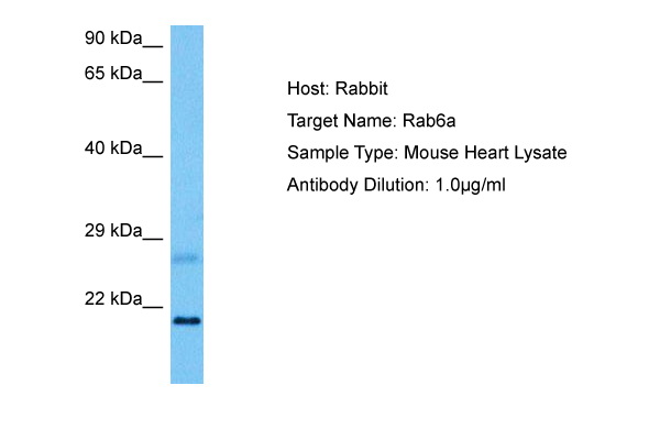
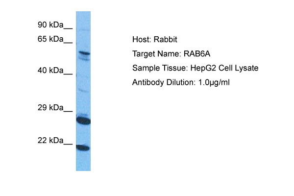 WB (Western Blot)
(Host: RabbitTarget Name: RAB6ASample Tissue: Human HepG2 Whole CellAntibody Dilution: 1.0ug/ml)
WB (Western Blot)
(Host: RabbitTarget Name: RAB6ASample Tissue: Human HepG2 Whole CellAntibody Dilution: 1.0ug/ml)
RAB6A, Polyclonal Antibody (Cat# AAA201546)
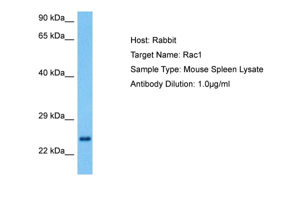
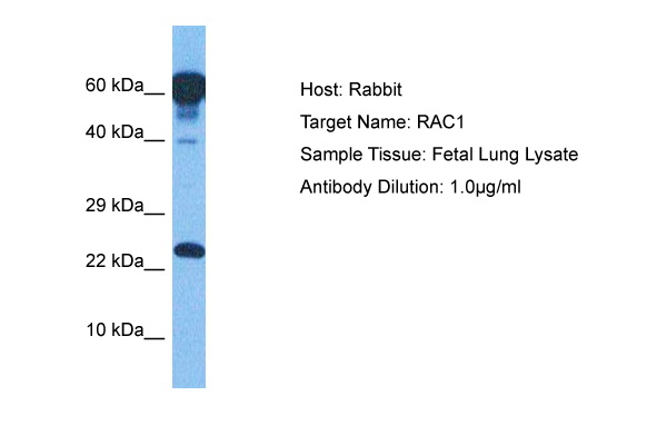 WB (Western Blot)
(Host: RabbitTarget Name: RAC1Sample Tissue: Human Fetal LungAntibody Dilution: 1.0ug/ml)
WB (Western Blot)
(Host: RabbitTarget Name: RAC1Sample Tissue: Human Fetal LungAntibody Dilution: 1.0ug/ml)
RAC1, Polyclonal Antibody (Cat# AAA201547)
 WB (Western Blot)
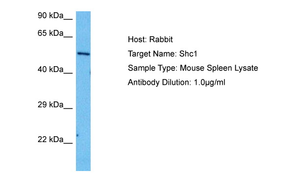
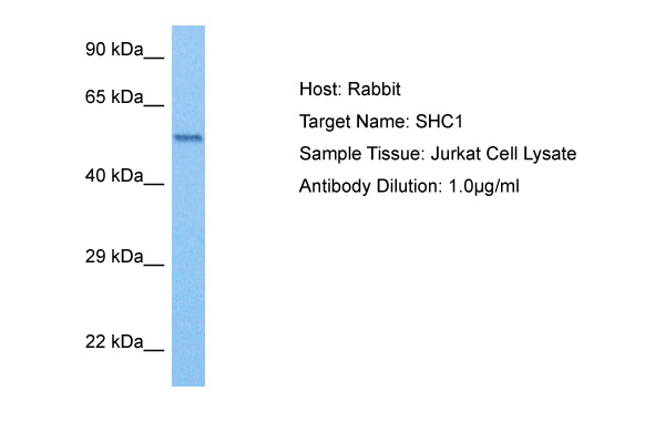
(Host: RabbitTarget Name: SHC1Sample Tissue: Human Jurkat Whole CellAntibody Dilution: 1.0ug/ml)
WB (Western Blot)
(Host: RabbitTarget Name: SHC1Sample Tissue: Human Jurkat Whole CellAntibody Dilution: 1.0ug/ml)
SHC1, Polyclonal Antibody (Cat# AAA201548)
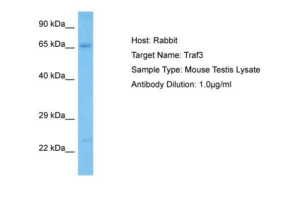
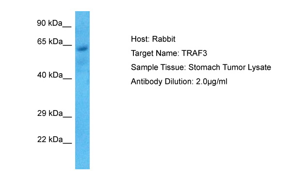 WB (Western Blot)
(Host: RabbitTarget Name: TRAF3Sample Tissue: Human Stomach TumorAntibody Dilution: 1.0ug/ml)
WB (Western Blot)
(Host: RabbitTarget Name: TRAF3Sample Tissue: Human Stomach TumorAntibody Dilution: 1.0ug/ml)
TRAF3, Polyclonal Antibody (Cat# AAA201550)
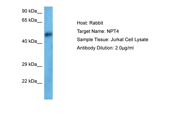 WB (Western Blot)
(Host: RabbitTarget Name: SLC17A3Sample Tissue: Human Jurkat Whole CellAntibody Dilution: 1.0ug/ml)
WB (Western Blot)
(Host: RabbitTarget Name: SLC17A3Sample Tissue: Human Jurkat Whole CellAntibody Dilution: 1.0ug/ml)
SLC17A3, Polyclonal Antibody (Cat# AAA201553)
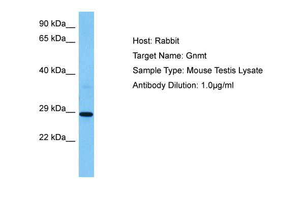
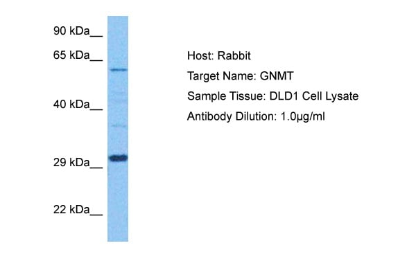 WB (Western Blot)
(Host: RabbitTarget Name: GNMTSample Tissue: Human DLD1 Whole CellAntibody Dilution: 1.0ug/ml)
WB (Western Blot)
(Host: RabbitTarget Name: GNMTSample Tissue: Human DLD1 Whole CellAntibody Dilution: 1.0ug/ml)
GNMT, Polyclonal Antibody (Cat# AAA201554)
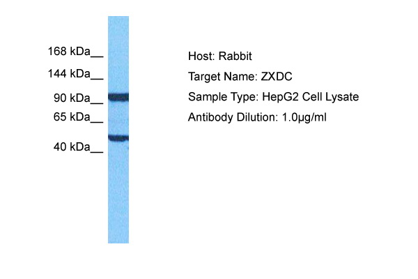 WB (Western Blot)
(Host: RabbitTarget Name: ZXDCSample Tissue: Human HepG2 Whole CellAntibody Dilution: 1.0ug/ml)
WB (Western Blot)
(Host: RabbitTarget Name: ZXDCSample Tissue: Human HepG2 Whole CellAntibody Dilution: 1.0ug/ml)
ZXDC, Polyclonal Antibody (Cat# AAA201555)
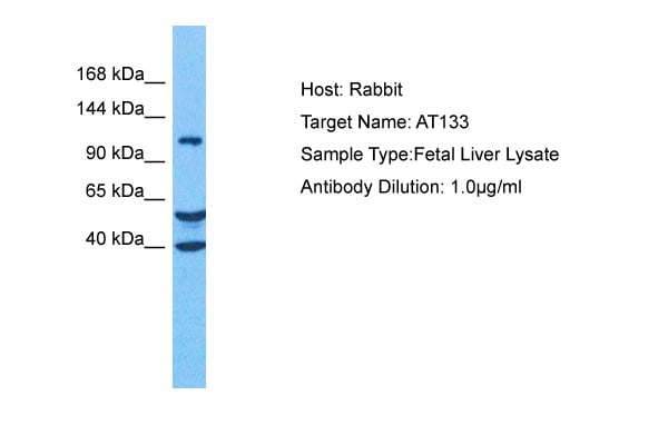 WB (Western Blot)
(Host: RabbitTarget Name: ATP13A3Sample Tissue: Human Fetal LiverAntibody Dilution: 1.0ug/ml)
WB (Western Blot)
(Host: RabbitTarget Name: ATP13A3Sample Tissue: Human Fetal LiverAntibody Dilution: 1.0ug/ml)
ATP13A3, Polyclonal Antibody (Cat# AAA201556)
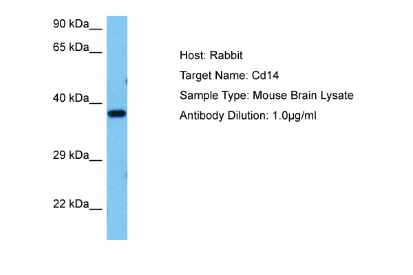
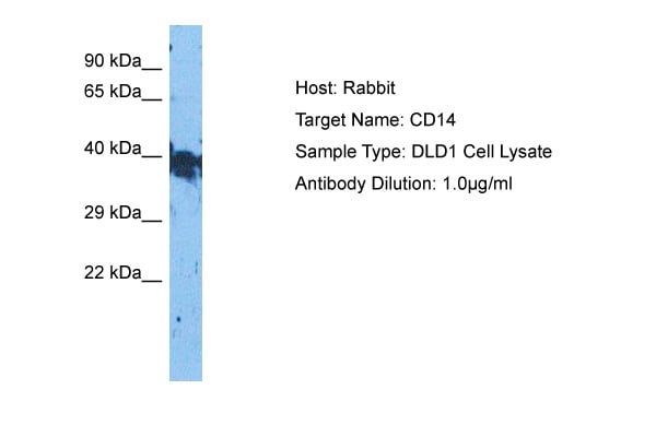 WB (Western Blot)
(Host: RabbitTarget Name: CD14Sample Tissue: Human DLD1 Whole CellAntibody Dilution: 1.0ug/ml)
WB (Western Blot)
(Host: RabbitTarget Name: CD14Sample Tissue: Human DLD1 Whole CellAntibody Dilution: 1.0ug/ml)
CD14, Polyclonal Antibody (Cat# AAA201558)
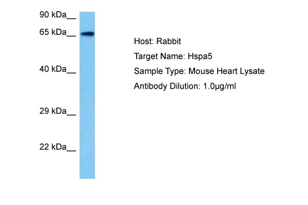
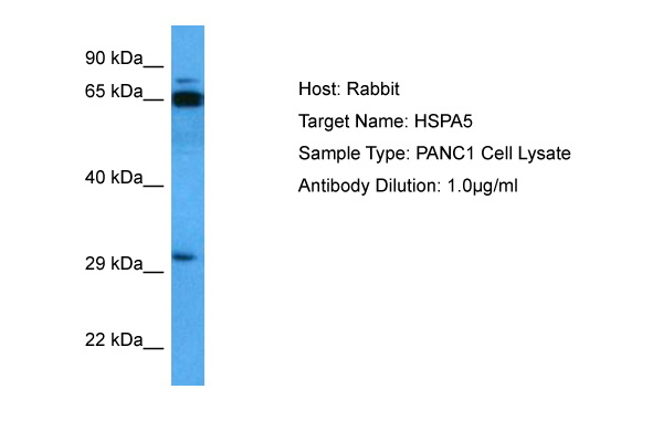 WB (Western Blot)
(Host: RabbitTarget Name: HSPA5Sample Tissue: PANC1 Whole Cell lysatesAntibody Dilution: 1ug/ml)
WB (Western Blot)
(Host: RabbitTarget Name: HSPA5Sample Tissue: PANC1 Whole Cell lysatesAntibody Dilution: 1ug/ml)
HSPA5, Polyclonal Antibody (Cat# AAA201563)
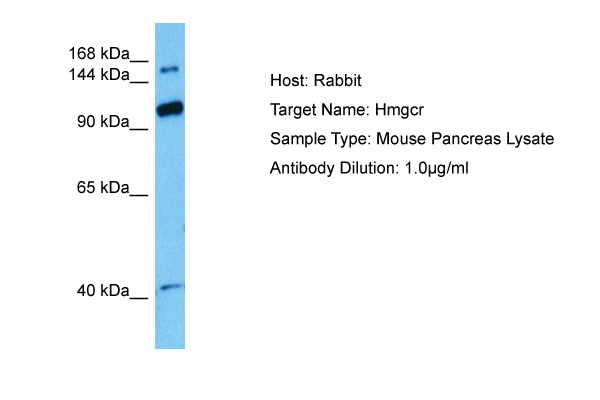
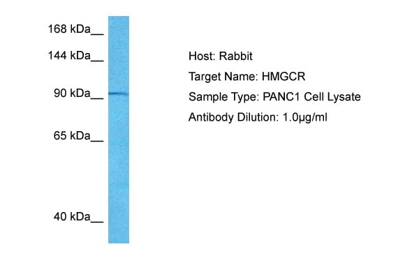 WB (Western Blot)
(Host: RabbitTarget Name: HMGCRSample Tissue: Human PANC1Whole Cell lysatesAntibody Dilution: 1ug/ml)
WB (Western Blot)
(Host: RabbitTarget Name: HMGCRSample Tissue: Human PANC1Whole Cell lysatesAntibody Dilution: 1ug/ml)
HMGCR, Polyclonal Antibody (Cat# AAA201564)
What are Polyclonal Antibodies?
Polyclonal antibodies are antibodies that come from multiple B cell clones of a host animal. The typical hosts used for the majority of polyclonal antibody production are rabbits, goats, sheep, and donkeys. These polyclonal antibodies, once having identified their target, will bind to different epitopes located at different regions or sequences on the same protein/antigen. As a result, they are ideal at locating and binding to the target, even if the target is in very low concentrations (due to many different antibodies being able to bind to the same target molecule, which allows for significant amplification of a downstream signal).
Polyclonal antibodies are typically produced by injecting an antigen into a host animal, which causes the animal’s immune system to attack the foreign antigen by mass generating antibodies against it. After a period of time, serum is collected from the animal and purified using physicochemical fractionation, class-specific affinity purification, and/or antigen-affinity purification.
Key Uses of Polyclonal Antibodies
- Western Blotting: This method is used to find specific proteins in biological samples after separating them by size.
- Immunohistochemistry: IHC helps visualize the location of proteins in tissue sections using various staining techniques.
- ELISA: (Enzyme-Linked Immunosorbent Assay) is typically used to identify specific protein quantities in a sample. ELISAs can be either “Quantitative” or “Qualitative”.
- Flow Cytometry: technique that identifies and measures the specific protein on the surface or inside the cells in a fluid suspension.
- Immunoprecipitation: IP isolates and studies a specific protein from a complex mixture using antibodies.
Why Buy Polyclonal Antibodies from AAA Biotech?
1. Ideal for Various Applications
Our antibodies are generally going to be validated for use in multiple types of assays, including ELISA, Western Blotting, Immunohistochemistry, Immunoprecipitation, amongst others. They are ideal for a wide range of research applications.
2. Rigorous Quality Control
All of the antibodies in our catalog undergo strict quality testing to ensure specificity, sensitivity, and consistent performance. We are confident in the ability of our antibodies to provide you with accurate results.
3. Wide Assortment of Antibodies
Antibodies in are catalog can be found for both common and exotic species, and these antibodies are also available in both conjugated and recombinant forms to suit many diverse experimental needs.
4. Highly Purified
Our antibodies are available in purified forms with over 85% purity, as confirmed by SDS-PAGE. They are also available with tags such as His, Flag, GST, or MBP. We cater to customers worldwide.
FAQ
1. How are polyclonal antibodies produced?
Traditionally, polyclonal antibodies are produced by injecting an antigen into a host animal (such as a rabbit or goat), which then triggers an immune response from the host animal. The animal’s B cells produce antibodies that will recognize different parts of the injected antigen. These antibodies are then collected from the animal’s blood and purified for use.
2. How do polyclonal antibodies differ from monoclonal antibodies?
Polyclonal antibodies are a mix of antibodies that bind to different locations (epitopes) of the same antigen, while monoclonal antibodies are identical and bind to just one specific epitope. This makes polyclonal antibodies more versatile and better at detecting proteins that may be present in low quantities or in altered/modified forms.
3. How should I store polyclonal antibodies?
Polyclonal antibodies should be stored at 4°C for short-term use (up to a few weeks) and at -20°C or -80°C for long-term storage. Avoid repeated freeze-thaw cycles by dividing them into small aliquots. Always check the datasheet for specific storage instructions.