Filters
▼Clonality
▼Type
▼Reactivity
▼Gene Name
▼Isotype
▼Host
▼Application
▼Clone
▼Polyclonal Antibodies
At AAA Biotech also known as AAA Bio or AAABio, we provide a broad range of purified polyclonal antibodies (pAbs) that are able to all be browsed online through our website. Due to their high specificity and strong binding affinity, these antibodies are ideal for wide swathes of research and experimental applications.
Our polyclonal antibodies can easily support your work, whether you use them for Western Blotting, Immunocytochemistry (with or without Immunofluorescence used in conjunction), Immunohistochemistry, Immunoprecipitation, and ELISA tests. We highly encourage you to browse our range of pAbs and choose the one that best suits your experimental model.
Viewing 1800-1850 of 96805 product results
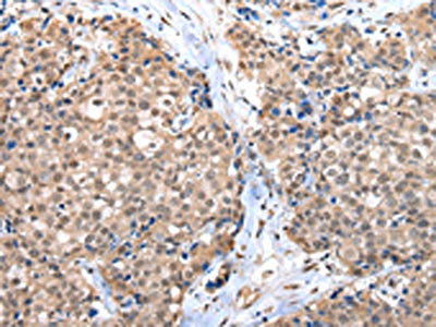
 IHC (Immunohiostchemistry)
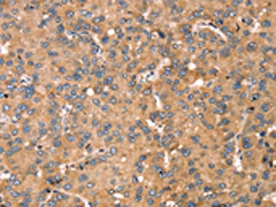
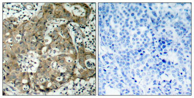
(The image on the left is immunohistochemistry of paraffin-embedded Human lung cancer tissue using AAA241833(TGFB2 Antibody) at dilution 1/30, on the right is treated with synthetic peptide. (Original magnification: ×200))
IHC (Immunohiostchemistry)
(The image on the left is immunohistochemistry of paraffin-embedded Human lung cancer tissue using AAA241833(TGFB2 Antibody) at dilution 1/30, on the right is treated with synthetic peptide. (Original magnification: ×200))
TGFB2, Polyclonal Antibody (Cat# AAA241833)
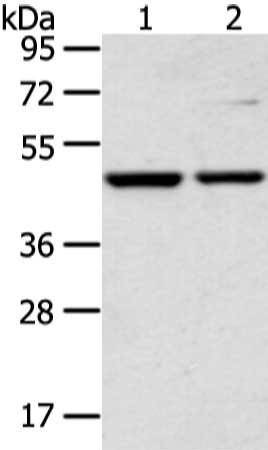
 SDS-PAGE
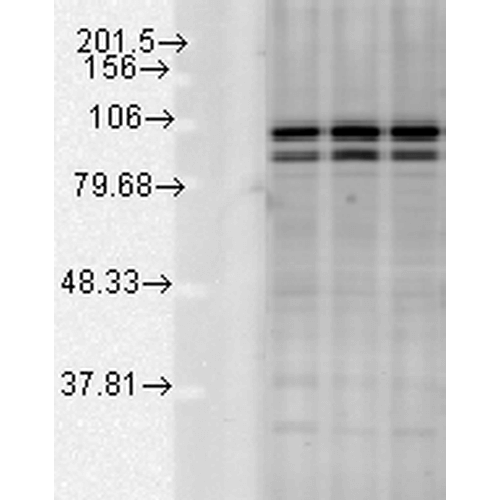
(Gel: 6%SDS-PAGE, Lysate: 80 ug, Lane 1-3: 293T, 231 and lo2 cell, Primary antibody: AAA241840(TRIM24 Antibody) at dilution 1/400 dilution, Secondary antibody: Goat anti rabbit IgG at 1/8000 dilution, Exposure time: 1 minute)
SDS-PAGE
(Gel: 6%SDS-PAGE, Lysate: 80 ug, Lane 1-3: 293T, 231 and lo2 cell, Primary antibody: AAA241840(TRIM24 Antibody) at dilution 1/400 dilution, Secondary antibody: Goat anti rabbit IgG at 1/8000 dilution, Exposure time: 1 minute)
TRIM24, Polyclonal Antibody (Cat# AAA241840)
 SDS-PAGE
(Gel: 8%SDS-PAGE, Lysate: 40 ug, Lane 1-2: 293T and NIH/3T3 cell, Primary antibody: AAA242049(CTBP1 Antibody) at dilution 1/650 dilution, Secondary antibody: Goat anti rabbit IgG at 1/8000 dilution, Exposure time: 5 seconds)
SDS-PAGE
(Gel: 8%SDS-PAGE, Lysate: 40 ug, Lane 1-2: 293T and NIH/3T3 cell, Primary antibody: AAA242049(CTBP1 Antibody) at dilution 1/650 dilution, Secondary antibody: Goat anti rabbit IgG at 1/8000 dilution, Exposure time: 5 seconds)
CTBP1, Polyclonal Antibody (Cat# AAA242049)
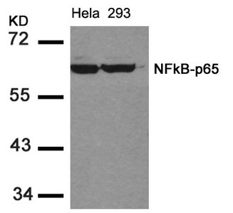
 IHC (Immunohiostchemistry)
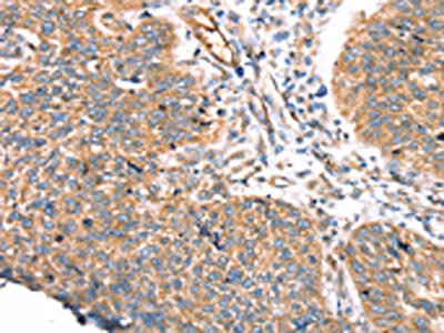
(Immunohistochemical analysis of paraffin-embedded human breast carcinoma tissue using NFkB-p65(Ab-536) Antibody(left) or the same antibody preincubated with blocking peptide(right).)
IHC (Immunohiostchemistry)
(Immunohistochemical analysis of paraffin-embedded human breast carcinoma tissue using NFkB-p65(Ab-536) Antibody(left) or the same antibody preincubated with blocking peptide(right).)
RELA, Polyclonal Antibody (Cat# AAA242058)
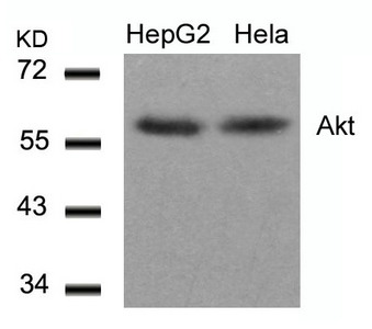
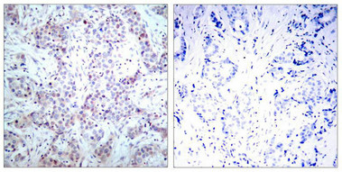
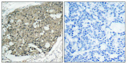 IHC (Immunohiostchemistry)
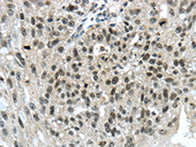
(Immunohistochemical analysis of paraffin-embedded human breast carcinoma tissue using Akt(Ab-308) Antibody(left) or the same antibody preincubated with blocking peptide(right).)
IHC (Immunohiostchemistry)
(Immunohistochemical analysis of paraffin-embedded human breast carcinoma tissue using Akt(Ab-308) Antibody(left) or the same antibody preincubated with blocking peptide(right).)
AKT1, Polyclonal Antibody (Cat# AAA242084)
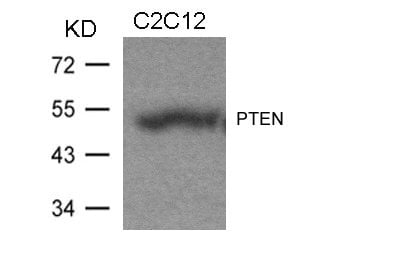
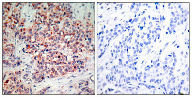 IHC (Immunohiostchemistry)
(Immunohistochemical analysis of paraffin-embedded human breast carcinoma tissue using PTEN(Ab-370) Antibody(left) or the same antibody preincubated with blocking peptide(right).)
IHC (Immunohiostchemistry)
(Immunohistochemical analysis of paraffin-embedded human breast carcinoma tissue using PTEN(Ab-370) Antibody(left) or the same antibody preincubated with blocking peptide(right).)
PTEN, Polyclonal Antibody (Cat# AAA242086)
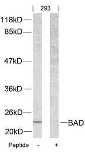
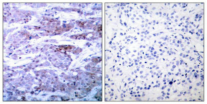 IHC (Immunohiostchemistry)
(Immunohistochemical analysis of paraffin-embedded human breast carcinoma tissue using BAD(Ab-112) Antibody(left) or the same antibody preincubated with blocking peptide(right).)
IHC (Immunohiostchemistry)
(Immunohistochemical analysis of paraffin-embedded human breast carcinoma tissue using BAD(Ab-112) Antibody(left) or the same antibody preincubated with blocking peptide(right).)
Bad, Polyclonal Antibody (Cat# AAA242088)
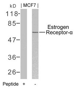
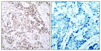 IHC (Immunohiostchemistry)
(Immunohistochemical analysis of paraffin-embedded human breast carcinoma tissue using Estrogen Receptor-a(Ab-106) Antibody(left) or the same antibody preincubated with blocking peptide(right).)
IHC (Immunohiostchemistry)
(Immunohistochemical analysis of paraffin-embedded human breast carcinoma tissue using Estrogen Receptor-a(Ab-106) Antibody(left) or the same antibody preincubated with blocking peptide(right).)
ESR1, Polyclonal Antibody (Cat# AAA242092)
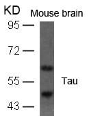
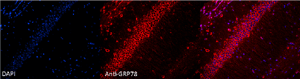
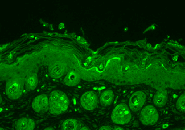
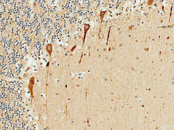
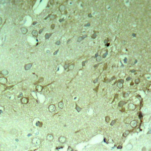 IHC (Immunohiostchemistry)
(Immunohistochemical analysis of paraffin-embedded rat hippocampal region tissue from a model with Alzheimer)
IHC (Immunohiostchemistry)
(Immunohistochemical analysis of paraffin-embedded rat hippocampal region tissue from a model with Alzheimer)
MAPT, Polyclonal Antibody (Cat# AAA242100)
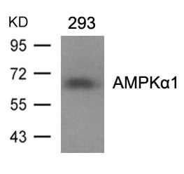
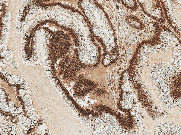
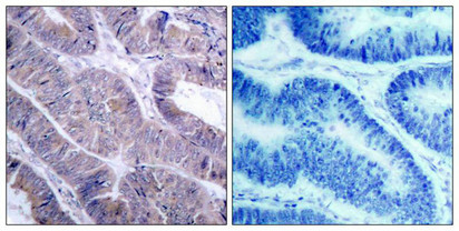 IHC (Immunohiostchemistry)
(Immunohistochemical analysis of paraffin-embedded human colon carcinoma tissue using AMPKa1(Ab-487)Antibody(left) or the same antibody preincubated with blocking peptide(right).)
IHC (Immunohiostchemistry)
(Immunohistochemical analysis of paraffin-embedded human colon carcinoma tissue using AMPKa1(Ab-487)Antibody(left) or the same antibody preincubated with blocking peptide(right).)
PRKAA1/PRKAA2, Polyclonal Antibody (Cat# AAA242110)

 IHC (Immunohiostchemistry)
(Immunohistochemical analysis of paraffin-embedded human breast carcinoma tissue using FKHR(Ab-319) Antibody(left) or the same antibody preincubated with blocking peptide(right).)
IHC (Immunohiostchemistry)
(Immunohistochemical analysis of paraffin-embedded human breast carcinoma tissue using FKHR(Ab-319) Antibody(left) or the same antibody preincubated with blocking peptide(right).)
FOXO1, Polyclonal Antibody (Cat# AAA242126)

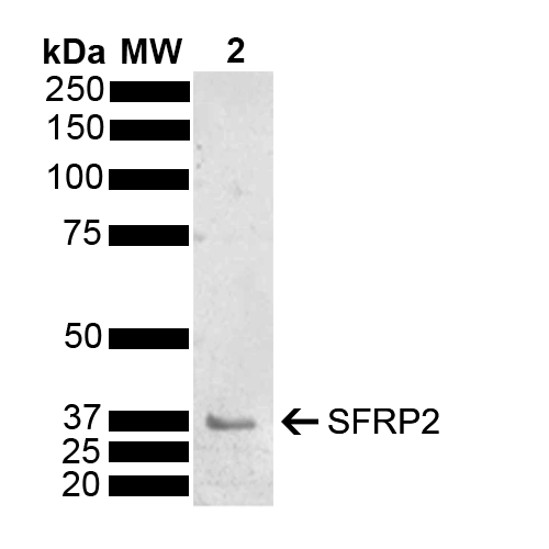
 WB (Western Blot)
(Western blot analysis of Mouse Brain showing detection of 33.5 kDa SFRP2 protein using Rabbit Anti-SFRP2 Polyclonal Antibody . Lane 1: Molecular Weight Ladder (MW). Lane 2: Mouse Brain . Load: 15 ug. Block: 5% Skim Milk powder in TBST. Primary Antibody: Rabbit Anti-SFRP2 Polyclonal Antibody at 1:1000 for 2 hours at RT with shaking. Secondary Antibody: Goat Anti-Rabbit IgG: HRP at 1:5000 for 1 hour at RT. Color Development: ECL solution for 5 min at RT. Predicted/Observed Size: 33.5 kDa. Other Band(s): 37 kDa.)
WB (Western Blot)
(Western blot analysis of Mouse Brain showing detection of 33.5 kDa SFRP2 protein using Rabbit Anti-SFRP2 Polyclonal Antibody . Lane 1: Molecular Weight Ladder (MW). Lane 2: Mouse Brain . Load: 15 ug. Block: 5% Skim Milk powder in TBST. Primary Antibody: Rabbit Anti-SFRP2 Polyclonal Antibody at 1:1000 for 2 hours at RT with shaking. Secondary Antibody: Goat Anti-Rabbit IgG: HRP at 1:5000 for 1 hour at RT. Color Development: ECL solution for 5 min at RT. Predicted/Observed Size: 33.5 kDa. Other Band(s): 37 kDa.)
SFRP2, Polyclonal Antibody (Cat# AAA253896)

 WB (Western Blot)
(Western blot analysis of Mouse Brain showing detection of 33.5 kDa SFRP2 protein using Rabbit Anti-SFRP2 Polyclonal Antibody . Lane 1: Molecular Weight Ladder (MW). Lane 2: Mouse Brain . Load: 15 ug. Block: 5% Skim Milk powder in TBST. Primary Antibody: Rabbit Anti-SFRP2 Polyclonal Antibody at 1:1000 for 2 hours at RT with shaking. Secondary Antibody: Goat Anti-Rabbit IgG: HRP at 1:5000 for 1 hour at RT. Color Development: ECL solution for 5 min at RT. Predicted/Observed Size: 33.5 kDa. Other Band(s): 37 kDa.)
WB (Western Blot)
(Western blot analysis of Mouse Brain showing detection of 33.5 kDa SFRP2 protein using Rabbit Anti-SFRP2 Polyclonal Antibody . Lane 1: Molecular Weight Ladder (MW). Lane 2: Mouse Brain . Load: 15 ug. Block: 5% Skim Milk powder in TBST. Primary Antibody: Rabbit Anti-SFRP2 Polyclonal Antibody at 1:1000 for 2 hours at RT with shaking. Secondary Antibody: Goat Anti-Rabbit IgG: HRP at 1:5000 for 1 hour at RT. Color Development: ECL solution for 5 min at RT. Predicted/Observed Size: 33.5 kDa. Other Band(s): 37 kDa.)
SFRP2, Polyclonal Antibody (Cat# AAA253897)

 WB (Western Blot)
(Western blot analysis of Mouse Brain showing detection of 33.5 kDa SFRP2 protein using Rabbit Anti-SFRP2 Polyclonal Antibody . Lane 1: Molecular Weight Ladder (MW). Lane 2: Mouse Brain . Load: 15 ug. Block: 5% Skim Milk powder in TBST. Primary Antibody: Rabbit Anti-SFRP2 Polyclonal Antibody at 1:1000 for 2 hours at RT with shaking. Secondary Antibody: Goat Anti-Rabbit IgG: HRP at 1:5000 for 1 hour at RT. Color Development: ECL solution for 5 min at RT. Predicted/Observed Size: 33.5 kDa. Other Band(s): 37 kDa.)
WB (Western Blot)
(Western blot analysis of Mouse Brain showing detection of 33.5 kDa SFRP2 protein using Rabbit Anti-SFRP2 Polyclonal Antibody . Lane 1: Molecular Weight Ladder (MW). Lane 2: Mouse Brain . Load: 15 ug. Block: 5% Skim Milk powder in TBST. Primary Antibody: Rabbit Anti-SFRP2 Polyclonal Antibody at 1:1000 for 2 hours at RT with shaking. Secondary Antibody: Goat Anti-Rabbit IgG: HRP at 1:5000 for 1 hour at RT. Color Development: ECL solution for 5 min at RT. Predicted/Observed Size: 33.5 kDa. Other Band(s): 37 kDa.)
SFRP2, Polyclonal Antibody (Cat# AAA253899)
 WB (Western Blot)
(Western blot analysis of Mouse Brain showing detection of 33.5 kDa SFRP2 protein using Rabbit Anti-SFRP2 Polyclonal Antibody . Lane 1: Molecular Weight Ladder (MW). Lane 2: Mouse Brain . Load: 15 ug. Block: 5% Skim Milk powder in TBST. Primary Antibody: Rabbit Anti-SFRP2 Polyclonal Antibody at 1:1000 for 2 hours at RT with shaking. Secondary Antibody: Goat Anti-Rabbit IgG: HRP at 1:5000 for 1 hour at RT. Color Development: ECL solution for 5 min at RT. Predicted/Observed Size: 33.5 kDa. Other Band(s): 37 kDa.)
WB (Western Blot)
(Western blot analysis of Mouse Brain showing detection of 33.5 kDa SFRP2 protein using Rabbit Anti-SFRP2 Polyclonal Antibody . Lane 1: Molecular Weight Ladder (MW). Lane 2: Mouse Brain . Load: 15 ug. Block: 5% Skim Milk powder in TBST. Primary Antibody: Rabbit Anti-SFRP2 Polyclonal Antibody at 1:1000 for 2 hours at RT with shaking. Secondary Antibody: Goat Anti-Rabbit IgG: HRP at 1:5000 for 1 hour at RT. Color Development: ECL solution for 5 min at RT. Predicted/Observed Size: 33.5 kDa. Other Band(s): 37 kDa.)
SFRP2, Polyclonal Antibody (Cat# AAA253902)
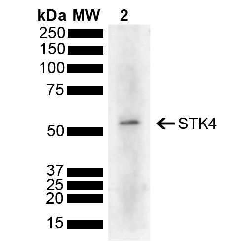
 WB (Western Blot)
(Western blot analysis of Human Cervical cancer cell line (HeLa) lysate showing detection of 55.6 kDa STK4 protein using Rabbit Anti-STK4 Polyclonal Antibody . Lane 1: Molecular Weight Ladder (MW). Lane 2: HeLa . Load: 10 ug. Block: 5% Skim Milk powder in TBST. Primary Antibody: Rabbit Anti-STK4 Polyclonal Antibody at 1:1000 for 2 hours at RT with shaking. Secondary Antibody: Goat Anti-Rabbit IgG: HRP at 1:5000 for 1 hour at RT. Color Development: ECL solution for 5 min at RT. Predicted/Observed Size: 55.6 kDa.)
WB (Western Blot)
(Western blot analysis of Human Cervical cancer cell line (HeLa) lysate showing detection of 55.6 kDa STK4 protein using Rabbit Anti-STK4 Polyclonal Antibody . Lane 1: Molecular Weight Ladder (MW). Lane 2: HeLa . Load: 10 ug. Block: 5% Skim Milk powder in TBST. Primary Antibody: Rabbit Anti-STK4 Polyclonal Antibody at 1:1000 for 2 hours at RT with shaking. Secondary Antibody: Goat Anti-Rabbit IgG: HRP at 1:5000 for 1 hour at RT. Color Development: ECL solution for 5 min at RT. Predicted/Observed Size: 55.6 kDa.)
STK4, Polyclonal Antibody (Cat# AAA253906)

 WB (Western Blot)
(Western blot analysis of Human Cervical cancer cell line (HeLa) lysate showing detection of 55.6 kDa STK4 protein using Rabbit Anti-STK4 Polyclonal Antibody . Lane 1: Molecular Weight Ladder (MW). Lane 2: HeLa . Load: 10 ug. Block: 5% Skim Milk powder in TBST. Primary Antibody: Rabbit Anti-STK4 Polyclonal Antibody at 1:1000 for 2 hours at RT with shaking. Secondary Antibody: Goat Anti-Rabbit IgG: HRP at 1:5000 for 1 hour at RT. Color Development: ECL solution for 5 min at RT. Predicted/Observed Size: 55.6 kDa.)
WB (Western Blot)
(Western blot analysis of Human Cervical cancer cell line (HeLa) lysate showing detection of 55.6 kDa STK4 protein using Rabbit Anti-STK4 Polyclonal Antibody . Lane 1: Molecular Weight Ladder (MW). Lane 2: HeLa . Load: 10 ug. Block: 5% Skim Milk powder in TBST. Primary Antibody: Rabbit Anti-STK4 Polyclonal Antibody at 1:1000 for 2 hours at RT with shaking. Secondary Antibody: Goat Anti-Rabbit IgG: HRP at 1:5000 for 1 hour at RT. Color Development: ECL solution for 5 min at RT. Predicted/Observed Size: 55.6 kDa.)
STK4, Polyclonal Antibody (Cat# AAA253907)

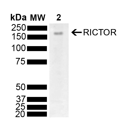
 WB (Western Blot)
(Western blot analysis of Mouse Brain showing detection of 192 kDa RICTOR protein using Rabbit Anti-RICTOR Polyclonal Antibody . Lane 1: Molecular Weight Ladder (MW). Lane 2: Mouse Brain. Load: 10 ug. Block: 5% Skim Milk powder in TBST. Primary Antibody: Rabbit Anti-RICTOR Polyclonal Antibody at 1:1000 for 2 hours at RT with shaking. Secondary Antibody: Goat Anti-Rabbit IgG: HRP at 1:5000 for 1 hour at RT. Color Development: ECL solution for 5 min at RT. Predicted/Observed Size: 192 kDa.)
WB (Western Blot)
(Western blot analysis of Mouse Brain showing detection of 192 kDa RICTOR protein using Rabbit Anti-RICTOR Polyclonal Antibody . Lane 1: Molecular Weight Ladder (MW). Lane 2: Mouse Brain. Load: 10 ug. Block: 5% Skim Milk powder in TBST. Primary Antibody: Rabbit Anti-RICTOR Polyclonal Antibody at 1:1000 for 2 hours at RT with shaking. Secondary Antibody: Goat Anti-Rabbit IgG: HRP at 1:5000 for 1 hour at RT. Color Development: ECL solution for 5 min at RT. Predicted/Observed Size: 192 kDa.)
RICTOR, Polyclonal Antibody (Cat# AAA253913)

 WB (Western Blot)
(Western blot analysis of Mouse Brain showing detection of 192 kDa RICTOR protein using Rabbit Anti-RICTOR Polyclonal Antibody . Lane 1: Molecular Weight Ladder (MW). Lane 2: Mouse Brain. Load: 10 ug. Block: 5% Skim Milk powder in TBST. Primary Antibody: Rabbit Anti-RICTOR Polyclonal Antibody at 1:1000 for 2 hours at RT with shaking. Secondary Antibody: Goat Anti-Rabbit IgG: HRP at 1:5000 for 1 hour at RT. Color Development: ECL solution for 5 min at RT. Predicted/Observed Size: 192 kDa.)
WB (Western Blot)
(Western blot analysis of Mouse Brain showing detection of 192 kDa RICTOR protein using Rabbit Anti-RICTOR Polyclonal Antibody . Lane 1: Molecular Weight Ladder (MW). Lane 2: Mouse Brain. Load: 10 ug. Block: 5% Skim Milk powder in TBST. Primary Antibody: Rabbit Anti-RICTOR Polyclonal Antibody at 1:1000 for 2 hours at RT with shaking. Secondary Antibody: Goat Anti-Rabbit IgG: HRP at 1:5000 for 1 hour at RT. Color Development: ECL solution for 5 min at RT. Predicted/Observed Size: 192 kDa.)
RICTOR, Polyclonal Antibody (Cat# AAA253914)

 WB (Western Blot)
(Western blot analysis of Mouse Brain showing detection of 192 kDa RICTOR protein using Rabbit Anti-RICTOR Polyclonal Antibody . Lane 1: Molecular Weight Ladder (MW). Lane 2: Mouse Brain. Load: 10 ug. Block: 5% Skim Milk powder in TBST. Primary Antibody: Rabbit Anti-RICTOR Polyclonal Antibody at 1:1000 for 2 hours at RT with shaking. Secondary Antibody: Goat Anti-Rabbit IgG: HRP at 1:5000 for 1 hour at RT. Color Development: ECL solution for 5 min at RT. Predicted/Observed Size: 192 kDa.)
WB (Western Blot)
(Western blot analysis of Mouse Brain showing detection of 192 kDa RICTOR protein using Rabbit Anti-RICTOR Polyclonal Antibody . Lane 1: Molecular Weight Ladder (MW). Lane 2: Mouse Brain. Load: 10 ug. Block: 5% Skim Milk powder in TBST. Primary Antibody: Rabbit Anti-RICTOR Polyclonal Antibody at 1:1000 for 2 hours at RT with shaking. Secondary Antibody: Goat Anti-Rabbit IgG: HRP at 1:5000 for 1 hour at RT. Color Development: ECL solution for 5 min at RT. Predicted/Observed Size: 192 kDa.)
RICTOR, Polyclonal Antibody (Cat# AAA253915)

 WB (Western Blot)
(Western blot analysis of Mouse Brain showing detection of 192 kDa RICTOR protein using Rabbit Anti-RICTOR Polyclonal Antibody . Lane 1: Molecular Weight Ladder (MW). Lane 2: Mouse Brain. Load: 10 ug. Block: 5% Skim Milk powder in TBST. Primary Antibody: Rabbit Anti-RICTOR Polyclonal Antibody at 1:1000 for 2 hours at RT with shaking. Secondary Antibody: Goat Anti-Rabbit IgG: HRP at 1:5000 for 1 hour at RT. Color Development: ECL solution for 5 min at RT. Predicted/Observed Size: 192 kDa.)
WB (Western Blot)
(Western blot analysis of Mouse Brain showing detection of 192 kDa RICTOR protein using Rabbit Anti-RICTOR Polyclonal Antibody . Lane 1: Molecular Weight Ladder (MW). Lane 2: Mouse Brain. Load: 10 ug. Block: 5% Skim Milk powder in TBST. Primary Antibody: Rabbit Anti-RICTOR Polyclonal Antibody at 1:1000 for 2 hours at RT with shaking. Secondary Antibody: Goat Anti-Rabbit IgG: HRP at 1:5000 for 1 hour at RT. Color Development: ECL solution for 5 min at RT. Predicted/Observed Size: 192 kDa.)
RICTOR, Polyclonal Antibody (Cat# AAA253917)
 WB (Western Blot)
(Western blot analysis of Mouse Brain showing detection of 192 kDa RICTOR protein using Rabbit Anti-RICTOR Polyclonal Antibody . Lane 1: Molecular Weight Ladder (MW). Lane 2: Mouse Brain. Load: 10 ug. Block: 5% Skim Milk powder in TBST. Primary Antibody: Rabbit Anti-RICTOR Polyclonal Antibody at 1:1000 for 2 hours at RT with shaking. Secondary Antibody: Goat Anti-Rabbit IgG: HRP at 1:5000 for 1 hour at RT. Color Development: ECL solution for 5 min at RT. Predicted/Observed Size: 192 kDa.)
WB (Western Blot)
(Western blot analysis of Mouse Brain showing detection of 192 kDa RICTOR protein using Rabbit Anti-RICTOR Polyclonal Antibody . Lane 1: Molecular Weight Ladder (MW). Lane 2: Mouse Brain. Load: 10 ug. Block: 5% Skim Milk powder in TBST. Primary Antibody: Rabbit Anti-RICTOR Polyclonal Antibody at 1:1000 for 2 hours at RT with shaking. Secondary Antibody: Goat Anti-Rabbit IgG: HRP at 1:5000 for 1 hour at RT. Color Development: ECL solution for 5 min at RT. Predicted/Observed Size: 192 kDa.)
RICTOR, Polyclonal Antibody (Cat# AAA253918)

 WB (Western Blot)
(Western blot analysis of Mouse Brain showing detection of 192 kDa RICTOR protein using Rabbit Anti-RICTOR Polyclonal Antibody . Lane 1: Molecular Weight Ladder (MW). Lane 2: Mouse Brain. Load: 10 ug. Block: 5% Skim Milk powder in TBST. Primary Antibody: Rabbit Anti-RICTOR Polyclonal Antibody at 1:1000 for 2 hours at RT with shaking. Secondary Antibody: Goat Anti-Rabbit IgG: HRP at 1:5000 for 1 hour at RT. Color Development: ECL solution for 5 min at RT. Predicted/Observed Size: 192 kDa.)
WB (Western Blot)
(Western blot analysis of Mouse Brain showing detection of 192 kDa RICTOR protein using Rabbit Anti-RICTOR Polyclonal Antibody . Lane 1: Molecular Weight Ladder (MW). Lane 2: Mouse Brain. Load: 10 ug. Block: 5% Skim Milk powder in TBST. Primary Antibody: Rabbit Anti-RICTOR Polyclonal Antibody at 1:1000 for 2 hours at RT with shaking. Secondary Antibody: Goat Anti-Rabbit IgG: HRP at 1:5000 for 1 hour at RT. Color Development: ECL solution for 5 min at RT. Predicted/Observed Size: 192 kDa.)
RICTOR, Polyclonal Antibody (Cat# AAA253920)
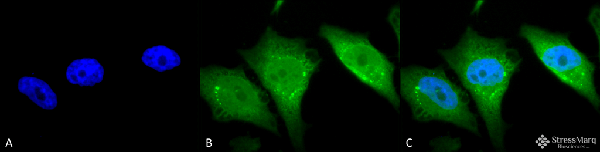
 ICC (Immunocytochemistry)
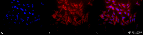
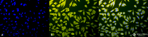
(Immunocytochemistry/Immunofluorescence analysis using Rabbit Anti-Hsp27 Polyclonal Antibody . Tissue: Heat Shocked Cervical cancer cell line (HeLa). Species: Human. Fixation: 2% Formaldehyde for 20 min at RT. Primary Antibody: Rabbit Anti-Hsp27 Polyclonal Antibody at 1:250 for 12 hours at 4 degree C. Secondary Antibody: APC Goat Anti-Rabbit (red) at 1:200 for 2 hours at RT. Counterstain: DAPI (blue) nuclear stain at 1:40000 for 2 hours at RT. Localization: Cytoplasm. Mitochondrion matrix. Magnification: 20x. (A) DAPI (blue) nuclear stain. (B) Anti-Hsp27 Antibody. (C) Composite. Heat Shocked at 42 degree C for 1h.)
ICC (Immunocytochemistry)
(Immunocytochemistry/Immunofluorescence analysis using Rabbit Anti-Hsp27 Polyclonal Antibody . Tissue: Heat Shocked Cervical cancer cell line (HeLa). Species: Human. Fixation: 2% Formaldehyde for 20 min at RT. Primary Antibody: Rabbit Anti-Hsp27 Polyclonal Antibody at 1:250 for 12 hours at 4 degree C. Secondary Antibody: APC Goat Anti-Rabbit (red) at 1:200 for 2 hours at RT. Counterstain: DAPI (blue) nuclear stain at 1:40000 for 2 hours at RT. Localization: Cytoplasm. Mitochondrion matrix. Magnification: 20x. (A) DAPI (blue) nuclear stain. (B) Anti-Hsp27 Antibody. (C) Composite. Heat Shocked at 42 degree C for 1h.)
HSP27, Polyclonal Antibody (Cat# AAA253943)
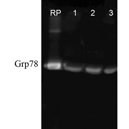
 WB (Western Blot)
(Western blot analysis of Rat Tissue lysates showing detection of GRP78 protein using Rabbit Anti-GRP78 Polyclonal Antibody . Load: 15 ugprotein. Block: 1.5% BSA. Primary Antibody: Rabbit Anti-GRP78 Polyclonal Antibody at 1:1000 for 2 hours at RT. Secondary Antibody: Donkey Anti-Rabbit IgG: HRP for 1 hour at RT.)
WB (Western Blot)
(Western blot analysis of Rat Tissue lysates showing detection of GRP78 protein using Rabbit Anti-GRP78 Polyclonal Antibody . Load: 15 ugprotein. Block: 1.5% BSA. Primary Antibody: Rabbit Anti-GRP78 Polyclonal Antibody at 1:1000 for 2 hours at RT. Secondary Antibody: Donkey Anti-Rabbit IgG: HRP for 1 hour at RT.)
GRP78, Polyclonal Antibody (Cat# AAA253944)
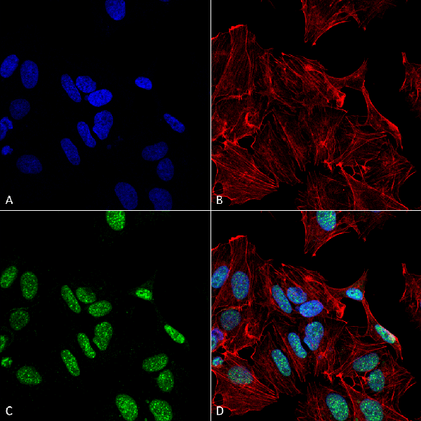
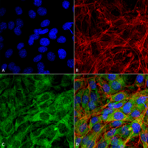
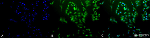 ICC (Immunocytochemistry)
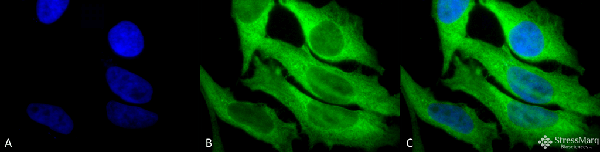
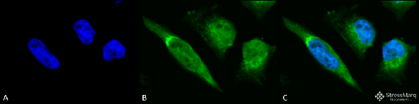
(Immunocytochemistry/Immunofluorescence analysis using Rabbit Anti-Ubiquitin Polyclonal Antibody . Tissue: Cervical cancer cell line (HeLa). Species: Human. Fixation: 2% Formaldehyde for 20 min at RT. Primary Antibody: Rabbit Anti-Ubiquitin Polyclonal Antibody at 1:200 for 12 hours at 4 degree C. Secondary Antibody: FITC Goat Anti-Rabbit (green) at 1:200 for 2 hours at RT. Counterstain: DAPI (blue) nuclear stain at 1:40000 for 2 hours at RT. Localization: Cytoplasm. Magnification: 20x. (A) DAPI (blue) nuclear stain. (B) Anti-Ubiquitin Antibody. (C) Composite.)
ICC (Immunocytochemistry)
(Immunocytochemistry/Immunofluorescence analysis using Rabbit Anti-Ubiquitin Polyclonal Antibody . Tissue: Cervical cancer cell line (HeLa). Species: Human. Fixation: 2% Formaldehyde for 20 min at RT. Primary Antibody: Rabbit Anti-Ubiquitin Polyclonal Antibody at 1:200 for 12 hours at 4 degree C. Secondary Antibody: FITC Goat Anti-Rabbit (green) at 1:200 for 2 hours at RT. Counterstain: DAPI (blue) nuclear stain at 1:40000 for 2 hours at RT. Localization: Cytoplasm. Magnification: 20x. (A) DAPI (blue) nuclear stain. (B) Anti-Ubiquitin Antibody. (C) Composite.)
Ubiquitin, Polyclonal Antibody (Cat# AAA253947)

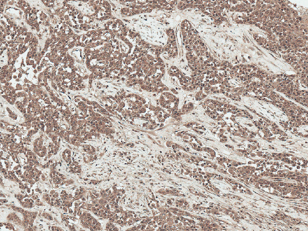
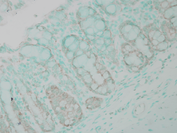
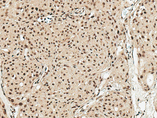
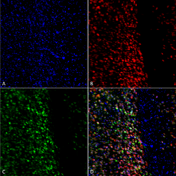
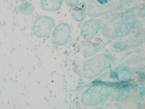 IHC (Immunohistochemistry)
(Immunohistochemistry analysis using Rabbit Anti-Calnexin Polyclonal Antibody . Tissue: colon colitis. Species: Mouse. Fixation: Formalin. Primary Antibody: Rabbit Anti-Calnexin Polyclonal Antibody at 1:100000 for 12 hours at 4 degree C. Secondary Antibody: Biotin Goat Anti-Rabbit at 1:2000 for 1 hour at RT. Counterstain: Methyl Green at 200uL for 2 min at RT. Localization: Inflammatory cells.)
IHC (Immunohistochemistry)
(Immunohistochemistry analysis using Rabbit Anti-Calnexin Polyclonal Antibody . Tissue: colon colitis. Species: Mouse. Fixation: Formalin. Primary Antibody: Rabbit Anti-Calnexin Polyclonal Antibody at 1:100000 for 12 hours at 4 degree C. Secondary Antibody: Biotin Goat Anti-Rabbit at 1:2000 for 1 hour at RT. Counterstain: Methyl Green at 200uL for 2 min at RT. Localization: Inflammatory cells.)
Calnexin-NT, Polyclonal Antibody (Cat# AAA253950)
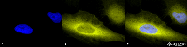
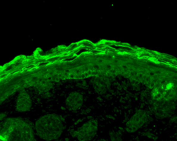
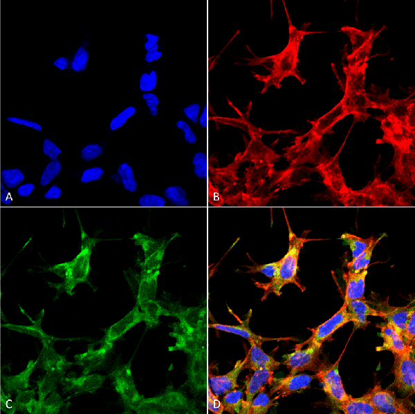
 ICC (Immunocytochemistry)
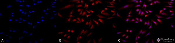
(Immunocytochemistry/Immunofluorescence analysis using Rabbit Anti-Calnexin-CT Polyclonal Antibody . Tissue: Heat Shocked Cervical cancer cell line (HeLa). Species: Human. Fixation: 2% Formaldehyde for 20 min at RT. Primary Antibody: Rabbit Anti-Calnexin-CT Polyclonal Antibody at 1:80 for 12 hours at 4 degree C. Secondary Antibody: R-PE Goat Anti-Rabbit (yellow) at 1:200 for 2 hours at RT. Counterstain: DAPI (blue) nuclear stain at 1:40000 for 2 hours at RT. Localization: Endoplasmic reticulum membrane. Melanosome. Magnification: 20x. (A) DAPI (blue) nuclear stain. (B) Anti-Calnexin-CT Antibody. (C) Composite. Heat Shocked at 42 degree C for 1h.)
ICC (Immunocytochemistry)
(Immunocytochemistry/Immunofluorescence analysis using Rabbit Anti-Calnexin-CT Polyclonal Antibody . Tissue: Heat Shocked Cervical cancer cell line (HeLa). Species: Human. Fixation: 2% Formaldehyde for 20 min at RT. Primary Antibody: Rabbit Anti-Calnexin-CT Polyclonal Antibody at 1:80 for 12 hours at 4 degree C. Secondary Antibody: R-PE Goat Anti-Rabbit (yellow) at 1:200 for 2 hours at RT. Counterstain: DAPI (blue) nuclear stain at 1:40000 for 2 hours at RT. Localization: Endoplasmic reticulum membrane. Melanosome. Magnification: 20x. (A) DAPI (blue) nuclear stain. (B) Anti-Calnexin-CT Antibody. (C) Composite. Heat Shocked at 42 degree C for 1h.)
Calnexin-CT, Polyclonal Antibody (Cat# AAA253951)

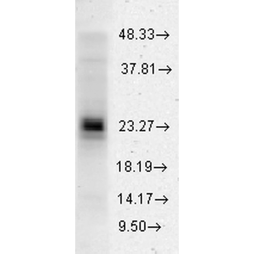
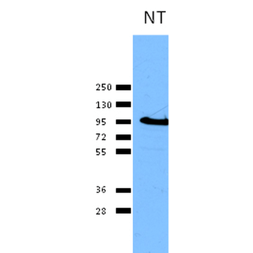
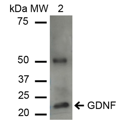 WB (Western Blot)
(Western blot analysis of Human Cervical cancer cell line (HeLa) lysate showing detection of ~23.7 kDa GDNF protein using Rabbit Anti-GDNF Polyclonal Antibody . Lane 1: Molecular Weight Ladder (MW). Lane 2: HeLa cell lysates. Load: 15 ug. Block: 5% Skim Milk in 1X TBST. Primary Antibody: Rabbit Anti-GDNF Polyclonal Antibody at 1:1000 for 2 hours at RT. Secondary Antibody: Goat Anti-Rabbit IgG: HRP at 1:1000 for 60 min at RT. Color Development: ECL solution for 6 min in RT. Predicted/Observed Size: ~23.7 kDa. Other Band(s): 50 kDa is a dimer.)
WB (Western Blot)
(Western blot analysis of Human Cervical cancer cell line (HeLa) lysate showing detection of ~23.7 kDa GDNF protein using Rabbit Anti-GDNF Polyclonal Antibody . Lane 1: Molecular Weight Ladder (MW). Lane 2: HeLa cell lysates. Load: 15 ug. Block: 5% Skim Milk in 1X TBST. Primary Antibody: Rabbit Anti-GDNF Polyclonal Antibody at 1:1000 for 2 hours at RT. Secondary Antibody: Goat Anti-Rabbit IgG: HRP at 1:1000 for 60 min at RT. Color Development: ECL solution for 6 min in RT. Predicted/Observed Size: ~23.7 kDa. Other Band(s): 50 kDa is a dimer.)
GDNF, Polyclonal Antibody (Cat# AAA253953)


 WB (Western Blot)
(Western blot analysis of Human Cervical cancer cell line (HeLa) lysate showing detection of ~23.7 kDa GDNF protein using Rabbit Anti-GDNF Polyclonal Antibody . Lane 1: Molecular Weight Ladder (MW). Lane 2: HeLa cell lysates. Load: 15 ug. Block: 5% Skim Milk in 1X TBST. Primary Antibody: Rabbit Anti-GDNF Polyclonal Antibody at 1:1000 for 2 hours at RT. Secondary Antibody: Goat Anti-Rabbit IgG: HRP at 1:1000 for 60 min at RT. Color Development: ECL solution for 6 min in RT. Predicted/Observed Size: ~23.7 kDa. Other Band(s): 50 kDa is a dimer.)
WB (Western Blot)
(Western blot analysis of Human Cervical cancer cell line (HeLa) lysate showing detection of ~23.7 kDa GDNF protein using Rabbit Anti-GDNF Polyclonal Antibody . Lane 1: Molecular Weight Ladder (MW). Lane 2: HeLa cell lysates. Load: 15 ug. Block: 5% Skim Milk in 1X TBST. Primary Antibody: Rabbit Anti-GDNF Polyclonal Antibody at 1:1000 for 2 hours at RT. Secondary Antibody: Goat Anti-Rabbit IgG: HRP at 1:1000 for 60 min at RT. Color Development: ECL solution for 6 min in RT. Predicted/Observed Size: ~23.7 kDa. Other Band(s): 50 kDa is a dimer.)
GDNF, Polyclonal Antibody (Cat# AAA253957)

 WB (Western Blot)
(Western blot analysis of Human Cervical cancer cell line (HeLa) lysate showing detection of ~23.7 kDa GDNF protein using Rabbit Anti-GDNF Polyclonal Antibody . Lane 1: Molecular Weight Ladder (MW). Lane 2: HeLa cell lysates. Load: 15 ug. Block: 5% Skim Milk in 1X TBST. Primary Antibody: Rabbit Anti-GDNF Polyclonal Antibody at 1:1000 for 2 hours at RT. Secondary Antibody: Goat Anti-Rabbit IgG: HRP at 1:1000 for 60 min at RT. Color Development: ECL solution for 6 min in RT. Predicted/Observed Size: ~23.7 kDa. Other Band(s): 50 kDa is a dimer.)
WB (Western Blot)
(Western blot analysis of Human Cervical cancer cell line (HeLa) lysate showing detection of ~23.7 kDa GDNF protein using Rabbit Anti-GDNF Polyclonal Antibody . Lane 1: Molecular Weight Ladder (MW). Lane 2: HeLa cell lysates. Load: 15 ug. Block: 5% Skim Milk in 1X TBST. Primary Antibody: Rabbit Anti-GDNF Polyclonal Antibody at 1:1000 for 2 hours at RT. Secondary Antibody: Goat Anti-Rabbit IgG: HRP at 1:1000 for 60 min at RT. Color Development: ECL solution for 6 min in RT. Predicted/Observed Size: ~23.7 kDa. Other Band(s): 50 kDa is a dimer.)
GDNF, Polyclonal Antibody (Cat# AAA253958)
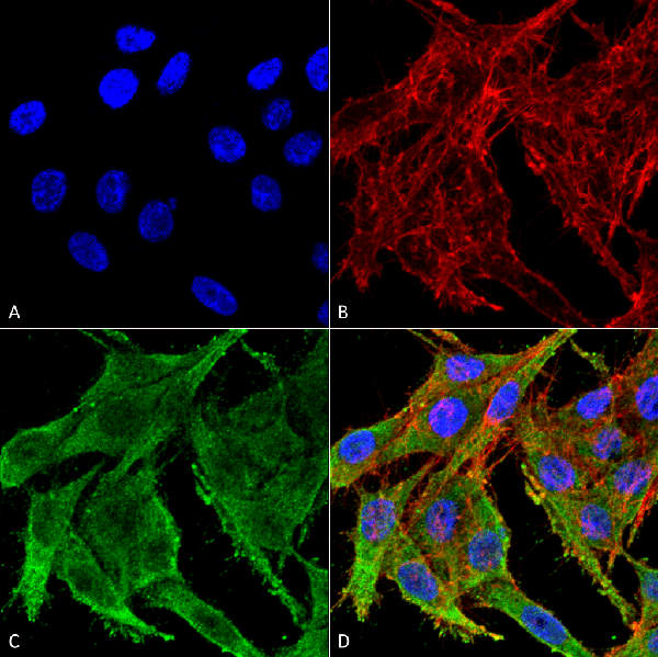
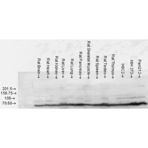
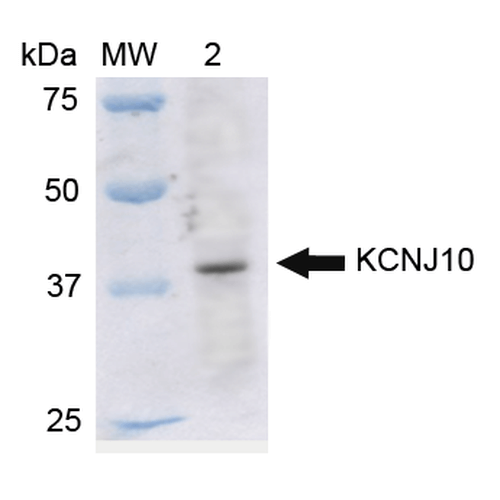 WB (Western Blot)
(Western blot analysis of Rat Liver cell lysates showing detection of ~42.5 kDa Kir4.1 protein using Rabbit Anti-Kir4.1 Polyclonal Antibody (SPC-700). Lane 1: Molecular Weight Ladder (MW). Lane 2: Rat Liver cell lysates. Load: 15 ug. Block: 5% Skim Milk in 1X TBST. Primary Antibody: Rabbit Anti-Kir4.1 Polyclonal Antibody (SPC-700) at 1:1000 for 2 hours at RT. Secondary Antibody: Goat Anti-Rabbit IgG: HRP at 1:2000 for 60 min at RT. Color Development: ECL solution for 6 min in RT. Predicted/Observed Size: ~42.5 kDa.)
WB (Western Blot)
(Western blot analysis of Rat Liver cell lysates showing detection of ~42.5 kDa Kir4.1 protein using Rabbit Anti-Kir4.1 Polyclonal Antibody (SPC-700). Lane 1: Molecular Weight Ladder (MW). Lane 2: Rat Liver cell lysates. Load: 15 ug. Block: 5% Skim Milk in 1X TBST. Primary Antibody: Rabbit Anti-Kir4.1 Polyclonal Antibody (SPC-700) at 1:1000 for 2 hours at RT. Secondary Antibody: Goat Anti-Rabbit IgG: HRP at 1:2000 for 60 min at RT. Color Development: ECL solution for 6 min in RT. Predicted/Observed Size: ~42.5 kDa.)
Kir4.1, Polyclonal Antibody (Cat# AAA253836)

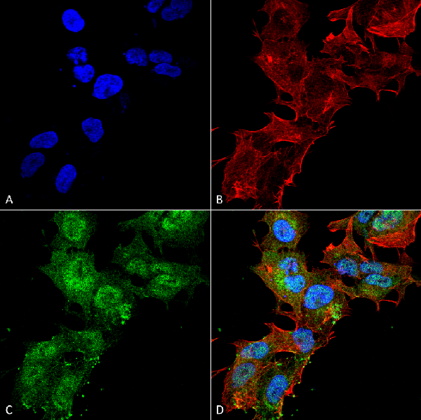
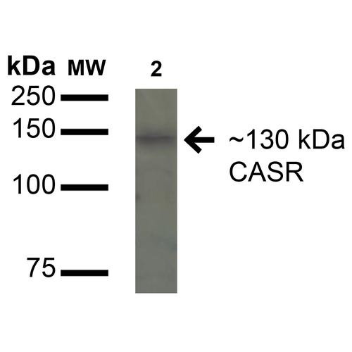
 WB (Western Blot)
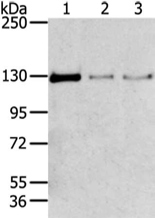
(Western blot analysis of Rat Kidney cell lysates showing detection of 130kDa Calcium Sensing Receptor protein using Rabbit Anti-Calcium Sensing Receptor Polyclonal Antibody (SPC-713). Lane 1: Molecular Weight Ladder (MW). Lane 2: Rat Kidney cell lysates. Load: 15 ug. Block: 5% Skim Milk in 1X TBST. Primary Antibody: Rabbit Anti-Calcium Sensing Receptor Polyclonal Antibody (SPC-713) at 1:1000 for 16 hours at 4 degree C. Secondary Antibody: Goat-Anti-Rabbit IgG: HRP at 1:200 for 60 min at RT. Color Development: TMB. Predicted/Observed Size: 130kDa.)
WB (Western Blot)
(Western blot analysis of Rat Kidney cell lysates showing detection of 130kDa Calcium Sensing Receptor protein using Rabbit Anti-Calcium Sensing Receptor Polyclonal Antibody (SPC-713). Lane 1: Molecular Weight Ladder (MW). Lane 2: Rat Kidney cell lysates. Load: 15 ug. Block: 5% Skim Milk in 1X TBST. Primary Antibody: Rabbit Anti-Calcium Sensing Receptor Polyclonal Antibody (SPC-713) at 1:1000 for 16 hours at 4 degree C. Secondary Antibody: Goat-Anti-Rabbit IgG: HRP at 1:200 for 60 min at RT. Color Development: TMB. Predicted/Observed Size: 130kDa.)
Calcium Sensing Receptor, Polyclonal Antibody (Cat# AAA253842)
 WB (Western Blot)
(Western blot analysis of Rat Kidney cell lysates showing detection of 130kDa Calcium Sensing Receptor protein using Rabbit Anti-Calcium Sensing Receptor Polyclonal Antibody (SPC-713). Lane 1: Molecular Weight Ladder (MW). Lane 2: Rat Kidney cell lysates. Load: 15 ug. Block: 5% Skim Milk in 1X TBST. Primary Antibody: Rabbit Anti-Calcium Sensing Receptor Polyclonal Antibody (SPC-713) at 1:1000 for 16 hours at 4 degree C. Secondary Antibody: Goat-Anti-Rabbit IgG: HRP at 1:200 for 60 min at RT. Color Development: TMB. Predicted/Observed Size: 130kDa.)
WB (Western Blot)
(Western blot analysis of Rat Kidney cell lysates showing detection of 130kDa Calcium Sensing Receptor protein using Rabbit Anti-Calcium Sensing Receptor Polyclonal Antibody (SPC-713). Lane 1: Molecular Weight Ladder (MW). Lane 2: Rat Kidney cell lysates. Load: 15 ug. Block: 5% Skim Milk in 1X TBST. Primary Antibody: Rabbit Anti-Calcium Sensing Receptor Polyclonal Antibody (SPC-713) at 1:1000 for 16 hours at 4 degree C. Secondary Antibody: Goat-Anti-Rabbit IgG: HRP at 1:200 for 60 min at RT. Color Development: TMB. Predicted/Observed Size: 130kDa.)
Calcium Sensing Receptor, Polyclonal Antibody (Cat# AAA253843)

 WB (Western Blot)
(Western blot analysis of Rat Kidney cell lysates showing detection of 130kDa Calcium Sensing Receptor protein using Rabbit Anti-Calcium Sensing Receptor Polyclonal Antibody (SPC-713). Lane 1: Molecular Weight Ladder (MW). Lane 2: Rat Kidney cell lysates. Load: 15 ug. Block: 5% Skim Milk in 1X TBST. Primary Antibody: Rabbit Anti-Calcium Sensing Receptor Polyclonal Antibody (SPC-713) at 1:1000 for 16 hours at 4 degree C. Secondary Antibody: Goat-Anti-Rabbit IgG: HRP at 1:200 for 60 min at RT. Color Development: TMB. Predicted/Observed Size: 130kDa.)
WB (Western Blot)
(Western blot analysis of Rat Kidney cell lysates showing detection of 130kDa Calcium Sensing Receptor protein using Rabbit Anti-Calcium Sensing Receptor Polyclonal Antibody (SPC-713). Lane 1: Molecular Weight Ladder (MW). Lane 2: Rat Kidney cell lysates. Load: 15 ug. Block: 5% Skim Milk in 1X TBST. Primary Antibody: Rabbit Anti-Calcium Sensing Receptor Polyclonal Antibody (SPC-713) at 1:1000 for 16 hours at 4 degree C. Secondary Antibody: Goat-Anti-Rabbit IgG: HRP at 1:200 for 60 min at RT. Color Development: TMB. Predicted/Observed Size: 130kDa.)
Calcium Sensing Receptor, Polyclonal Antibody (Cat# AAA253844)

 WB (Western Blot)
(Western blot analysis of Rat Kidney cell lysates showing detection of 130kDa Calcium Sensing Receptor protein using Rabbit Anti-Calcium Sensing Receptor Polyclonal Antibody (SPC-713). Lane 1: Molecular Weight Ladder (MW). Lane 2: Rat Kidney cell lysates. Load: 15 ug. Block: 5% Skim Milk in 1X TBST. Primary Antibody: Rabbit Anti-Calcium Sensing Receptor Polyclonal Antibody (SPC-713) at 1:1000 for 16 hours at 4 degree C. Secondary Antibody: Goat-Anti-Rabbit IgG: HRP at 1:200 for 60 min at RT. Color Development: TMB. Predicted/Observed Size: 130kDa.)
WB (Western Blot)
(Western blot analysis of Rat Kidney cell lysates showing detection of 130kDa Calcium Sensing Receptor protein using Rabbit Anti-Calcium Sensing Receptor Polyclonal Antibody (SPC-713). Lane 1: Molecular Weight Ladder (MW). Lane 2: Rat Kidney cell lysates. Load: 15 ug. Block: 5% Skim Milk in 1X TBST. Primary Antibody: Rabbit Anti-Calcium Sensing Receptor Polyclonal Antibody (SPC-713) at 1:1000 for 16 hours at 4 degree C. Secondary Antibody: Goat-Anti-Rabbit IgG: HRP at 1:200 for 60 min at RT. Color Development: TMB. Predicted/Observed Size: 130kDa.)
Calcium Sensing Receptor, Polyclonal Antibody (Cat# AAA253848)

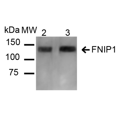 WB (Western Blot)
(Western blot analysis of Mouse, Rat Kidney showing detection of ~131 kDa FNIP1 protein using Rabbit Anti-FNIP1 Polyclonal Antibody (SPC-718). Lane 1: Molecular Weight Ladder (MW). Lane 2: Mouse Kidney cell lysates. Lane 3: Rat Kidney cell lysates. Load: 20 ug. Block: 5% Skim Milk in 1X TBST. Primary Antibody: Rabbit Anti-FNIP1 Polyclonal Antibody (SPC-718) at 1:1000 for 16 hours at 4 degree C. Secondary Antibody: Goat Anti-Rabbit IgG: HRP at 1:2000 for 60 min at RT. Color Development: ECL solution for 6 min at RT. Predicted/Observed Size: ~131 kDa.)
WB (Western Blot)
(Western blot analysis of Mouse, Rat Kidney showing detection of ~131 kDa FNIP1 protein using Rabbit Anti-FNIP1 Polyclonal Antibody (SPC-718). Lane 1: Molecular Weight Ladder (MW). Lane 2: Mouse Kidney cell lysates. Lane 3: Rat Kidney cell lysates. Load: 20 ug. Block: 5% Skim Milk in 1X TBST. Primary Antibody: Rabbit Anti-FNIP1 Polyclonal Antibody (SPC-718) at 1:1000 for 16 hours at 4 degree C. Secondary Antibody: Goat Anti-Rabbit IgG: HRP at 1:2000 for 60 min at RT. Color Development: ECL solution for 6 min at RT. Predicted/Observed Size: ~131 kDa.)
FNIP1, Polyclonal Antibody (Cat# AAA253851)

 WB (Western Blot)
(Western blot analysis of Mouse, Rat Kidney showing detection of ~131 kDa FNIP1 protein using Rabbit Anti-FNIP1 Polyclonal Antibody (SPC-718). Lane 1: Molecular Weight Ladder (MW). Lane 2: Mouse Kidney cell lysates. Lane 3: Rat Kidney cell lysates. Load: 20 ug. Block: 5% Skim Milk in 1X TBST. Primary Antibody: Rabbit Anti-FNIP1 Polyclonal Antibody (SPC-718) at 1:1000 for 16 hours at 4 degree C. Secondary Antibody: Goat Anti-Rabbit IgG: HRP at 1:2000 for 60 min at RT. Color Development: ECL solution for 6 min at RT. Predicted/Observed Size: ~131 kDa.)
WB (Western Blot)
(Western blot analysis of Mouse, Rat Kidney showing detection of ~131 kDa FNIP1 protein using Rabbit Anti-FNIP1 Polyclonal Antibody (SPC-718). Lane 1: Molecular Weight Ladder (MW). Lane 2: Mouse Kidney cell lysates. Lane 3: Rat Kidney cell lysates. Load: 20 ug. Block: 5% Skim Milk in 1X TBST. Primary Antibody: Rabbit Anti-FNIP1 Polyclonal Antibody (SPC-718) at 1:1000 for 16 hours at 4 degree C. Secondary Antibody: Goat Anti-Rabbit IgG: HRP at 1:2000 for 60 min at RT. Color Development: ECL solution for 6 min at RT. Predicted/Observed Size: ~131 kDa.)
FNIP1, Polyclonal Antibody (Cat# AAA253852)
 WB (Western Blot)
(Western blot analysis of Mouse, Rat Kidney showing detection of ~131 kDa FNIP1 protein using Rabbit Anti-FNIP1 Polyclonal Antibody (SPC-718). Lane 1: Molecular Weight Ladder (MW). Lane 2: Mouse Kidney cell lysates. Lane 3: Rat Kidney cell lysates. Load: 20 ug. Block: 5% Skim Milk in 1X TBST. Primary Antibody: Rabbit Anti-FNIP1 Polyclonal Antibody (SPC-718) at 1:1000 for 16 hours at 4 degree C. Secondary Antibody: Goat Anti-Rabbit IgG: HRP at 1:2000 for 60 min at RT. Color Development: ECL solution for 6 min at RT. Predicted/Observed Size: ~131 kDa.)
WB (Western Blot)
(Western blot analysis of Mouse, Rat Kidney showing detection of ~131 kDa FNIP1 protein using Rabbit Anti-FNIP1 Polyclonal Antibody (SPC-718). Lane 1: Molecular Weight Ladder (MW). Lane 2: Mouse Kidney cell lysates. Lane 3: Rat Kidney cell lysates. Load: 20 ug. Block: 5% Skim Milk in 1X TBST. Primary Antibody: Rabbit Anti-FNIP1 Polyclonal Antibody (SPC-718) at 1:1000 for 16 hours at 4 degree C. Secondary Antibody: Goat Anti-Rabbit IgG: HRP at 1:2000 for 60 min at RT. Color Development: ECL solution for 6 min at RT. Predicted/Observed Size: ~131 kDa.)
FNIP1, Polyclonal Antibody (Cat# AAA253854)

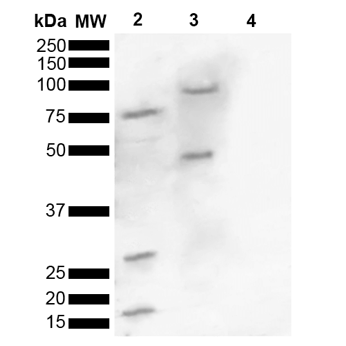
 WB (Western Blot)
(Western blot analysis of Human, Mouse brain lysate showing detection of ~16 kDa Alpha Synuclein pSer129 protein using Rabbit Anti-Alpha Synuclein pSer129 Polyclonal Antibody (SPC-742). Lane 1: Molecular Weight Ladder (MW). Lane 2: Human brain lysate. Lane 3: Mouse brain lysate. Lane 4: Alpha Synuclein Monomer (0.5 ug). Load: 15 ug. Block: 5% Skim Milk in 1X TBST. Primary Antibody: Rabbit Anti-Alpha Synuclein pSer129 Polyclonal Antibody (SPC-742) at 1:1000 for 2 hours at RT. Secondary Antibody: Goat Anti-Rabbit HRP:IgG at 1:3000 for 1 hour at RT. Color Development: ECL solution for 5 min at RT. Predicted/Observed Size: ~16 kDa. Other Band(s): 100, 75, 45, 30,16 kDa.)
WB (Western Blot)
(Western blot analysis of Human, Mouse brain lysate showing detection of ~16 kDa Alpha Synuclein pSer129 protein using Rabbit Anti-Alpha Synuclein pSer129 Polyclonal Antibody (SPC-742). Lane 1: Molecular Weight Ladder (MW). Lane 2: Human brain lysate. Lane 3: Mouse brain lysate. Lane 4: Alpha Synuclein Monomer (0.5 ug). Load: 15 ug. Block: 5% Skim Milk in 1X TBST. Primary Antibody: Rabbit Anti-Alpha Synuclein pSer129 Polyclonal Antibody (SPC-742) at 1:1000 for 2 hours at RT. Secondary Antibody: Goat Anti-Rabbit HRP:IgG at 1:3000 for 1 hour at RT. Color Development: ECL solution for 5 min at RT. Predicted/Observed Size: ~16 kDa. Other Band(s): 100, 75, 45, 30,16 kDa.)
Alpha Synuclein, Polyclonal Antibody (Cat# AAA253859)

 WB (Western Blot)
(Western blot analysis of Human, Mouse brain lysate showing detection of ~16 kDa Alpha Synuclein pSer129 protein using Rabbit Anti-Alpha Synuclein pSer129 Polyclonal Antibody (SPC-742). Lane 1: Molecular Weight Ladder (MW). Lane 2: Human brain lysate. Lane 3: Mouse brain lysate. Lane 4: Alpha Synuclein Monomer (0.5 ug). Load: 15 ug. Block: 5% Skim Milk in 1X TBST. Primary Antibody: Rabbit Anti-Alpha Synuclein pSer129 Polyclonal Antibody (SPC-742) at 1:1000 for 2 hours at RT. Secondary Antibody: Goat Anti-Rabbit HRP:IgG at 1:3000 for 1 hour at RT. Color Development: ECL solution for 5 min at RT. Predicted/Observed Size: ~16 kDa. Other Band(s): 100, 75, 45, 30,16 kDa.)
WB (Western Blot)
(Western blot analysis of Human, Mouse brain lysate showing detection of ~16 kDa Alpha Synuclein pSer129 protein using Rabbit Anti-Alpha Synuclein pSer129 Polyclonal Antibody (SPC-742). Lane 1: Molecular Weight Ladder (MW). Lane 2: Human brain lysate. Lane 3: Mouse brain lysate. Lane 4: Alpha Synuclein Monomer (0.5 ug). Load: 15 ug. Block: 5% Skim Milk in 1X TBST. Primary Antibody: Rabbit Anti-Alpha Synuclein pSer129 Polyclonal Antibody (SPC-742) at 1:1000 for 2 hours at RT. Secondary Antibody: Goat Anti-Rabbit HRP:IgG at 1:3000 for 1 hour at RT. Color Development: ECL solution for 5 min at RT. Predicted/Observed Size: ~16 kDa. Other Band(s): 100, 75, 45, 30,16 kDa.)
Alpha Synuclein, Polyclonal Antibody (Cat# AAA253860)

 WB (Western Blot)
(Western blot analysis of Human, Mouse brain lysate showing detection of ~16 kDa Alpha Synuclein pSer129 protein using Rabbit Anti-Alpha Synuclein pSer129 Polyclonal Antibody (SPC-742). Lane 1: Molecular Weight Ladder (MW). Lane 2: Human brain lysate. Lane 3: Mouse brain lysate. Lane 4: Alpha Synuclein Monomer (0.5 ug). Load: 15 ug. Block: 5% Skim Milk in 1X TBST. Primary Antibody: Rabbit Anti-Alpha Synuclein pSer129 Polyclonal Antibody (SPC-742) at 1:1000 for 2 hours at RT. Secondary Antibody: Goat Anti-Rabbit HRP:IgG at 1:3000 for 1 hour at RT. Color Development: ECL solution for 5 min at RT. Predicted/Observed Size: ~16 kDa. Other Band(s): 100, 75, 45, 30,16 kDa.)
WB (Western Blot)
(Western blot analysis of Human, Mouse brain lysate showing detection of ~16 kDa Alpha Synuclein pSer129 protein using Rabbit Anti-Alpha Synuclein pSer129 Polyclonal Antibody (SPC-742). Lane 1: Molecular Weight Ladder (MW). Lane 2: Human brain lysate. Lane 3: Mouse brain lysate. Lane 4: Alpha Synuclein Monomer (0.5 ug). Load: 15 ug. Block: 5% Skim Milk in 1X TBST. Primary Antibody: Rabbit Anti-Alpha Synuclein pSer129 Polyclonal Antibody (SPC-742) at 1:1000 for 2 hours at RT. Secondary Antibody: Goat Anti-Rabbit HRP:IgG at 1:3000 for 1 hour at RT. Color Development: ECL solution for 5 min at RT. Predicted/Observed Size: ~16 kDa. Other Band(s): 100, 75, 45, 30,16 kDa.)
Alpha Synuclein, Polyclonal Antibody (Cat# AAA253866)
 WB (Western Blot)
(Western blot analysis of Human, Mouse brain lysate showing detection of ~16 kDa Alpha Synuclein pSer129 protein using Rabbit Anti-Alpha Synuclein pSer129 Polyclonal Antibody (SPC-742). Lane 1: Molecular Weight Ladder (MW). Lane 2: Human brain lysate. Lane 3: Mouse brain lysate. Lane 4: Alpha Synuclein Monomer (0.5 ug). Load: 15 ug. Block: 5% Skim Milk in 1X TBST. Primary Antibody: Rabbit Anti-Alpha Synuclein pSer129 Polyclonal Antibody (SPC-742) at 1:1000 for 2 hours at RT. Secondary Antibody: Goat Anti-Rabbit HRP:IgG at 1:3000 for 1 hour at RT. Color Development: ECL solution for 5 min at RT. Predicted/Observed Size: ~16 kDa. Other Band(s): 100, 75, 45, 30,16 kDa.)
WB (Western Blot)
(Western blot analysis of Human, Mouse brain lysate showing detection of ~16 kDa Alpha Synuclein pSer129 protein using Rabbit Anti-Alpha Synuclein pSer129 Polyclonal Antibody (SPC-742). Lane 1: Molecular Weight Ladder (MW). Lane 2: Human brain lysate. Lane 3: Mouse brain lysate. Lane 4: Alpha Synuclein Monomer (0.5 ug). Load: 15 ug. Block: 5% Skim Milk in 1X TBST. Primary Antibody: Rabbit Anti-Alpha Synuclein pSer129 Polyclonal Antibody (SPC-742) at 1:1000 for 2 hours at RT. Secondary Antibody: Goat Anti-Rabbit HRP:IgG at 1:3000 for 1 hour at RT. Color Development: ECL solution for 5 min at RT. Predicted/Observed Size: ~16 kDa. Other Band(s): 100, 75, 45, 30,16 kDa.)
Alpha Synuclein, Polyclonal Antibody (Cat# AAA253867)

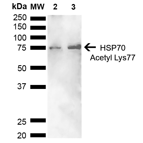
 WB (Western Blot)
(Western blot analysis of Human Cervical cancer cell line (HeLa) lysate showing detection of ~70 kDa HSP70 Acetyl Lys77 protein using Rabbit Anti-HSP70 Acetyl Lys77 Polyclonal Antibody (SPC-743). Lane 1: Molecular Weight Ladder (MW). Lane 2: Cervical Cancer cell line (HeLa) lysate. Lane 3: H2O2 Cervical Cancer cell line (HeLa) lysate. Load: 10 ug. Block: 5% Skim Milk in 1X TBST. Primary Antibody: Rabbit Anti-HSP70 Acetyl Lys77 Polyclonal Antibody (SPC-743) at 1:1000 for 2 hours at RT. Secondary Antibody: Goat Anti-Rabbit HRP:IgG at 1:3000 for 1 hour at RT. Color Development: ECL solution for 5 min at RT. Predicted/Observed Size: ~70 kDa.)
WB (Western Blot)
(Western blot analysis of Human Cervical cancer cell line (HeLa) lysate showing detection of ~70 kDa HSP70 Acetyl Lys77 protein using Rabbit Anti-HSP70 Acetyl Lys77 Polyclonal Antibody (SPC-743). Lane 1: Molecular Weight Ladder (MW). Lane 2: Cervical Cancer cell line (HeLa) lysate. Lane 3: H2O2 Cervical Cancer cell line (HeLa) lysate. Load: 10 ug. Block: 5% Skim Milk in 1X TBST. Primary Antibody: Rabbit Anti-HSP70 Acetyl Lys77 Polyclonal Antibody (SPC-743) at 1:1000 for 2 hours at RT. Secondary Antibody: Goat Anti-Rabbit HRP:IgG at 1:3000 for 1 hour at RT. Color Development: ECL solution for 5 min at RT. Predicted/Observed Size: ~70 kDa.)
HSP70, Polyclonal Antibody (Cat# AAA253872)
 WB (Western Blot)
(Western blot analysis of Human Cervical cancer cell line (HeLa) lysate showing detection of ~70 kDa HSP70 Acetyl Lys77 protein using Rabbit Anti-HSP70 Acetyl Lys77 Polyclonal Antibody (SPC-743). Lane 1: Molecular Weight Ladder (MW). Lane 2: Cervical Cancer cell line (HeLa) lysate. Lane 3: H2O2 Cervical Cancer cell line (HeLa) lysate. Load: 10 ug. Block: 5% Skim Milk in 1X TBST. Primary Antibody: Rabbit Anti-HSP70 Acetyl Lys77 Polyclonal Antibody (SPC-743) at 1:1000 for 2 hours at RT. Secondary Antibody: Goat Anti-Rabbit HRP:IgG at 1:3000 for 1 hour at RT. Color Development: ECL solution for 5 min at RT. Predicted/Observed Size: ~70 kDa.)
WB (Western Blot)
(Western blot analysis of Human Cervical cancer cell line (HeLa) lysate showing detection of ~70 kDa HSP70 Acetyl Lys77 protein using Rabbit Anti-HSP70 Acetyl Lys77 Polyclonal Antibody (SPC-743). Lane 1: Molecular Weight Ladder (MW). Lane 2: Cervical Cancer cell line (HeLa) lysate. Lane 3: H2O2 Cervical Cancer cell line (HeLa) lysate. Load: 10 ug. Block: 5% Skim Milk in 1X TBST. Primary Antibody: Rabbit Anti-HSP70 Acetyl Lys77 Polyclonal Antibody (SPC-743) at 1:1000 for 2 hours at RT. Secondary Antibody: Goat Anti-Rabbit HRP:IgG at 1:3000 for 1 hour at RT. Color Development: ECL solution for 5 min at RT. Predicted/Observed Size: ~70 kDa.)
HSP70, Polyclonal Antibody (Cat# AAA253874)
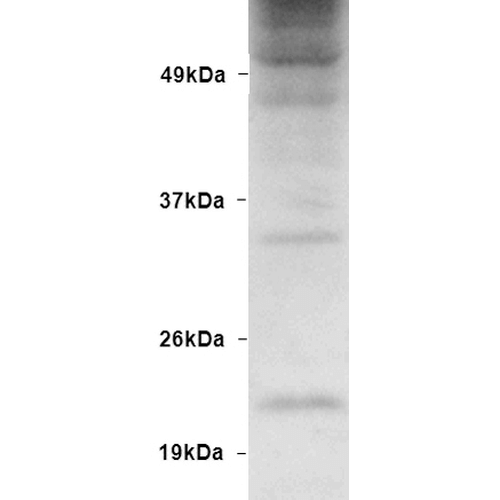
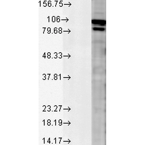
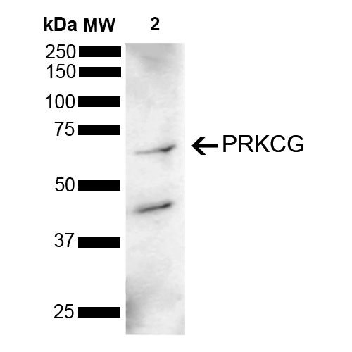
 WB (Western Blot)
(Western blot analysis of Mouse Brain showing detection of 78 kDa PRKCG protein using Rabbit Anti-PRKCG Polyclonal Antibody . Lane 1: Molecular Weight Ladder (MW). Lane 2: Mouse Brain . Load: 15 ug. Block: 5% Skim Milk powder in TBST. Primary Antibody: Rabbit Anti-PRKCG Polyclonal Antibody at 1:1000 for 2 hours at RT with shaking. Secondary Antibody: Goat Anti-Rabbit IgG: HRP at 1:5000 for 1 hour at RT. Color Development: ECL solution for 5 min at RT. Predicted/Observed Size: 78 kDa. Other Band(s): 62 kDa, 40 kDa.)
WB (Western Blot)
(Western blot analysis of Mouse Brain showing detection of 78 kDa PRKCG protein using Rabbit Anti-PRKCG Polyclonal Antibody . Lane 1: Molecular Weight Ladder (MW). Lane 2: Mouse Brain . Load: 15 ug. Block: 5% Skim Milk powder in TBST. Primary Antibody: Rabbit Anti-PRKCG Polyclonal Antibody at 1:1000 for 2 hours at RT with shaking. Secondary Antibody: Goat Anti-Rabbit IgG: HRP at 1:5000 for 1 hour at RT. Color Development: ECL solution for 5 min at RT. Predicted/Observed Size: 78 kDa. Other Band(s): 62 kDa, 40 kDa.)
PKC gamma, Polyclonal Antibody (Cat# AAA253878)

 WB (Western Blot)
(Western blot analysis of Mouse Brain showing detection of 78 kDa PRKCG protein using Rabbit Anti-PRKCG Polyclonal Antibody . Lane 1: Molecular Weight Ladder (MW). Lane 2: Mouse Brain . Load: 15 ug. Block: 5% Skim Milk powder in TBST. Primary Antibody: Rabbit Anti-PRKCG Polyclonal Antibody at 1:1000 for 2 hours at RT with shaking. Secondary Antibody: Goat Anti-Rabbit IgG: HRP at 1:5000 for 1 hour at RT. Color Development: ECL solution for 5 min at RT. Predicted/Observed Size: 78 kDa. Other Band(s): 62 kDa, 40 kDa.)
WB (Western Blot)
(Western blot analysis of Mouse Brain showing detection of 78 kDa PRKCG protein using Rabbit Anti-PRKCG Polyclonal Antibody . Lane 1: Molecular Weight Ladder (MW). Lane 2: Mouse Brain . Load: 15 ug. Block: 5% Skim Milk powder in TBST. Primary Antibody: Rabbit Anti-PRKCG Polyclonal Antibody at 1:1000 for 2 hours at RT with shaking. Secondary Antibody: Goat Anti-Rabbit IgG: HRP at 1:5000 for 1 hour at RT. Color Development: ECL solution for 5 min at RT. Predicted/Observed Size: 78 kDa. Other Band(s): 62 kDa, 40 kDa.)
PKC gamma, Polyclonal Antibody (Cat# AAA253879)

 WB (Western Blot)
(Western blot analysis of Mouse Brain showing detection of 78 kDa PRKCG protein using Rabbit Anti-PRKCG Polyclonal Antibody . Lane 1: Molecular Weight Ladder (MW). Lane 2: Mouse Brain . Load: 15 ug. Block: 5% Skim Milk powder in TBST. Primary Antibody: Rabbit Anti-PRKCG Polyclonal Antibody at 1:1000 for 2 hours at RT with shaking. Secondary Antibody: Goat Anti-Rabbit IgG: HRP at 1:5000 for 1 hour at RT. Color Development: ECL solution for 5 min at RT. Predicted/Observed Size: 78 kDa. Other Band(s): 62 kDa, 40 kDa.)
WB (Western Blot)
(Western blot analysis of Mouse Brain showing detection of 78 kDa PRKCG protein using Rabbit Anti-PRKCG Polyclonal Antibody . Lane 1: Molecular Weight Ladder (MW). Lane 2: Mouse Brain . Load: 15 ug. Block: 5% Skim Milk powder in TBST. Primary Antibody: Rabbit Anti-PRKCG Polyclonal Antibody at 1:1000 for 2 hours at RT with shaking. Secondary Antibody: Goat Anti-Rabbit IgG: HRP at 1:5000 for 1 hour at RT. Color Development: ECL solution for 5 min at RT. Predicted/Observed Size: 78 kDa. Other Band(s): 62 kDa, 40 kDa.)
PKC gamma, Polyclonal Antibody (Cat# AAA253880)

 WB (Western Blot)
(Western blot analysis of Mouse Brain showing detection of 78 kDa PRKCG protein using Rabbit Anti-PRKCG Polyclonal Antibody . Lane 1: Molecular Weight Ladder (MW). Lane 2: Mouse Brain . Load: 15 ug. Block: 5% Skim Milk powder in TBST. Primary Antibody: Rabbit Anti-PRKCG Polyclonal Antibody at 1:1000 for 2 hours at RT with shaking. Secondary Antibody: Goat Anti-Rabbit IgG: HRP at 1:5000 for 1 hour at RT. Color Development: ECL solution for 5 min at RT. Predicted/Observed Size: 78 kDa. Other Band(s): 62 kDa, 40 kDa.)
WB (Western Blot)
(Western blot analysis of Mouse Brain showing detection of 78 kDa PRKCG protein using Rabbit Anti-PRKCG Polyclonal Antibody . Lane 1: Molecular Weight Ladder (MW). Lane 2: Mouse Brain . Load: 15 ug. Block: 5% Skim Milk powder in TBST. Primary Antibody: Rabbit Anti-PRKCG Polyclonal Antibody at 1:1000 for 2 hours at RT with shaking. Secondary Antibody: Goat Anti-Rabbit IgG: HRP at 1:5000 for 1 hour at RT. Color Development: ECL solution for 5 min at RT. Predicted/Observed Size: 78 kDa. Other Band(s): 62 kDa, 40 kDa.)
PKC gamma, Polyclonal Antibody (Cat# AAA253882)

 WB (Western Blot)
(Western blot analysis of Mouse Brain showing detection of 78 kDa PRKCG protein using Rabbit Anti-PRKCG Polyclonal Antibody . Lane 1: Molecular Weight Ladder (MW). Lane 2: Mouse Brain . Load: 15 ug. Block: 5% Skim Milk powder in TBST. Primary Antibody: Rabbit Anti-PRKCG Polyclonal Antibody at 1:1000 for 2 hours at RT with shaking. Secondary Antibody: Goat Anti-Rabbit IgG: HRP at 1:5000 for 1 hour at RT. Color Development: ECL solution for 5 min at RT. Predicted/Observed Size: 78 kDa. Other Band(s): 62 kDa, 40 kDa.)
WB (Western Blot)
(Western blot analysis of Mouse Brain showing detection of 78 kDa PRKCG protein using Rabbit Anti-PRKCG Polyclonal Antibody . Lane 1: Molecular Weight Ladder (MW). Lane 2: Mouse Brain . Load: 15 ug. Block: 5% Skim Milk powder in TBST. Primary Antibody: Rabbit Anti-PRKCG Polyclonal Antibody at 1:1000 for 2 hours at RT with shaking. Secondary Antibody: Goat Anti-Rabbit IgG: HRP at 1:5000 for 1 hour at RT. Color Development: ECL solution for 5 min at RT. Predicted/Observed Size: 78 kDa. Other Band(s): 62 kDa, 40 kDa.)
PKC gamma, Polyclonal Antibody (Cat# AAA253883)
What are Polyclonal Antibodies?
Polyclonal antibodies are antibodies that come from multiple B cell clones of a host animal. The typical hosts used for the majority of polyclonal antibody production are rabbits, goats, sheep, and donkeys. These polyclonal antibodies, once having identified their target, will bind to different epitopes located at different regions or sequences on the same protein/antigen. As a result, they are ideal at locating and binding to the target, even if the target is in very low concentrations (due to many different antibodies being able to bind to the same target molecule, which allows for significant amplification of a downstream signal).
Polyclonal antibodies are typically produced by injecting an antigen into a host animal, which causes the animal’s immune system to attack the foreign antigen by mass generating antibodies against it. After a period of time, serum is collected from the animal and purified using physicochemical fractionation, class-specific affinity purification, and/or antigen-affinity purification.
Key Uses of Polyclonal Antibodies
- Western Blotting: This method is used to find specific proteins in biological samples after separating them by size.
- Immunohistochemistry: IHC helps visualize the location of proteins in tissue sections using various staining techniques.
- ELISA: (Enzyme-Linked Immunosorbent Assay) is typically used to identify specific protein quantities in a sample. ELISAs can be either “Quantitative” or “Qualitative”.
- Flow Cytometry: technique that identifies and measures the specific protein on the surface or inside the cells in a fluid suspension.
- Immunoprecipitation: IP isolates and studies a specific protein from a complex mixture using antibodies.
Why Buy Polyclonal Antibodies from AAA Biotech?
1. Ideal for Various Applications
Our antibodies are generally going to be validated for use in multiple types of assays, including ELISA, Western Blotting, Immunohistochemistry, Immunoprecipitation, amongst others. They are ideal for a wide range of research applications.
2. Rigorous Quality Control
All of the antibodies in our catalog undergo strict quality testing to ensure specificity, sensitivity, and consistent performance. We are confident in the ability of our antibodies to provide you with accurate results.
3. Wide Assortment of Antibodies
Antibodies in are catalog can be found for both common and exotic species, and these antibodies are also available in both conjugated and recombinant forms to suit many diverse experimental needs.
4. Highly Purified
Our antibodies are available in purified forms with over 85% purity, as confirmed by SDS-PAGE. They are also available with tags such as His, Flag, GST, or MBP. We cater to customers worldwide.
FAQ
1. How are polyclonal antibodies produced?
Traditionally, polyclonal antibodies are produced by injecting an antigen into a host animal (such as a rabbit or goat), which then triggers an immune response from the host animal. The animal’s B cells produce antibodies that will recognize different parts of the injected antigen. These antibodies are then collected from the animal’s blood and purified for use.
2. How do polyclonal antibodies differ from monoclonal antibodies?
Polyclonal antibodies are a mix of antibodies that bind to different locations (epitopes) of the same antigen, while monoclonal antibodies are identical and bind to just one specific epitope. This makes polyclonal antibodies more versatile and better at detecting proteins that may be present in low quantities or in altered/modified forms.
3. How should I store polyclonal antibodies?
Polyclonal antibodies should be stored at 4°C for short-term use (up to a few weeks) and at -20°C or -80°C for long-term storage. Avoid repeated freeze-thaw cycles by dividing them into small aliquots. Always check the datasheet for specific storage instructions.