Filters
▼Clonality
▼Type
▼Reactivity
▼Gene Name
▼Isotype
▼Host
▼Application
▼Clone
▼Polyclonal Antibodies
At AAA Biotech also known as AAA Bio or AAABio, we provide a broad range of purified polyclonal antibodies (pAbs) that are able to all be browsed online through our website. Due to their high specificity and strong binding affinity, these antibodies are ideal for wide swathes of research and experimental applications.
Our polyclonal antibodies can easily support your work, whether you use them for Western Blotting, Immunocytochemistry (with or without Immunofluorescence used in conjunction), Immunohistochemistry, Immunoprecipitation, and ELISA tests. We highly encourage you to browse our range of pAbs and choose the one that best suits your experimental model.
Viewing 1600-1650 of 96805 product results
 IHC (Immunohistochemisry)
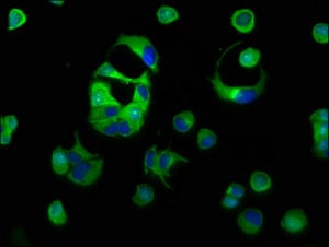
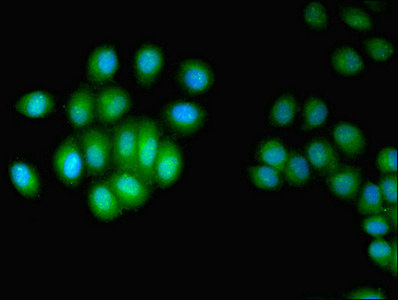
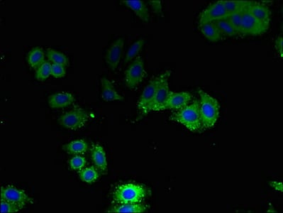
(Immunofluorescent analysis of HepG2 cells using AAA118011 at a dilution of 1:100 and Alexa Fluor 488-congugated AffiniPure Goat Anti-Rabbit IgG(H+L))
IHC (Immunohistochemisry)
(Immunofluorescent analysis of HepG2 cells using AAA118011 at a dilution of 1:100 and Alexa Fluor 488-congugated AffiniPure Goat Anti-Rabbit IgG(H+L))
General transcription factor IIH subunit 5, Polyclonal Antibody (Cat# AAA118011)
 IHC (Immunohistochemisry)
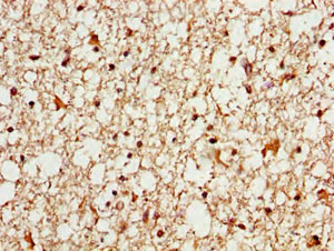
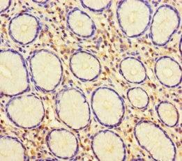
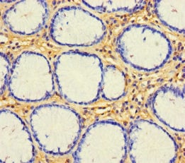
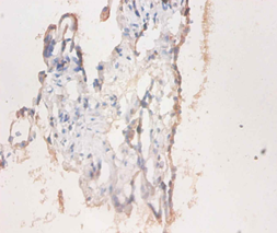
(Immunohistochemistry of paraffin-embedded human placenta tissue using AAA118014 at dilution of 1:100)
IHC (Immunohistochemisry)
(Immunohistochemistry of paraffin-embedded human placenta tissue using AAA118014 at dilution of 1:100)
52 kDa repressor of the inhibitor of the protein kinase, Polyclonal Antibody (Cat# AAA118014)
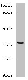
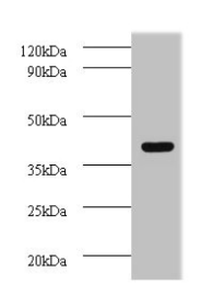
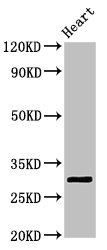 WB (Western Blot)
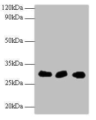
(Western BlotPositive WB detected in: Rat heart tissueAll lanes: Tpsb2 antibody at 2.5ug/mlSecondaryGoat polyclonal to rabbit IgG at 1/50000 dilutionPredicted band size: 31 KDaObserved band size: 31 KDa)
WB (Western Blot)
(Western BlotPositive WB detected in: Rat heart tissueAll lanes: Tpsb2 antibody at 2.5ug/mlSecondaryGoat polyclonal to rabbit IgG at 1/50000 dilutionPredicted band size: 31 KDaObserved band size: 31 KDa)
Tryptase beta-2, Polyclonal Antibody (Cat# AAA118017)
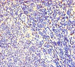
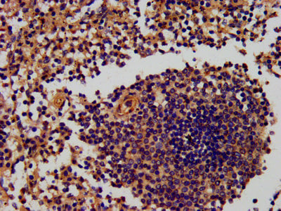 IHC (Immunohiostchemistry)
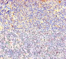
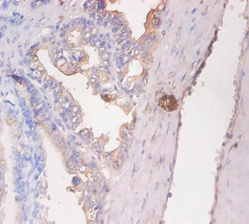
(IHC image of AAA118018 diluted at 1:600 and staining in paraffin-embedded human spleen tissue performed on a Leica BondTM system. After dewaxing and hydration, antigen retrieval was mediated by high pressure in a citrate buffer (pH 6.0). Section was blocked with 10% normal goat serum 30min at RT. Then primary antibody (1% BSA) was incubated at 4 degree C overnight. The primary is detected by a biotinylated secondary antibody and visualized using an HRP conjugated SP system.)
IHC (Immunohiostchemistry)
(IHC image of AAA118018 diluted at 1:600 and staining in paraffin-embedded human spleen tissue performed on a Leica BondTM system. After dewaxing and hydration, antigen retrieval was mediated by high pressure in a citrate buffer (pH 6.0). Section was blocked with 10% normal goat serum 30min at RT. Then primary antibody (1% BSA) was incubated at 4 degree C overnight. The primary is detected by a biotinylated secondary antibody and visualized using an HRP conjugated SP system.)
RING-box protein 2, Polyclonal Antibody (Cat# AAA118018)

 IHC (Immunohistochemisry)
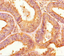
(Immunohistochemistry of paraffin-embedded human stomach tissue using AAA118021 at dilution of 1:100)
IHC (Immunohistochemisry)
(Immunohistochemistry of paraffin-embedded human stomach tissue using AAA118021 at dilution of 1:100)
WD repeat-containing protein 54, Polyclonal Antibody (Cat# AAA118021)
 WB (Western Blot)
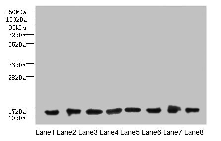
(western blot All lanes: Apolipoprotein C-III antibody at 2ug/ml+rat liver tissue Secondary Goat polyclonal to Rabbit lgG at 1/15000 dilution Predicted band size:11kda Observed band size:11kda)
WB (Western Blot)
(western blot All lanes: Apolipoprotein C-III antibody at 2ug/ml+rat liver tissue Secondary Goat polyclonal to Rabbit lgG at 1/15000 dilution Predicted band size:11kda Observed band size:11kda)
Apolipoprotein C-III, Polyclonal Antibody (Cat# AAA118025)
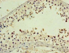
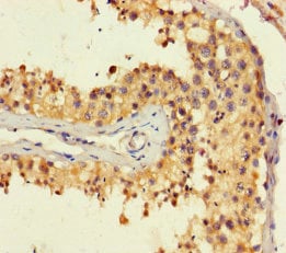 IHC (Immunohiostchemistry)
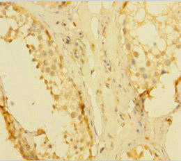
(Immunohistochemistry of paraffin-embedded human testis tissue using AAA118030 at dilution of 1:100)
IHC (Immunohiostchemistry)
(Immunohistochemistry of paraffin-embedded human testis tissue using AAA118030 at dilution of 1:100)
Retrotransposon-derived protein PEG10, Polyclonal Antibody (Cat# AAA118030)
Plasmodium falciparum Apical membrane antigen 1, Polyclonal Antibody (Cat# AAA118032)

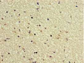
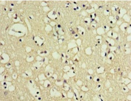
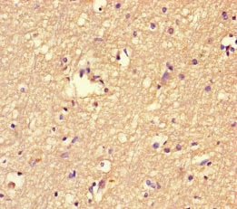
 IHC (Immunohistochemistry)
(IHC image of AAA118036 diluted at1:600 and staining in paraffin-embedded humanbrain tissue performed on a Leica BondTMsystem. After dewaxing and hydration, antigenretrieval was mediated by high pressure in acitrate buffer (pH 6.0). Section was blocked with10% normal goat serum 30min at RT. Thenprimary antibody (1% BSA) was incubated at4°C overnight. The primary is detected by abiotinylated secondary antibody and visualizedtissue using an HRP conjugated SP system)
IHC (Immunohistochemistry)
(IHC image of AAA118036 diluted at1:600 and staining in paraffin-embedded humanbrain tissue performed on a Leica BondTMsystem. After dewaxing and hydration, antigenretrieval was mediated by high pressure in acitrate buffer (pH 6.0). Section was blocked with10% normal goat serum 30min at RT. Thenprimary antibody (1% BSA) was incubated at4°C overnight. The primary is detected by abiotinylated secondary antibody and visualizedtissue using an HRP conjugated SP system)
Myelin-associated glycoprotein, Polyclonal Antibody (Cat# AAA118036)
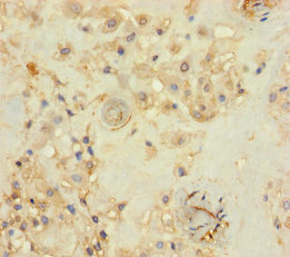
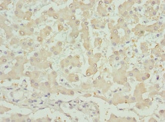
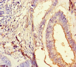
 IHC (Immunohiostchemistry)
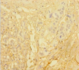
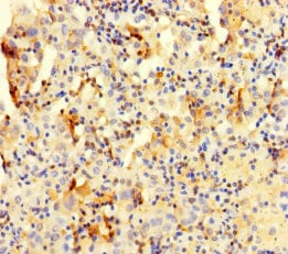
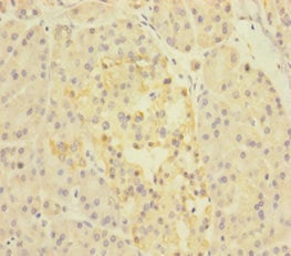
(Immunohistochemistry of paraffin-embedded human pancreatic cancer using AAA118037 at dilution 1:100)
IHC (Immunohiostchemistry)
(Immunohistochemistry of paraffin-embedded human pancreatic cancer using AAA118037 at dilution 1:100)
Kallikrein-1, Polyclonal Antibody (Cat# AAA118037)
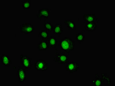
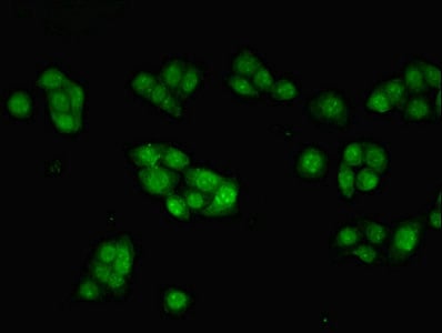
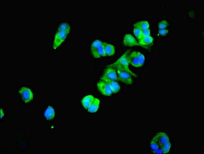 IF (Immunofluorescence)
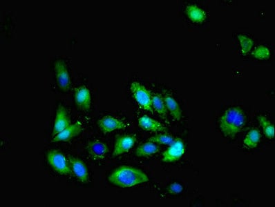
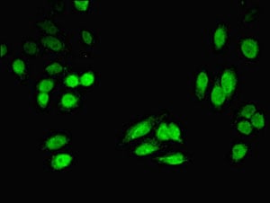
(Immunofluorescent analysis of MCF-7 cells using AAA118040 at a dilution of 1:100 and Alexa Fluor 488-congugated AffiniPure Goat Anti-Rabbit IgG(H+L))
IF (Immunofluorescence)
(Immunofluorescent analysis of MCF-7 cells using AAA118040 at a dilution of 1:100 and Alexa Fluor 488-congugated AffiniPure Goat Anti-Rabbit IgG(H+L))
Proteasome subunit beta type-4, Polyclonal Antibody (Cat# AAA118040)


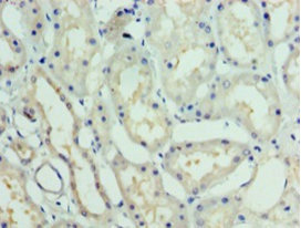
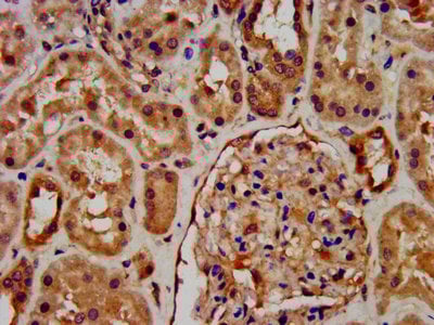
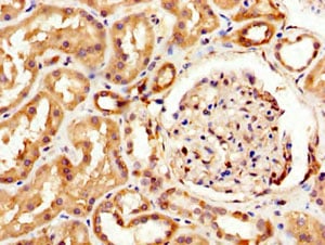
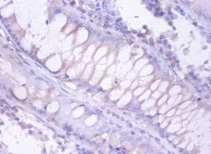
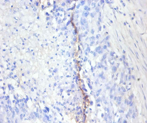
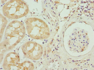
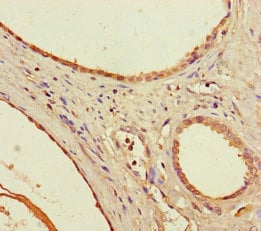
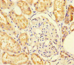 IHC (Immunohistochemisry)
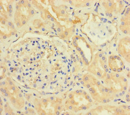
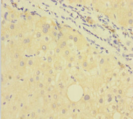
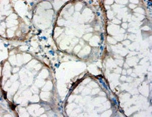
(Immunohistochemistry of paraffin-embedded human kidney tissue using AAA118041 at dilution 1:100)
IHC (Immunohistochemisry)
(Immunohistochemistry of paraffin-embedded human kidney tissue using AAA118041 at dilution 1:100)
Sulfite oxidase, Polyclonal Antibody (Cat# AAA118041)
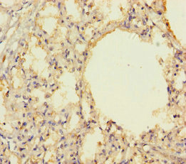
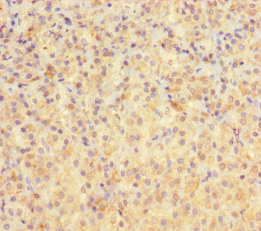
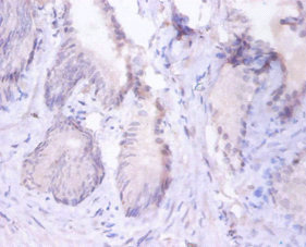
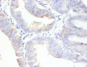
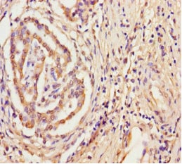
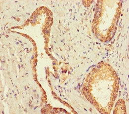 IHC (Immunohistochemisry)
(Immunohistochemistry of paraffin-embedded human prostate cancer using AAA118042 at dilution of 1:100)
IHC (Immunohistochemisry)
(Immunohistochemistry of paraffin-embedded human prostate cancer using AAA118042 at dilution of 1:100)
Ubiquitin-conjugating enzyme E2 Q2, Polyclonal Antibody (Cat# AAA118042)
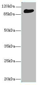
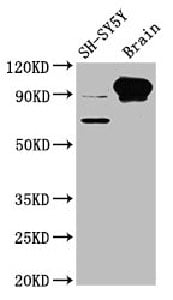
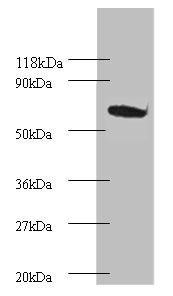
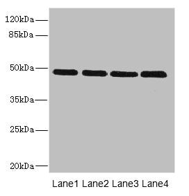
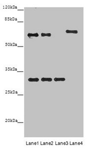
 WB (Western Blot)
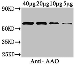
(Western BlotPositive WB detected in: Cucurbita maxima(40ug,20ug,10ug,5ug)All lanes: AAO antibody at 3ug/mlSecondaryGoat polyclonal to rabbit IgG at 1/50000 dilutionPredicted band size: 65 kDaObserved band size: 65 kDa)
WB (Western Blot)
(Western BlotPositive WB detected in: Cucurbita maxima(40ug,20ug,10ug,5ug)All lanes: AAO antibody at 3ug/mlSecondaryGoat polyclonal to rabbit IgG at 1/50000 dilutionPredicted band size: 65 kDaObserved band size: 65 kDa)
Cucurbita maxima L-ascorbate oxidase, Polyclonal Antibody (Cat# AAA118043)
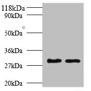
 WB (Western Blot)
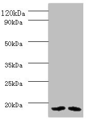
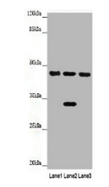
(Western blotAll lanes: Mitochondrial import inner membrane translocase subunit TIM16 antibody at 2ug/mlLane 1:HL-60 whole cell lysateLane 2:HepG2 whole cell lysateSecondaryGoat polyclonal to rabbit at 1/10000 dilutionPredicted band size: 14kDaObserved band size: 14kDa)
WB (Western Blot)
(Western blotAll lanes: Mitochondrial import inner membrane translocase subunit TIM16 antibody at 2ug/mlLane 1:HL-60 whole cell lysateLane 2:HepG2 whole cell lysateSecondaryGoat polyclonal to rabbit at 1/10000 dilutionPredicted band size: 14kDaObserved band size: 14kDa)
Mitochondrial import inner membrane translocase subunit TIM16, Polyclonal Antibody (Cat# AAA118046)
Betatrophin, Polyclonal Antibody (Cat# AAA118054)
Apolipoprotein A-I, Polyclonal Antibody (Cat# AAA118057)
 IHC (Immunohistochemisry)
(Immunofluorescent analysis of A549 cells using AAA118058 at a dilution of 1:100 and Alexa Fluor 488-congugated AffiniPure Goat Anti-Rabbit IgG(H+L))
IHC (Immunohistochemisry)
(Immunofluorescent analysis of A549 cells using AAA118058 at a dilution of 1:100 and Alexa Fluor 488-congugated AffiniPure Goat Anti-Rabbit IgG(H+L))
Checkpoint protein HUS1, Polyclonal Antibody (Cat# AAA118058)
 IHC (Immunohistochemisry)
(Immunofluorescent analysis of Hela cells cells using AAA118059 at a dilution of 1:100 and Alexa Fluor 488-congugated AffiniPure Goat Anti-Rabbit IgG(H+L))
IHC (Immunohistochemisry)
(Immunofluorescent analysis of Hela cells cells using AAA118059 at a dilution of 1:100 and Alexa Fluor 488-congugated AffiniPure Goat Anti-Rabbit IgG(H+L))
Fractalkine, Polyclonal Antibody (Cat# AAA118059)
 IHC (Immunohiostchemistry)
(Immunohistochemistry of paraffin-embedded human bladder carcinoma at dilution 1:100)
IHC (Immunohiostchemistry)
(Immunohistochemistry of paraffin-embedded human bladder carcinoma at dilution 1:100)
Antigen KI-67, Polyclonal Antibody (Cat# AAA118061)
Complement C3, Polyclonal Antibody (Cat# AAA118062)
 IF (Immunofluorescence)
(Immunofluorescent analysis of PC3 cells using AAA118064 at a dilution of 1:100 and Alexa Fluor 488-congugated AffiniPure Goat Anti-Rabbit IgG(H+L))
IF (Immunofluorescence)
(Immunofluorescent analysis of PC3 cells using AAA118064 at a dilution of 1:100 and Alexa Fluor 488-congugated AffiniPure Goat Anti-Rabbit IgG(H+L))
Inhibin beta B chain, Polyclonal Antibody (Cat# AAA118064)
Mycobacterium tuberculosis Phosphate-binding protein PstS 1, Polyclonal Antibody (Cat# AAA118065)
IgG receptor FcRn large subunit p51, Polyclonal Antibody (Cat# AAA118067)
Myosin-binding protein C, Polyclonal Antibody (Cat# AAA118070)
 IHC (Immunohiostchemistry)
(Immunohistochemistry of paraffin-embedded human brain tissue using AAA118071 at dilution 1:100)
IHC (Immunohiostchemistry)
(Immunohistochemistry of paraffin-embedded human brain tissue using AAA118071 at dilution 1:100)
Paraneoplastic antigen-like protein 5, Polyclonal Antibody (Cat# AAA118071)
 IHC (Immunohistochemisry)
(Immunohistochemistry of paraffin-embedded human lung cancer using AAA118077 at dilution of 1:100)
IHC (Immunohistochemisry)
(Immunohistochemistry of paraffin-embedded human lung cancer using AAA118077 at dilution of 1:100)
Coactosin-like protein, Polyclonal Antibody (Cat# AAA118077)
Interleukin-8, Polyclonal Antibody (Cat# AAA118078)
 IHC (Immunohiostchemistry)
(Immunohistochemistry of paraffin-embedded human pancreatic tissue using AAA118080 at dilution 1:100)
IHC (Immunohiostchemistry)
(Immunohistochemistry of paraffin-embedded human pancreatic tissue using AAA118080 at dilution 1:100)
Growth/differentiation factor 9, Polyclonal Antibody (Cat# AAA118080)
 IHC (Immunohistochemisry)
(Immunofluorescent analysis of HepG2 cells using AAA117787 at a dilution of 1:100 and Alexa Fluor 488-congugated AffiniPure Goat Anti-Rabbit IgG(H+L))
IHC (Immunohistochemisry)
(Immunofluorescent analysis of HepG2 cells using AAA117787 at a dilution of 1:100 and Alexa Fluor 488-congugated AffiniPure Goat Anti-Rabbit IgG(H+L))
4-hydroxyphenylpyruvate dioxygenase, Polyclonal Antibody (Cat# AAA117787)
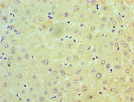
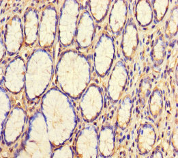
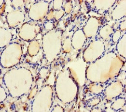
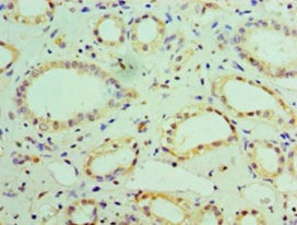 IHC (Immunohiostchemistry)
(Immunohistochemistry of paraffin-embedded human kidney tissue using AAA117792 at dilution 1:100)
IHC (Immunohiostchemistry)
(Immunohistochemistry of paraffin-embedded human kidney tissue using AAA117792 at dilution 1:100)
NADH dehydrogenase [ubiquinone] 1 beta subcomplex subunit 7, Polyclonal Antibody (Cat# AAA117792)
Vitellogenin-2, Polyclonal Antibody (Cat# AAA117796)
Nuclear pore membrane glycoprotein 210, Polyclonal Antibody (Cat# AAA117800)
 IHC (Immunohistochemisry)
(Immunofluorescent analysis of MCF-7 cells using AAA117804 at a dilution of 1:100 and Alexa Fluor 488-congugated AffiniPure Goat Anti-Rabbit IgG(H+L))
IHC (Immunohistochemisry)
(Immunofluorescent analysis of MCF-7 cells using AAA117804 at a dilution of 1:100 and Alexa Fluor 488-congugated AffiniPure Goat Anti-Rabbit IgG(H+L))
Lipopolysaccharide-responsive and beige-like anchor protein, Polyclonal Antibody (Cat# AAA117804)
 IHC (Immunohiostchemistry)
(Immunohistochemistry of paraffin-embedded human melanoma cancer using AAA117805 at dilution of 1:100)
IHC (Immunohiostchemistry)
(Immunohistochemistry of paraffin-embedded human melanoma cancer using AAA117805 at dilution of 1:100)
Protein FAM114A2, Polyclonal Antibody (Cat# AAA117805)

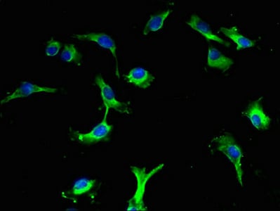
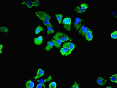
 IF (Immunofluorescence)
(Immunofluorescent analysis of HepG2 cells using AAA117808 at a dilution of 1:100 and Alexa Fluor 488-congugated AffiniPure Goat Anti-Rabbit IgG(H+L))
IF (Immunofluorescence)
(Immunofluorescent analysis of HepG2 cells using AAA117808 at a dilution of 1:100 and Alexa Fluor 488-congugated AffiniPure Goat Anti-Rabbit IgG(H+L))
Aquaporin-1, Polyclonal Antibody (Cat# AAA117808)
Flagellin, Polyclonal Antibody (Cat# AAA117810)
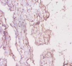
 IHC (Immunohiostchemistry)
(Immunohistochemistry of paraffin-embedded human bladder carcinoma at dilution 1:100)
IHC (Immunohiostchemistry)
(Immunohistochemistry of paraffin-embedded human bladder carcinoma at dilution 1:100)
CD59 glycoprotein, Polyclonal Antibody (Cat# AAA117811)
 IHC (Immunohiostchemistry)
(Immunohistochemistry of paraffin-embedded human pancreatic cancer using AAA117813 at dilution 1:100)
IHC (Immunohiostchemistry)
(Immunohistochemistry of paraffin-embedded human pancreatic cancer using AAA117813 at dilution 1:100)
Protein-tyrosine sulfotransferase 2, Polyclonal Antibody (Cat# AAA117813)
Periostin, Polyclonal Antibody (Cat# AAA117815)
 IHC (Immunohistochemisry)
(Immunohistochemistry of paraffin-embedded human prostate cancer using AAA117819 at dilution of 1:100)
IHC (Immunohistochemisry)
(Immunohistochemistry of paraffin-embedded human prostate cancer using AAA117819 at dilution of 1:100)
Homeobox protein OTX2, Polyclonal Antibody (Cat# AAA117819)
N-acetylgalactosamine-6-sulfatase, Polyclonal Antibody (Cat# AAA117821)
Epiplakin, Polyclonal Antibody (Cat# AAA117828)
subsp Parvalbumin beta, Polyclonal Antibody (Cat# AAA117830)
Salmonella typhi Outer membrane protein A, Polyclonal Antibody (Cat# AAA117834)
cAMP-activated global transcriptional regulator CRP, Polyclonal Antibody (Cat# AAA117835)
Tryptase beta-2, Polyclonal Antibody (Cat# AAA117837)
 IHC (Immunohistochemisry)
(Immunofluorescent analysis of A549 cells using AAA117838 at a dilution of 1:100 and Alexa Fluor 488-congugated AffiniPure Goat Anti-Rabbit IgG(H+L))
IHC (Immunohistochemisry)
(Immunofluorescent analysis of A549 cells using AAA117838 at a dilution of 1:100 and Alexa Fluor 488-congugated AffiniPure Goat Anti-Rabbit IgG(H+L))
F-box/LRR-repeat protein 18, Polyclonal Antibody (Cat# AAA117838)
HLA class II histocompatibility antigen, DQ alpha 1 chain, Polyclonal Antibody (Cat# AAA117839)
Scavenger receptor cysteine-rich type 1 protein M130, Polyclonal Antibody (Cat# AAA117842)
What are Polyclonal Antibodies?
Polyclonal antibodies are antibodies that come from multiple B cell clones of a host animal. The typical hosts used for the majority of polyclonal antibody production are rabbits, goats, sheep, and donkeys. These polyclonal antibodies, once having identified their target, will bind to different epitopes located at different regions or sequences on the same protein/antigen. As a result, they are ideal at locating and binding to the target, even if the target is in very low concentrations (due to many different antibodies being able to bind to the same target molecule, which allows for significant amplification of a downstream signal).
Polyclonal antibodies are typically produced by injecting an antigen into a host animal, which causes the animal’s immune system to attack the foreign antigen by mass generating antibodies against it. After a period of time, serum is collected from the animal and purified using physicochemical fractionation, class-specific affinity purification, and/or antigen-affinity purification.
Key Uses of Polyclonal Antibodies
- Western Blotting: This method is used to find specific proteins in biological samples after separating them by size.
- Immunohistochemistry: IHC helps visualize the location of proteins in tissue sections using various staining techniques.
- ELISA: (Enzyme-Linked Immunosorbent Assay) is typically used to identify specific protein quantities in a sample. ELISAs can be either “Quantitative” or “Qualitative”.
- Flow Cytometry: technique that identifies and measures the specific protein on the surface or inside the cells in a fluid suspension.
- Immunoprecipitation: IP isolates and studies a specific protein from a complex mixture using antibodies.
Why Buy Polyclonal Antibodies from AAA Biotech?
1. Ideal for Various Applications
Our antibodies are generally going to be validated for use in multiple types of assays, including ELISA, Western Blotting, Immunohistochemistry, Immunoprecipitation, amongst others. They are ideal for a wide range of research applications.
2. Rigorous Quality Control
All of the antibodies in our catalog undergo strict quality testing to ensure specificity, sensitivity, and consistent performance. We are confident in the ability of our antibodies to provide you with accurate results.
3. Wide Assortment of Antibodies
Antibodies in are catalog can be found for both common and exotic species, and these antibodies are also available in both conjugated and recombinant forms to suit many diverse experimental needs.
4. Highly Purified
Our antibodies are available in purified forms with over 85% purity, as confirmed by SDS-PAGE. They are also available with tags such as His, Flag, GST, or MBP. We cater to customers worldwide.
FAQ
1. How are polyclonal antibodies produced?
Traditionally, polyclonal antibodies are produced by injecting an antigen into a host animal (such as a rabbit or goat), which then triggers an immune response from the host animal. The animal’s B cells produce antibodies that will recognize different parts of the injected antigen. These antibodies are then collected from the animal’s blood and purified for use.
2. How do polyclonal antibodies differ from monoclonal antibodies?
Polyclonal antibodies are a mix of antibodies that bind to different locations (epitopes) of the same antigen, while monoclonal antibodies are identical and bind to just one specific epitope. This makes polyclonal antibodies more versatile and better at detecting proteins that may be present in low quantities or in altered/modified forms.
3. How should I store polyclonal antibodies?
Polyclonal antibodies should be stored at 4°C for short-term use (up to a few weeks) and at -20°C or -80°C for long-term storage. Avoid repeated freeze-thaw cycles by dividing them into small aliquots. Always check the datasheet for specific storage instructions.