Filters
▼Clonality
▼Type
▼Reactivity
▼Gene Name
▼Isotype
▼Host
▼Application
▼Clone
▼Polyclonal Antibodies
At AAA Biotech also known as AAA Bio or AAABio, we provide a broad range of purified polyclonal antibodies (pAbs) that are able to all be browsed online through our website. Due to their high specificity and strong binding affinity, these antibodies are ideal for wide swathes of research and experimental applications.
Our polyclonal antibodies can easily support your work, whether you use them for Western Blotting, Immunocytochemistry (with or without Immunofluorescence used in conjunction), Immunohistochemistry, Immunoprecipitation, and ELISA tests. We highly encourage you to browse our range of pAbs and choose the one that best suits your experimental model.
Viewing 1550-1600 of 96805 product results
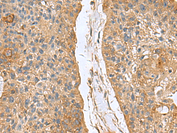
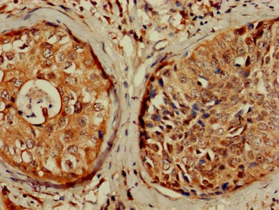
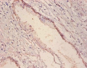
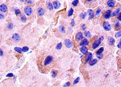
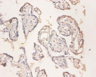
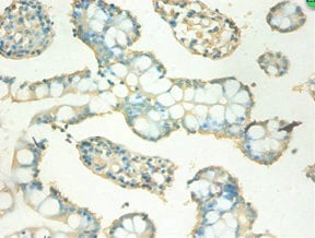
 IHC (Immunohiostchemistry)
(Immunohistochemistry of paraffin-embedded Human cervical cancer tissue using GCSH Polyclonal Antibody at dilution of 1:60(×200))
IHC (Immunohiostchemistry)
(Immunohistochemistry of paraffin-embedded Human cervical cancer tissue using GCSH Polyclonal Antibody at dilution of 1:60(×200))
GCSH, Polyclonal Antibody (Cat# AAA176661)

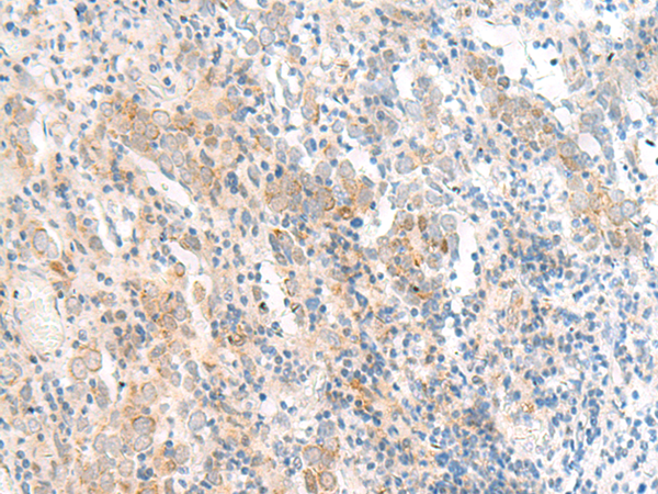
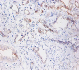
 IHC (Immunohiostchemistry)
(Immunohistochemistry of paraffin-embedded Human colorectal cancer tissue using RNF208 Polyclonal Antibody at dilution of 1:55(×200))
IHC (Immunohiostchemistry)
(Immunohistochemistry of paraffin-embedded Human colorectal cancer tissue using RNF208 Polyclonal Antibody at dilution of 1:55(×200))
RNF208, Polyclonal Antibody (Cat# AAA176666)

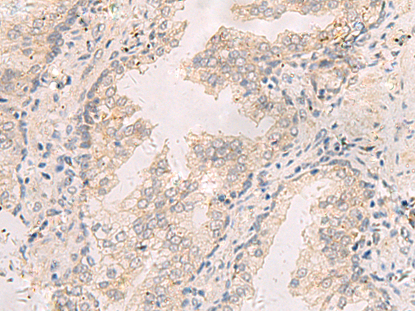
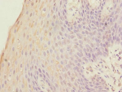
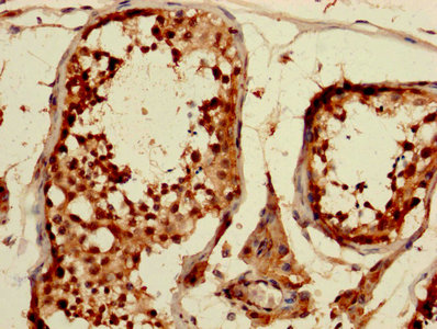
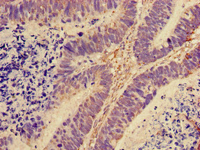
 IHC (Immunohistochemisry)
(Immunohistochemistry of paraffin-embedded Human colorectal cancer tissue using HPCAL1 Polyclonal Antibody at dilution of 1:90(×200))
IHC (Immunohistochemisry)
(Immunohistochemistry of paraffin-embedded Human colorectal cancer tissue using HPCAL1 Polyclonal Antibody at dilution of 1:90(×200))
HPCAL1, Polyclonal Antibody (Cat# AAA176670)
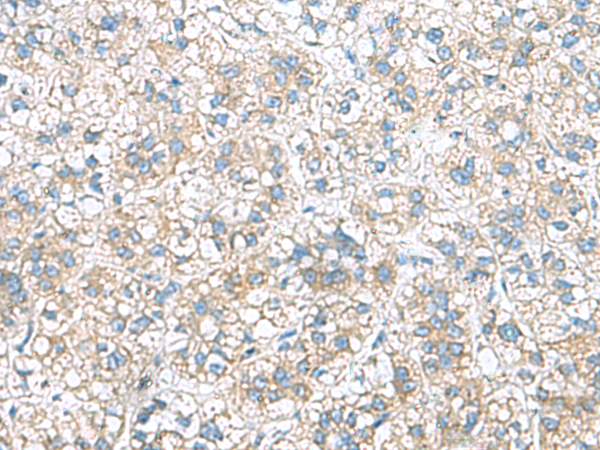
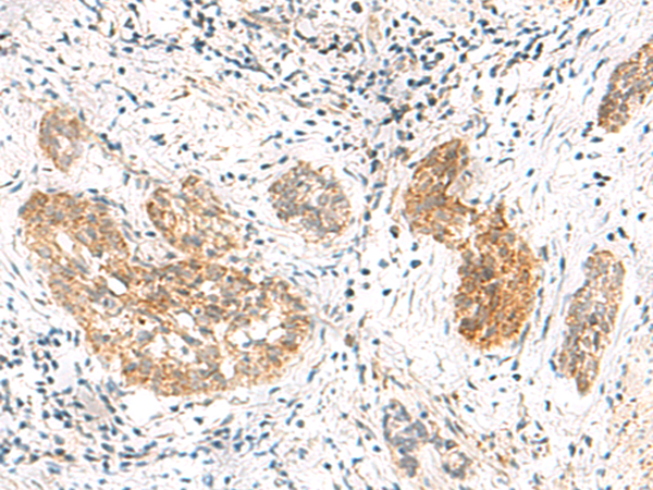
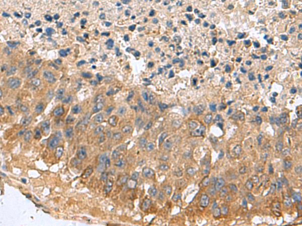 IHC (Immunohiostchemistry)
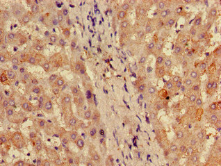
(Immunohistochemistry of paraffin-embedded Human liver cancer tissue using MARCKSL1 Polyclonal Antibody at dilution of 1:70(×200))
IHC (Immunohiostchemistry)
(Immunohistochemistry of paraffin-embedded Human liver cancer tissue using MARCKSL1 Polyclonal Antibody at dilution of 1:70(×200))
MARCKSL1, Polyclonal Antibody (Cat# AAA176671)
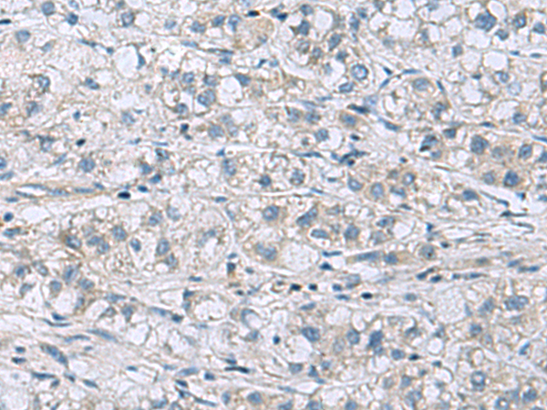
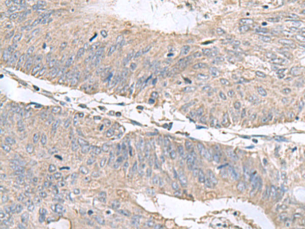
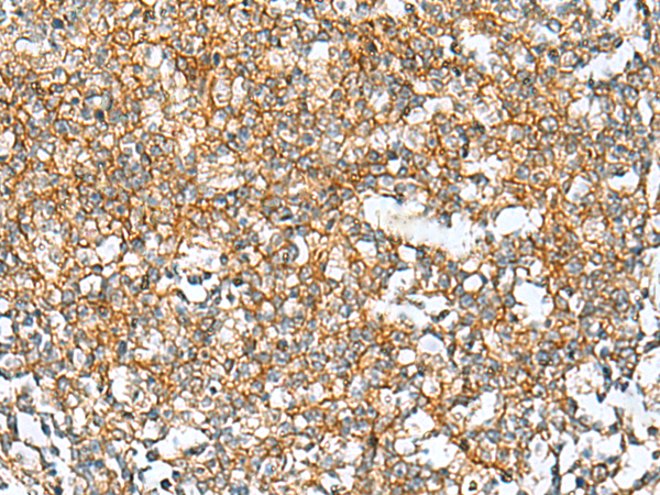
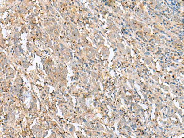
 IHC (Immunohiostchemistry)
(Immunohistochemistry of paraffin-embedded Human gastric cancer tissue using ITGB1BP1 Polyclonal Antibody at dilution of 1:60(×200))
IHC (Immunohiostchemistry)
(Immunohistochemistry of paraffin-embedded Human gastric cancer tissue using ITGB1BP1 Polyclonal Antibody at dilution of 1:60(×200))
ITGB1BP1, Polyclonal Antibody (Cat# AAA176673)
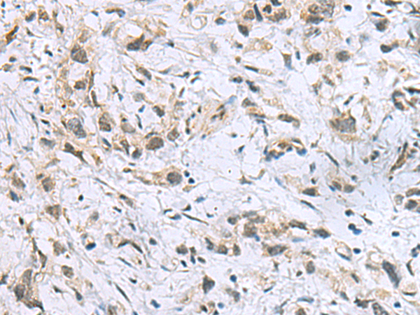
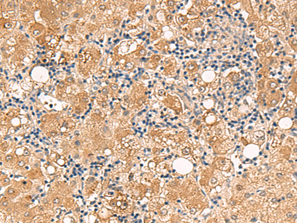
 IHC (Immunohiostchemistry)
(Immunohistochemistry of paraffin-embedded Human liver cancer tissue using RAB13 Polyclonal Antibody at dilution of 1:50(×200))
IHC (Immunohiostchemistry)
(Immunohistochemistry of paraffin-embedded Human liver cancer tissue using RAB13 Polyclonal Antibody at dilution of 1:50(×200))
RAB13, Polyclonal Antibody (Cat# AAA176677)
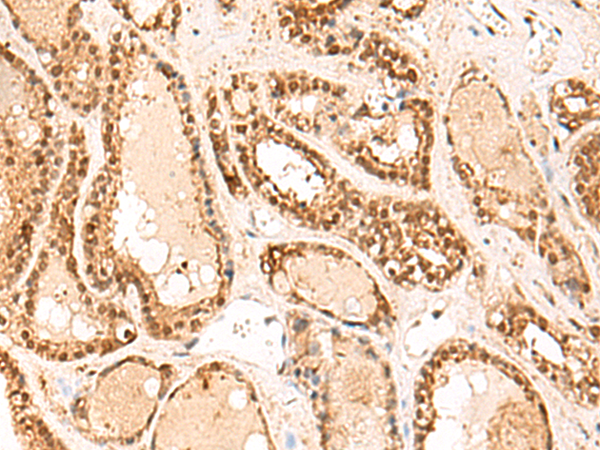
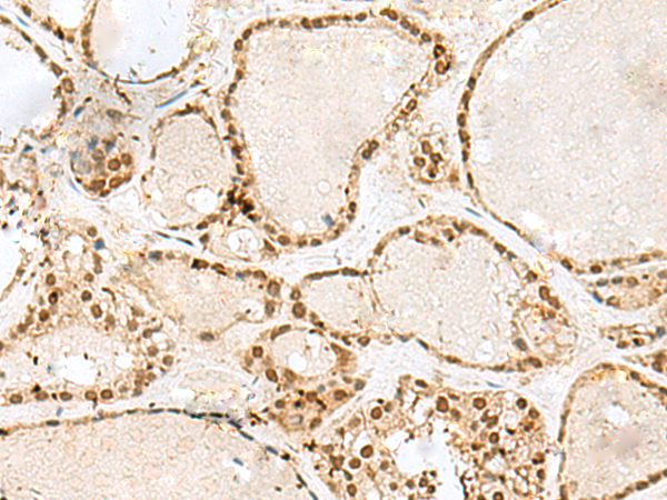
 IHC (Immunohiostchemistry)
(Immunohistochemistry of paraffin-embedded Human thyroid cancer tissue using ATP5O Polyclonal Antibody at dilution of 1:90(×200))
IHC (Immunohiostchemistry)
(Immunohistochemistry of paraffin-embedded Human thyroid cancer tissue using ATP5O Polyclonal Antibody at dilution of 1:90(×200))
ATP5O, Polyclonal Antibody (Cat# AAA176684)
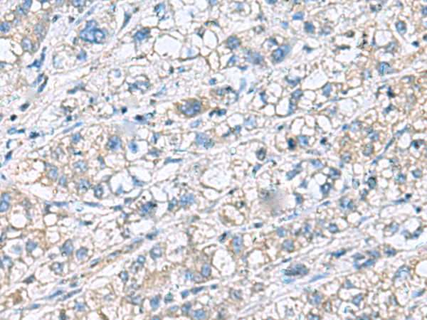 IHC (Immunohiostchemistry)
(Immunohistochemistry of paraffin-embedded Human liver cancer tissue using KRCC1 Polyclonal Antibody at dilution of 1:60(×200))
IHC (Immunohiostchemistry)
(Immunohistochemistry of paraffin-embedded Human liver cancer tissue using KRCC1 Polyclonal Antibody at dilution of 1:60(×200))
KRCC1, Polyclonal Antibody (Cat# AAA176709)
 IHC (Immunohistochemisry)
(Immunohistochemistry of paraffin-embedded Human liver cancer tissue using TNNT1 Polyclonal Antibody at dilution of 1:35(×200))
IHC (Immunohistochemisry)
(Immunohistochemistry of paraffin-embedded Human liver cancer tissue using TNNT1 Polyclonal Antibody at dilution of 1:35(×200))
TNNT1, Polyclonal Antibody (Cat# AAA176710)
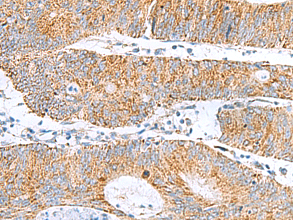
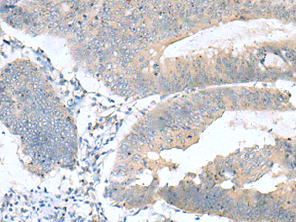
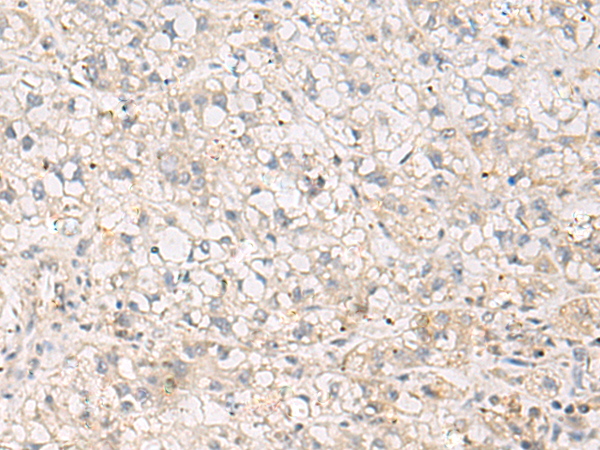
 IHC (Immunohiostchemistry)
(Immunohistochemistry of paraffin-embedded Human cervical cancer tissue using MRPL50 Polyclonal Antibody at dilution of 1:35(×200))
IHC (Immunohiostchemistry)
(Immunohistochemistry of paraffin-embedded Human cervical cancer tissue using MRPL50 Polyclonal Antibody at dilution of 1:35(×200))
MRPL50, Polyclonal Antibody (Cat# AAA176512)
 IHC (Immunohiostchemistry)
(Immunohistochemistry of paraffin-embedded Human thyroid cancer tissue using FUNDC2 Polyclonal Antibody at dilution of 1:60(×200))
IHC (Immunohiostchemistry)
(Immunohistochemistry of paraffin-embedded Human thyroid cancer tissue using FUNDC2 Polyclonal Antibody at dilution of 1:60(×200))
FUNDC2, Polyclonal Antibody (Cat# AAA176523)
 IHC (Immunohiostchemistry)
(Immunohistochemistry of paraffin-embedded Human cervical cancer tissue using FLOT2 Polyclonal Antibody at dilution of 1:50(×200))
IHC (Immunohiostchemistry)
(Immunohistochemistry of paraffin-embedded Human cervical cancer tissue using FLOT2 Polyclonal Antibody at dilution of 1:50(×200))
FLOT2, Polyclonal Antibody (Cat# AAA176464)
 IHC (Immunohiostchemistry)
(Immunohistochemistry of paraffin-embedded Human cervical cancer tissue using GTF3C2 Polyclonal Antibody at dilution of 1:45(×200))
IHC (Immunohiostchemistry)
(Immunohistochemistry of paraffin-embedded Human cervical cancer tissue using GTF3C2 Polyclonal Antibody at dilution of 1:45(×200))
GTF3C2, Polyclonal Antibody (Cat# AAA176478)
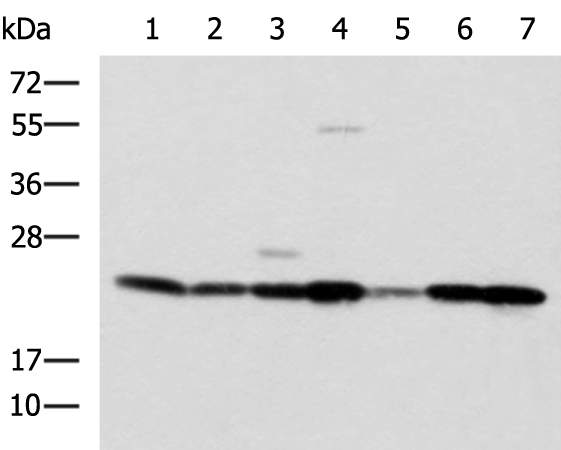
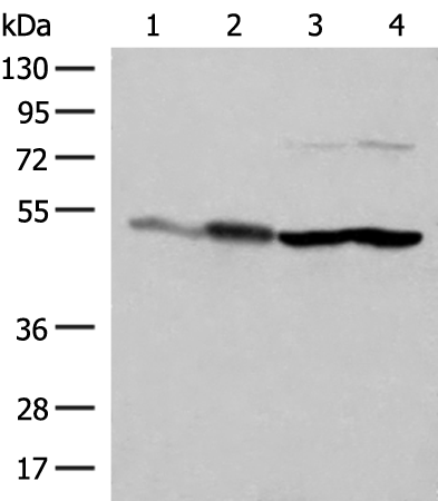
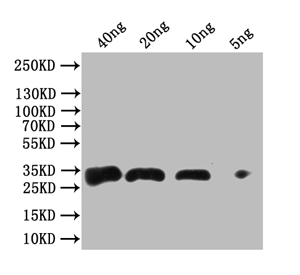
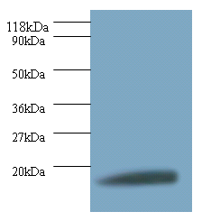
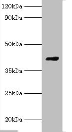
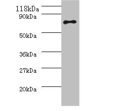
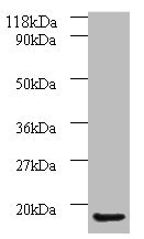 WB (Western Blot)
(Western blotAll lanes: AP-1 complex subunit sigma-3 antibody at 2ug/ml+A549 whole cell lysateSecondaryGoat polyclonal to Rabbit IgG at 1/10000 dilutionPredicted band size: 19,20,13 kDaObserved band size: 19 kDa)
WB (Western Blot)
(Western blotAll lanes: AP-1 complex subunit sigma-3 antibody at 2ug/ml+A549 whole cell lysateSecondaryGoat polyclonal to Rabbit IgG at 1/10000 dilutionPredicted band size: 19,20,13 kDaObserved band size: 19 kDa)
AP-1 complex subunit sigma-3, Polyclonal Antibody (Cat# AAA118081)
S-ribosylhomocysteine lyase, Polyclonal Antibody (Cat# AAA118082)
Angiopoietin-2, Polyclonal Antibody (Cat# AAA118083)
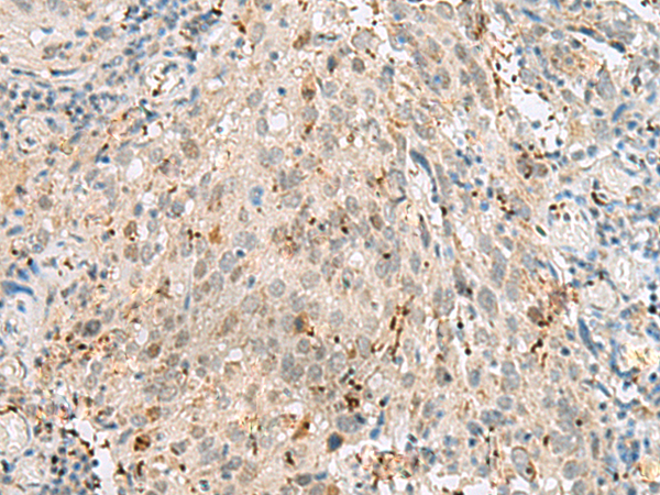
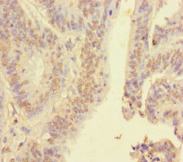
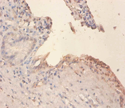
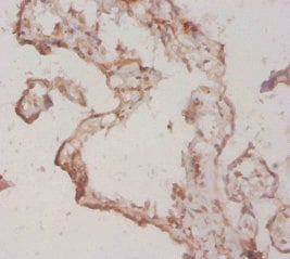
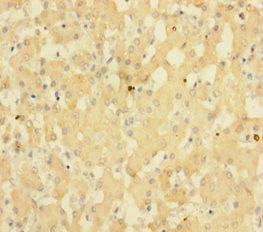
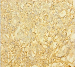
 IHC (Immunohiostchemistry)
(Immunohistochemistry of paraffin-embedded human bladder carcinoma at dilution 1:100)
IHC (Immunohiostchemistry)
(Immunohistochemistry of paraffin-embedded human bladder carcinoma at dilution 1:100)
Antigen KI-67, Polyclonal Antibody (Cat# AAA118084)
 WB (Western Blot)
(Positive WB detected in Recombinant Mycobacterium tuberculosis Antigen 85-A proteinAll lanes: Antigen 85-A antibody at 1:1000SecondaryGoat polyclonal to rabbit IgG at 1/50000 dilutionPredicted band size: 34 kDaObserved band size: 34 kDa)
WB (Western Blot)
(Positive WB detected in Recombinant Mycobacterium tuberculosis Antigen 85-A proteinAll lanes: Antigen 85-A antibody at 1:1000SecondaryGoat polyclonal to rabbit IgG at 1/50000 dilutionPredicted band size: 34 kDaObserved band size: 34 kDa)
Mycobacterium tuberculosis Antigen 85-A, Polyclonal Antibody (Cat# AAA118088)

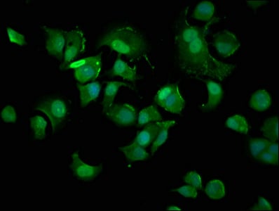
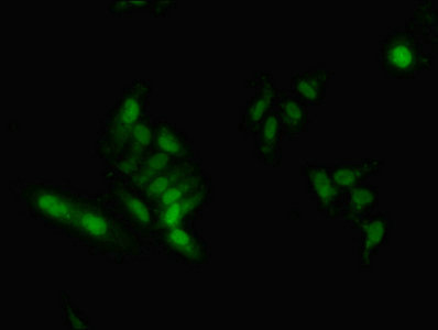 IHC (Immunohistochemisry)
(Immunofluorescent analysis of Hela cells using AAA118095 at a dilution of 1:100 and Alexa Fluor 488-congugated AffiniPure Goat Anti-Rabbit IgG(H+L))
IHC (Immunohistochemisry)
(Immunofluorescent analysis of Hela cells using AAA118095 at a dilution of 1:100 and Alexa Fluor 488-congugated AffiniPure Goat Anti-Rabbit IgG(H+L))
Heterogeneous nuclear ribonucleoproteins A2/B1, Polyclonal Antibody (Cat# AAA118095)
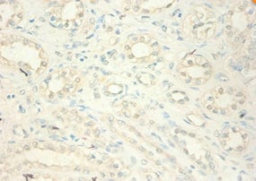
 IHC (Immunohiostchemistry)
(Immunohistochemistry of paraffin-embedded human kidney tissue using AAA118096 at dilution of 1:100)
IHC (Immunohiostchemistry)
(Immunohistochemistry of paraffin-embedded human kidney tissue using AAA118096 at dilution of 1:100)
Lutropin subunit beta, Polyclonal Antibody (Cat# AAA118096)
 ChIP (Chromatin Immunoprecipitation)
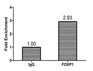
(Chromatin Immunoprecipitation Hela(1.1^106)were cross-linked with formaldehyde, sonicated, and immunoprecipitated with 4ug anti-FOXP1 or a control normal rabbit IgG . The resulting ChIP DNA was quantified using real-time PCR with primers against the HSP90B promoter.)
ChIP (Chromatin Immunoprecipitation)
(Chromatin Immunoprecipitation Hela(1.1^106)were cross-linked with formaldehyde, sonicated, and immunoprecipitated with 4ug anti-FOXP1 or a control normal rabbit IgG . The resulting ChIP DNA was quantified using real-time PCR with primers against the HSP90B promoter.)
Forkhead box protein P1, Polyclonal Antibody (Cat# AAA118098)
 IHC (Immunohistochemisry)
(Immunohistochemistry of paraffin-embedded human spleen tissue using AAA118101 at dilution of 1:100)
IHC (Immunohistochemisry)
(Immunohistochemistry of paraffin-embedded human spleen tissue using AAA118101 at dilution of 1:100)
Talin-2, Polyclonal Antibody (Cat# AAA118101)
 IHC (Immunohistochemisry)
(Immunofluorescence staining of MCF-7 cells with AAA118102 at 1:400,counter-stained with DAPI. The cells were fixed in 4% formaldehyde, permeabilized using 0.2% Triton X-100 and blocked in 10% normal Goat Serum. The cells were then incubated with the antibody overnight at 4 degree C.The secondary antibody was Alexa Fluor 488-congugated AffiniPure Goat Anti-Rabbit IgG (H+L).)
IHC (Immunohistochemisry)
(Immunofluorescence staining of MCF-7 cells with AAA118102 at 1:400,counter-stained with DAPI. The cells were fixed in 4% formaldehyde, permeabilized using 0.2% Triton X-100 and blocked in 10% normal Goat Serum. The cells were then incubated with the antibody overnight at 4 degree C.The secondary antibody was Alexa Fluor 488-congugated AffiniPure Goat Anti-Rabbit IgG (H+L).)
Cyclin-dependent kinase inhibitor 1, Polyclonal Antibody (Cat# AAA118102)
 IHC (Immunohiostchemistry)
(Immunohistochemistry of paraffin embedded human prostate using AAA118104 at dilution of 1:100)
IHC (Immunohiostchemistry)
(Immunohistochemistry of paraffin embedded human prostate using AAA118104 at dilution of 1:100)
Alpha-galactosidase A, Polyclonal Antibody (Cat# AAA118104)
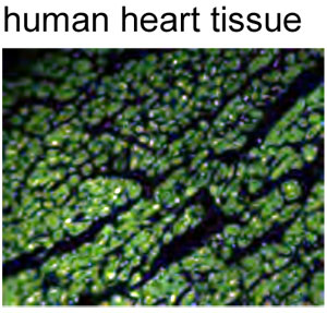 IF (Immunofluorescence)
(anti-human Myoglobin polyclonal Antibody,FITC conjugatedPrimary antibodiesPositive IF detected in human heart tissue)
IF (Immunofluorescence)
(anti-human Myoglobin polyclonal Antibody,FITC conjugatedPrimary antibodiesPositive IF detected in human heart tissue)
Myoglobin conjugated, Polyclonal Antibody (Cat# AAA118106)
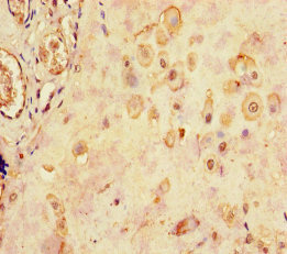
 IHC (Immunohistochemisry)
(Immunohistochemistry of paraffin-embedded mouse brain using AAA118108 at dilution 1:50)
IHC (Immunohistochemisry)
(Immunohistochemistry of paraffin-embedded mouse brain using AAA118108 at dilution 1:50)
Cyclic AMP-responsive element-binding protein 1, Polyclonal Antibody (Cat# AAA118108)
Gastrin-releasing peptide, Polyclonal Antibody (Cat# AAA118111)
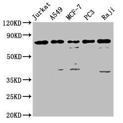
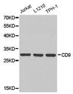 WB (Western Blot)
(Western Blot analysis of extracts of various cell lines, using CD9 antibody)
WB (Western Blot)
(Western Blot analysis of extracts of various cell lines, using CD9 antibody)
CD9, Polyclonal Antibody (Cat# AAA118115)
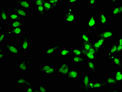
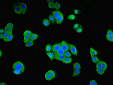 IF (Immunofluorescence)
(Immunofluorescent analysis of PC3 cells using AAA118118 at a dilution of 1:100 and Alexa Fluor 488-congugated AffiniPure Goat Anti-Rabbit IgG(H+L))
IF (Immunofluorescence)
(Immunofluorescent analysis of PC3 cells using AAA118118 at a dilution of 1:100 and Alexa Fluor 488-congugated AffiniPure Goat Anti-Rabbit IgG(H+L))
Origin recognition complex subunit 4, Polyclonal Antibody (Cat# AAA118118)
Decorin, Polyclonal Antibody (Cat# AAA118121)
Lactobacillus plantarum L-lactate dehydrogenase 1, Polyclonal Antibody (Cat# AAA118122)
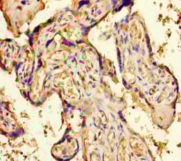
 IHC (Immunohiostchemistry)
(Immunohistochemistry of paraffin-embedded human placenta at dilution 1:100)
IHC (Immunohiostchemistry)
(Immunohistochemistry of paraffin-embedded human placenta at dilution 1:100)
40S ribosomal protein S11, Polyclonal Antibody (Cat# AAA118127)
Rabies virus Nucleoprotein, Polyclonal Antibody (Cat# AAA118131)
JC polyomavirus Minor capsid protein VP2, Polyclonal Antibody (Cat# AAA118134)
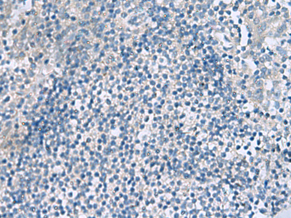
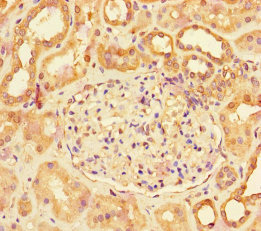
 IHC (Immunohiostchemistry)
(Immunohistochemistry of paraffin-embedded human kidney at dilution 1:100)
IHC (Immunohiostchemistry)
(Immunohistochemistry of paraffin-embedded human kidney at dilution 1:100)
Phospholipid transfer, Polyclonal Antibody (Cat# AAA118138)
 IHC (Immunohiostchemistry)
(Immunohistochemistry of paraffin-embedded human ovarian cancer using AAA118140 at dilution of 1:100)
IHC (Immunohiostchemistry)
(Immunohistochemistry of paraffin-embedded human ovarian cancer using AAA118140 at dilution of 1:100)
60S ribosomal protein L28, Polyclonal Antibody (Cat# AAA118140)

 IHC (Immunohiostchemistry)
(Immunohistochemistry of paraffin-embedded human placenta using AAA118144 at dilution of 1:20)
IHC (Immunohiostchemistry)
(Immunohistochemistry of paraffin-embedded human placenta using AAA118144 at dilution of 1:20)
Fibroblast growth factor 1, Polyclonal Antibody (Cat# AAA118144)
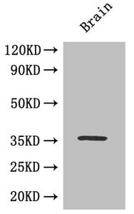
 WB (Western Blot)
(Western BlotPositive WB detected in: Rat brain tissue,Mouse brain tissueAll lanes: Gnao1 antibody at 3ug/mlSecondaryGoat polyclonal to rabbit IgG at 1/50000 dilutionPredicted band size: 41 KDaObserved band size: 41 KDa)
WB (Western Blot)
(Western BlotPositive WB detected in: Rat brain tissue,Mouse brain tissueAll lanes: Gnao1 antibody at 3ug/mlSecondaryGoat polyclonal to rabbit IgG at 1/50000 dilutionPredicted band size: 41 KDaObserved band size: 41 KDa)
Guanine nucleotide-binding protein G, Polyclonal Antibody (Cat# AAA118146)
Stress-induced-phosphoprotein 1, Polyclonal Antibody (Cat# AAA118153)
Rabies virus Nucleoprotein, Polyclonal Antibody (Cat# AAA118154)
 WB (Western Blot)
(Western blotAll lanes: H-2 class II histocompatibility antigen gamma chain antibody at 2ug/ml+mouse lung tissueSecondaryGoat polyclonal to rabbit at 1/10000 dilutionPredicted band size: 32,25kDaObserved band size: 34 kDa)
WB (Western Blot)
(Western blotAll lanes: H-2 class II histocompatibility antigen gamma chain antibody at 2ug/ml+mouse lung tissueSecondaryGoat polyclonal to rabbit at 1/10000 dilutionPredicted band size: 32,25kDaObserved band size: 34 kDa)
H-2 class II histocompatibility antigen gamma chain, Polyclonal Antibody (Cat# AAA118157)
 IHC (Immunohistochemistry)
(Immunohistochemistry of paraffin-embedded human pancreatic tissue using AAA118159 at dilution 1:100)
IHC (Immunohistochemistry)
(Immunohistochemistry of paraffin-embedded human pancreatic tissue using AAA118159 at dilution 1:100)
Insulin, Polyclonal Antibody (Cat# AAA118159)
Cation-dependent mannose-6-phosphate receptor, Polyclonal Antibody (Cat# AAA118160)
Metallothionein-1G, Polyclonal Antibody (Cat# AAA118161)
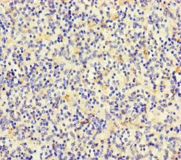
 IHC (Immunohiostchemistry)
(Immunohistochemistry of paraffin-embedded human colon cancer using AAA118164 at dilution 1:100)
IHC (Immunohiostchemistry)
(Immunohistochemistry of paraffin-embedded human colon cancer using AAA118164 at dilution 1:100)
Avpr1a protein, Polyclonal Antibody (Cat# AAA118164)
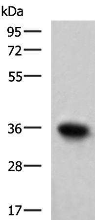
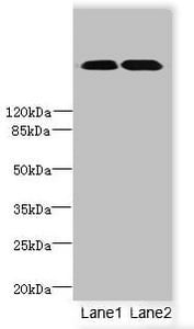
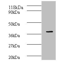
 WB (Western Blot)
(Western blotAll lanes: Urease subunit antibody at 2ug/ml+293T whole cell lysateSecondaryGoat polyclonal to Rabbit IgG at 1/10000 dilutionPredicted band size: 62kDaObserved band size: 62kDa)
WB (Western Blot)
(Western blotAll lanes: Urease subunit antibody at 2ug/ml+293T whole cell lysateSecondaryGoat polyclonal to Rabbit IgG at 1/10000 dilutionPredicted band size: 62kDaObserved band size: 62kDa)
Helicobacter pylori Urease subunit beta, Polyclonal Antibody (Cat# AAA117997)
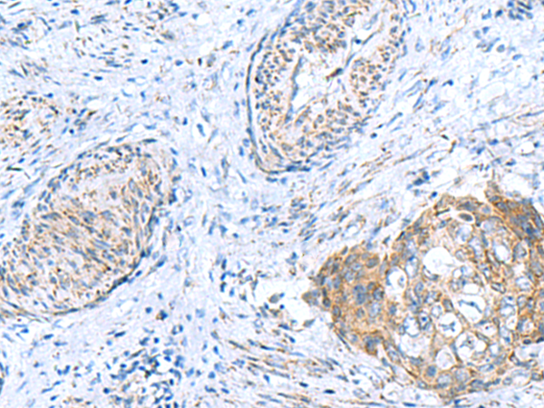
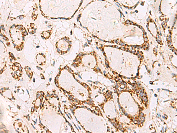
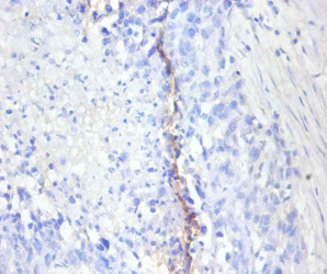
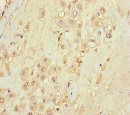
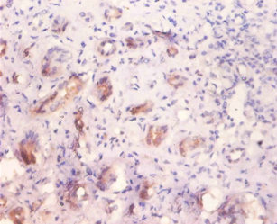
 IHC (Immunohiostchemistry)
(Immunohistochemistry of paraffin-embedded human pancreatic cancer using AAA118000 at dilution 1:100)
IHC (Immunohiostchemistry)
(Immunohistochemistry of paraffin-embedded human pancreatic cancer using AAA118000 at dilution 1:100)
Activated RNA polymerase II transcriptional coactivator p15, Polyclonal Antibody (Cat# AAA118000)
Glutamate decarboxylase alpha, Polyclonal Antibody (Cat# AAA118001)
Protein Wnt-3a, Polyclonal Antibody (Cat# AAA118008)
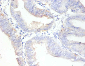
 IHC (Immunohiostchemistry)
(Immunohistochemistry of paraffin-embedded human small intestine using AAA118009 at dilution 1:100)
IHC (Immunohiostchemistry)
(Immunohistochemistry of paraffin-embedded human small intestine using AAA118009 at dilution 1:100)
Metallothionein-2, Polyclonal Antibody (Cat# AAA118009)
What are Polyclonal Antibodies?
Polyclonal antibodies are antibodies that come from multiple B cell clones of a host animal. The typical hosts used for the majority of polyclonal antibody production are rabbits, goats, sheep, and donkeys. These polyclonal antibodies, once having identified their target, will bind to different epitopes located at different regions or sequences on the same protein/antigen. As a result, they are ideal at locating and binding to the target, even if the target is in very low concentrations (due to many different antibodies being able to bind to the same target molecule, which allows for significant amplification of a downstream signal).
Polyclonal antibodies are typically produced by injecting an antigen into a host animal, which causes the animal’s immune system to attack the foreign antigen by mass generating antibodies against it. After a period of time, serum is collected from the animal and purified using physicochemical fractionation, class-specific affinity purification, and/or antigen-affinity purification.
Key Uses of Polyclonal Antibodies
- Western Blotting: This method is used to find specific proteins in biological samples after separating them by size.
- Immunohistochemistry: IHC helps visualize the location of proteins in tissue sections using various staining techniques.
- ELISA: (Enzyme-Linked Immunosorbent Assay) is typically used to identify specific protein quantities in a sample. ELISAs can be either “Quantitative” or “Qualitative”.
- Flow Cytometry: technique that identifies and measures the specific protein on the surface or inside the cells in a fluid suspension.
- Immunoprecipitation: IP isolates and studies a specific protein from a complex mixture using antibodies.
Why Buy Polyclonal Antibodies from AAA Biotech?
1. Ideal for Various Applications
Our antibodies are generally going to be validated for use in multiple types of assays, including ELISA, Western Blotting, Immunohistochemistry, Immunoprecipitation, amongst others. They are ideal for a wide range of research applications.
2. Rigorous Quality Control
All of the antibodies in our catalog undergo strict quality testing to ensure specificity, sensitivity, and consistent performance. We are confident in the ability of our antibodies to provide you with accurate results.
3. Wide Assortment of Antibodies
Antibodies in are catalog can be found for both common and exotic species, and these antibodies are also available in both conjugated and recombinant forms to suit many diverse experimental needs.
4. Highly Purified
Our antibodies are available in purified forms with over 85% purity, as confirmed by SDS-PAGE. They are also available with tags such as His, Flag, GST, or MBP. We cater to customers worldwide.
FAQ
1. How are polyclonal antibodies produced?
Traditionally, polyclonal antibodies are produced by injecting an antigen into a host animal (such as a rabbit or goat), which then triggers an immune response from the host animal. The animal’s B cells produce antibodies that will recognize different parts of the injected antigen. These antibodies are then collected from the animal’s blood and purified for use.
2. How do polyclonal antibodies differ from monoclonal antibodies?
Polyclonal antibodies are a mix of antibodies that bind to different locations (epitopes) of the same antigen, while monoclonal antibodies are identical and bind to just one specific epitope. This makes polyclonal antibodies more versatile and better at detecting proteins that may be present in low quantities or in altered/modified forms.
3. How should I store polyclonal antibodies?
Polyclonal antibodies should be stored at 4°C for short-term use (up to a few weeks) and at -20°C or -80°C for long-term storage. Avoid repeated freeze-thaw cycles by dividing them into small aliquots. Always check the datasheet for specific storage instructions.