Filters
▼Clonality
▼Type
▼Reactivity
▼Gene Name
▼Isotype
▼Host
▼Application
▼Clone
▼Polyclonal Antibodies
At AAA Biotech also known as AAA Bio or AAABio, we provide a broad range of purified polyclonal antibodies (pAbs) that are able to all be browsed online through our website. Due to their high specificity and strong binding affinity, these antibodies are ideal for wide swathes of research and experimental applications.
Our polyclonal antibodies can easily support your work, whether you use them for Western Blotting, Immunocytochemistry (with or without Immunofluorescence used in conjunction), Immunohistochemistry, Immunoprecipitation, and ELISA tests. We highly encourage you to browse our range of pAbs and choose the one that best suits your experimental model.
Viewing 1500-1550 of 96805 product results
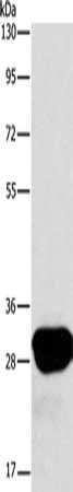
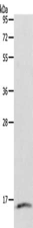
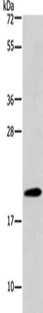
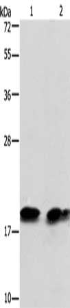
 SDS-PAGE
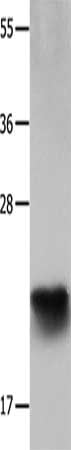
(Gel: 12%SDS-PAGE, Lysate: 30 ug, Lane: Human colon cancer tissue, Primary antibody: AAA237494(IL18 Antibody) at dilution 1/2000, Secondary antibody: Goat anti rabbit IgG at 1/8000 dilution, Exposure time: 1 minute)
SDS-PAGE
(Gel: 12%SDS-PAGE, Lysate: 30 ug, Lane: Human colon cancer tissue, Primary antibody: AAA237494(IL18 Antibody) at dilution 1/2000, Secondary antibody: Goat anti rabbit IgG at 1/8000 dilution, Exposure time: 1 minute)
IL18, Polyclonal Antibody (Cat# AAA237494)
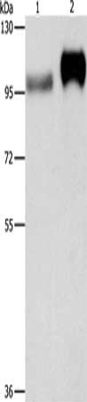
 SDS-PAGE
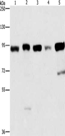
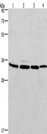
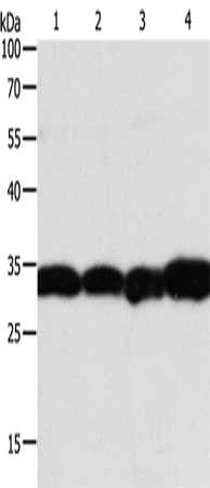
(Gel: 6%SDS-PAGE,Lysate: 40 ug,Lane 1-5: Raji cells, 231 cells, HepG2 cells, Hela cells, 293T cells,Primary antibody: AAA237498(IMMT Antibody) at dilution 1/270 dilution,Secondary antibody: Goat anti rabbit IgG at 1/8000 dilution,Exposure time: 5 seconds)
SDS-PAGE
(Gel: 6%SDS-PAGE,Lysate: 40 ug,Lane 1-5: Raji cells, 231 cells, HepG2 cells, Hela cells, 293T cells,Primary antibody: AAA237498(IMMT Antibody) at dilution 1/270 dilution,Secondary antibody: Goat anti rabbit IgG at 1/8000 dilution,Exposure time: 5 seconds)
IMMT, Polyclonal Antibody (Cat# AAA237498)
 SDS-PAGE
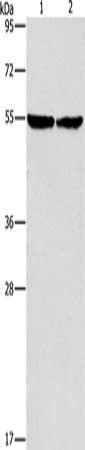
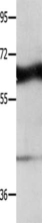
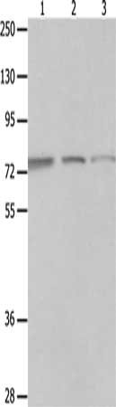
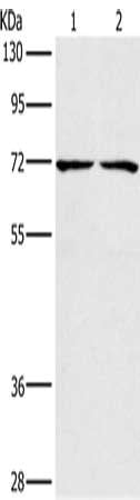
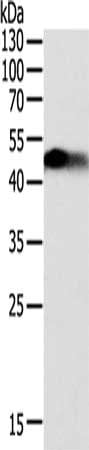
(Gel: 8%SDS-PAGE, Lysate: 40 ug, Lane 1-2: Mouse heart tissue, hela cells, Primary antibody: AAA237501(ILK Antibody) at dilution 1/600, Secondary antibody: Goat anti rabbit IgG at 1/8000 dilution, Exposure time: 40 seconds)
SDS-PAGE
(Gel: 8%SDS-PAGE, Lysate: 40 ug, Lane 1-2: Mouse heart tissue, hela cells, Primary antibody: AAA237501(ILK Antibody) at dilution 1/600, Secondary antibody: Goat anti rabbit IgG at 1/8000 dilution, Exposure time: 40 seconds)
ILK, Polyclonal Antibody (Cat# AAA237501)
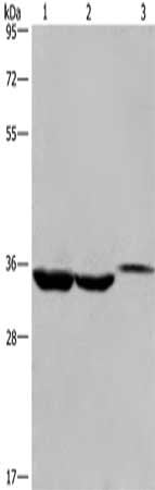 SDS-PAGE
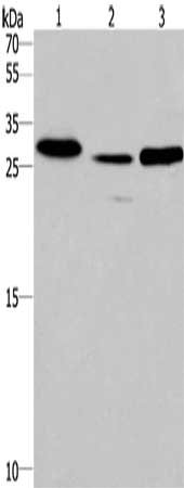
(Gel: 10%SDS-PAGE, Lysate: 40 ug, Lane 1-3: Lovo cells, human liver cancer tissue, mouse kidney tissue, Primary antibody: AAA237503(ING2 Antibody) at dilution 1/425, Secondary antibody: Goat anti rabbit IgG at 1/8000 dilution, Exposure time: 40 seconds)
SDS-PAGE
(Gel: 10%SDS-PAGE, Lysate: 40 ug, Lane 1-3: Lovo cells, human liver cancer tissue, mouse kidney tissue, Primary antibody: AAA237503(ING2 Antibody) at dilution 1/425, Secondary antibody: Goat anti rabbit IgG at 1/8000 dilution, Exposure time: 40 seconds)
ING2, Polyclonal Antibody (Cat# AAA237503)
 SDS-PAGE
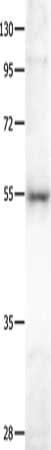
(Gel: 8%SDS-PAGE, Lysate: 40 ug, Lane: Mouse fat tissue, Primary antibody: AAA237505(INHBA Antibody) at dilution 1/701, Secondary antibody: Goat anti rabbit IgG at 1/8000 dilution, Exposure time: 1 minute)
SDS-PAGE
(Gel: 8%SDS-PAGE, Lysate: 40 ug, Lane: Mouse fat tissue, Primary antibody: AAA237505(INHBA Antibody) at dilution 1/701, Secondary antibody: Goat anti rabbit IgG at 1/8000 dilution, Exposure time: 1 minute)
INHBA, Polyclonal Antibody (Cat# AAA237505)
 SDS-PAGE
(Gel: 8%SDS-PAGE, Lysate: 40 ug, Lane 1-2: Huvec cells, human placenta tissue, Primary antibody: AAA237508(ITGB3 Antibody) at dilution 1/500, Secondary antibody: Goat anti rabbit IgG at 1/8000 dilution, Exposure time: 10 seconds)
SDS-PAGE
(Gel: 8%SDS-PAGE, Lysate: 40 ug, Lane 1-2: Huvec cells, human placenta tissue, Primary antibody: AAA237508(ITGB3 Antibody) at dilution 1/500, Secondary antibody: Goat anti rabbit IgG at 1/8000 dilution, Exposure time: 10 seconds)
ITGB3, Polyclonal Antibody (Cat# AAA237508)
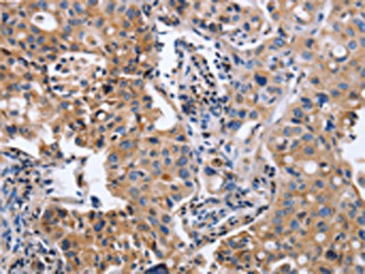
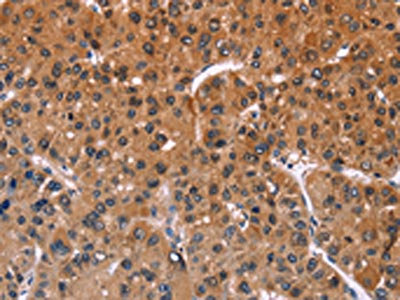
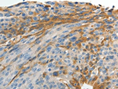
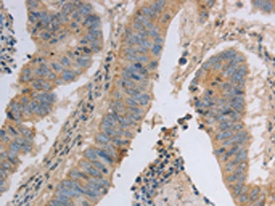
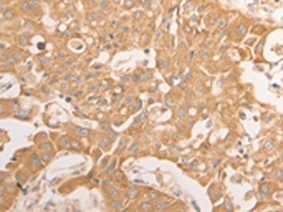
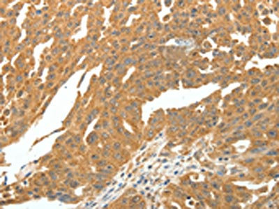
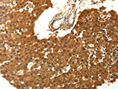
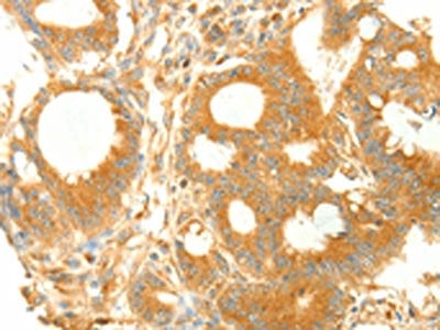
 IHC (Immunohiostchemistry)
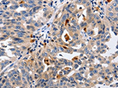
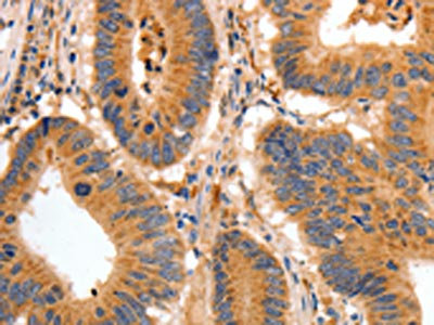
(The image on the left is immunohistochemistry of paraffin-embedded Human colon cancer tissue using AAA237514(ITGB5 Antibody) at dilution 1/40, on the right is treated with fusion protein. (Original magnification: ×200))
IHC (Immunohiostchemistry)
(The image on the left is immunohistochemistry of paraffin-embedded Human colon cancer tissue using AAA237514(ITGB5 Antibody) at dilution 1/40, on the right is treated with fusion protein. (Original magnification: ×200))
ITGB5, Polyclonal Antibody (Cat# AAA237514)
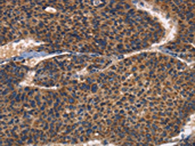
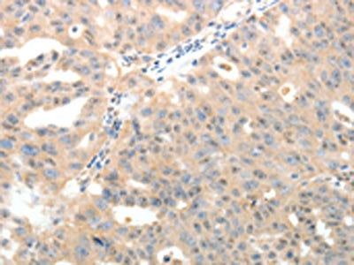
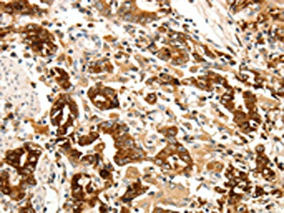
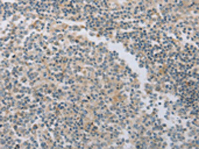
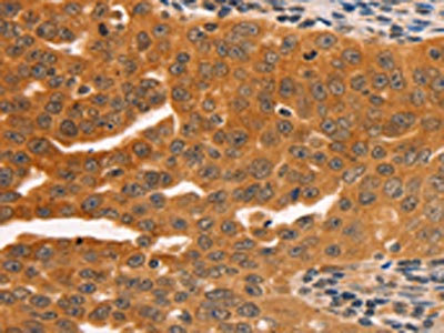
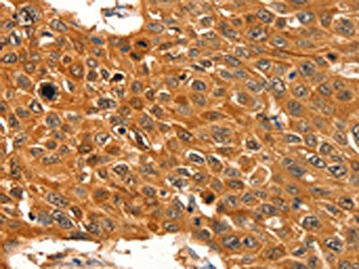
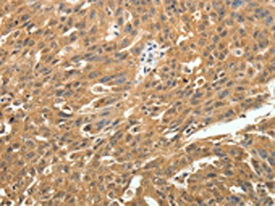
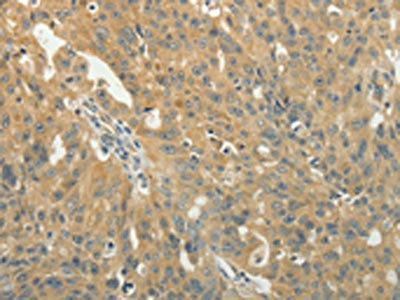
 IHC (Immunohiostchemistry)
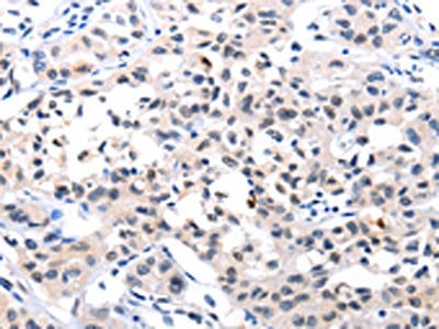
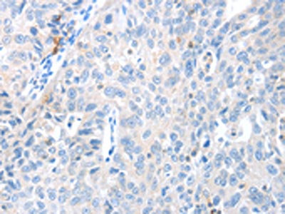
(The image on the left is immunohistochemistry of paraffin-embedded Human esophagus cancer tissue using AAA237521(IRAK3 Antibody) at dilution 1/50, on the right is treated with fusion protein. (Original magnification: ×200))
IHC (Immunohiostchemistry)
(The image on the left is immunohistochemistry of paraffin-embedded Human esophagus cancer tissue using AAA237521(IRAK3 Antibody) at dilution 1/50, on the right is treated with fusion protein. (Original magnification: ×200))
IRAK3, Polyclonal Antibody (Cat# AAA237521)
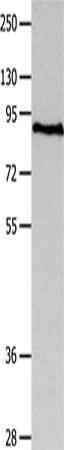
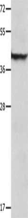
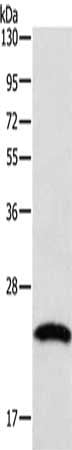
 SDS-PAGE
(Gel: 10%SDS-PAGE, Lysate: 40 ug, Lane 1-3: Human fetal liver tissue, mouse heart tissue, human testis tissue, Primary antibody: AAA237522(IRAK1BP1 Antibody) at dilution 1/475, Secondary antibody: Goat anti rabbit IgG at 1/8000 dilution, Exposure time: 1 minute)
SDS-PAGE
(Gel: 10%SDS-PAGE, Lysate: 40 ug, Lane 1-3: Human fetal liver tissue, mouse heart tissue, human testis tissue, Primary antibody: AAA237522(IRAK1BP1 Antibody) at dilution 1/475, Secondary antibody: Goat anti rabbit IgG at 1/8000 dilution, Exposure time: 1 minute)
IRAK1BP1, Polyclonal Antibody (Cat# AAA237522)

 SDS-PAGE
(Gel: 10%SDS-PAGE, Lysate: 30 ug, Lane: Mouse lung tissue, Primary antibody: AAA237524(IRAK2 Antibody) at dilution 1/1500, Secondary antibody: Goat anti rabbit IgG at 1/8000 dilution, Exposure time: 20 seconds)
SDS-PAGE
(Gel: 10%SDS-PAGE, Lysate: 30 ug, Lane: Mouse lung tissue, Primary antibody: AAA237524(IRAK2 Antibody) at dilution 1/1500, Secondary antibody: Goat anti rabbit IgG at 1/8000 dilution, Exposure time: 20 seconds)
IRAK2, Polyclonal Antibody (Cat# AAA237524)
 SDS-PAGE
(Gel: 8%SDS-PAGE, Lysate: 40 ug, Lane: SKOV3 cells, Primary antibody: AAA237528(BAIAP2 Antibody) at dilution 1/350, Secondary antibody: Goat anti rabbit IgG at 1/8000 dilution, Exposure time: 40 seconds)
SDS-PAGE
(Gel: 8%SDS-PAGE, Lysate: 40 ug, Lane: SKOV3 cells, Primary antibody: AAA237528(BAIAP2 Antibody) at dilution 1/350, Secondary antibody: Goat anti rabbit IgG at 1/8000 dilution, Exposure time: 40 seconds)
BAIAP2, Polyclonal Antibody (Cat# AAA237528)
 SDS-PAGE
(Gel: 12%SDS-PAGE, Lysate: 30 ug, Lane: Mouse liver tissue, Primary antibody: AAA237529(COPS5 Antibody) at dilution 1/1550, Secondary antibody: Goat anti rabbit IgG at 1/8000 dilution, Exposure time: 5 seconds)
SDS-PAGE
(Gel: 12%SDS-PAGE, Lysate: 30 ug, Lane: Mouse liver tissue, Primary antibody: AAA237529(COPS5 Antibody) at dilution 1/1550, Secondary antibody: Goat anti rabbit IgG at 1/8000 dilution, Exposure time: 5 seconds)
COPS5, Polyclonal Antibody (Cat# AAA237529)
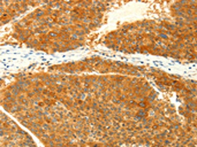
 IHC (Immunohiostchemistry)
(The image on the left is immunohistochemistry of paraffin-embedded Human ovarian cancer tissue using AAA237540(KLK7 Antibody) at dilution 1/10, on the right is treated with fusion protein. (Original magnification: ×200))
IHC (Immunohiostchemistry)
(The image on the left is immunohistochemistry of paraffin-embedded Human ovarian cancer tissue using AAA237540(KLK7 Antibody) at dilution 1/10, on the right is treated with fusion protein. (Original magnification: ×200))
KLK7, Polyclonal Antibody (Cat# AAA237540)
 IHC (Immunohiostchemistry)
(The image on the left is immunohistochemistry of paraffin-embedded Human ovarian cancer tissue using AAA237543(XRCC6 Antibody) at dilution 1/10, on the right is treated with fusion protein. (Original magnification: ×200))
IHC (Immunohiostchemistry)
(The image on the left is immunohistochemistry of paraffin-embedded Human ovarian cancer tissue using AAA237543(XRCC6 Antibody) at dilution 1/10, on the right is treated with fusion protein. (Original magnification: ×200))
XRCC6, Polyclonal Antibody (Cat# AAA237543)
 IHC (Immunohiostchemistry)
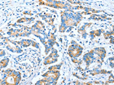
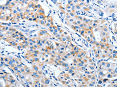
(The image on the left is immunohistochemistry of paraffin-embedded Human lung cancer tissue using AAA237552(GNRH1 Antibody) at dilution 1/65, on the right is treated with fusion protein. (Original magnification: ×200))
IHC (Immunohiostchemistry)
(The image on the left is immunohistochemistry of paraffin-embedded Human lung cancer tissue using AAA237552(GNRH1 Antibody) at dilution 1/65, on the right is treated with fusion protein. (Original magnification: ×200))
GNRH1, Polyclonal Antibody (Cat# AAA237552)
 IHC (Immunohiostchemistry)
(The image on the left is immunohistochemistry of paraffin-embedded Human cervical cancer tissue using AAA237558(LRP12 Antibody) at dilution 1/20, on the right is treated with fusion protein. (Original magnification: ×200))
IHC (Immunohiostchemistry)
(The image on the left is immunohistochemistry of paraffin-embedded Human cervical cancer tissue using AAA237558(LRP12 Antibody) at dilution 1/20, on the right is treated with fusion protein. (Original magnification: ×200))
LRP12, Polyclonal Antibody (Cat# AAA237558)
 SDS-PAGE
(Gel: 10%SDS-PAGE, Lysate: 40 ug, Lane: A549 cells, Primary antibody: AAA237561(LXN Antibody) at dilution 1/667, Secondary antibody: Goat anti rabbit IgG at 1/8000 dilution, Exposure time: 10 seconds)
SDS-PAGE
(Gel: 10%SDS-PAGE, Lysate: 40 ug, Lane: A549 cells, Primary antibody: AAA237561(LXN Antibody) at dilution 1/667, Secondary antibody: Goat anti rabbit IgG at 1/8000 dilution, Exposure time: 10 seconds)
LXN, Polyclonal Antibody (Cat# AAA237561)
 SDS-PAGE
(Gel: 12%SDS-PAGE, Lysate: 40 ug, Lane: Human fetal lung tissue, Primary antibody: AAA237563(LYZ Antibody) at dilution 1/200, Secondary antibody: Goat anti rabbit IgG at 1/8000 dilution, Exposure time: 45 seconds)
SDS-PAGE
(Gel: 12%SDS-PAGE, Lysate: 40 ug, Lane: Human fetal lung tissue, Primary antibody: AAA237563(LYZ Antibody) at dilution 1/200, Secondary antibody: Goat anti rabbit IgG at 1/8000 dilution, Exposure time: 45 seconds)
LYZ, Polyclonal Antibody (Cat# AAA237563)
 SDS-PAGE
(Gel: 10%SDS-PAGE, Lysate: 40 ug, Lane: Human seminoma tissue, Primary antibody: AAA237567(MAD2L1 Antibody) at dilution 1/100, Secondary antibody: Goat anti rabbit IgG at 1/8000 dilution, Exposure time: 30 seconds)
SDS-PAGE
(Gel: 10%SDS-PAGE, Lysate: 40 ug, Lane: Human seminoma tissue, Primary antibody: AAA237567(MAD2L1 Antibody) at dilution 1/100, Secondary antibody: Goat anti rabbit IgG at 1/8000 dilution, Exposure time: 30 seconds)
MAD2L1, Polyclonal Antibody (Cat# AAA237567)
 SDS-PAGE
(Gel: 10%SDS-PAGE, Lysate: 40 ug, Lane 1-4: A549 cells, 231 cells, PC3 cells, hela cells, Primary antibody: AAA237569(MAD2L1BP Antibody) at dilution 1/675, Secondary antibody: Goat anti rabbit IgG at 1/8000 dilution, Exposure time: 20 seconds)
SDS-PAGE
(Gel: 10%SDS-PAGE, Lysate: 40 ug, Lane 1-4: A549 cells, 231 cells, PC3 cells, hela cells, Primary antibody: AAA237569(MAD2L1BP Antibody) at dilution 1/675, Secondary antibody: Goat anti rabbit IgG at 1/8000 dilution, Exposure time: 20 seconds)
MAD2L1BP, Polyclonal Antibody (Cat# AAA237569)
 SDS-PAGE
(Gel: 10%SDS-PAGE, Lysate: 40 ug, Lane: Human brain glioma tissue, Primary antibody: AAA237573(MAGEB4 Antibody) at dilution 1/550, Secondary antibody: Goat anti rabbit IgG at 1/8000 dilution, Exposure time: 2 minutes)
SDS-PAGE
(Gel: 10%SDS-PAGE, Lysate: 40 ug, Lane: Human brain glioma tissue, Primary antibody: AAA237573(MAGEB4 Antibody) at dilution 1/550, Secondary antibody: Goat anti rabbit IgG at 1/8000 dilution, Exposure time: 2 minutes)
MAGEB4, Polyclonal Antibody (Cat# AAA237573)

 SDS-PAGE
(Gel: 10%SDS-PAGE, Lysate: 40 ug, Lane 1-3: A549 cells, Jurkat cells, 293T cells, Primary antibody: AAA237575(MAGED1 Antibody) at dilution 1/1200, Secondary antibody: Goat anti rabbit IgG at 1/8000 dilution, Exposure time: 3 minutes)
SDS-PAGE
(Gel: 10%SDS-PAGE, Lysate: 40 ug, Lane 1-3: A549 cells, Jurkat cells, 293T cells, Primary antibody: AAA237575(MAGED1 Antibody) at dilution 1/1200, Secondary antibody: Goat anti rabbit IgG at 1/8000 dilution, Exposure time: 3 minutes)
MAGED1, Polyclonal Antibody (Cat# AAA237575)
 SDS-PAGE
(Gel: 10%SDS-PAGE, Lysate: 40 ug, Lane: Human fetal brain tissue, Primary antibody: AAA237576(MAGED1 Antibody) at dilution 1/1200, Secondary antibody: Goat anti rabbit IgG at 1/8000 dilution, Exposure time: 90 seconds)
SDS-PAGE
(Gel: 10%SDS-PAGE, Lysate: 40 ug, Lane: Human fetal brain tissue, Primary antibody: AAA237576(MAGED1 Antibody) at dilution 1/1200, Secondary antibody: Goat anti rabbit IgG at 1/8000 dilution, Exposure time: 90 seconds)
MAGED1, Polyclonal Antibody (Cat# AAA237576)
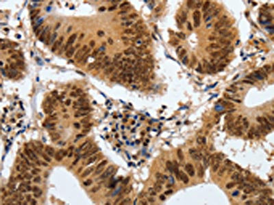
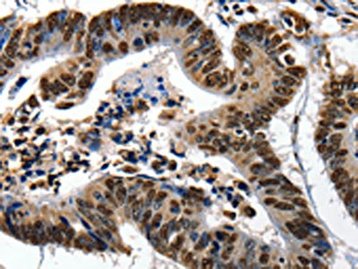
 IHC (Immunohiostchemistry)
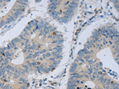
(The image on the left is immunohistochemistry of paraffin-embedded Human colon cancer tissue using AAA237577(MAP2K1 Antibody) at dilution 1/20, on the right is treated with fusion protein. (Original magnification: ×200))
IHC (Immunohiostchemistry)
(The image on the left is immunohistochemistry of paraffin-embedded Human colon cancer tissue using AAA237577(MAP2K1 Antibody) at dilution 1/20, on the right is treated with fusion protein. (Original magnification: ×200))
MAP2K1, Polyclonal Antibody (Cat# AAA237577)
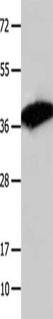
 SDS-PAGE
(Gel: 12%SDS-PAGE, Lysate: 30 ug, Lane: Mouse muscle tissue, Primary antibody: AAA237578(MAP2K6 Antibody) at dilution 1/500, Secondary antibody: Goat anti rabbit IgG at 1/8000 dilution, Exposure time: 20 seconds)
SDS-PAGE
(Gel: 12%SDS-PAGE, Lysate: 30 ug, Lane: Mouse muscle tissue, Primary antibody: AAA237578(MAP2K6 Antibody) at dilution 1/500, Secondary antibody: Goat anti rabbit IgG at 1/8000 dilution, Exposure time: 20 seconds)
MAP2K6, Polyclonal Antibody (Cat# AAA237578)
 SDS-PAGE
(Gel: 12%SDS-PAGE, Lysate: 40 ug, Lane: Mouse lung tissue, Primary antibody: AAA237582(SERPINB5 Antibody) at dilution 1/1000, Secondary antibody: Goat anti rabbit IgG at 1/8000 dilution, Exposure time: 10 seconds)
SDS-PAGE
(Gel: 12%SDS-PAGE, Lysate: 40 ug, Lane: Mouse lung tissue, Primary antibody: AAA237582(SERPINB5 Antibody) at dilution 1/1000, Secondary antibody: Goat anti rabbit IgG at 1/8000 dilution, Exposure time: 10 seconds)
SERPINB5, Polyclonal Antibody (Cat# AAA237582)
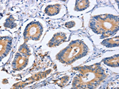
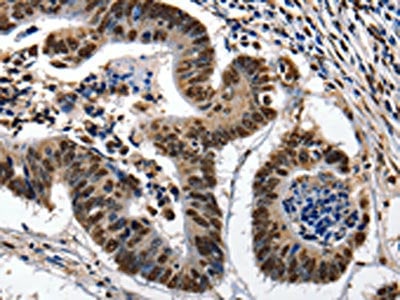
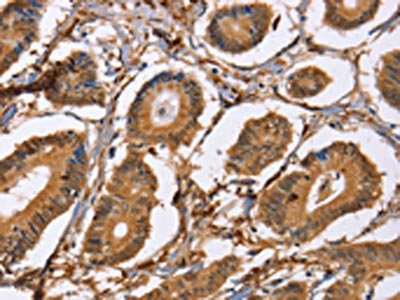
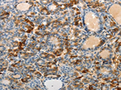
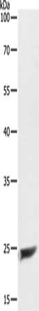
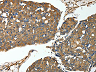
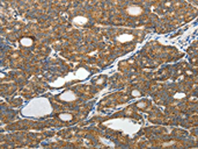
 IHC (Immunohiostchemistry)
(The image on the left is immunohistochemistry of paraffin-embedded Human thyroid cancer tissue using AAA237583(ST14 Antibody) at dilution 1/70, on the right is treated with fusion protein. (Original magnification: ×200))
IHC (Immunohiostchemistry)
(The image on the left is immunohistochemistry of paraffin-embedded Human thyroid cancer tissue using AAA237583(ST14 Antibody) at dilution 1/70, on the right is treated with fusion protein. (Original magnification: ×200))
ST14, Polyclonal Antibody (Cat# AAA237583)
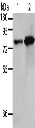
 SDS-PAGE
(Gel: 6%SDS-PAGE, Lysate: 40 ug, Lane 1-5: HepG2 cells, K562 cells, Jurkat cells, 231 cells, hela cells, Primary antibody: AAA237585(MCM5 Antibody) at dilution 1/300, Secondary antibody: Goat anti rabbit IgG at 1/8000 dilution, Exposure time: 20 seconds)
SDS-PAGE
(Gel: 6%SDS-PAGE, Lysate: 40 ug, Lane 1-5: HepG2 cells, K562 cells, Jurkat cells, 231 cells, hela cells, Primary antibody: AAA237585(MCM5 Antibody) at dilution 1/300, Secondary antibody: Goat anti rabbit IgG at 1/8000 dilution, Exposure time: 20 seconds)
MCM5, Polyclonal Antibody (Cat# AAA237585)
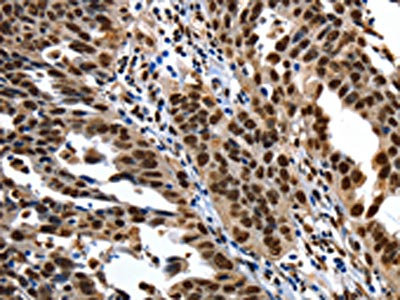
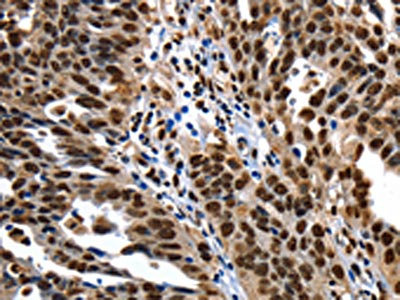
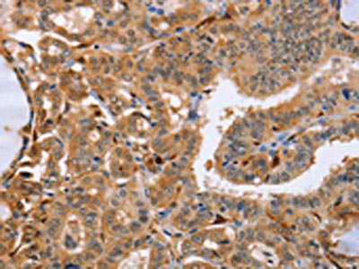
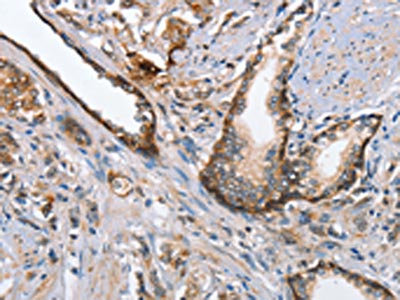
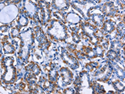
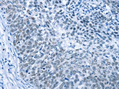 IHC (Immunohiostchemistry)
(The image on the left is immunohistochemistry of paraffin-embedded Human cervical cancer tissue using AAA237590(MCM7 Antibody) at dilution 1/40, on the right is treated with fusion protein. (Original magnification: ×200))
IHC (Immunohiostchemistry)
(The image on the left is immunohistochemistry of paraffin-embedded Human cervical cancer tissue using AAA237590(MCM7 Antibody) at dilution 1/40, on the right is treated with fusion protein. (Original magnification: ×200))
MCM7, Polyclonal Antibody (Cat# AAA237590)

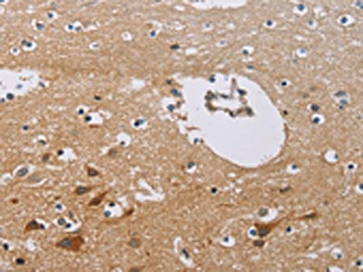
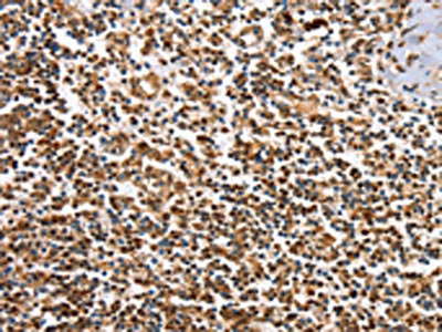
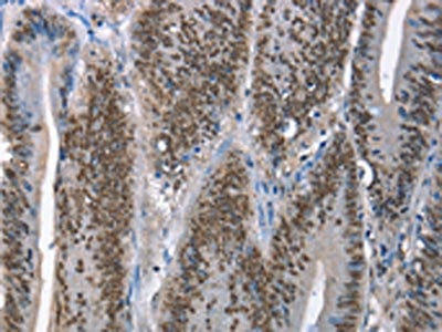
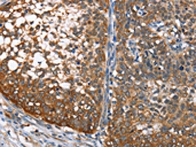
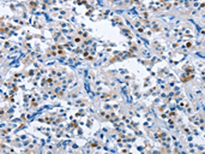
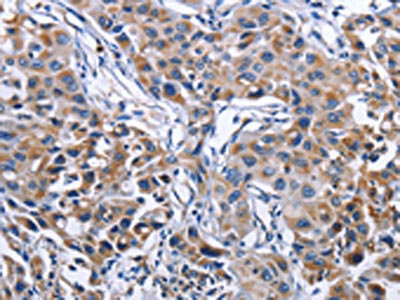
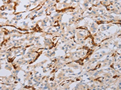
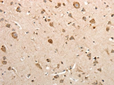
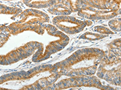
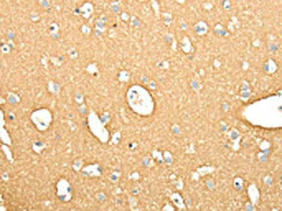
 IHC (Immunohiostchemistry)
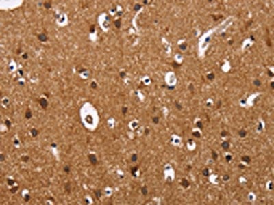
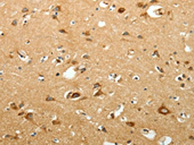
(The image on the left is immunohistochemistry of paraffin-embedded Human brain tissue using AAA237599(MMP16 Antibody) at dilution 1/40, on the right is treated with fusion protein. (Original magnification: ×200))
IHC (Immunohiostchemistry)
(The image on the left is immunohistochemistry of paraffin-embedded Human brain tissue using AAA237599(MMP16 Antibody) at dilution 1/40, on the right is treated with fusion protein. (Original magnification: ×200))
MMP16, Polyclonal Antibody (Cat# AAA237599)
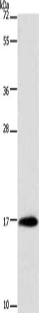
 SDS-PAGE
(Gel: 10%SDS-PAGE, Lysate: 40 ug, Lane 1-2: 293T cells, A431 cells, Primary antibody: AAA237601(MSN Antibody) at dilution 1/500, Secondary antibody: Goat anti rabbit IgG at 1/8000 dilution, Exposure time: 1 minute)
SDS-PAGE
(Gel: 10%SDS-PAGE, Lysate: 40 ug, Lane 1-2: 293T cells, A431 cells, Primary antibody: AAA237601(MSN Antibody) at dilution 1/500, Secondary antibody: Goat anti rabbit IgG at 1/8000 dilution, Exposure time: 1 minute)
MSN, Polyclonal Antibody (Cat# AAA237601)
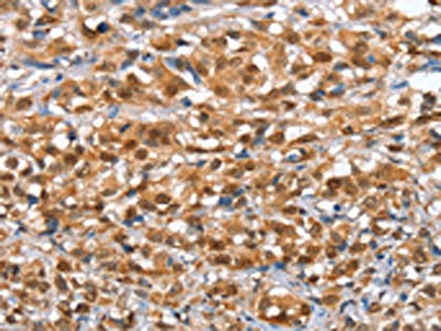
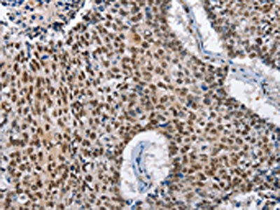
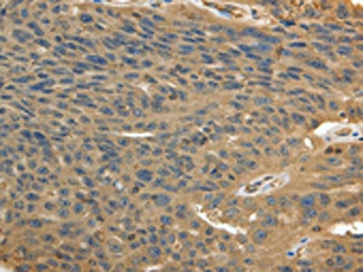
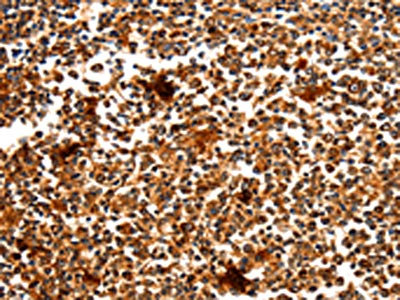
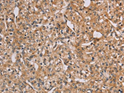
 IHC (Immunohiostchemistry)
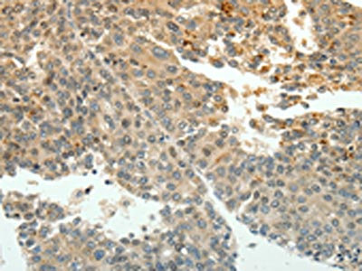
(The image on the left is immunohistochemistry of paraffin-embedded Human lung cancer tissue using AAA237603(MPO Antibody) at dilution 1/50, on the right is treated with fusion protein. (Original magnification: ×200))
IHC (Immunohiostchemistry)
(The image on the left is immunohistochemistry of paraffin-embedded Human lung cancer tissue using AAA237603(MPO Antibody) at dilution 1/50, on the right is treated with fusion protein. (Original magnification: ×200))
MPO, Polyclonal Antibody (Cat# AAA237603)
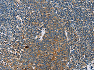
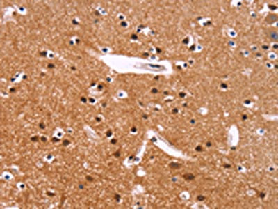
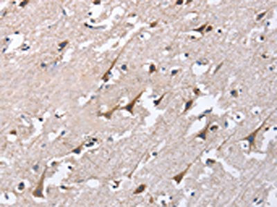
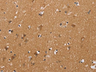
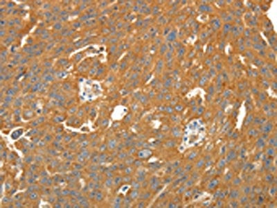
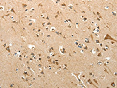
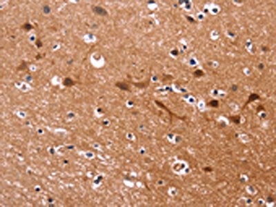 IHC (Immunohiostchemistry)
(The image on the left is immunohistochemistry of paraffin-embedded Human brain tissue using AAA237604(MSH4 Antibody) at dilution 1/30, on the right is treated with fusion protein. (Original magnification: ×200))
IHC (Immunohiostchemistry)
(The image on the left is immunohistochemistry of paraffin-embedded Human brain tissue using AAA237604(MSH4 Antibody) at dilution 1/30, on the right is treated with fusion protein. (Original magnification: ×200))
MSH4, Polyclonal Antibody (Cat# AAA237604)
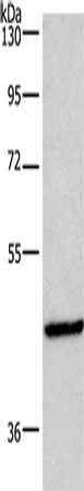
 SDS-PAGE
(Gel: 12%SDS-PAGE, Lysate: 40 ug, Lane: Mouse muscle tissue, Primary antibody: AAA237619(MYOZ1 Antibody) at dilution 1/1000, Secondary antibody: Goat anti rabbit IgG at 1/8000 dilution, Exposure time: 10 seconds)
SDS-PAGE
(Gel: 12%SDS-PAGE, Lysate: 40 ug, Lane: Mouse muscle tissue, Primary antibody: AAA237619(MYOZ1 Antibody) at dilution 1/1000, Secondary antibody: Goat anti rabbit IgG at 1/8000 dilution, Exposure time: 10 seconds)
MYOZ1, Polyclonal Antibody (Cat# AAA237619)
 SDS-PAGE
(Gel: 8%SDS-PAGE, Lysate: 40 ug, Lane: Hela cells, Primary antibody: AAA237621(MUTYH Antibody) at dilution 1/500, Secondary antibody: Goat anti rabbit IgG at 1/8000 dilution, Exposure time: 1 minute)
SDS-PAGE
(Gel: 8%SDS-PAGE, Lysate: 40 ug, Lane: Hela cells, Primary antibody: AAA237621(MUTYH Antibody) at dilution 1/500, Secondary antibody: Goat anti rabbit IgG at 1/8000 dilution, Exposure time: 1 minute)
MUTYH, Polyclonal Antibody (Cat# AAA237621)
 SDS-PAGE
(Gel: 10%SDS-PAGE, Lysate: 40 ug, Lane: Mouse stomach tissue, Primary antibody: AAA237625(MYL6 Antibody) at dilution 1/262.5, Secondary antibody: Goat anti rabbit IgG at 1/8000 dilution, Exposure time: 30 seconds)
SDS-PAGE
(Gel: 10%SDS-PAGE, Lysate: 40 ug, Lane: Mouse stomach tissue, Primary antibody: AAA237625(MYL6 Antibody) at dilution 1/262.5, Secondary antibody: Goat anti rabbit IgG at 1/8000 dilution, Exposure time: 30 seconds)
MYL6, Polyclonal Antibody (Cat# AAA237625)
 SDS-PAGE
(Gel: 8%SDS-PAGE, Lysate: 40 ug, Lane 1-2: Lovo cells, 293T cells, Primary antibody: AAA237627(CHRNA4 Antibody) at dilution 1/200, Secondary antibody: Goat anti rabbit IgG at 1/8000 dilution, Exposure time: 5 seconds)
SDS-PAGE
(Gel: 8%SDS-PAGE, Lysate: 40 ug, Lane 1-2: Lovo cells, 293T cells, Primary antibody: AAA237627(CHRNA4 Antibody) at dilution 1/200, Secondary antibody: Goat anti rabbit IgG at 1/8000 dilution, Exposure time: 5 seconds)
CHRNA4, Polyclonal Antibody (Cat# AAA237627)
 SDS-PAGE
(Gel: 10%SDS-PAGE, Lysate: 40 ug, Lane: Jurkat cells, Primary antibody: AAA237630(NAP1L1 Antibody) at dilution 1/650, Secondary antibody: Goat anti rabbit IgG at 1/8000 dilution, Exposure time: 40 seconds)
SDS-PAGE
(Gel: 10%SDS-PAGE, Lysate: 40 ug, Lane: Jurkat cells, Primary antibody: AAA237630(NAP1L1 Antibody) at dilution 1/650, Secondary antibody: Goat anti rabbit IgG at 1/8000 dilution, Exposure time: 40 seconds)
NAP1L1, Polyclonal Antibody (Cat# AAA237630)
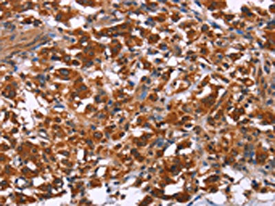
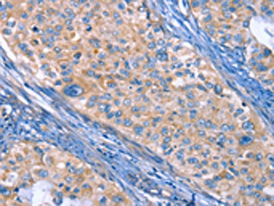
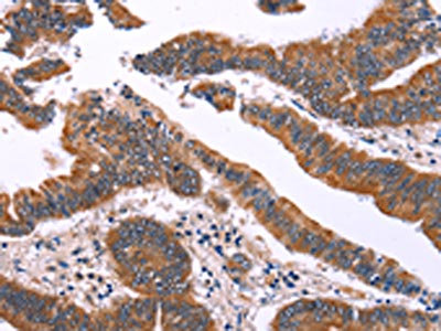
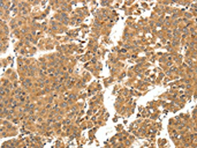
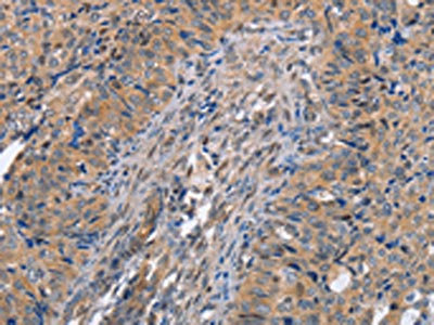
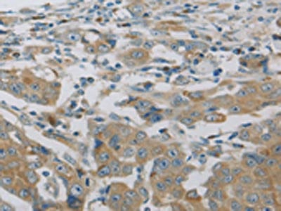
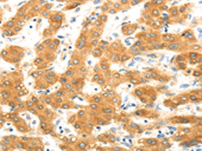
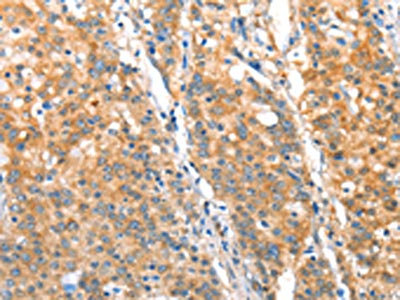
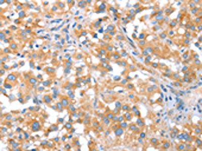
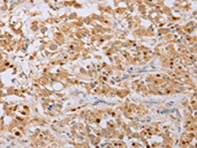
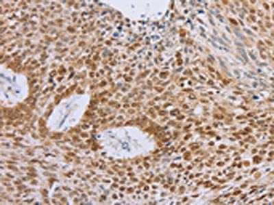
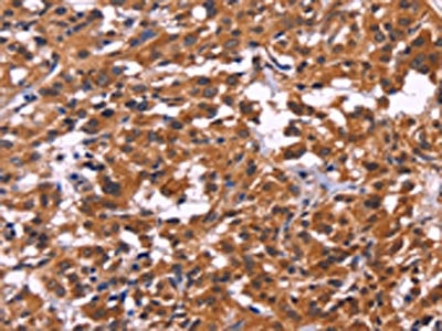
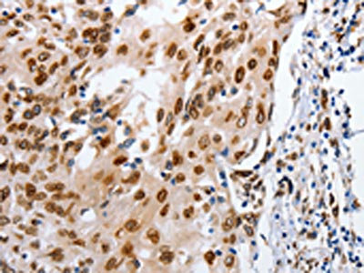
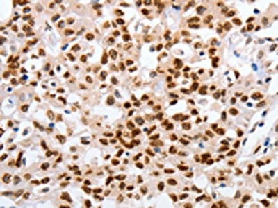
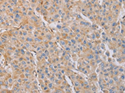
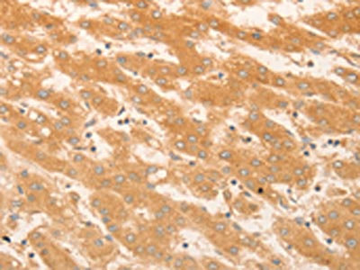
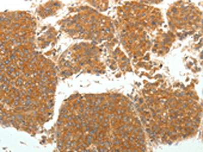
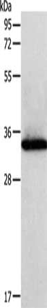
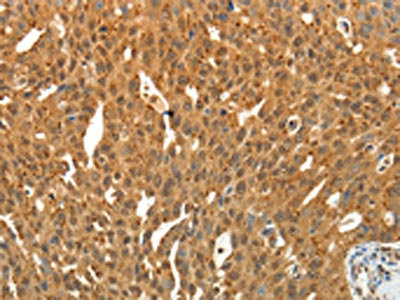
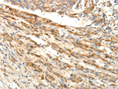
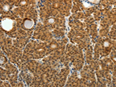
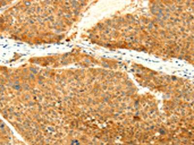
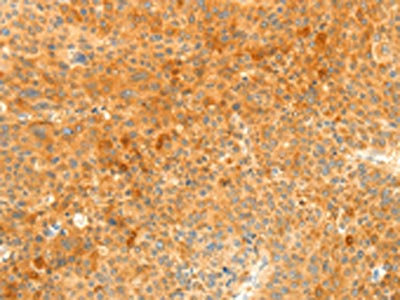
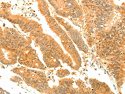
 IHC (Immunohiostchemistry)
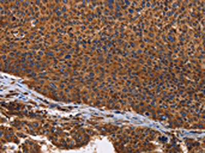
(The image on the left is immunohistochemistry of paraffin-embedded Human liver cancer tissue using AAA237799(SIRT3 Antibody) at dilution 1/15, on the right is treated with fusion protein. (Original magnification: ×200))
IHC (Immunohiostchemistry)
(The image on the left is immunohistochemistry of paraffin-embedded Human liver cancer tissue using AAA237799(SIRT3 Antibody) at dilution 1/15, on the right is treated with fusion protein. (Original magnification: ×200))
SIRT3, Polyclonal Antibody (Cat# AAA237799)
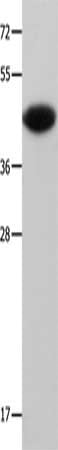
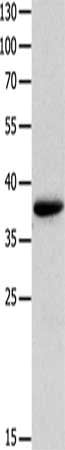
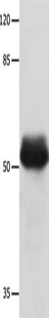
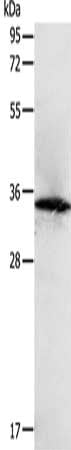
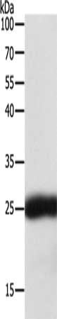
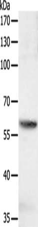
 SDS-PAGE
(Gel: 12%SDS-PAGE, Lysate: 40 ug, Lane: MCF7 cells, Primary antibody: AAA237801(SIRT4 Antibody) at dilution 1/450, Secondary antibody: Goat anti rabbit IgG at 1/8000 dilution, Exposure time: 1 minute)
SDS-PAGE
(Gel: 12%SDS-PAGE, Lysate: 40 ug, Lane: MCF7 cells, Primary antibody: AAA237801(SIRT4 Antibody) at dilution 1/450, Secondary antibody: Goat anti rabbit IgG at 1/8000 dilution, Exposure time: 1 minute)
SIRT4, Polyclonal Antibody (Cat# AAA237801)
 SDS-PAGE
(Gel: 12%SDS-PAGE, Lysate: 40 ug, Lane: MCF7 cells, Primary antibody: AAA237802(SIRT4 Antibody) at dilution 1/400, Secondary antibody: Goat anti rabbit IgG at 1/8000 dilution, Exposure time: 1 minute)
SDS-PAGE
(Gel: 12%SDS-PAGE, Lysate: 40 ug, Lane: MCF7 cells, Primary antibody: AAA237802(SIRT4 Antibody) at dilution 1/400, Secondary antibody: Goat anti rabbit IgG at 1/8000 dilution, Exposure time: 1 minute)
SIRT4, Polyclonal Antibody (Cat# AAA237802)
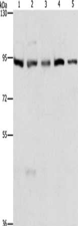
 SDS-PAGE
(Gel: 12%SDS-PAGE, Lysate: 40 ug, Lane: Hela cells, Primary antibody: AAA237803(SKP1 Antibody) at dilution 1/400, Secondary antibody: Goat anti rabbit IgG at 1/8000 dilution, Exposure time: 20 seconds)
SDS-PAGE
(Gel: 12%SDS-PAGE, Lysate: 40 ug, Lane: Hela cells, Primary antibody: AAA237803(SKP1 Antibody) at dilution 1/400, Secondary antibody: Goat anti rabbit IgG at 1/8000 dilution, Exposure time: 20 seconds)
SKP1, Polyclonal Antibody (Cat# AAA237803)
 SDS-PAGE
(Gel: 12%SDS-PAGE, Lysate: 40 ug, Lane 1-2: Mouse brain tissue, NIH/3T3 cells, Primary antibody: AAA237804(SKP1 Antibody) at dilution 1/550, Secondary antibody: Goat anti rabbit IgG at 1/8000 dilution, Exposure time: 20 seconds)
SDS-PAGE
(Gel: 12%SDS-PAGE, Lysate: 40 ug, Lane 1-2: Mouse brain tissue, NIH/3T3 cells, Primary antibody: AAA237804(SKP1 Antibody) at dilution 1/550, Secondary antibody: Goat anti rabbit IgG at 1/8000 dilution, Exposure time: 20 seconds)
SKP1, Polyclonal Antibody (Cat# AAA237804)

 SDS-PAGE
(Gel: 12%SDS-PAGE, Lysate: 40 ug, Lane: Human fetal kidney tissue, Primary antibody: AAA237807(SLC12A1 Antibody) at dilution 1/200, Secondary antibody: Goat anti rabbit IgG at 1/8000 dilution, Exposure time: 1 minute)
SDS-PAGE
(Gel: 12%SDS-PAGE, Lysate: 40 ug, Lane: Human fetal kidney tissue, Primary antibody: AAA237807(SLC12A1 Antibody) at dilution 1/200, Secondary antibody: Goat anti rabbit IgG at 1/8000 dilution, Exposure time: 1 minute)
SLC12A1, Polyclonal Antibody (Cat# AAA237807)

 IHC (Immunohiostchemistry)
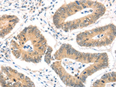
(The image on the left is immunohistochemistry of paraffin-embedded Human colon cancer tissue using AAA237808(SLC12A4 Antibody) at dilution 1/30, on the right is treated with fusion protein. (Original magnification: ×200))
IHC (Immunohiostchemistry)
(The image on the left is immunohistochemistry of paraffin-embedded Human colon cancer tissue using AAA237808(SLC12A4 Antibody) at dilution 1/30, on the right is treated with fusion protein. (Original magnification: ×200))
SLC12A4, Polyclonal Antibody (Cat# AAA237808)
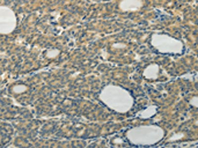
 IHC (Immunohiostchemistry)
(The image on the left is immunohistochemistry of paraffin-embedded Human brain tissue using AAA237809(SLC12A4 Antibody) at dilution 1/30, on the right is treated with fusion protein. (Original magnification: ×200))
IHC (Immunohiostchemistry)
(The image on the left is immunohistochemistry of paraffin-embedded Human brain tissue using AAA237809(SLC12A4 Antibody) at dilution 1/30, on the right is treated with fusion protein. (Original magnification: ×200))
SLC12A4, Polyclonal Antibody (Cat# AAA237809)
 SDS-PAGE
(Gel: 10%SDS-PAGE, Lysate: 40 ug, Lane: A549 cells, Primary antibody: AAA237815(TAGLN2 Antibody) at dilution 1/250, Secondary antibody: Goat anti rabbit IgG at 1/8000 dilution, Exposure time: 2 minutes)
SDS-PAGE
(Gel: 10%SDS-PAGE, Lysate: 40 ug, Lane: A549 cells, Primary antibody: AAA237815(TAGLN2 Antibody) at dilution 1/250, Secondary antibody: Goat anti rabbit IgG at 1/8000 dilution, Exposure time: 2 minutes)
TAGLN2, Polyclonal Antibody (Cat# AAA237815)
 SDS-PAGE
(Gel: 12%SDS-PAGE, Lysate: 40 ug, Lane 1-4: Hela cells, Jurkat cells, human liver cancer tissue, mouse brain tissue, Primary antibody: AAA237817(NAPA Antibody) at dilution 1/350, Secondary antibody: Goat anti rabbit IgG at 1/8000 dilution, Exposure time: 3 seconds)
SDS-PAGE
(Gel: 12%SDS-PAGE, Lysate: 40 ug, Lane 1-4: Hela cells, Jurkat cells, human liver cancer tissue, mouse brain tissue, Primary antibody: AAA237817(NAPA Antibody) at dilution 1/350, Secondary antibody: Goat anti rabbit IgG at 1/8000 dilution, Exposure time: 3 seconds)
NAPA, Polyclonal Antibody (Cat# AAA237817)
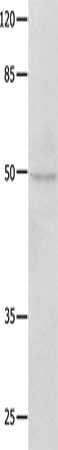
 SDS-PAGE
(Gel: 12%SDS-PAGE, Lysate: 40 ug, Lane: Hela cells, Primary antibody: AAA237819(SNAP25 Antibody) at dilution 1/400, Secondary antibody: Goat anti rabbit IgG at 1/8000 dilution, Exposure time: 3 seconds)
SDS-PAGE
(Gel: 12%SDS-PAGE, Lysate: 40 ug, Lane: Hela cells, Primary antibody: AAA237819(SNAP25 Antibody) at dilution 1/400, Secondary antibody: Goat anti rabbit IgG at 1/8000 dilution, Exposure time: 3 seconds)
SNAP25, Polyclonal Antibody (Cat# AAA237819)
 SDS-PAGE
(Gel: 10%SDS-PAGE, Lysate: 40 ug, Lane: jurkat cells, Primary antibody: AAA237825(SOCS6 Antibody) at dilution 1/600, Secondary antibody: Goat anti rabbit IgG at 1/8000 dilution, Exposure time: 1 second)
SDS-PAGE
(Gel: 10%SDS-PAGE, Lysate: 40 ug, Lane: jurkat cells, Primary antibody: AAA237825(SOCS6 Antibody) at dilution 1/600, Secondary antibody: Goat anti rabbit IgG at 1/8000 dilution, Exposure time: 1 second)
SOCS6, Polyclonal Antibody (Cat# AAA237825)
What are Polyclonal Antibodies?
Polyclonal antibodies are antibodies that come from multiple B cell clones of a host animal. The typical hosts used for the majority of polyclonal antibody production are rabbits, goats, sheep, and donkeys. These polyclonal antibodies, once having identified their target, will bind to different epitopes located at different regions or sequences on the same protein/antigen. As a result, they are ideal at locating and binding to the target, even if the target is in very low concentrations (due to many different antibodies being able to bind to the same target molecule, which allows for significant amplification of a downstream signal).
Polyclonal antibodies are typically produced by injecting an antigen into a host animal, which causes the animal’s immune system to attack the foreign antigen by mass generating antibodies against it. After a period of time, serum is collected from the animal and purified using physicochemical fractionation, class-specific affinity purification, and/or antigen-affinity purification.
Key Uses of Polyclonal Antibodies
- Western Blotting: This method is used to find specific proteins in biological samples after separating them by size.
- Immunohistochemistry: IHC helps visualize the location of proteins in tissue sections using various staining techniques.
- ELISA: (Enzyme-Linked Immunosorbent Assay) is typically used to identify specific protein quantities in a sample. ELISAs can be either “Quantitative” or “Qualitative”.
- Flow Cytometry: technique that identifies and measures the specific protein on the surface or inside the cells in a fluid suspension.
- Immunoprecipitation: IP isolates and studies a specific protein from a complex mixture using antibodies.
Why Buy Polyclonal Antibodies from AAA Biotech?
1. Ideal for Various Applications
Our antibodies are generally going to be validated for use in multiple types of assays, including ELISA, Western Blotting, Immunohistochemistry, Immunoprecipitation, amongst others. They are ideal for a wide range of research applications.
2. Rigorous Quality Control
All of the antibodies in our catalog undergo strict quality testing to ensure specificity, sensitivity, and consistent performance. We are confident in the ability of our antibodies to provide you with accurate results.
3. Wide Assortment of Antibodies
Antibodies in are catalog can be found for both common and exotic species, and these antibodies are also available in both conjugated and recombinant forms to suit many diverse experimental needs.
4. Highly Purified
Our antibodies are available in purified forms with over 85% purity, as confirmed by SDS-PAGE. They are also available with tags such as His, Flag, GST, or MBP. We cater to customers worldwide.
FAQ
1. How are polyclonal antibodies produced?
Traditionally, polyclonal antibodies are produced by injecting an antigen into a host animal (such as a rabbit or goat), which then triggers an immune response from the host animal. The animal’s B cells produce antibodies that will recognize different parts of the injected antigen. These antibodies are then collected from the animal’s blood and purified for use.
2. How do polyclonal antibodies differ from monoclonal antibodies?
Polyclonal antibodies are a mix of antibodies that bind to different locations (epitopes) of the same antigen, while monoclonal antibodies are identical and bind to just one specific epitope. This makes polyclonal antibodies more versatile and better at detecting proteins that may be present in low quantities or in altered/modified forms.
3. How should I store polyclonal antibodies?
Polyclonal antibodies should be stored at 4°C for short-term use (up to a few weeks) and at -20°C or -80°C for long-term storage. Avoid repeated freeze-thaw cycles by dividing them into small aliquots. Always check the datasheet for specific storage instructions.