Filters
▼Clonality
▼Type
▼Reactivity
▼Gene Name
▼Isotype
▼Host
▼Application
▼Clone
▼Polyclonal Antibodies
At AAA Biotech also known as AAA Bio or AAABio, we provide a broad range of purified polyclonal antibodies (pAbs) that are able to all be browsed online through our website. Due to their high specificity and strong binding affinity, these antibodies are ideal for wide swathes of research and experimental applications.
Our polyclonal antibodies can easily support your work, whether you use them for Western Blotting, Immunocytochemistry (with or without Immunofluorescence used in conjunction), Immunohistochemistry, Immunoprecipitation, and ELISA tests. We highly encourage you to browse our range of pAbs and choose the one that best suits your experimental model.
Viewing 1300-1350 of 96805 product results
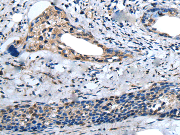
 IHC (Immunohiostchemistry)
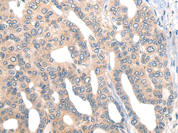
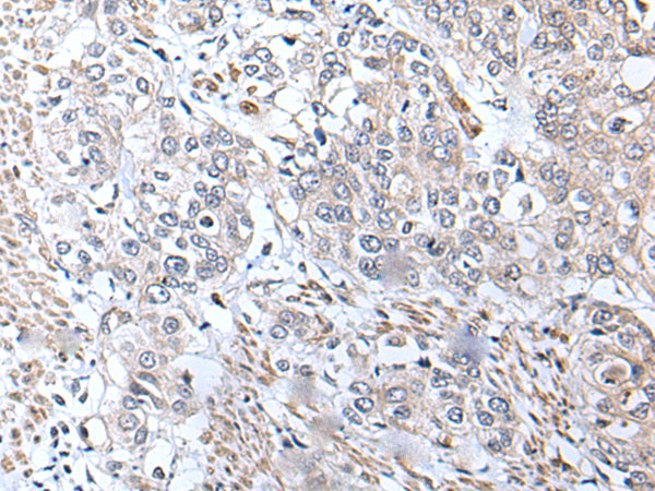
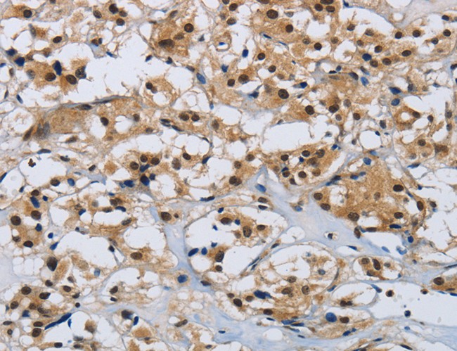
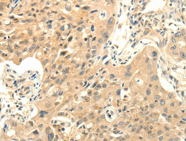
(Immunohistochemistry of paraffin-embedded Human cervical cancer tissue using KBTBD11 Polyclonal Antibody at dilution of 1:35(×200))
IHC (Immunohiostchemistry)
(Immunohistochemistry of paraffin-embedded Human cervical cancer tissue using KBTBD11 Polyclonal Antibody at dilution of 1:35(×200))
KBTBD11, Polyclonal Antibody (Cat# AAA176093)
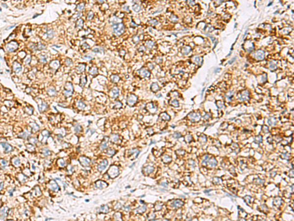
 IHC (Immunohiostchemistry)
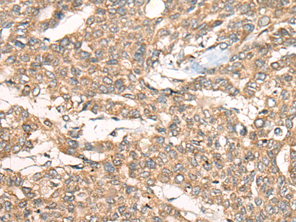
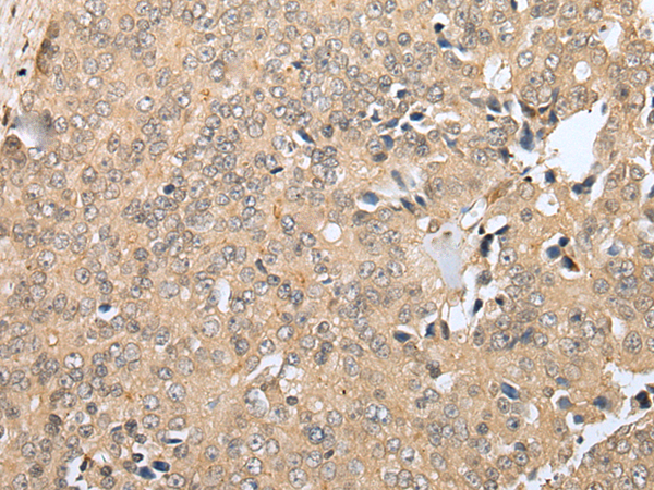
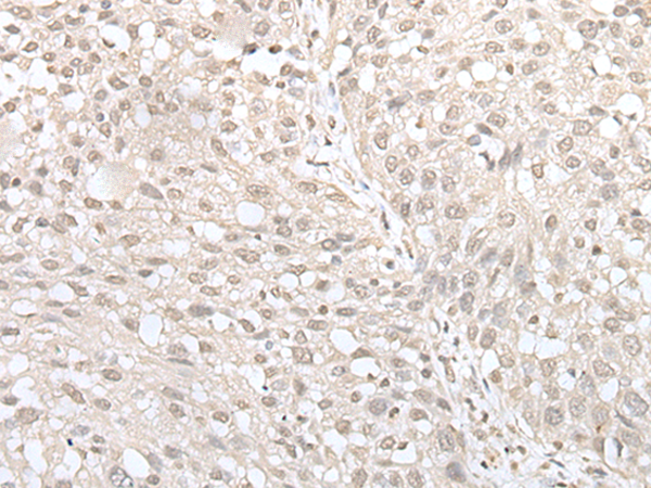
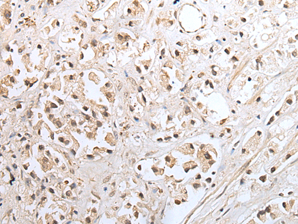
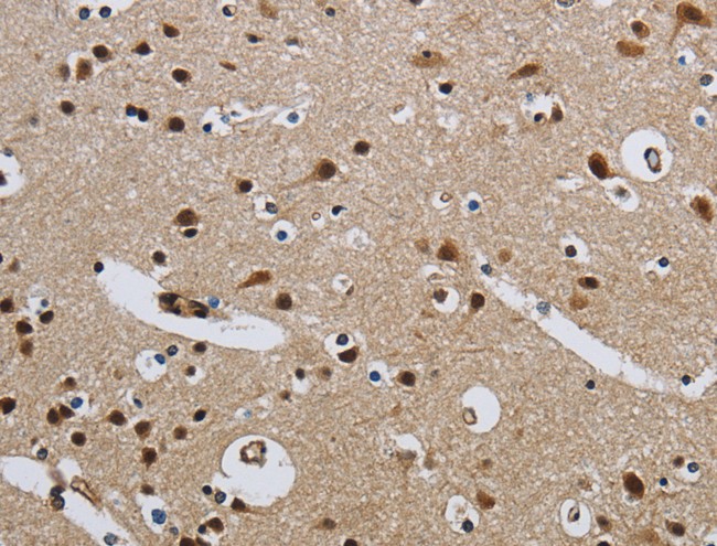
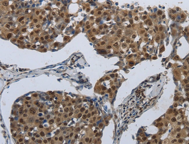
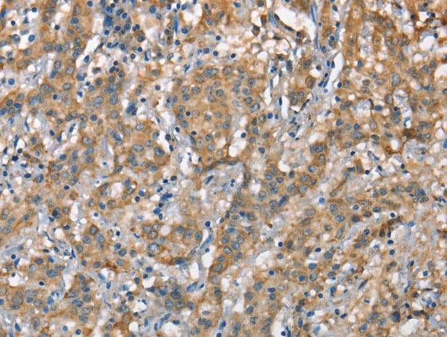
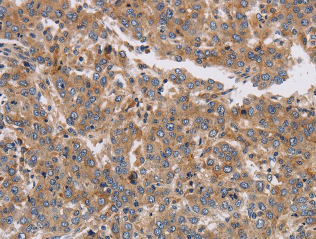
(Immunohistochemistry of paraffin-embedded Human liver cancer tissue using CUL5 Polyclonal Antibody at dilution of 1:55(×200))
IHC (Immunohiostchemistry)
(Immunohistochemistry of paraffin-embedded Human liver cancer tissue using CUL5 Polyclonal Antibody at dilution of 1:55(×200))
CUL5, Polyclonal Antibody (Cat# AAA176116)
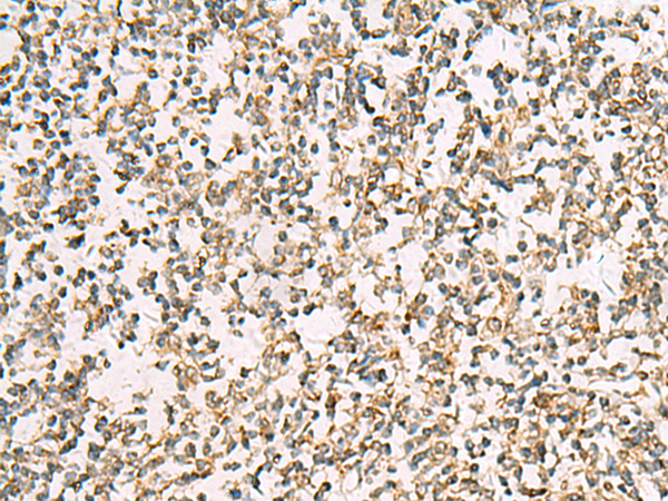
 IHC (Immunohiostchemistry)
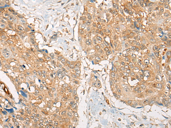
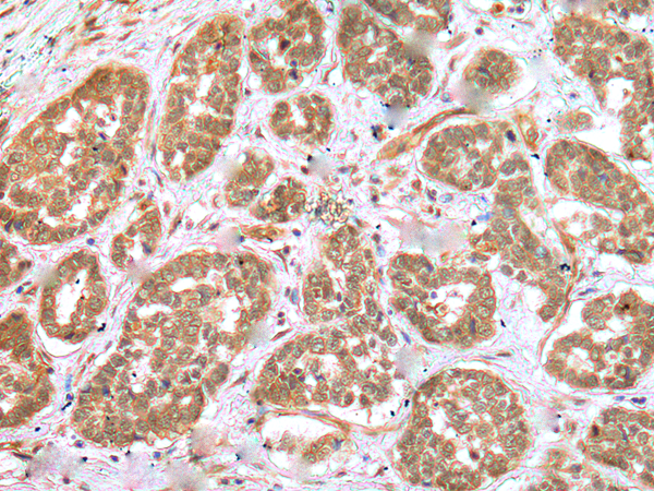
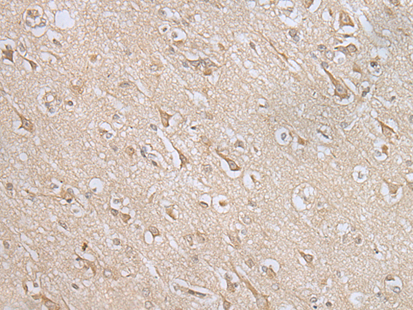
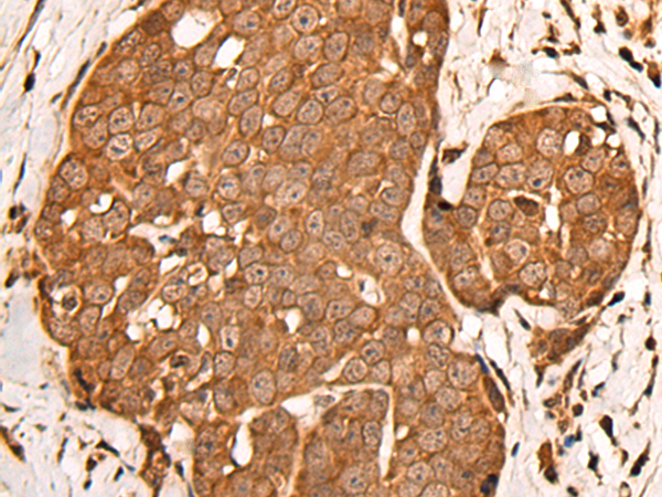
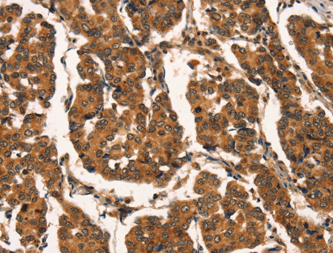
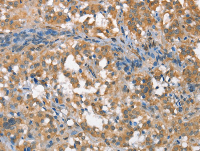
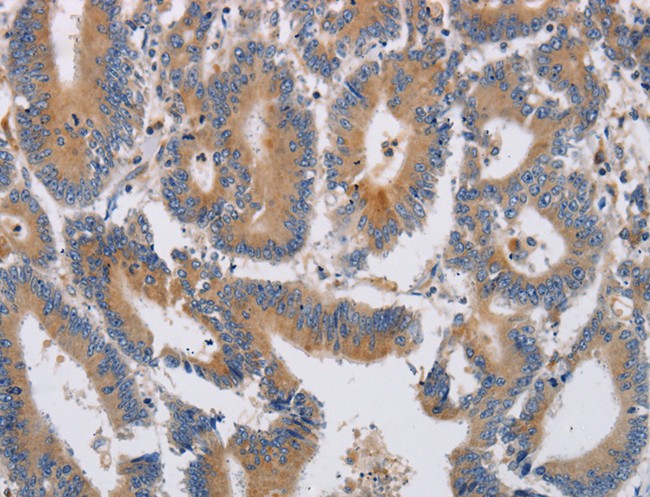
(Immunohistochemistry of paraffin-embedded Human esophagus cancer tissue using ZXDA Polyclonal Antibody at dilution of 1:30(×200))
IHC (Immunohiostchemistry)
(Immunohistochemistry of paraffin-embedded Human esophagus cancer tissue using ZXDA Polyclonal Antibody at dilution of 1:30(×200))
ZXDA, Polyclonal Antibody (Cat# AAA175821)
 IHC (Immunohiostchemistry)
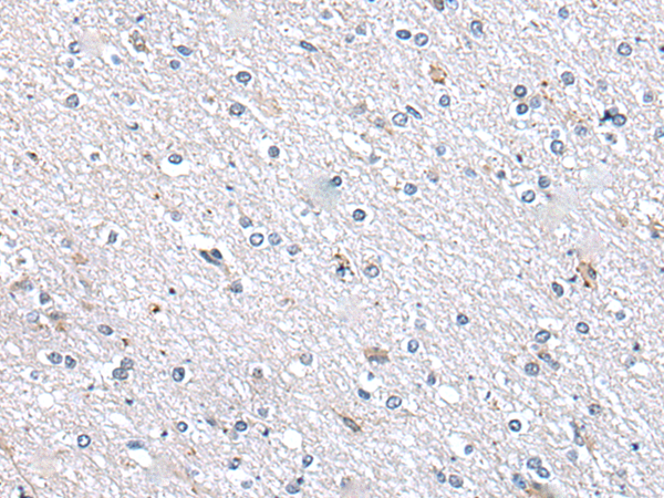
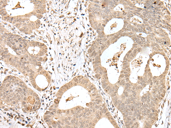
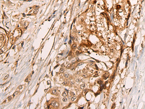
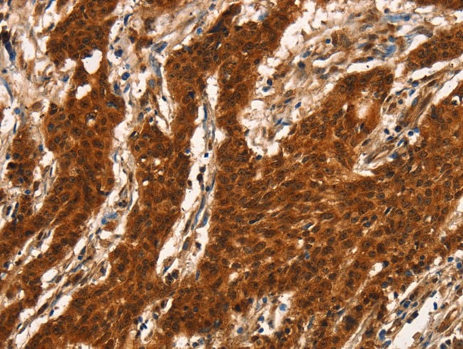
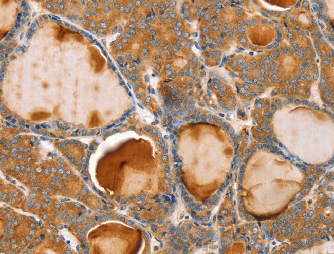
(Immunohistochemistry of paraffin-embedded Human thyroid cancer tissue using YPEL4 Polyclonal Antibody at dilution of 1:25(×200))
IHC (Immunohiostchemistry)
(Immunohistochemistry of paraffin-embedded Human thyroid cancer tissue using YPEL4 Polyclonal Antibody at dilution of 1:25(×200))
YPEL4, Polyclonal Antibody (Cat# AAA175830)

 IHC (Immunohiostchemistry)
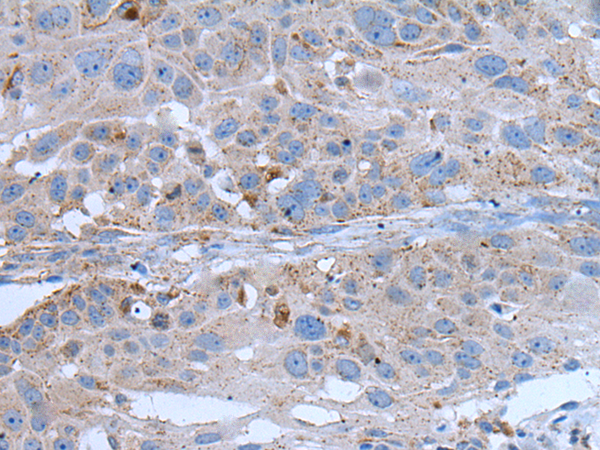
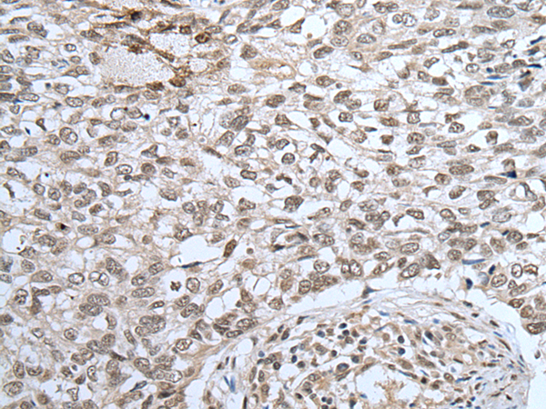
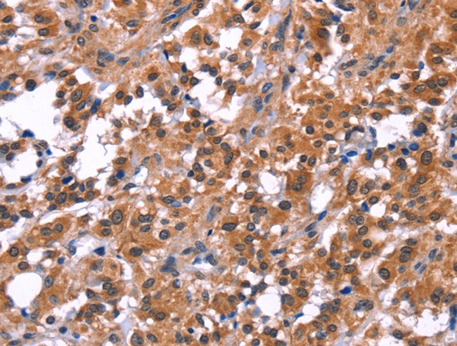
(Immunohistochemistry of paraffin-embedded Human liver cancer tissue using CORO2B Polyclonal Antibody at dilution of 1:50(×200))
IHC (Immunohiostchemistry)
(Immunohistochemistry of paraffin-embedded Human liver cancer tissue using CORO2B Polyclonal Antibody at dilution of 1:50(×200))
CORO2B, Polyclonal Antibody (Cat# AAA175933)
 IHC (Immunohiostchemistry)
(Immunohistochemistry of paraffin-embedded Human esophagus cancer tissue using RNF114 Polyclonal Antibody at dilution of 1:25(×200))
IHC (Immunohiostchemistry)
(Immunohistochemistry of paraffin-embedded Human esophagus cancer tissue using RNF114 Polyclonal Antibody at dilution of 1:25(×200))
RNF114, Polyclonal Antibody (Cat# AAA176219)
 IHC (Immunohiostchemistry)
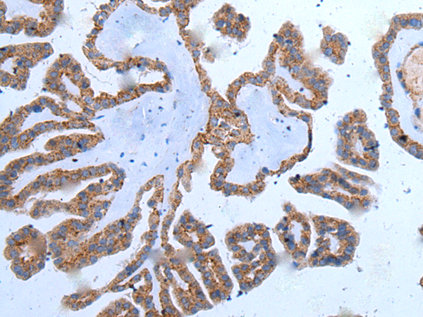
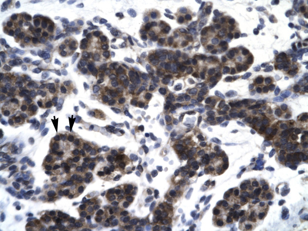
(Immunohistochemistry of paraffin-embedded Human prost ate cancer tissue using METTL7A Polyclonal Antibody at dilution of 1:40(×200))
IHC (Immunohiostchemistry)
(Immunohistochemistry of paraffin-embedded Human prost ate cancer tissue using METTL7A Polyclonal Antibody at dilution of 1:40(×200))
METTL7A, Polyclonal Antibody (Cat# AAA176228)
 IHC (Immunohiostchemistry)
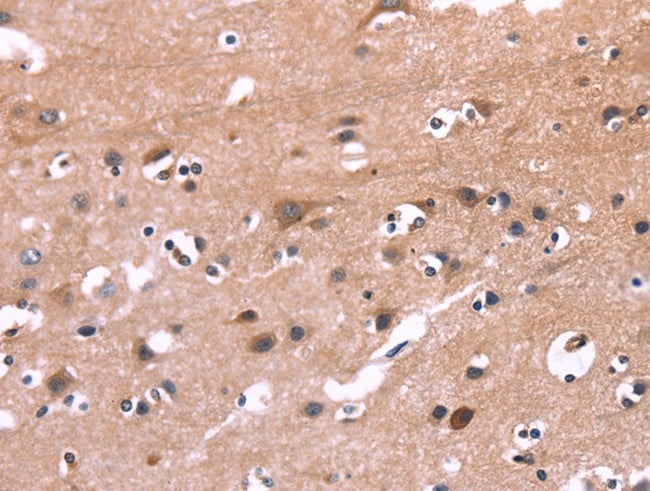
(Immunohistochemistry of paraffin-embedded Human brain tissue using MIF4GD Polyclonal Antibody at dilution of 1:30(×200))
IHC (Immunohiostchemistry)
(Immunohistochemistry of paraffin-embedded Human brain tissue using MIF4GD Polyclonal Antibody at dilution of 1:30(×200))
MIF4GD, Polyclonal Antibody (Cat# AAA176233)
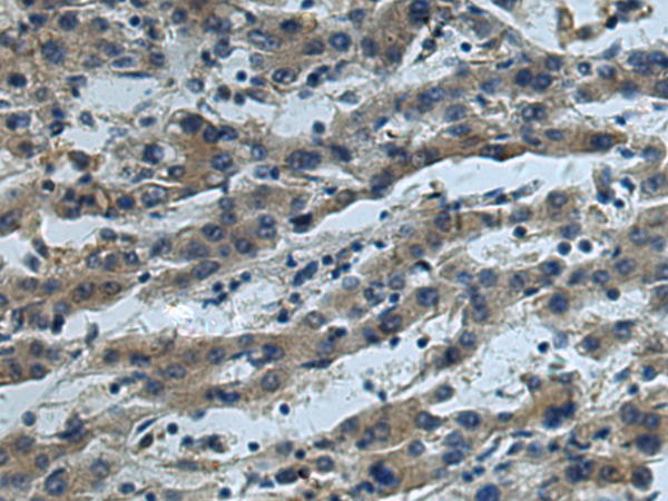
 IHC (Immunohiostchemistry)
(Immunohistochemistry of paraffin-embedded Human prost ate cancer tissue using SNF8 Polyclonal Antibody at dilution of 1:30(×200))
IHC (Immunohiostchemistry)
(Immunohistochemistry of paraffin-embedded Human prost ate cancer tissue using SNF8 Polyclonal Antibody at dilution of 1:30(×200))
SNF8, Polyclonal Antibody (Cat# AAA176234)
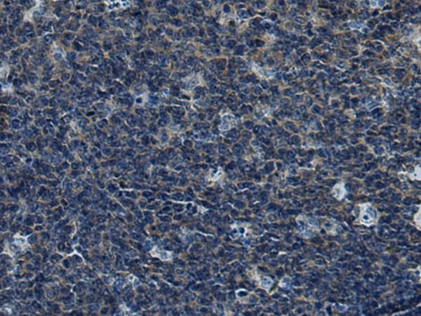
 IHC (Immunohiostchemistry)
(Immunohistochemistry of paraffin-embedded Human prost ate cancer tissue using ZPR1 Polyclonal Antibody at dilution of 1:30(×200))
IHC (Immunohiostchemistry)
(Immunohistochemistry of paraffin-embedded Human prost ate cancer tissue using ZPR1 Polyclonal Antibody at dilution of 1:30(×200))
ZPR1, Polyclonal Antibody (Cat# AAA176247)
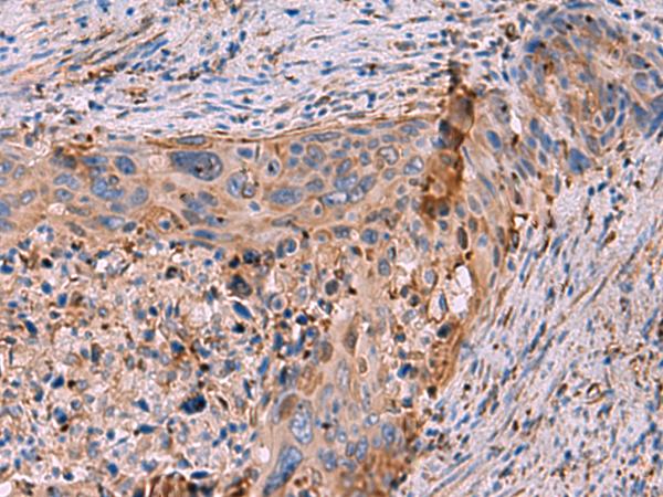
 IHC (Immunohiostchemistry)
(Immunohistochemistry of paraffin-embedded Human cervical cancer tissue using ZNF37A Polyclonal Antibody at dilution of 1:30(×200))
IHC (Immunohiostchemistry)
(Immunohistochemistry of paraffin-embedded Human cervical cancer tissue using ZNF37A Polyclonal Antibody at dilution of 1:30(×200))
ZNF37A, Polyclonal Antibody (Cat# AAA176254)
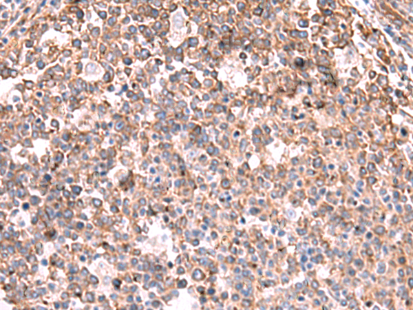
 IHC (Immunohiostchemistry)
(Immunohistochemistry of paraffin-embedded Human tonsil tissue using ZPBP2 Polyclonal Antibody at dilution of 1:50(×200))
IHC (Immunohiostchemistry)
(Immunohistochemistry of paraffin-embedded Human tonsil tissue using ZPBP2 Polyclonal Antibody at dilution of 1:50(×200))
ZPBP2, Polyclonal Antibody (Cat# AAA176264)
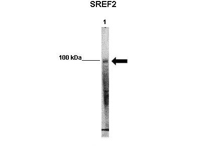
 WB (Western Blot)
(WB Suggested Anti-SREBF2 Antibody Titration: 0.2-1 ug/mlELISA Titer: 1:62500Positive Control: Raji cell lysateSREBF2 is strongly supported by BioGPS gene expression data to be expressed in Human Raji cells)
WB (Western Blot)
(WB Suggested Anti-SREBF2 Antibody Titration: 0.2-1 ug/mlELISA Titer: 1:62500Positive Control: Raji cell lysateSREBF2 is strongly supported by BioGPS gene expression data to be expressed in Human Raji cells)
SREBF2, Polyclonal Antibody (Cat# AAA201702)
 WB (Western Blot)
(WB Suggested Anti-SREBF1 Antibody Titration: 0.2-1 ug/mlELISA Titer: 1:62500Positive Control: Transfected 293T)
WB (Western Blot)
(WB Suggested Anti-SREBF1 Antibody Titration: 0.2-1 ug/mlELISA Titer: 1:62500Positive Control: Transfected 293T)
SREBF1, Polyclonal Antibody (Cat# AAA201704)

 WB (Western Blot)
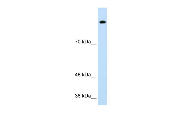
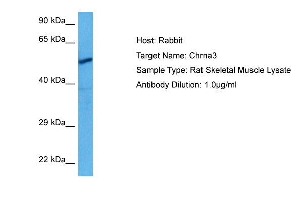
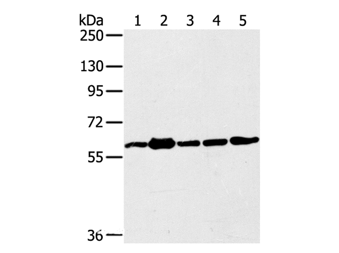
(WB Suggested Anti-CHRNA3 Antibody Titration: 0.1ug/mlPositive Control: Human Thymus)
WB (Western Blot)
(WB Suggested Anti-CHRNA3 Antibody Titration: 0.1ug/mlPositive Control: Human Thymus)
CHRNA3, Polyclonal Antibody (Cat# AAA201709)
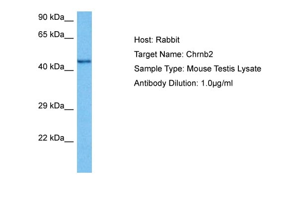
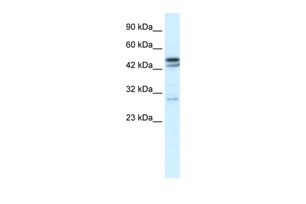
 WB (Western Blot)
(WB Suggested Anti-CHRNB2 Antibody Titration: 0.2-1 ug/mlELISA Titer: 1:62500Positive Control: HepG2 cell lysate)
WB (Western Blot)
(WB Suggested Anti-CHRNB2 Antibody Titration: 0.2-1 ug/mlELISA Titer: 1:62500Positive Control: HepG2 cell lysate)
CHRNB2, Polyclonal Antibody (Cat# AAA201711)
 WB (Western Blot)
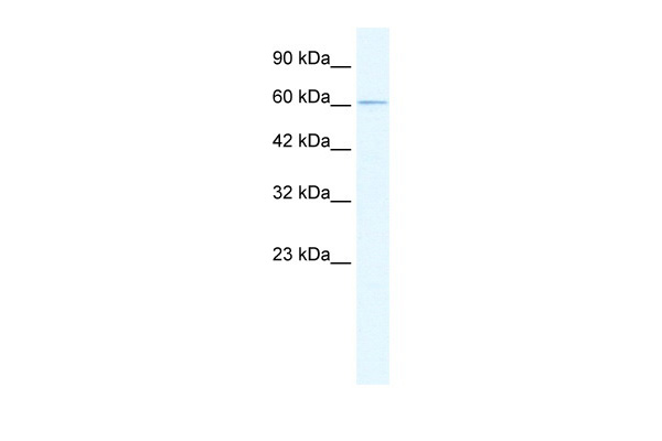
(WB Suggested Anti-CHRND Antibody Titration: 0.2-1 ug/mlELISA Titer: 1:312500Positive Control: HepG2 cell lysate)
WB (Western Blot)
(WB Suggested Anti-CHRND Antibody Titration: 0.2-1 ug/mlELISA Titer: 1:312500Positive Control: HepG2 cell lysate)
CHRND, Polyclonal Antibody (Cat# AAA201712)
 WB (Western Blot)
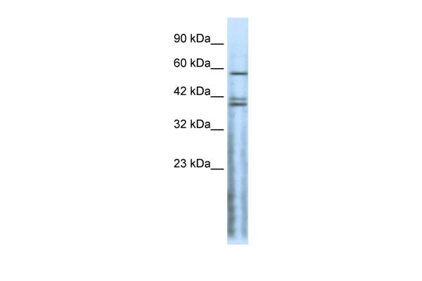
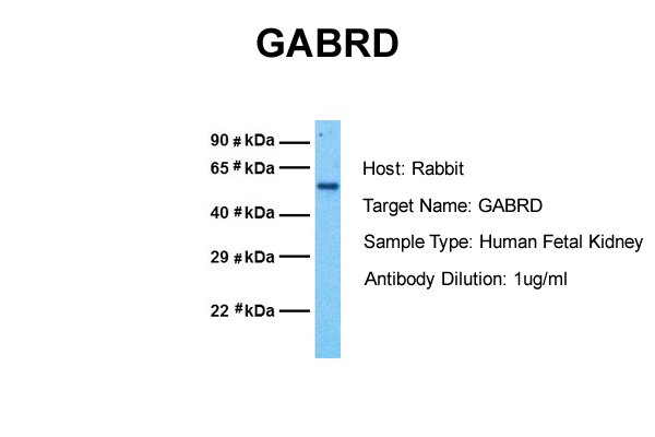
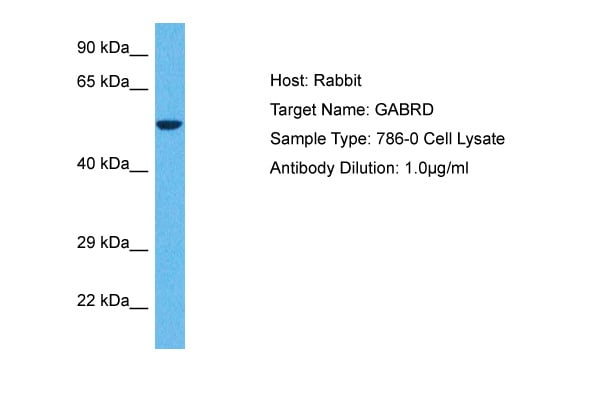
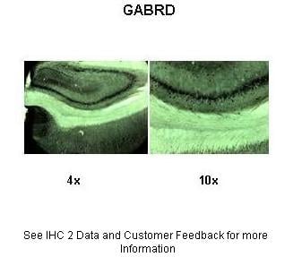
(WB Suggested Anti-GABRD Antibody Titration: 0.2-1 ug/mlPositive Control: HepG2 cell lysate)
WB (Western Blot)
(WB Suggested Anti-GABRD Antibody Titration: 0.2-1 ug/mlPositive Control: HepG2 cell lysate)
GABRD, Polyclonal Antibody (Cat# AAA201713)
 WB (Western Blot)
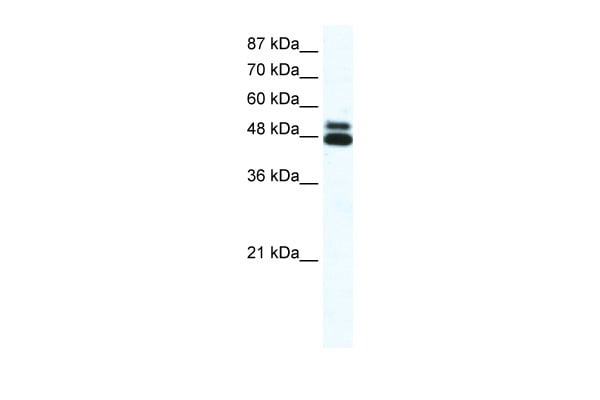
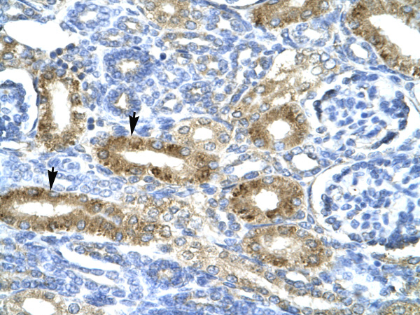
(WB Suggested Anti-OPRK1 Antibody Titration: 1 ug/mlPositive Control: HuFetal Kidney)
WB (Western Blot)
(WB Suggested Anti-OPRK1 Antibody Titration: 1 ug/mlPositive Control: HuFetal Kidney)
OPRK1, Polyclonal Antibody (Cat# AAA201719)

 WB (Western Blot)
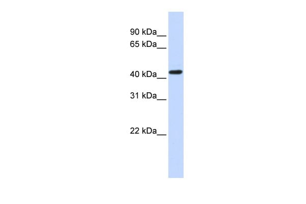
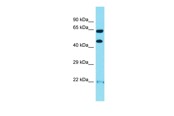
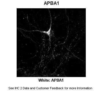
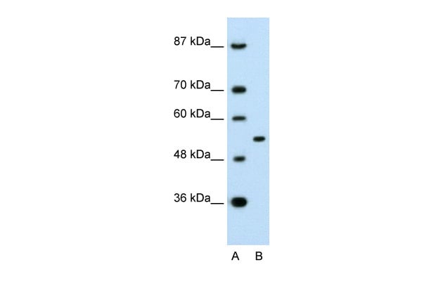
(WB Suggested Anti-APBA1 Antibody Titration: 0.2-1 ug/mlPositive Control: Raji cell lysateAPBA1 is supported by BioGPS gene expression data to be expressed in Raji)
WB (Western Blot)
(WB Suggested Anti-APBA1 Antibody Titration: 0.2-1 ug/mlPositive Control: Raji cell lysateAPBA1 is supported by BioGPS gene expression data to be expressed in Raji)
APBA1, Polyclonal Antibody (Cat# AAA201722)
 WB (Western Blot)
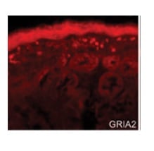
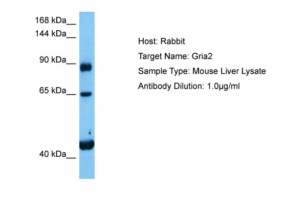
(Host: MouseTarget Name: GRIA2Sample Tissue: Mouse LiverAntibody Dilution: 1ug/ml)
WB (Western Blot)
(Host: MouseTarget Name: GRIA2Sample Tissue: Mouse LiverAntibody Dilution: 1ug/ml)
GRIA2, Polyclonal Antibody (Cat# AAA201723)
 WB (Western Blot)
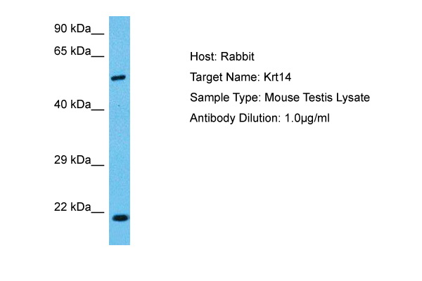
(WB Suggested Anti-KRT14 Antibody Titration: 1.25ug/mlELISA Titer: 1:312500Positive Control: Human Thymus)
WB (Western Blot)
(WB Suggested Anti-KRT14 Antibody Titration: 1.25ug/mlELISA Titer: 1:312500Positive Control: Human Thymus)
KRT14, Polyclonal Antibody (Cat# AAA201729)
 WB (Western Blot)
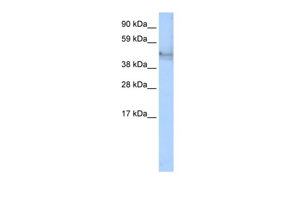
(WB Suggested Anti-SERPINF1 Antibody Titration: 0.2-1 ug/mlPositive Control: Human Placenta)
WB (Western Blot)
(WB Suggested Anti-SERPINF1 Antibody Titration: 0.2-1 ug/mlPositive Control: Human Placenta)
SERPINF1, Polyclonal Antibody (Cat# AAA201732)

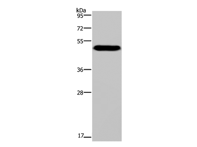
 WB (Western Blot)
(WB Suggested Anti-SMAD3 Antibody Titration: 2.0ug/mlPositive Control: Human Liver)
WB (Western Blot)
(WB Suggested Anti-SMAD3 Antibody Titration: 2.0ug/mlPositive Control: Human Liver)
SMAD3, Polyclonal Antibody (Cat# AAA201736)
 WB (Western Blot)
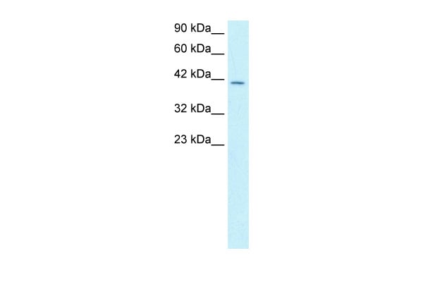
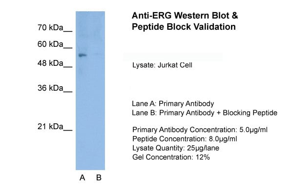
(WB Suggested Anti-ERG Antibody Titration: 1.0ug/mlPositive Control: Jurkat cell lysate)
WB (Western Blot)
(WB Suggested Anti-ERG Antibody Titration: 1.0ug/mlPositive Control: Jurkat cell lysate)
ERG, Polyclonal Antibody (Cat# AAA201737)
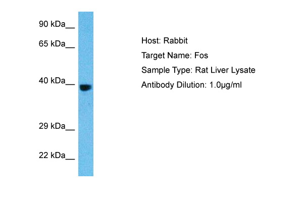
 WB (Western Blot)
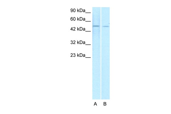
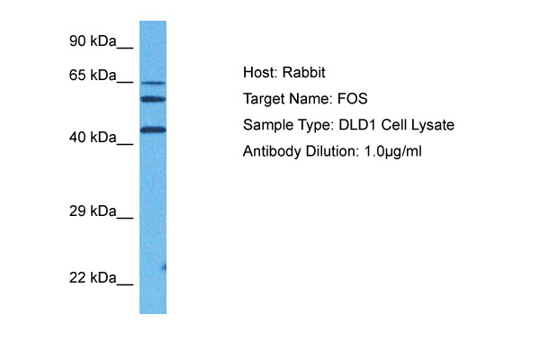
(WB Suggested Anti-FOS Antibody Titration: 0.2-1 ug/mlELISA Titer: 1:62500Positive Control: 293T cell lysate)
WB (Western Blot)
(WB Suggested Anti-FOS Antibody Titration: 0.2-1 ug/mlELISA Titer: 1:62500Positive Control: 293T cell lysate)
FOS, Polyclonal Antibody (Cat# AAA201738)
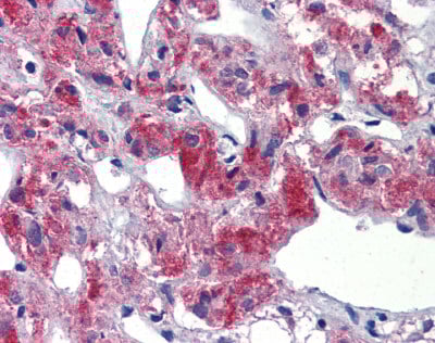
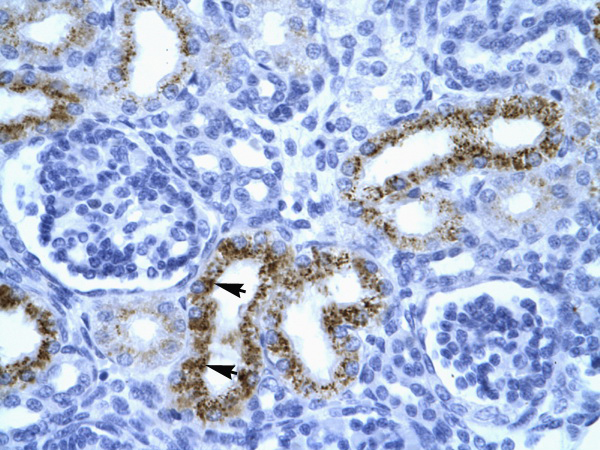
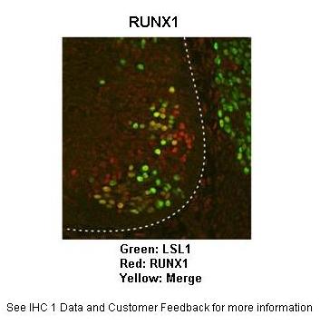
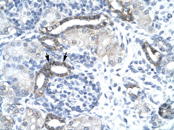
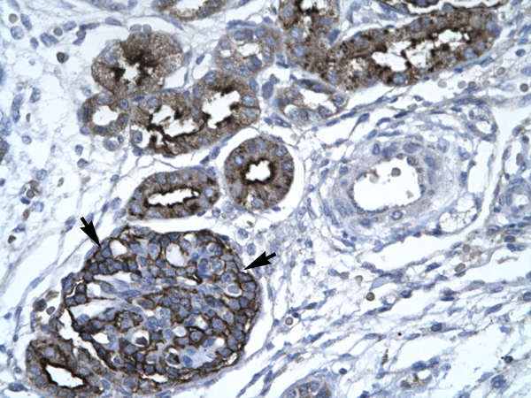
 IHC (Immunohiostchemistry)
(Rabbit Anti-TAL1 AntibodyParaffin Embedded Tissue: Human hepatocyte cellCellular Data: Epithelial cells of renal tubuleAntibody Concentration: 4.0-8.0 ug/mlMagnification: 400X)
IHC (Immunohiostchemistry)
(Rabbit Anti-TAL1 AntibodyParaffin Embedded Tissue: Human hepatocyte cellCellular Data: Epithelial cells of renal tubuleAntibody Concentration: 4.0-8.0 ug/mlMagnification: 400X)
TAL1, Polyclonal Antibody (Cat# AAA201739)
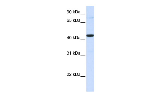
 WB (Western Blot)
(WB Suggested Anti-FOS Antibody Titration: 1.25ug/mlELISA Titer: 1:312500Positive Control: HepG2 cell lysate)
WB (Western Blot)
(WB Suggested Anti-FOS Antibody Titration: 1.25ug/mlELISA Titer: 1:312500Positive Control: HepG2 cell lysate)
FOS, Polyclonal Antibody (Cat# AAA201740)
 WB (Western Blot)
(WB Suggested Anti-NFYC Antibody Titration: 0.2-1 ug/mlELISA Titer: 1:62500Positive Control: Jurkat cell lysateNFYC is strongly supported by BioGPS gene expression data to be expressed in Human Jurkat cells)
WB (Western Blot)
(WB Suggested Anti-NFYC Antibody Titration: 0.2-1 ug/mlELISA Titer: 1:62500Positive Control: Jurkat cell lysateNFYC is strongly supported by BioGPS gene expression data to be expressed in Human Jurkat cells)
NFYC, Polyclonal Antibody (Cat# AAA201742)
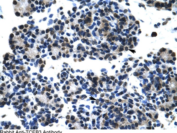
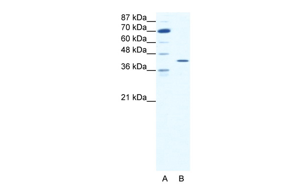
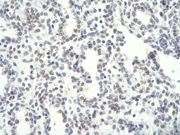
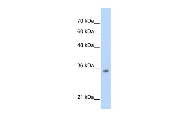
 WB (Western Blot)
(WB Suggested Anti-TCEB3 Antibody Titration: 7.5ug/mlPositive Control: Transfected 293TTCEB3 is strongly supported by BioGPS gene expression data to be expressed in Human HEK293T cells)
WB (Western Blot)
(WB Suggested Anti-TCEB3 Antibody Titration: 7.5ug/mlPositive Control: Transfected 293TTCEB3 is strongly supported by BioGPS gene expression data to be expressed in Human HEK293T cells)
ELOA, Polyclonal Antibody (Cat# AAA201744)
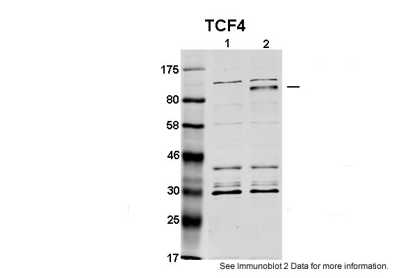
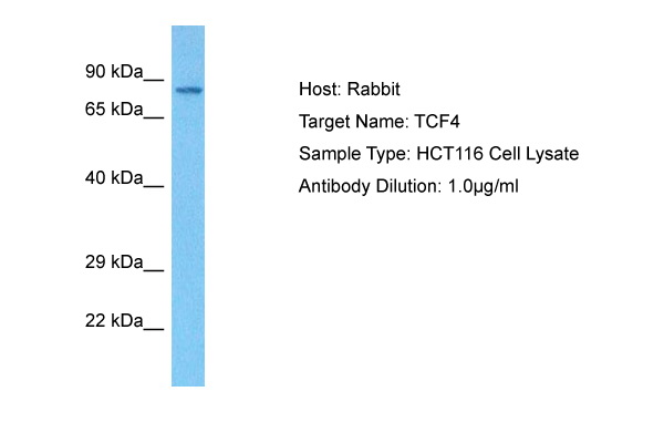
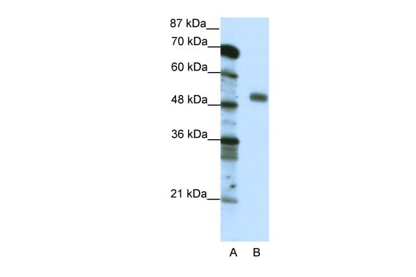
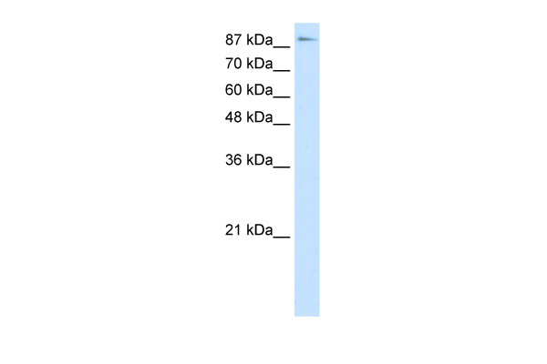
 WB (Western Blot)
(WB Suggested Anti-TCF4 AntibodyTitration: 0.2-1 ug/mlPositive Control: HepG2)
WB (Western Blot)
(WB Suggested Anti-TCF4 AntibodyTitration: 0.2-1 ug/mlPositive Control: HepG2)
TCF4, Polyclonal Antibody (Cat# AAA201752)
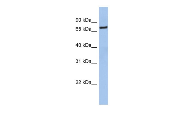 WB (Western Blot)
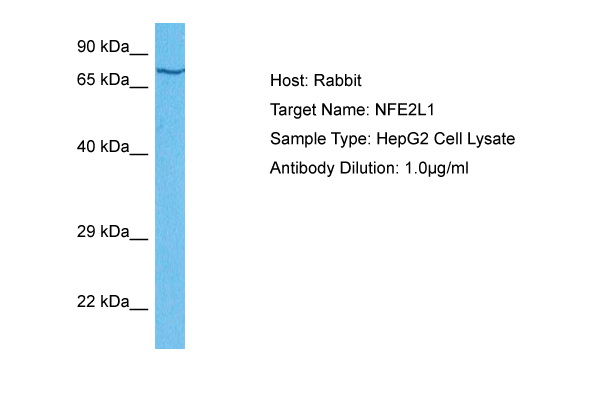
(NFE2L1 (nuclear factor (erythroid-derived 2)-like 1) Antibody (against the middle region of NFE2L1) (50ug) validated by WB using Jurkat cell lysate at 0.2-1 ug/ml.)
WB (Western Blot)
(NFE2L1 (nuclear factor (erythroid-derived 2)-like 1) Antibody (against the middle region of NFE2L1) (50ug) validated by WB using Jurkat cell lysate at 0.2-1 ug/ml.)
NFE2L1, Polyclonal Antibody (Cat# AAA201753)
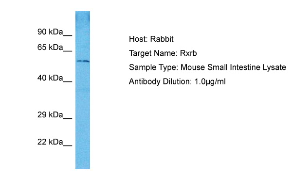
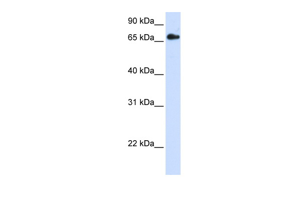 WB (Western Blot)
(WB Suggested Anti-RXRB Antibody Titration: 0.2-1 ug/mlELISA Titer: 1:312500Positive Control: Transfected 293TRXRB is strongly supported by BioGPS gene expression data to be expressed in Human HEK293T cells)
WB (Western Blot)
(WB Suggested Anti-RXRB Antibody Titration: 0.2-1 ug/mlELISA Titer: 1:312500Positive Control: Transfected 293TRXRB is strongly supported by BioGPS gene expression data to be expressed in Human HEK293T cells)
RXRB, Polyclonal Antibody (Cat# AAA201755)
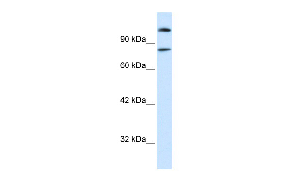
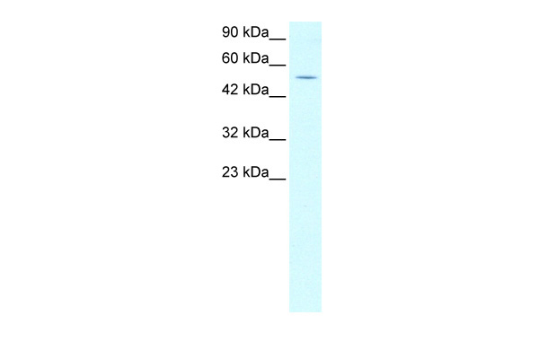
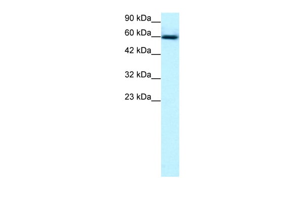
 WB (Western Blot)
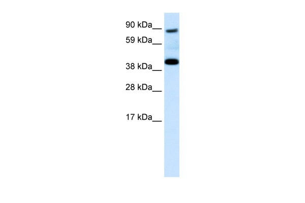
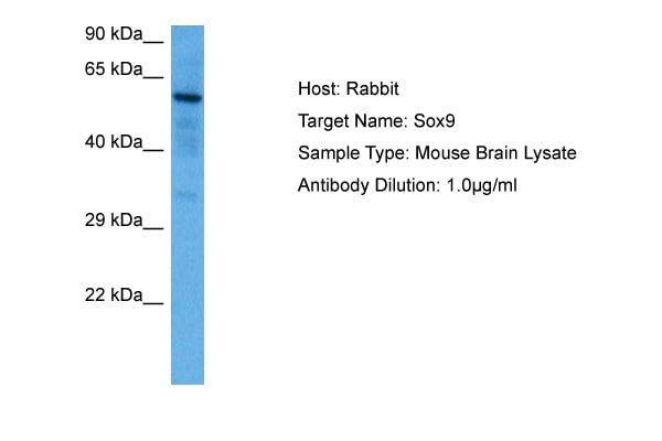
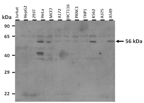
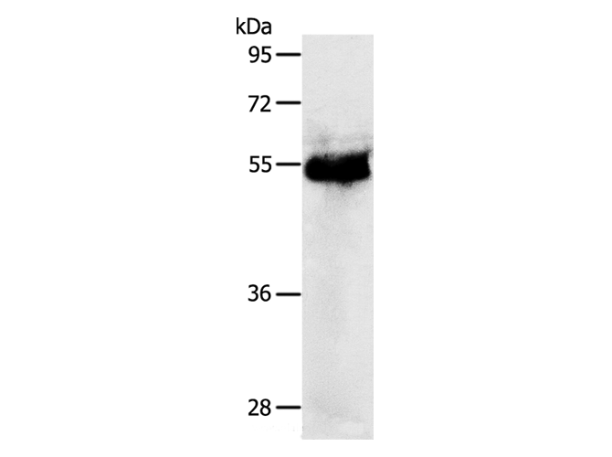
(25 ug of the indicated Human whole cell extracts was loaded onto a 12% SDS-PAGE gel. 3 ug/mL of the antibody was used in this experiment. The protein is modified by phosphorylation, and a putative 49 kDa isoform also contains this immunizing peptide sequence.)
WB (Western Blot)
(25 ug of the indicated Human whole cell extracts was loaded onto a 12% SDS-PAGE gel. 3 ug/mL of the antibody was used in this experiment. The protein is modified by phosphorylation, and a putative 49 kDa isoform also contains this immunizing peptide sequence.)
SOX9, Polyclonal Antibody (Cat# AAA201756)
Predicted Species Reactivity: Human, Mouse, Rat, Cow, Dog, Goat, Guinea Pig, Rabbit, Zebrafish
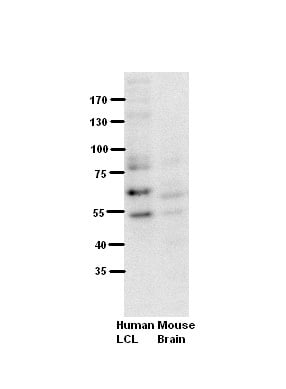 WB (Western Blot)
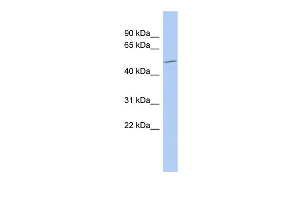
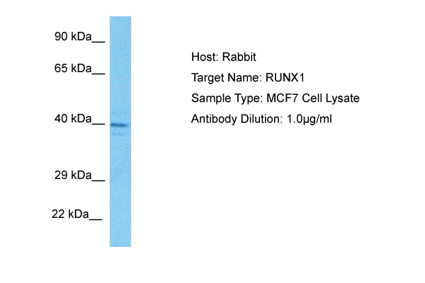
(WB Suggested Anti-RUNX1 Antibody Titration: 5% MilkELISA Titer: dilution: 1:500Positive Control: Human LCL and mouse brains)
WB (Western Blot)
(WB Suggested Anti-RUNX1 Antibody Titration: 5% MilkELISA Titer: dilution: 1:500Positive Control: Human LCL and mouse brains)
RUNX1, Polyclonal Antibody (Cat# AAA201761)
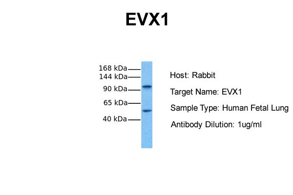
 WB (Western Blot)
(WB Suggested Anti-EVX1 Antibody Titration: 1.0-2.0ug/mlPositive Control: Jurkat cell lysate)
WB (Western Blot)
(WB Suggested Anti-EVX1 Antibody Titration: 1.0-2.0ug/mlPositive Control: Jurkat cell lysate)
EVX1, Polyclonal Antibody (Cat# AAA201764)

 WB (Western Blot)
(WB Suggested Anti-GABPA Antibody Titration: 0.5-1.0ug/mlPositive Control: Jurkat cell lysateGABPA is supported by BioGPS gene expression data to be expressed in Jurkat)
WB (Western Blot)
(WB Suggested Anti-GABPA Antibody Titration: 0.5-1.0ug/mlPositive Control: Jurkat cell lysateGABPA is supported by BioGPS gene expression data to be expressed in Jurkat)
GABPA, Polyclonal Antibody (Cat# AAA201765)
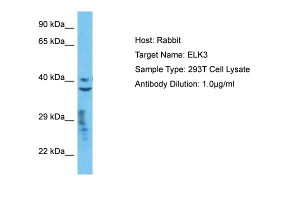
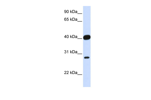 WB (Western Blot)
(WB Suggested Anti-ELK3 Antibody Titration: 0.2-1 ug/mlELISA Titer: 1:62500Positive Control: Human heart)
WB (Western Blot)
(WB Suggested Anti-ELK3 Antibody Titration: 0.2-1 ug/mlELISA Titer: 1:62500Positive Control: Human heart)
ELK3, Polyclonal Antibody (Cat# AAA201773)
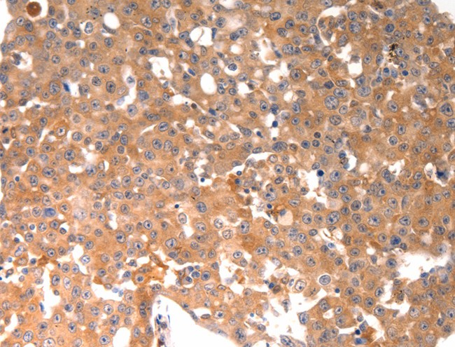
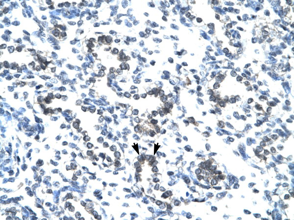
 IHC (Immunohiostchemistry)
(Immunohistochemistry of paraffin-embedded Human ovarian cancer using BGLAP Polyclonal Antibody at dilution of 1:40)
IHC (Immunohiostchemistry)
(Immunohistochemistry of paraffin-embedded Human ovarian cancer using BGLAP Polyclonal Antibody at dilution of 1:40)
BGLAP, Polyclonal Antibody (Cat# AAA168490)
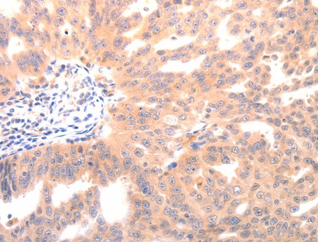
 IHC (Immunohiostchemistry)
(Immunohistochemistry of paraffin-embedded Human brain tissue using CMTM8 Polyclonal Antibody at dilution 1:40)
IHC (Immunohiostchemistry)
(Immunohistochemistry of paraffin-embedded Human brain tissue using CMTM8 Polyclonal Antibody at dilution 1:40)
CMTM8, Polyclonal Antibody (Cat# AAA168498)
 IHC (Immunohiostchemistry)
(Immunohistochemistry of paraffin-embedded Human gastric cancer tissue using CTNNBIP1 Polyclonal Antibody at dilution 1:20)
IHC (Immunohiostchemistry)
(Immunohistochemistry of paraffin-embedded Human gastric cancer tissue using CTNNBIP1 Polyclonal Antibody at dilution 1:20)
CTNNBIP1, Polyclonal Antibody (Cat# AAA168509)
 IHC (Immunohistochemisry)
(Immunohistochemistry of paraffin-embedded Human lung cancer using PRKAR1B Polyclonal Antibody at dilution of 1:70)
IHC (Immunohistochemisry)
(Immunohistochemistry of paraffin-embedded Human lung cancer using PRKAR1B Polyclonal Antibody at dilution of 1:70)
PRKAR1B, Polyclonal Antibody (Cat# AAA168510)
 IHC (Immunohiostchemistry)
(Immunohistochemistry of paraffin-embedded Human brain tissue using PIP5K1B Polyclonal Antibody at dilution 1:30)
IHC (Immunohiostchemistry)
(Immunohistochemistry of paraffin-embedded Human brain tissue using PIP5K1B Polyclonal Antibody at dilution 1:30)
PIP5K1B, Polyclonal Antibody (Cat# AAA168511)
 IHC (Immunohiostchemistry)
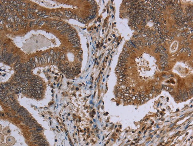
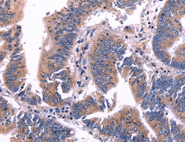
(Immunohistochemistry of paraffin-embedded Human colon cancer using MAVS Polyclonal Antibody at dilution of 1:30)
IHC (Immunohiostchemistry)
(Immunohistochemistry of paraffin-embedded Human colon cancer using MAVS Polyclonal Antibody at dilution of 1:30)
MAVS, Polyclonal Antibody (Cat# AAA168512)
 IHC (Immunohistochemisry)
(Immunohistochemistry of paraffin-embedded Human thyroid cancer using TRIM45 Polyclonal Antibody at dilution of 1:25)
IHC (Immunohistochemisry)
(Immunohistochemistry of paraffin-embedded Human thyroid cancer using TRIM45 Polyclonal Antibody at dilution of 1:25)
TRIM45, Polyclonal Antibody (Cat# AAA168516)
 IHC (Immunohiostchemistry)
(Immunohistochemistry of paraffin-embedded Human thyroid cancer tissue using CSMD1 Polyclonal Antibody at dilution 1:40)
IHC (Immunohiostchemistry)
(Immunohistochemistry of paraffin-embedded Human thyroid cancer tissue using CSMD1 Polyclonal Antibody at dilution 1:40)
CSMD1, Polyclonal Antibody (Cat# AAA168517)
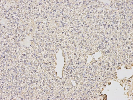
 IHC (Immunohiostchemistry)
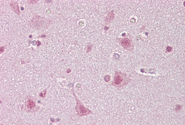
(Immunohistochemistry of paraffin-embedded Human brain tissue using ARC Polyclonal Antibody at dilution 1:50)
IHC (Immunohiostchemistry)
(Immunohistochemistry of paraffin-embedded Human brain tissue using ARC Polyclonal Antibody at dilution 1:50)
ARC, Polyclonal Antibody (Cat# AAA168518)

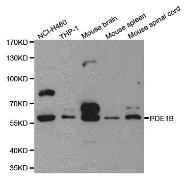
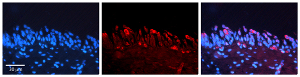
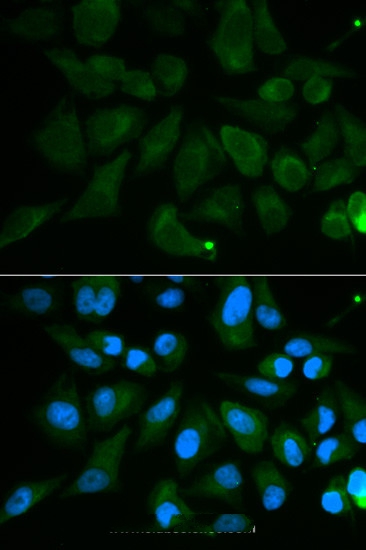 IF (Immunofluorescence)
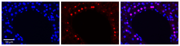
(Immunofluorescence analysis of MCF7 cell using PDE1B antibody. Blue: DAPI for nuclear staining.)
IF (Immunofluorescence)
(Immunofluorescence analysis of MCF7 cell using PDE1B antibody. Blue: DAPI for nuclear staining.)
PDE1B, Polyclonal Antibody (Cat# AAA168521)

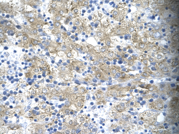
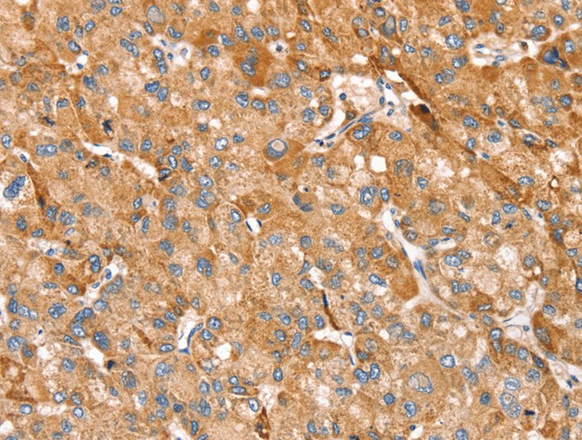
 IHC (Immunohiostchemistry)
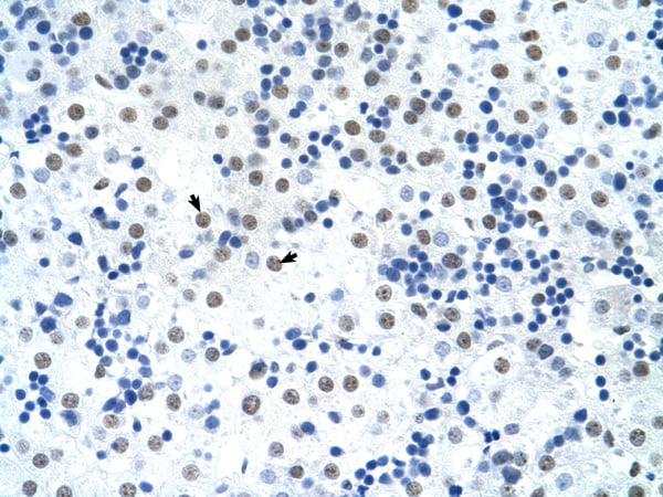
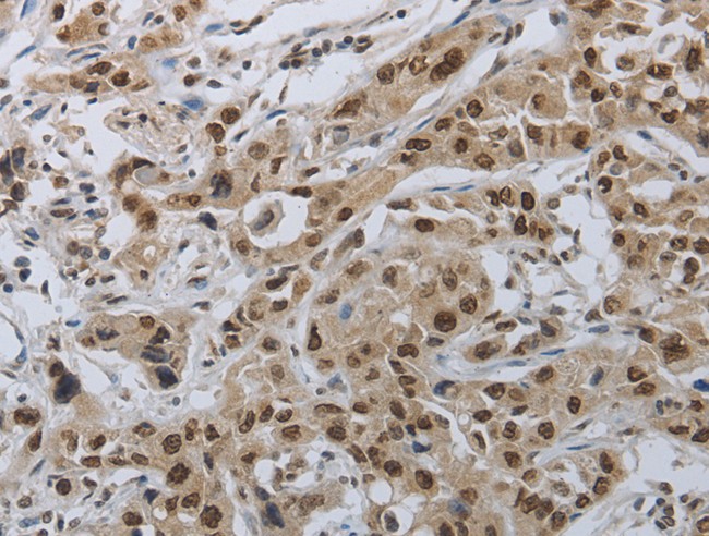
(Immunohistochemistry of paraffin-embedded Human liver cancer tissue using PRKACG Polyclonal Antibody at dilution 1:25)
IHC (Immunohiostchemistry)
(Immunohistochemistry of paraffin-embedded Human liver cancer tissue using PRKACG Polyclonal Antibody at dilution 1:25)
PRKACG, Polyclonal Antibody (Cat# AAA168522)
 IHC (Immunohiostchemistry)
(Immunohistochemistry of paraffin-embedded Human colon cancer tissue using KCNG1 Polyclonal Antibody at dilution 1:40)
IHC (Immunohiostchemistry)
(Immunohistochemistry of paraffin-embedded Human colon cancer tissue using KCNG1 Polyclonal Antibody at dilution 1:40)
KCNG1, Polyclonal Antibody (Cat# AAA168524)
What are Polyclonal Antibodies?
Polyclonal antibodies are antibodies that come from multiple B cell clones of a host animal. The typical hosts used for the majority of polyclonal antibody production are rabbits, goats, sheep, and donkeys. These polyclonal antibodies, once having identified their target, will bind to different epitopes located at different regions or sequences on the same protein/antigen. As a result, they are ideal at locating and binding to the target, even if the target is in very low concentrations (due to many different antibodies being able to bind to the same target molecule, which allows for significant amplification of a downstream signal).
Polyclonal antibodies are typically produced by injecting an antigen into a host animal, which causes the animal’s immune system to attack the foreign antigen by mass generating antibodies against it. After a period of time, serum is collected from the animal and purified using physicochemical fractionation, class-specific affinity purification, and/or antigen-affinity purification.
Key Uses of Polyclonal Antibodies
- Western Blotting: This method is used to find specific proteins in biological samples after separating them by size.
- Immunohistochemistry: IHC helps visualize the location of proteins in tissue sections using various staining techniques.
- ELISA: (Enzyme-Linked Immunosorbent Assay) is typically used to identify specific protein quantities in a sample. ELISAs can be either “Quantitative” or “Qualitative”.
- Flow Cytometry: technique that identifies and measures the specific protein on the surface or inside the cells in a fluid suspension.
- Immunoprecipitation: IP isolates and studies a specific protein from a complex mixture using antibodies.
Why Buy Polyclonal Antibodies from AAA Biotech?
1. Ideal for Various Applications
Our antibodies are generally going to be validated for use in multiple types of assays, including ELISA, Western Blotting, Immunohistochemistry, Immunoprecipitation, amongst others. They are ideal for a wide range of research applications.
2. Rigorous Quality Control
All of the antibodies in our catalog undergo strict quality testing to ensure specificity, sensitivity, and consistent performance. We are confident in the ability of our antibodies to provide you with accurate results.
3. Wide Assortment of Antibodies
Antibodies in are catalog can be found for both common and exotic species, and these antibodies are also available in both conjugated and recombinant forms to suit many diverse experimental needs.
4. Highly Purified
Our antibodies are available in purified forms with over 85% purity, as confirmed by SDS-PAGE. They are also available with tags such as His, Flag, GST, or MBP. We cater to customers worldwide.
FAQ
1. How are polyclonal antibodies produced?
Traditionally, polyclonal antibodies are produced by injecting an antigen into a host animal (such as a rabbit or goat), which then triggers an immune response from the host animal. The animal’s B cells produce antibodies that will recognize different parts of the injected antigen. These antibodies are then collected from the animal’s blood and purified for use.
2. How do polyclonal antibodies differ from monoclonal antibodies?
Polyclonal antibodies are a mix of antibodies that bind to different locations (epitopes) of the same antigen, while monoclonal antibodies are identical and bind to just one specific epitope. This makes polyclonal antibodies more versatile and better at detecting proteins that may be present in low quantities or in altered/modified forms.
3. How should I store polyclonal antibodies?
Polyclonal antibodies should be stored at 4°C for short-term use (up to a few weeks) and at -20°C or -80°C for long-term storage. Avoid repeated freeze-thaw cycles by dividing them into small aliquots. Always check the datasheet for specific storage instructions.