Filters
▼Clonality
▼Type
▼Reactivity
▼Gene Name
▼Isotype
▼Host
▼Application
▼Clone
▼Polyclonal Antibodies
At AAA Biotech also known as AAA Bio or AAABio, we provide a broad range of purified polyclonal antibodies (pAbs) that are able to all be browsed online through our website. Due to their high specificity and strong binding affinity, these antibodies are ideal for wide swathes of research and experimental applications.
Our polyclonal antibodies can easily support your work, whether you use them for Western Blotting, Immunocytochemistry (with or without Immunofluorescence used in conjunction), Immunohistochemistry, Immunoprecipitation, and ELISA tests. We highly encourage you to browse our range of pAbs and choose the one that best suits your experimental model.
Viewing 1150-1200 of 96805 product results
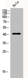
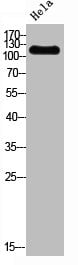
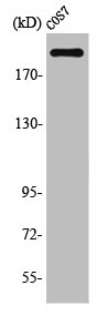
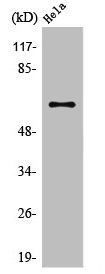
 WB (Western Blot)
(Western Blot analysis of various cells using p23 Polyclonal Antibody)
WB (Western Blot)
(Western Blot analysis of various cells using p23 Polyclonal Antibody)
PTGES3, Polyclonal Antibody (Cat# AAA236383)
 WB (Western Blot)
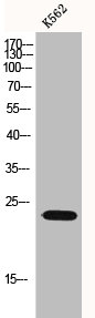
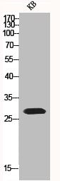
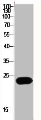
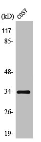
(Western Blot analysis of KB cells using p27 Polyclonal Antibody)
WB (Western Blot)
(Western Blot analysis of KB cells using p27 Polyclonal Antibody)
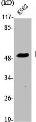
CDKN1B, Polyclonal Antibody (Cat# AAA236384)
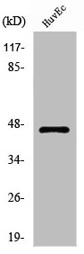
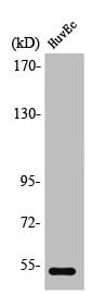
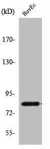
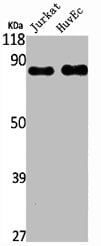
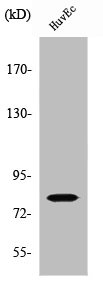
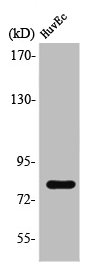
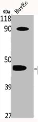 WB (Western Blot)
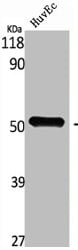
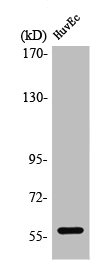
(Western Blot analysis of HuvEc cells using p47-phox Polyclonal Antibody)
WB (Western Blot)
(Western Blot analysis of HuvEc cells using p47-phox Polyclonal Antibody)
NCF1, Polyclonal Antibody (Cat# AAA236387)
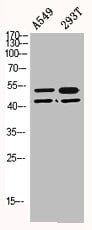 WB (Western Blot)
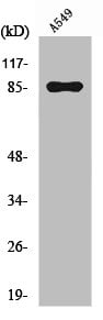
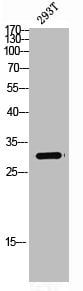
(Western Blot analysis of A549 293T cells using p53 Polyclonal Antibody)
WB (Western Blot)
(Western Blot analysis of A549 293T cells using p53 Polyclonal Antibody)
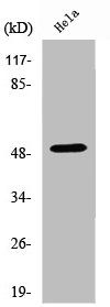
TP53, Polyclonal Antibody (Cat# AAA236389)
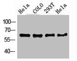
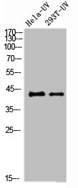
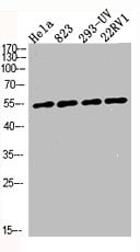 WB (Western Blot)
(Western Blot analysis of HELA 823 293-UV 22RV1 cells using p53 Polyclonal Antibody)
WB (Western Blot)
(Western Blot analysis of HELA 823 293-UV 22RV1 cells using p53 Polyclonal Antibody)
TP53, Polyclonal Antibody (Cat# AAA236390)
 WB (Western Blot)
(Western Blot analysis of HELA cells using PAI-3 Polyclonal Antibody)
WB (Western Blot)
(Western Blot analysis of HELA cells using PAI-3 Polyclonal Antibody)
SERPINA5, Polyclonal Antibody (Cat# AAA236398)

 WB (Western Blot)
(Western Blot analysis of various cells using PAK5 Polyclonal Antibody)
WB (Western Blot)
(Western Blot analysis of various cells using PAK5 Polyclonal Antibody)
PAK7, Polyclonal Antibody (Cat# AAA236399)

 WB (Western Blot)
(Western Blot analysis of HuvEc cells using PDK1 Polyclonal Antibody)
WB (Western Blot)
(Western Blot analysis of HuvEc cells using PDK1 Polyclonal Antibody)
PDK1, Polyclonal Antibody (Cat# AAA236403)
 WB (Western Blot)
(Western Blot analysis of HELA COLO 293T cells using PDK1 Polyclonal Antibody)
WB (Western Blot)
(Western Blot analysis of HELA COLO 293T cells using PDK1 Polyclonal Antibody)
PDPK1, Polyclonal Antibody (Cat# AAA236404)
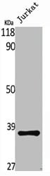
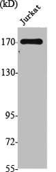
 WB (Western Blot)
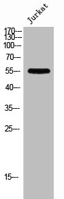
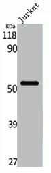
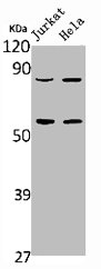
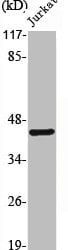
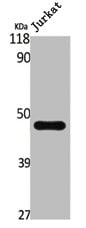
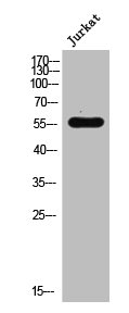
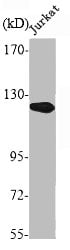
(Western Blot analysis of Jurkat cells using PFK-2 liv Polyclonal Antibody)
WB (Western Blot)
(Western Blot analysis of Jurkat cells using PFK-2 liv Polyclonal Antibody)
PFKFB1, Polyclonal Antibody (Cat# AAA236405)
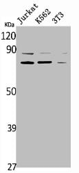
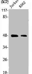
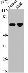
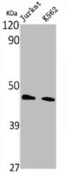 WB (Western Blot)
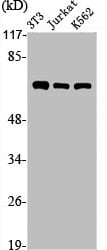
(Western Blot analysis of Jurkat K562 cells using PHKG1 Polyclonal Antibody)
WB (Western Blot)
(Western Blot analysis of Jurkat K562 cells using PHKG1 Polyclonal Antibody)
PHKG1, Polyclonal Antibody (Cat# AAA236408)
 WB (Western Blot)
(Western Blot analysis of HELA cells using PI 3-kinase p110alpha Polyclonal Antibody)
WB (Western Blot)
(Western Blot analysis of HELA cells using PI 3-kinase p110alpha Polyclonal Antibody)
PIK3CA, Polyclonal Antibody (Cat# AAA236409)
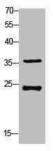
 WB (Western Blot)
(Western blot analysis of 293T COLO lysis using PI 3-kinase p85alpha/gamma antibody.)
WB (Western Blot)
(Western blot analysis of 293T COLO lysis using PI 3-kinase p85alpha/gamma antibody.)
PIK3R1/PIK3R3, Polyclonal Antibody (Cat# AAA236411)
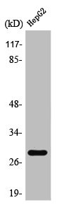
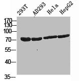 WB (Western Blot)
(Western Blot analysis of 293T AD293 HELA HepG2 cells using PI 3-kinase p85beta Polyclonal Antibody)
WB (Western Blot)
(Western Blot analysis of 293T AD293 HELA HepG2 cells using PI 3-kinase p85beta Polyclonal Antibody)
PIK3R2, Polyclonal Antibody (Cat# AAA236412)
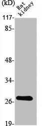
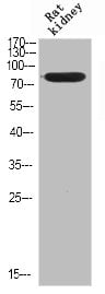
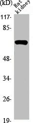 WB (Western Blot)
(Western Blot analysis of RAT-KIDNEY cells using PIAS 3 Polyclonal Antibody)
WB (Western Blot)
(Western Blot analysis of RAT-KIDNEY cells using PIAS 3 Polyclonal Antibody)
PIAS3, Polyclonal Antibody (Cat# AAA236413)
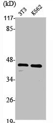
 WB (Western Blot)
(Western Blot analysis of NIH-3T3 Jurkat K562 cells using PIASx Polyclonal Antibody)
WB (Western Blot)
(Western Blot analysis of NIH-3T3 Jurkat K562 cells using PIASx Polyclonal Antibody)
PIAS2, Polyclonal Antibody (Cat# AAA236414)
 WB (Western Blot)
(Western Blot analysis of various cells using PIPK I gamma Polyclonal Antibody)
WB (Western Blot)
(Western Blot analysis of various cells using PIPK I gamma Polyclonal Antibody)
PIP5K1C, Polyclonal Antibody (Cat# AAA236415)
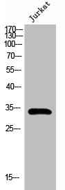
 WB (Western Blot)
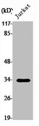
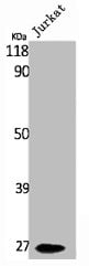
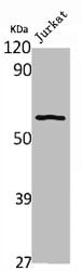
(Western Blot analysis of Jurkat cells using Pitx1 Polyclonal Antibody)
WB (Western Blot)
(Western Blot analysis of Jurkat cells using Pitx1 Polyclonal Antibody)
PITX1, Polyclonal Antibody (Cat# AAA236417)
 WB (Western Blot)
(Western Blot analysis of NIH-3T3 K562 cells using PKA Ialpha reg Polyclonal Antibody)
WB (Western Blot)
(Western Blot analysis of NIH-3T3 K562 cells using PKA Ialpha reg Polyclonal Antibody)
PRKAR1A, Polyclonal Antibody (Cat# AAA236420)
 WB (Western Blot)
(Western Blot analysis of K562 cells using PKA Ibeta reg Polyclonal Antibody)
WB (Western Blot)
(Western Blot analysis of K562 cells using PKA Ibeta reg Polyclonal Antibody)
PRKAR1B, Polyclonal Antibody (Cat# AAA236421)
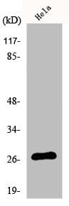
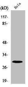
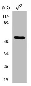
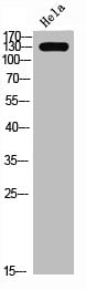
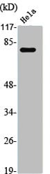
 WB (Western Blot)
(Western Blot analysis of Hela cells using PKC zeta Polyclonal Antibody)
WB (Western Blot)
(Western Blot analysis of Hela cells using PKC zeta Polyclonal Antibody)
PRKCZ, Polyclonal Antibody (Cat# AAA236426)
 WB (Western Blot)
(Western Blot analysis of Jurkat cells using PKR1 Polyclonal Antibody)
WB (Western Blot)
(Western Blot analysis of Jurkat cells using PKR1 Polyclonal Antibody)
PROKR1, Polyclonal Antibody (Cat# AAA236428)
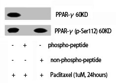 WB (Western Blot)
(Western Blot analysis of various cells using PPAR-gamma Polyclonal Antibody)
WB (Western Blot)
(Western Blot analysis of various cells using PPAR-gamma Polyclonal Antibody)
PPARG, Polyclonal Antibody (Cat# AAA236437)
 WB (Western Blot)
(Western Blot analysis of Jurkat cells using PRX1 Polyclonal Antibody)
WB (Western Blot)
(Western Blot analysis of Jurkat cells using PRX1 Polyclonal Antibody)
PRRX1, Polyclonal Antibody (Cat# AAA236441)
 WB (Western Blot)
(Western Blot analysis of RAT-KIDNEY cells using PSCA Polyclonal Antibody)
WB (Western Blot)
(Western Blot analysis of RAT-KIDNEY cells using PSCA Polyclonal Antibody)
PSCA, Polyclonal Antibody (Cat# AAA236442)
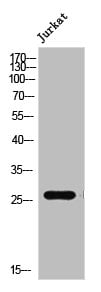
 WB (Western Blot)
(Western Blot analysis of Jurkat cells using PTEN Polyclonal Antibody)
WB (Western Blot)
(Western Blot analysis of Jurkat cells using PTEN Polyclonal Antibody)
PTEN, Polyclonal Antibody (Cat# AAA236445)
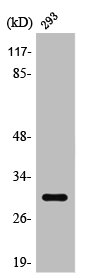
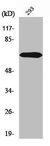
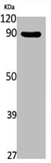
 WB (Western Blot)
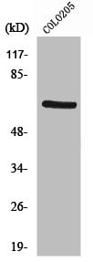
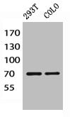
(Western blot analysis of 293T lysis using Rab 34 antibody.)
WB (Western Blot)
(Western blot analysis of 293T lysis using Rab 34 antibody.)
RAB34, Polyclonal Antibody (Cat# AAA236448)
 WB (Western Blot)
(Western Blot analysis of Hela cells using Rac1/2/3/CDC42 Polyclonal Antibody)
WB (Western Blot)
(Western Blot analysis of Hela cells using Rac1/2/3/CDC42 Polyclonal Antibody)
RAC1/RAC2/RAC3/CDC42, Polyclonal Antibody (Cat# AAA236450)
 WB (Western Blot)
(Western Blot analysis of Jurkat cells using Rad18 Polyclonal Antibody)
WB (Western Blot)
(Western Blot analysis of Jurkat cells using Rad18 Polyclonal Antibody)
RAD18, Polyclonal Antibody (Cat# AAA236451)
 WB (Western Blot)
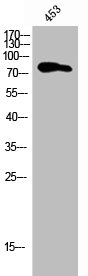
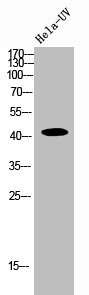
(Western Blot analysis of 293T-UV HELA-UV cells using Rad51 Polyclonal Antibody)
WB (Western Blot)
(Western Blot analysis of 293T-UV HELA-UV cells using Rad51 Polyclonal Antibody)
RAD51, Polyclonal Antibody (Cat# AAA236453)
 WB (Western Blot)
(Western Blot analysis of Jurkat cells using Radixin Polyclonal Antibody)
WB (Western Blot)
(Western Blot analysis of Jurkat cells using Radixin Polyclonal Antibody)
RDX, Polyclonal Antibody (Cat# AAA236455)
 WB (Western Blot)
(Western Blot analysis of PC3 NIH-3T3 cells using Rap1GAP Polyclonal Antibody)
WB (Western Blot)
(Western Blot analysis of PC3 NIH-3T3 cells using Rap1GAP Polyclonal Antibody)
RAP1GAP, Polyclonal Antibody (Cat# AAA236457)
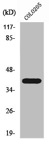
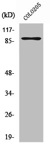
 WB (Western Blot)
(Western Blot analysis of customer's (cat sample) using Ribosomal Protein S4X Polyclonal Antibody)
WB (Western Blot)
(Western Blot analysis of customer's (cat sample) using Ribosomal Protein S4X Polyclonal Antibody)
RPS4X, Polyclonal Antibody (Cat# AAA236461)
 WB (Western Blot)
(Western Blot analysis of Jurkat cells using RNF113B Polyclonal Antibody)
WB (Western Blot)
(Western Blot analysis of Jurkat cells using RNF113B Polyclonal Antibody)
RNF113B, Polyclonal Antibody (Cat# AAA236466)
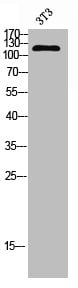
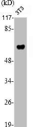 WB (Western Blot)
(Western Blot analysis of NIH-3T3 cells using RUNX2 Polyclonal Antibody)
WB (Western Blot)
(Western Blot analysis of NIH-3T3 cells using RUNX2 Polyclonal Antibody)
RUNX2, Polyclonal Antibody (Cat# AAA236468)
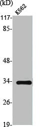
 WB (Western Blot)
(Western Blot analysis of K562 cells using RXRgamma Polyclonal Antibody)
WB (Western Blot)
(Western Blot analysis of K562 cells using RXRgamma Polyclonal Antibody)
RXRG, Polyclonal Antibody (Cat# AAA236469)
 WB (Western Blot)
(Western Blot analysis of NIH-3T3 K562 Jurkat RAT-intestine cells using SMAP45 Polyclonal Antibody)
WB (Western Blot)
(Western Blot analysis of NIH-3T3 K562 Jurkat RAT-intestine cells using SMAP45 Polyclonal Antibody)
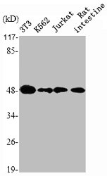
HDAC3, Polyclonal Antibody (Cat# AAA236477)
 WB (Western Blot)
(Western Blot analysis of Jurkat cells using SMG7 Polyclonal Antibody)
WB (Western Blot)
(Western Blot analysis of Jurkat cells using SMG7 Polyclonal Antibody)
SMG7, Polyclonal Antibody (Cat# AAA236478)
 WB (Western Blot)
(Western Blot analysis of RAT-KIDNEY cells using SMIT Polyclonal Antibody)
WB (Western Blot)
(Western Blot analysis of RAT-KIDNEY cells using SMIT Polyclonal Antibody)
SLC5A3, Polyclonal Antibody (Cat# AAA236479)
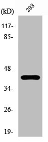
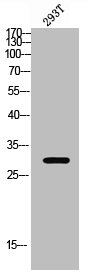
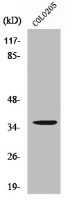
 WB (Western Blot)
(Western Blot analysis of 293T cells using Spindlin-1 Polyclonal Antibody)
WB (Western Blot)
(Western Blot analysis of 293T cells using Spindlin-1 Polyclonal Antibody)
SPIN1, Polyclonal Antibody (Cat# AAA236482)
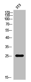
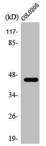
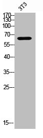
 WB (Western Blot)
(Western blot analysis of 3T3 lysis using SREBP-1 antibody.)
WB (Western Blot)
(Western blot analysis of 3T3 lysis using SREBP-1 antibody.)
SREBF1, Polyclonal Antibody (Cat# AAA236483)
 WB (Western Blot)
(Western Blot analysis of Jurkat K562 cells using SSB Polyclonal Antibody)
WB (Western Blot)
(Western Blot analysis of Jurkat K562 cells using SSB Polyclonal Antibody)
SSB, Polyclonal Antibody (Cat# AAA236484)
 WB (Western Blot)
(Western Blot analysis of NIH-3T3 cells using Stat3 Polyclonal Antibody)
WB (Western Blot)
(Western Blot analysis of NIH-3T3 cells using Stat3 Polyclonal Antibody)
STAT3, Polyclonal Antibody (Cat# AAA236487)
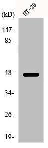
 WB (Western Blot)
(Western Blot analysis of HELA cells using Tau Polyclonal Antibody)
WB (Western Blot)
(Western Blot analysis of HELA cells using Tau Polyclonal Antibody)
MAPT, Polyclonal Antibody (Cat# AAA236493)

 WB (Western Blot)
(Western blot analysis of 3T3 lysis using TGFbeta RII antibody.)
WB (Western Blot)
(Western blot analysis of 3T3 lysis using TGFbeta RII antibody.)
TGFBR2, Polyclonal Antibody (Cat# AAA236496)
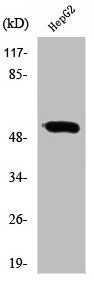
 WB (Western Blot)
(Western Blot analysis of NIH-3T3 cells using TIF1alpha Polyclonal Antibody)
WB (Western Blot)
(Western Blot analysis of NIH-3T3 cells using TIF1alpha Polyclonal Antibody)
TRIM24, Polyclonal Antibody (Cat# AAA236498)
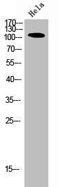
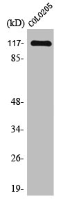
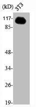
 WB (Western Blot)
(Western Blot analysis of Jurkat cells using Topo IIalpha Polyclonal Antibody)
WB (Western Blot)
(Western Blot analysis of Jurkat cells using Topo IIalpha Polyclonal Antibody)
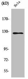
TOP2A, Polyclonal Antibody (Cat# AAA236499)
 WB (Western Blot)
(Western Blot analysis of K562 cells using TRADD Polyclonal Antibody)
WB (Western Blot)
(Western Blot analysis of K562 cells using TRADD Polyclonal Antibody)
TRADD, Polyclonal Antibody (Cat# AAA236500)
 WB (Western Blot)
(Western Blot analysis of Jurkat K562 cells using TRAF3 Polyclonal Antibody)
WB (Western Blot)
(Western Blot analysis of Jurkat K562 cells using TRAF3 Polyclonal Antibody)
TRAF3, Polyclonal Antibody (Cat# AAA236501)
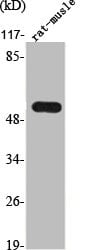 WB (Western Blot)
(Western Blot analysis of RAT-MUSCLE cells using TrxR2 Polyclonal Antibody)
WB (Western Blot)
(Western Blot analysis of RAT-MUSCLE cells using TrxR2 Polyclonal Antibody)
TXNRD2, Polyclonal Antibody (Cat# AAA236503)
What are Polyclonal Antibodies?
Polyclonal antibodies are antibodies that come from multiple B cell clones of a host animal. The typical hosts used for the majority of polyclonal antibody production are rabbits, goats, sheep, and donkeys. These polyclonal antibodies, once having identified their target, will bind to different epitopes located at different regions or sequences on the same protein/antigen. As a result, they are ideal at locating and binding to the target, even if the target is in very low concentrations (due to many different antibodies being able to bind to the same target molecule, which allows for significant amplification of a downstream signal).
Polyclonal antibodies are typically produced by injecting an antigen into a host animal, which causes the animal’s immune system to attack the foreign antigen by mass generating antibodies against it. After a period of time, serum is collected from the animal and purified using physicochemical fractionation, class-specific affinity purification, and/or antigen-affinity purification.
Key Uses of Polyclonal Antibodies
- Western Blotting: This method is used to find specific proteins in biological samples after separating them by size.
- Immunohistochemistry: IHC helps visualize the location of proteins in tissue sections using various staining techniques.
- ELISA: (Enzyme-Linked Immunosorbent Assay) is typically used to identify specific protein quantities in a sample. ELISAs can be either “Quantitative” or “Qualitative”.
- Flow Cytometry: technique that identifies and measures the specific protein on the surface or inside the cells in a fluid suspension.
- Immunoprecipitation: IP isolates and studies a specific protein from a complex mixture using antibodies.
Why Buy Polyclonal Antibodies from AAA Biotech?
1. Ideal for Various Applications
Our antibodies are generally going to be validated for use in multiple types of assays, including ELISA, Western Blotting, Immunohistochemistry, Immunoprecipitation, amongst others. They are ideal for a wide range of research applications.
2. Rigorous Quality Control
All of the antibodies in our catalog undergo strict quality testing to ensure specificity, sensitivity, and consistent performance. We are confident in the ability of our antibodies to provide you with accurate results.
3. Wide Assortment of Antibodies
Antibodies in are catalog can be found for both common and exotic species, and these antibodies are also available in both conjugated and recombinant forms to suit many diverse experimental needs.
4. Highly Purified
Our antibodies are available in purified forms with over 85% purity, as confirmed by SDS-PAGE. They are also available with tags such as His, Flag, GST, or MBP. We cater to customers worldwide.
FAQ
1. How are polyclonal antibodies produced?
Traditionally, polyclonal antibodies are produced by injecting an antigen into a host animal (such as a rabbit or goat), which then triggers an immune response from the host animal. The animal’s B cells produce antibodies that will recognize different parts of the injected antigen. These antibodies are then collected from the animal’s blood and purified for use.
2. How do polyclonal antibodies differ from monoclonal antibodies?
Polyclonal antibodies are a mix of antibodies that bind to different locations (epitopes) of the same antigen, while monoclonal antibodies are identical and bind to just one specific epitope. This makes polyclonal antibodies more versatile and better at detecting proteins that may be present in low quantities or in altered/modified forms.
3. How should I store polyclonal antibodies?
Polyclonal antibodies should be stored at 4°C for short-term use (up to a few weeks) and at -20°C or -80°C for long-term storage. Avoid repeated freeze-thaw cycles by dividing them into small aliquots. Always check the datasheet for specific storage instructions.