Filters
▼Clonality
▼Type
▼Reactivity
▼Gene Name
▼Isotype
▼Host
▼Application
▼Clone
▼Polyclonal Antibodies
At AAA Biotech also known as AAA Bio or AAABio, we provide a broad range of purified polyclonal antibodies (pAbs) that are able to all be browsed online through our website. Due to their high specificity and strong binding affinity, these antibodies are ideal for wide swathes of research and experimental applications.
Our polyclonal antibodies can easily support your work, whether you use them for Western Blotting, Immunocytochemistry (with or without Immunofluorescence used in conjunction), Immunohistochemistry, Immunoprecipitation, and ELISA tests. We highly encourage you to browse our range of pAbs and choose the one that best suits your experimental model.
Viewing 1000-1050 of 96805 product results
 IHC (Immunohiostchemistry)
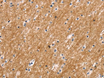
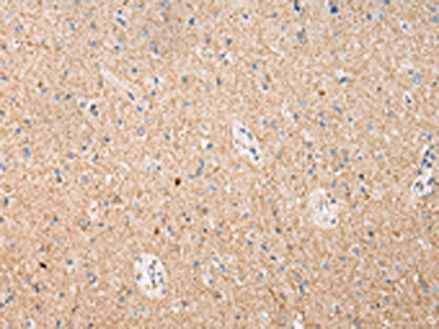
(The image on the left is immunohistochemistry of paraffin-embedded Human brain tissue using AAA239877(FOXN2 Antibody) at dilution 1/15, on the right is treated with synthetic peptide. (Original magnification: ×200))
IHC (Immunohiostchemistry)
(The image on the left is immunohistochemistry of paraffin-embedded Human brain tissue using AAA239877(FOXN2 Antibody) at dilution 1/15, on the right is treated with synthetic peptide. (Original magnification: ×200))
FOXN2, Polyclonal Antibody (Cat# AAA239877)
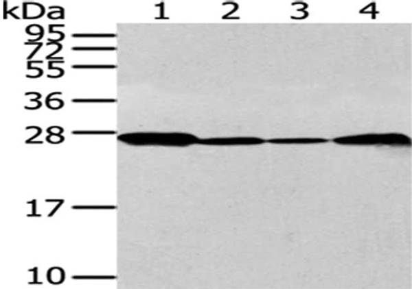
 SDS-PAGE
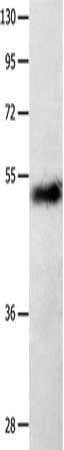
(Gel: 10%SDS-PAGE, Lysate: 50 ug, Lane: Hela cells, Primary antibody: AAA239884(IGF2BP1 Antibody) at dilution 1/750, Secondary antibody: Goat anti rabbit IgG at 1/8000 dilution, Exposure time: 1 minute)
SDS-PAGE
(Gel: 10%SDS-PAGE, Lysate: 50 ug, Lane: Hela cells, Primary antibody: AAA239884(IGF2BP1 Antibody) at dilution 1/750, Secondary antibody: Goat anti rabbit IgG at 1/8000 dilution, Exposure time: 1 minute)
IGF2BP1, Polyclonal Antibody (Cat# AAA239884)
 IHC (Immunohiostchemistry)
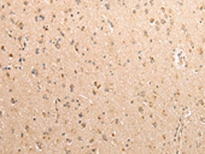
(The image on the left is immunohistochemistry of paraffin-embedded Human brain tissue using AAA239885(TRPM1 Antibody) at dilution 1/20, on the right is treated with synthetic peptide. (Original magnification: ×200))
IHC (Immunohiostchemistry)
(The image on the left is immunohistochemistry of paraffin-embedded Human brain tissue using AAA239885(TRPM1 Antibody) at dilution 1/20, on the right is treated with synthetic peptide. (Original magnification: ×200))
TRPM1, Polyclonal Antibody (Cat# AAA239885)
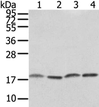
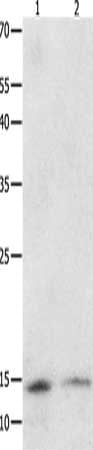 SDS-PAGE
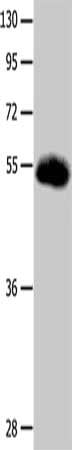
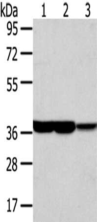
(Gel: 10+12+15%SDS-PAGE, Lysate: 40 ug, Lane 1-2: Jukart cells, A549 cells, Primary antibody: AAA239893(SUMO1 Antibody) at dilution 1/350, Secondary antibody: Goat anti rabbit IgG at 1/8000 dilution, Exposure time: 10 minutes)
SDS-PAGE
(Gel: 10+12+15%SDS-PAGE, Lysate: 40 ug, Lane 1-2: Jukart cells, A549 cells, Primary antibody: AAA239893(SUMO1 Antibody) at dilution 1/350, Secondary antibody: Goat anti rabbit IgG at 1/8000 dilution, Exposure time: 10 minutes)
SUMO1, Polyclonal Antibody (Cat# AAA239893)
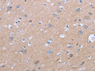 IHC (Immunohiostchemistry)
(The image on the left is immunohistochemistry of paraffin-embedded Human brain tissue using AAA239897(CXCR3 Antibody) at dilution 1/20, on the right is treated with synthetic peptide. (Original magnification: ×200))
IHC (Immunohiostchemistry)
(The image on the left is immunohistochemistry of paraffin-embedded Human brain tissue using AAA239897(CXCR3 Antibody) at dilution 1/20, on the right is treated with synthetic peptide. (Original magnification: ×200))
CXCR3, Polyclonal Antibody (Cat# AAA239897)
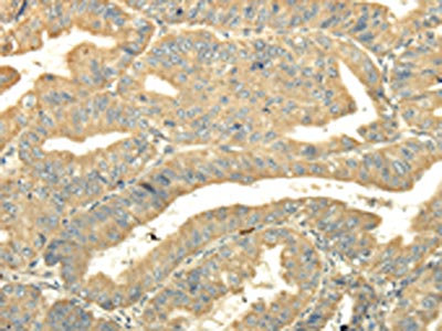
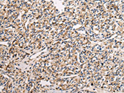
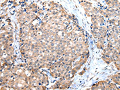
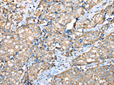
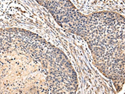
 IHC (Immunohiostchemistry)
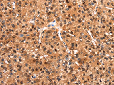
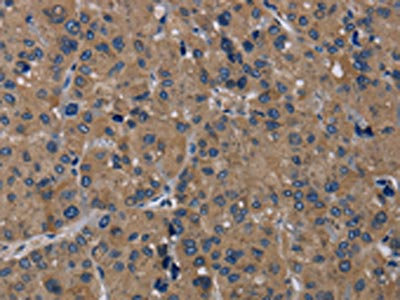
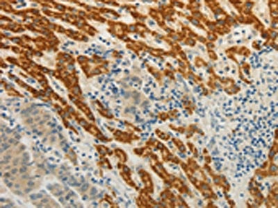
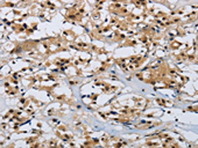
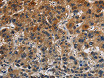
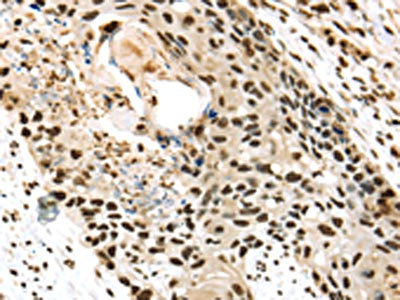
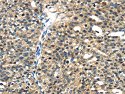
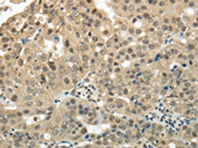
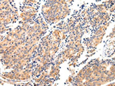
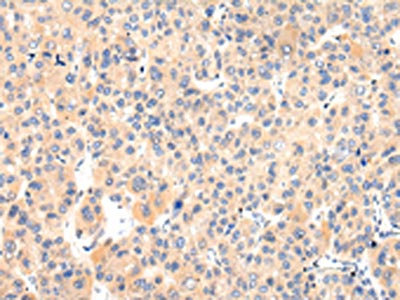
(The image on the left is immunohistochemistry of paraffin-embedded Human liver cancer tissue using AAA239900(GATA5 Antibody) at dilution 1/50, on the right is treated with synthetic peptide. (Original magnification: ×200))
IHC (Immunohiostchemistry)
(The image on the left is immunohistochemistry of paraffin-embedded Human liver cancer tissue using AAA239900(GATA5 Antibody) at dilution 1/50, on the right is treated with synthetic peptide. (Original magnification: ×200))
GATA5, Polyclonal Antibody (Cat# AAA239900)
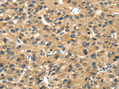
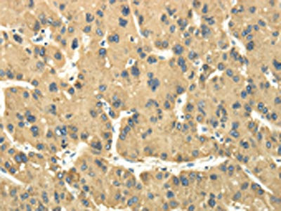
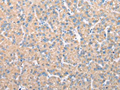
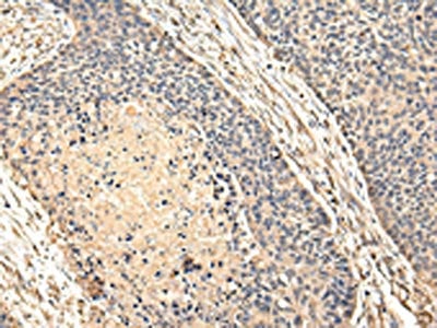
 IHC (Immunohiostchemistry)
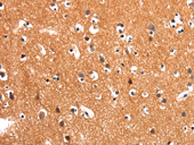
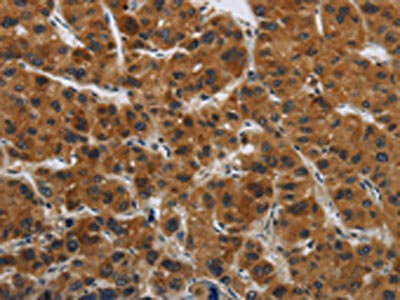
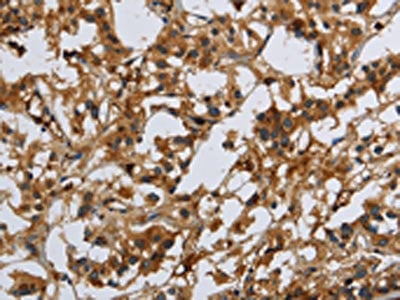
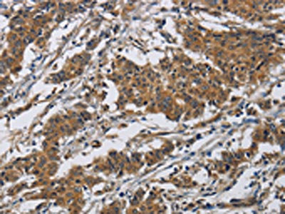
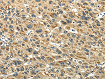
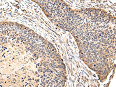
(The image on the left is immunohistochemistry of paraffin-embedded Human liver cancer tissue using AAA239902(CMTM7 Antibody) at dilution 1/30, on the right is treated with synthetic peptide. (Original magnification: ×200))
IHC (Immunohiostchemistry)
(The image on the left is immunohistochemistry of paraffin-embedded Human liver cancer tissue using AAA239902(CMTM7 Antibody) at dilution 1/30, on the right is treated with synthetic peptide. (Original magnification: ×200))
CMTM7, Polyclonal Antibody (Cat# AAA239902)
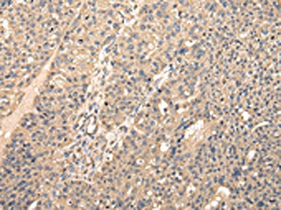
 IHC (Immunohiostchemistry)
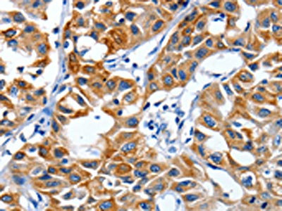
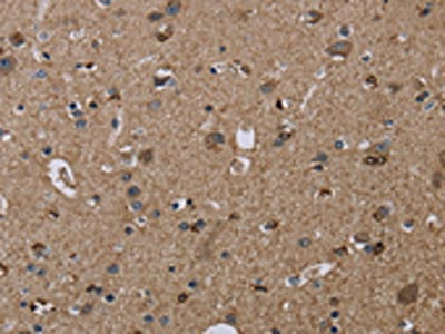
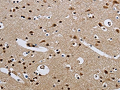
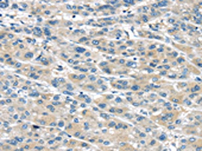
(The image on the left is immunohistochemistry of paraffin-embedded Human brain tissue using AAA239905(CMTM8 Antibody) at dilution 1/40, on the right is treated with synthetic peptide. (Original magnification: ×200))
IHC (Immunohiostchemistry)
(The image on the left is immunohistochemistry of paraffin-embedded Human brain tissue using AAA239905(CMTM8 Antibody) at dilution 1/40, on the right is treated with synthetic peptide. (Original magnification: ×200))
CMTM8, Polyclonal Antibody (Cat# AAA239905)
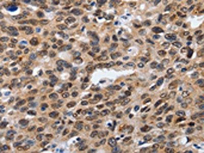
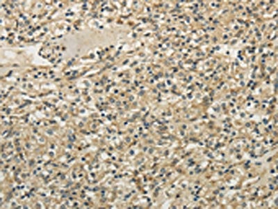
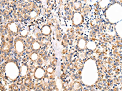
 IHC (Immunohiostchemistry)
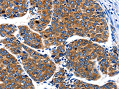
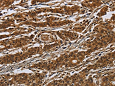
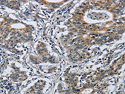
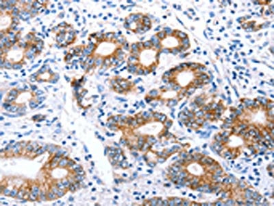
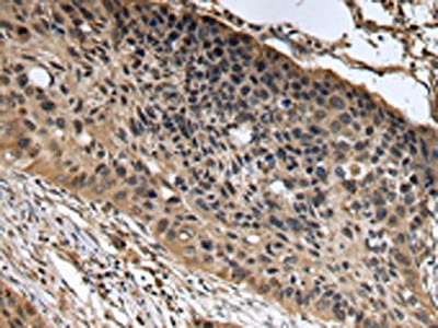
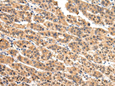
(The image on the left is immunohistochemistry of paraffin-embedded Human gastric cancer tissue using AAA239906(CMTM8 Antibody) at dilution 1/40, on the right is treated with synthetic peptide. (Original magnification: ×200))
IHC (Immunohiostchemistry)
(The image on the left is immunohistochemistry of paraffin-embedded Human gastric cancer tissue using AAA239906(CMTM8 Antibody) at dilution 1/40, on the right is treated with synthetic peptide. (Original magnification: ×200))
CMTM8, Polyclonal Antibody (Cat# AAA239906)
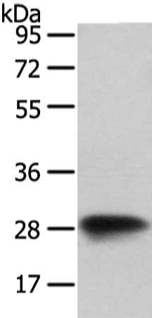
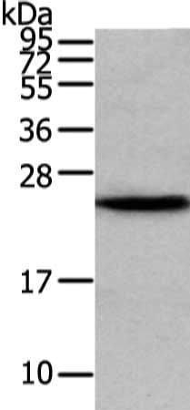
 SDS-PAGE
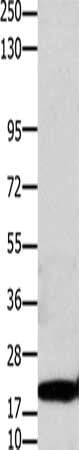
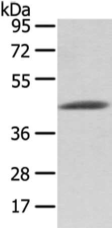
(Gel: 10+12%SDS-PAGE, Lysate: 40 ug, Lane: 231 cells, Primary antibody: AAA239907(CMTM3 Antibody) at dilution 1/350, Secondary antibody: Goat anti rabbit IgG at 1/8000 dilution, Exposure time: 1 minute)
SDS-PAGE
(Gel: 10+12%SDS-PAGE, Lysate: 40 ug, Lane: 231 cells, Primary antibody: AAA239907(CMTM3 Antibody) at dilution 1/350, Secondary antibody: Goat anti rabbit IgG at 1/8000 dilution, Exposure time: 1 minute)
CMTM3, Polyclonal Antibody (Cat# AAA239907)
 SDS-PAGE
(Gel: 10+12%SDS-PAGE, Lysate: 40 ug, Lane: Human fetal kidney tissue, Primary antibody: AAA239908(CMTM3 Antibody) at dilution 1/200, Secondary antibody: Goat anti rabbit IgG at 1/8000 dilution, Exposure time: 1 minute)
SDS-PAGE
(Gel: 10+12%SDS-PAGE, Lysate: 40 ug, Lane: Human fetal kidney tissue, Primary antibody: AAA239908(CMTM3 Antibody) at dilution 1/200, Secondary antibody: Goat anti rabbit IgG at 1/8000 dilution, Exposure time: 1 minute)
CMTM3, Polyclonal Antibody (Cat# AAA239908)
 SDS-PAGE
(Gel: 10%SDS-PAGE, Lysate: 30 ug, Lane: Hela cells, Primary antibody: AAA239914(CLDN1 Antibody) at dilution 1/1100, Secondary antibody: Goat anti rabbit IgG at 1/8000 dilution, Exposure time: 40 seconds)
SDS-PAGE
(Gel: 10%SDS-PAGE, Lysate: 30 ug, Lane: Hela cells, Primary antibody: AAA239914(CLDN1 Antibody) at dilution 1/1100, Secondary antibody: Goat anti rabbit IgG at 1/8000 dilution, Exposure time: 40 seconds)
CLDN1, Polyclonal Antibody (Cat# AAA239914)
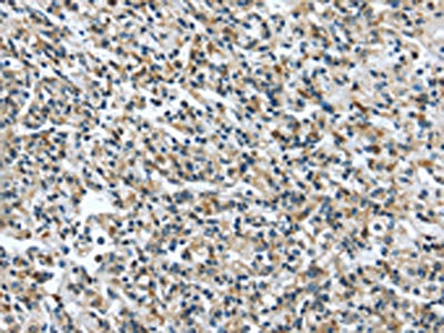
 IHC (Immunohiostchemistry)
(The image on the left is immunohistochemistry of paraffin-embedded Human gastric cancer tissue using AAA239918(CLDN8 Antibody) at dilution 1/50, on the right is treated with synthetic peptide. (Original magnification: ×200))
IHC (Immunohiostchemistry)
(The image on the left is immunohistochemistry of paraffin-embedded Human gastric cancer tissue using AAA239918(CLDN8 Antibody) at dilution 1/50, on the right is treated with synthetic peptide. (Original magnification: ×200))
CLDN8, Polyclonal Antibody (Cat# AAA239918)
 SDS-PAGE
(Gel: 10%SDS-PAGE, Lysate: 30 ug, Lane: Human liver cancer tissue, Primary antibody: AAA239920(CLDN7 Antibody) at dilution 1/1050, Secondary antibody: Goat anti rabbit IgG at 1/8000 dilution, Exposure time: 40 seconds)
SDS-PAGE
(Gel: 10%SDS-PAGE, Lysate: 30 ug, Lane: Human liver cancer tissue, Primary antibody: AAA239920(CLDN7 Antibody) at dilution 1/1050, Secondary antibody: Goat anti rabbit IgG at 1/8000 dilution, Exposure time: 40 seconds)
CLDN7, Polyclonal Antibody (Cat# AAA239920)
 SDS-PAGE
(Gel: 10%SDS-PAGE, Lysate: 40 ug, Lane: Hela cells, Primary antibody: AAA239922(GJB3 Antibody) at dilution 1/300, Secondary antibody: Goat anti rabbit IgG at 1/8000 dilution, Exposure time: 30 seconds)
SDS-PAGE
(Gel: 10%SDS-PAGE, Lysate: 40 ug, Lane: Hela cells, Primary antibody: AAA239922(GJB3 Antibody) at dilution 1/300, Secondary antibody: Goat anti rabbit IgG at 1/8000 dilution, Exposure time: 30 seconds)
GJB3, Polyclonal Antibody (Cat# AAA239922)
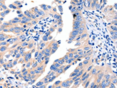
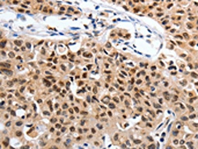
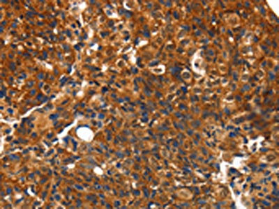
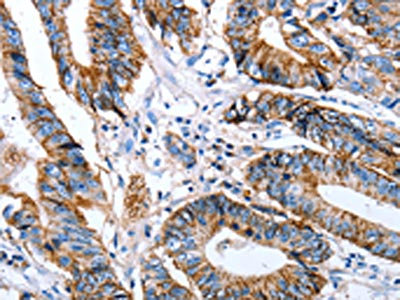
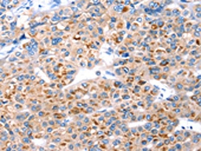
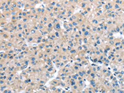
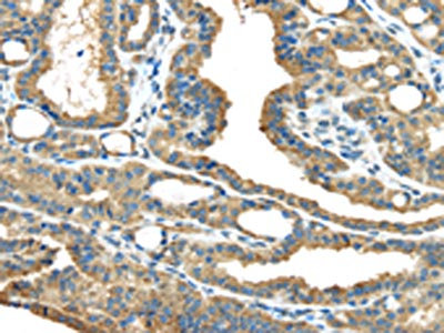
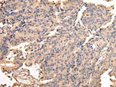
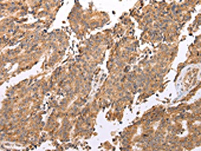
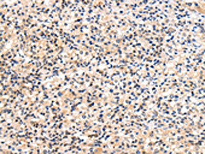
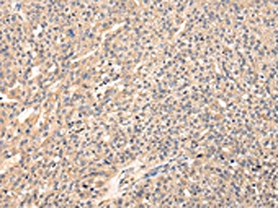
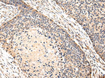
 IHC (Immunohiostchemistry)
(The image on the left is immunohistochemistry of paraffin-embedded Human lung cancer tissue using AAA239927(MSTN Antibody) at dilution 1/8, on the right is treated with synthetic peptide. (Original magnification: ×200))
IHC (Immunohiostchemistry)
(The image on the left is immunohistochemistry of paraffin-embedded Human lung cancer tissue using AAA239927(MSTN Antibody) at dilution 1/8, on the right is treated with synthetic peptide. (Original magnification: ×200))
MSTN, Polyclonal Antibody (Cat# AAA239927)
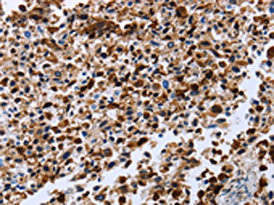
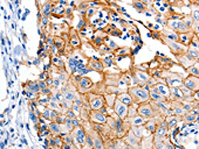
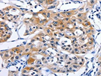
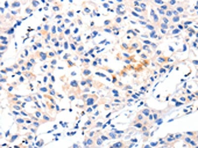
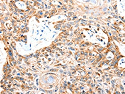
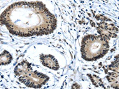
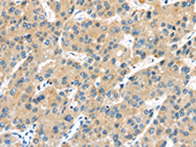
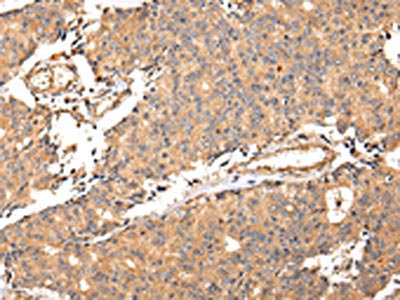
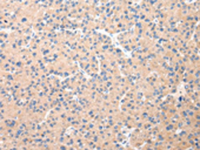
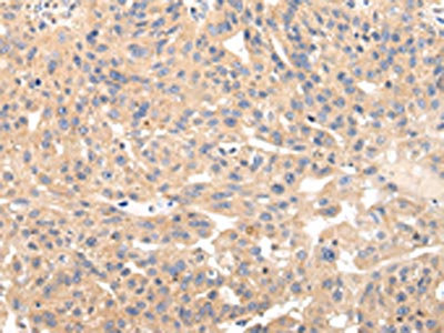
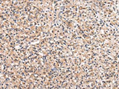
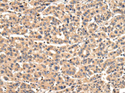
 IHC (Immunohiostchemistry)
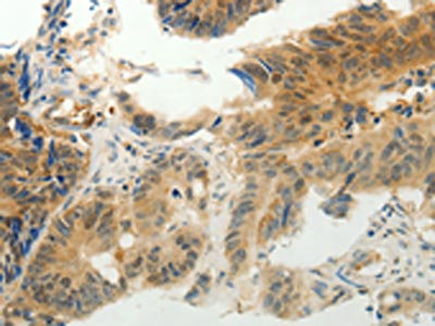
(The image on the left is immunohistochemistry of paraffin-embedded Human colon cancer tissue using AAA239931(GSN Antibody) at dilution 1/40, on the right is treated with synthetic peptide. (Original magnification: ×200))
IHC (Immunohiostchemistry)
(The image on the left is immunohistochemistry of paraffin-embedded Human colon cancer tissue using AAA239931(GSN Antibody) at dilution 1/40, on the right is treated with synthetic peptide. (Original magnification: ×200))
GSN, Polyclonal Antibody (Cat# AAA239931)
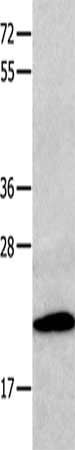
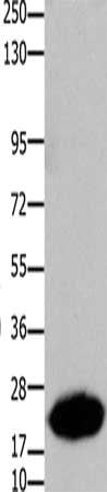
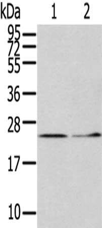
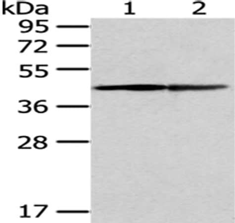
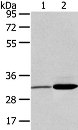
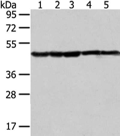
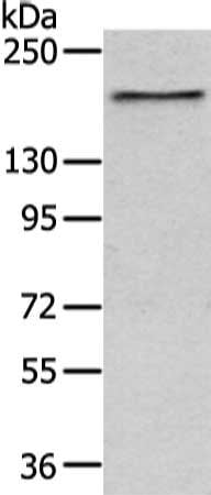
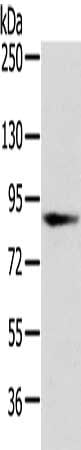
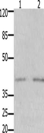 SDS-PAGE
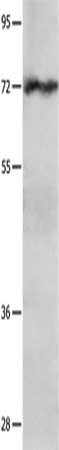
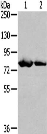
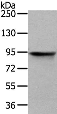
(Gel: 10%SDS-PAGE, Lysate: 40 ug, Lane 1-2: Human fetal kidney tissue, Human brain tissue, Primary antibody: AAA239932(GFRA3 Antibody) at dilution 1/1000, Secondary antibody: Goat anti rabbit IgG at 1/8000 dilution, Exposure time: 5 minutes)
SDS-PAGE
(Gel: 10%SDS-PAGE, Lysate: 40 ug, Lane 1-2: Human fetal kidney tissue, Human brain tissue, Primary antibody: AAA239932(GFRA3 Antibody) at dilution 1/1000, Secondary antibody: Goat anti rabbit IgG at 1/8000 dilution, Exposure time: 5 minutes)
GFRA3, Polyclonal Antibody (Cat# AAA239932)
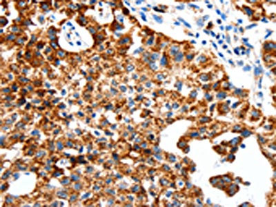
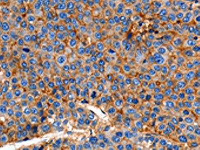
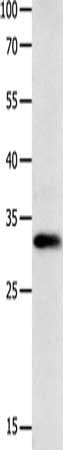
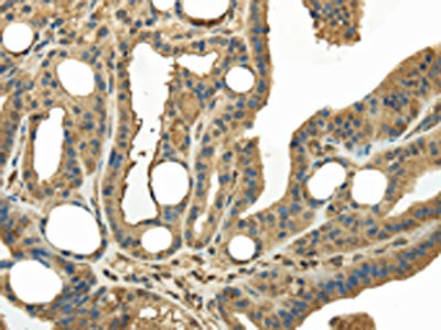
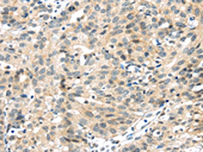
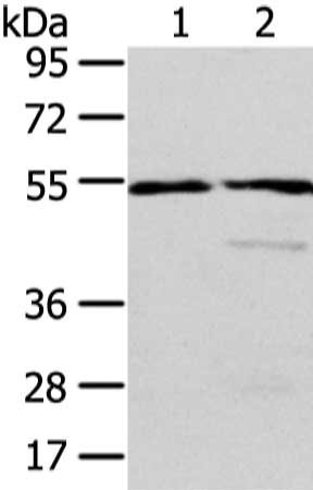
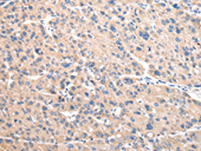
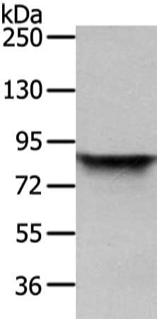
 SDS-PAGE
(Gel: 10%SDS-PAGE, Lysate: 40 ug, Lane: Human seminoma tissue, Primary antibody: AAA239934(SLC2A1 Antibody) at dilution 1/850, Secondary antibody: Goat anti rabbit IgG at 1/8000 dilution, Exposure time: 10 minutes)
SDS-PAGE
(Gel: 10%SDS-PAGE, Lysate: 40 ug, Lane: Human seminoma tissue, Primary antibody: AAA239934(SLC2A1 Antibody) at dilution 1/850, Secondary antibody: Goat anti rabbit IgG at 1/8000 dilution, Exposure time: 10 minutes)
SLC2A1, Polyclonal Antibody (Cat# AAA239934)
 SDS-PAGE
(Gel: 10%SDS-PAGE, Lysate: 40 ug, Lane: Human colon cancer tissue, Primary antibody: AAA239937(SLC2A3 Antibody) at dilution 1/400, Secondary antibody: Goat anti rabbit IgG at 1/8000 dilution, Exposure time: 2 minutes)
SDS-PAGE
(Gel: 10%SDS-PAGE, Lysate: 40 ug, Lane: Human colon cancer tissue, Primary antibody: AAA239937(SLC2A3 Antibody) at dilution 1/400, Secondary antibody: Goat anti rabbit IgG at 1/8000 dilution, Exposure time: 2 minutes)
SLC2A3, Polyclonal Antibody (Cat# AAA239937)
 SDS-PAGE
(Gel: 8%SDS-PAGE,Lysate: 40 ug,Lane 1-3: Human fetal brain tissue, Human placenta tissue, Mouse brain tissue,Primary antibody: AAA239529(TALDO1 Antibody) at dilution 1/400 dilution,Secondary antibody: Goat anti rabbit IgG at 1/8000 dilution,Exposure time: 10 seconds)
SDS-PAGE
(Gel: 8%SDS-PAGE,Lysate: 40 ug,Lane 1-3: Human fetal brain tissue, Human placenta tissue, Mouse brain tissue,Primary antibody: AAA239529(TALDO1 Antibody) at dilution 1/400 dilution,Secondary antibody: Goat anti rabbit IgG at 1/8000 dilution,Exposure time: 10 seconds)
TALDO1, Polyclonal Antibody (Cat# AAA239529)
 SDS-PAGE
(Gel: 6%SDS-PAGE,Lysate: 40 ug,Lane 1-2: Mouse heart tissue, MCF7 cells,Primary antibody: AAA239533(TGM5 Antibody) at dilution 1/400 dilution,Secondary antibody: Goat anti rabbit IgG at 1/8000 dilution,Exposure time: 2 minutes)
SDS-PAGE
(Gel: 6%SDS-PAGE,Lysate: 40 ug,Lane 1-2: Mouse heart tissue, MCF7 cells,Primary antibody: AAA239533(TGM5 Antibody) at dilution 1/400 dilution,Secondary antibody: Goat anti rabbit IgG at 1/8000 dilution,Exposure time: 2 minutes)
TGM5, Polyclonal Antibody (Cat# AAA239533)
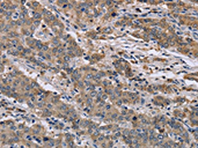
 IHC (Immunohiostchemistry)
(The image on the left is immunohistochemistry of paraffin-embedded Human breast cancer tissue using AAA239538(MED4 Antibody) at dilution 1/25, on the right is treated with fusion protein. (Original magnification: ×200))
IHC (Immunohiostchemistry)
(The image on the left is immunohistochemistry of paraffin-embedded Human breast cancer tissue using AAA239538(MED4 Antibody) at dilution 1/25, on the right is treated with fusion protein. (Original magnification: ×200))
MED4, Polyclonal Antibody (Cat# AAA239538)
 SDS-PAGE
(Gel: 12%SDS-PAGE,Lysate: 40 ug,Lane 1-2: Hela cells, Human fetal liver tissue,Primary antibody: AAA239546(ITPA Antibody) at dilution 1/400 dilution,Secondary antibody: Goat anti rabbit IgG at 1/8000 dilution,Exposure time: 5 minutes)
SDS-PAGE
(Gel: 12%SDS-PAGE,Lysate: 40 ug,Lane 1-2: Hela cells, Human fetal liver tissue,Primary antibody: AAA239546(ITPA Antibody) at dilution 1/400 dilution,Secondary antibody: Goat anti rabbit IgG at 1/8000 dilution,Exposure time: 5 minutes)
ITPA, Polyclonal Antibody (Cat# AAA239546)
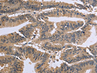
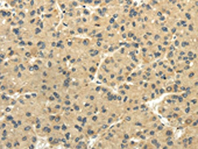
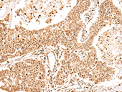
 IHC (Immunohiostchemistry)
(The image on the left is immunohistochemistry of paraffin-embedded Human breast cancer tissue using AAA239548(KEAP1 Antibody) at dilution 1/20, on the right is treated with fusion protein. (Original magnification: ×200))
IHC (Immunohiostchemistry)
(The image on the left is immunohistochemistry of paraffin-embedded Human breast cancer tissue using AAA239548(KEAP1 Antibody) at dilution 1/20, on the right is treated with fusion protein. (Original magnification: ×200))
KEAP1, Polyclonal Antibody (Cat# AAA239548)
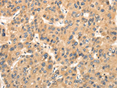
 IHC (Immunohiostchemistry)
(The image on the left is immunohistochemistry of paraffin-embedded Human esophagus cancer tissue using AAA239549(KEAP1 Antibody) at dilution 1/20, on the right is treated with fusion protein. (Original magnification: ×200))
IHC (Immunohiostchemistry)
(The image on the left is immunohistochemistry of paraffin-embedded Human esophagus cancer tissue using AAA239549(KEAP1 Antibody) at dilution 1/20, on the right is treated with fusion protein. (Original magnification: ×200))
KEAP1, Polyclonal Antibody (Cat# AAA239549)
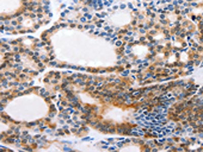
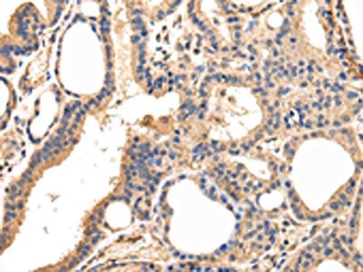
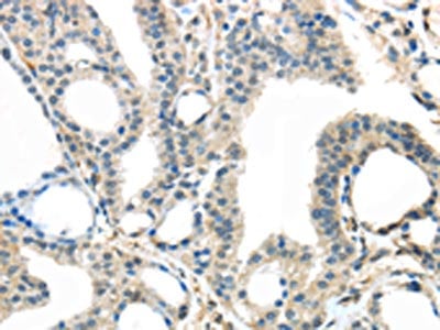 IHC (Immunohiostchemistry)
(The image on the left is immunohistochemistry of paraffin-embedded Human thyroid cancer tissue using AAA239551(TRPM2 Antibody) at dilution 1/30, on the right is treated with fusion protein. (Original magnification: ×200))
IHC (Immunohiostchemistry)
(The image on the left is immunohistochemistry of paraffin-embedded Human thyroid cancer tissue using AAA239551(TRPM2 Antibody) at dilution 1/30, on the right is treated with fusion protein. (Original magnification: ×200))
TRPM2, Polyclonal Antibody (Cat# AAA239551)
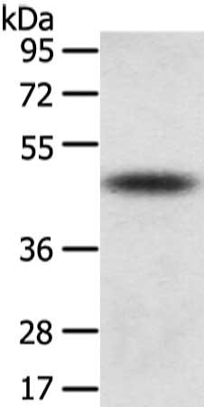 SDS-PAGE
(Gel: 8%SDS-PAGE, Lysate: 40 ug, Lane: Mouse adrenal gland tissue, Primary antibody: AAA239552(TTC23 Antibody) at dilution 1/800 dilution, Secondary antibody: Goat anti rabbit IgG at 1/8000 dilution, Exposure time: 10 seconds)
SDS-PAGE
(Gel: 8%SDS-PAGE, Lysate: 40 ug, Lane: Mouse adrenal gland tissue, Primary antibody: AAA239552(TTC23 Antibody) at dilution 1/800 dilution, Secondary antibody: Goat anti rabbit IgG at 1/8000 dilution, Exposure time: 10 seconds)
TTC23, Polyclonal Antibody (Cat# AAA239552)
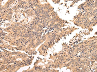
 IHC (Immunohiostchemistry)
(The image on the left is immunohistochemistry of paraffin-embedded Human esophagus cancer tissue using AAA239556(TULP2 Antibody) at dilution 1/20, on the right is treated with fusion protein. (Original magnification: ×200))
IHC (Immunohiostchemistry)
(The image on the left is immunohistochemistry of paraffin-embedded Human esophagus cancer tissue using AAA239556(TULP2 Antibody) at dilution 1/20, on the right is treated with fusion protein. (Original magnification: ×200))
TULP2, Polyclonal Antibody (Cat# AAA239556)
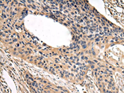
 IHC (Immunohiostchemistry)
(The image on the left is immunohistochemistry of paraffin-embedded Human prostate cancer tissue using AAA239557(TULP2 Antibody) at dilution 1/30, on the right is treated with fusion protein. (Original magnification: ×200))
IHC (Immunohiostchemistry)
(The image on the left is immunohistochemistry of paraffin-embedded Human prostate cancer tissue using AAA239557(TULP2 Antibody) at dilution 1/30, on the right is treated with fusion protein. (Original magnification: ×200))
TULP2, Polyclonal Antibody (Cat# AAA239557)
 SDS-PAGE
(Gel: 8%SDS-PAGE, Lysate: 40 ug, Lane 1-2: A431 and hela cell, Primary antibody: AAA239558(ERP44 Antibody) at dilution 1/450 dilution, Secondary antibody: Goat anti rabbit IgG at 1/8000 dilution, Exposure time: 2 minutes)
SDS-PAGE
(Gel: 8%SDS-PAGE, Lysate: 40 ug, Lane 1-2: A431 and hela cell, Primary antibody: AAA239558(ERP44 Antibody) at dilution 1/450 dilution, Secondary antibody: Goat anti rabbit IgG at 1/8000 dilution, Exposure time: 2 minutes)
ERP44, Polyclonal Antibody (Cat# AAA239558)
 IHC (Immunohiostchemistry)
(The image on the left is immunohistochemistry of paraffin-embedded Human breast cancer tissue using AAA239566(UBL3 Antibody) at dilution 1/30, on the right is treated with fusion protein. (Original magnification: ×200))
IHC (Immunohiostchemistry)
(The image on the left is immunohistochemistry of paraffin-embedded Human breast cancer tissue using AAA239566(UBL3 Antibody) at dilution 1/30, on the right is treated with fusion protein. (Original magnification: ×200))
UBL3, Polyclonal Antibody (Cat# AAA239566)


 SDS-PAGE
(Gel: 12%SDS-PAGE, Lysate: 40 ug, Lane 1-4: 293T, K562, Jurkat and 231 cell, Primary antibody: AAA239568(UCHL3 Antibody) at dilution 1/300 dilution, Secondary antibody: Goat anti rabbit IgG at 1/8000 dilution, Exposure time: 30 seconds)
SDS-PAGE
(Gel: 12%SDS-PAGE, Lysate: 40 ug, Lane 1-4: 293T, K562, Jurkat and 231 cell, Primary antibody: AAA239568(UCHL3 Antibody) at dilution 1/300 dilution, Secondary antibody: Goat anti rabbit IgG at 1/8000 dilution, Exposure time: 30 seconds)
UCHL3, Polyclonal Antibody (Cat# AAA239568)
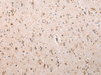
 IHC (Immunohiostchemistry)
(The image on the left is immunohistochemistry of paraffin-embedded Human brain tissue using AAA239573(MAP2 Antibody) at dilution 1/20, on the right is treated with fusion protein. (Original magnification: ×200))
IHC (Immunohiostchemistry)
(The image on the left is immunohistochemistry of paraffin-embedded Human brain tissue using AAA239573(MAP2 Antibody) at dilution 1/20, on the right is treated with fusion protein. (Original magnification: ×200))
MAP2, Polyclonal Antibody (Cat# AAA239573)
 SDS-PAGE
(Gel: 8%SDS-PAGE, Lysate: 40 ug, Lane 1-2: Human fetal brain tissue and hela cell, Primary antibody: AAA239577(SDCBP Antibody) at dilution 1/450 dilution, Secondary antibody: Goat anti rabbit IgG at 1/8000 dilution, Exposure time: 3 seconds)
SDS-PAGE
(Gel: 8%SDS-PAGE, Lysate: 40 ug, Lane 1-2: Human fetal brain tissue and hela cell, Primary antibody: AAA239577(SDCBP Antibody) at dilution 1/450 dilution, Secondary antibody: Goat anti rabbit IgG at 1/8000 dilution, Exposure time: 3 seconds)
SDCBP, Polyclonal Antibody (Cat# AAA239577)
 SDS-PAGE
(Gel: 6%SDS-PAGE, Lysate: 40 ug, Lane 1-2: Human normal liver and fetal liver tissue, Primary antibody: AAA239578(ALDH3A2 Antibody) at dilution 1/250 dilution, Secondary antibody: Goat anti rabbit IgG at 1/8000 dilution, Exposure time: 10 seconds)
SDS-PAGE
(Gel: 6%SDS-PAGE, Lysate: 40 ug, Lane 1-2: Human normal liver and fetal liver tissue, Primary antibody: AAA239578(ALDH3A2 Antibody) at dilution 1/250 dilution, Secondary antibody: Goat anti rabbit IgG at 1/8000 dilution, Exposure time: 10 seconds)
ALDH3A2, Polyclonal Antibody (Cat# AAA239578)
 SDS-PAGE
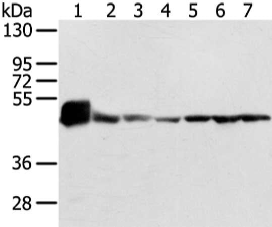
(Gel: 8%SDS-PAGE, Lysate: 40 ug, Lane 1-7: Mouse heart tissue and lo2 cell, 231 cell and human fetal brain tissue, A431, hela and 293T cell, Primary antibody: AAA239589(UQCRC1 Antibody) at dilution 1/200 dilution, Secondary antibody: Goat anti rabbit IgG at 1/8000 dilution, Exposure time: 1 second)
SDS-PAGE
(Gel: 8%SDS-PAGE, Lysate: 40 ug, Lane 1-7: Mouse heart tissue and lo2 cell, 231 cell and human fetal brain tissue, A431, hela and 293T cell, Primary antibody: AAA239589(UQCRC1 Antibody) at dilution 1/200 dilution, Secondary antibody: Goat anti rabbit IgG at 1/8000 dilution, Exposure time: 1 second)
UQCRC1, Polyclonal Antibody (Cat# AAA239589)
 SDS-PAGE
(Gel: 8%SDS-PAGE, Lysate: 40 ug, Lane 1-5: Jurkat, Raji, hepg2, A431 and HT-29 cell, Primary antibody: AAA239591(UQCRC2 Antibody) at dilution 1/400 dilution, Secondary antibody: Goat anti rabbit IgG at 1/8000 dilution, Exposure time: 10 seconds)
SDS-PAGE
(Gel: 8%SDS-PAGE, Lysate: 40 ug, Lane 1-5: Jurkat, Raji, hepg2, A431 and HT-29 cell, Primary antibody: AAA239591(UQCRC2 Antibody) at dilution 1/400 dilution, Secondary antibody: Goat anti rabbit IgG at 1/8000 dilution, Exposure time: 10 seconds)
UQCRC2, Polyclonal Antibody (Cat# AAA239591)
 IHC (Immunohiostchemistry)
(The image on the left is immunohistochemistry of paraffin-embedded Human breast cancer tissue using AAA239593(DUSP11 Antibody) at dilution 1/25, on the right is treated with fusion protein. (Original magnification: ×200))
IHC (Immunohiostchemistry)
(The image on the left is immunohistochemistry of paraffin-embedded Human breast cancer tissue using AAA239593(DUSP11 Antibody) at dilution 1/25, on the right is treated with fusion protein. (Original magnification: ×200))
DUSP11, Polyclonal Antibody (Cat# AAA239593)
 IHC (Immunohiostchemistry)
(The image on the left is immunohistochemistry of paraffin-embedded Human thyroid cancer tissue using AAA239596(USP15 Antibody) at dilution 1/35, on the right is treated with fusion protein. (Original magnification: ×200))
IHC (Immunohiostchemistry)
(The image on the left is immunohistochemistry of paraffin-embedded Human thyroid cancer tissue using AAA239596(USP15 Antibody) at dilution 1/35, on the right is treated with fusion protein. (Original magnification: ×200))
USP15, Polyclonal Antibody (Cat# AAA239596)
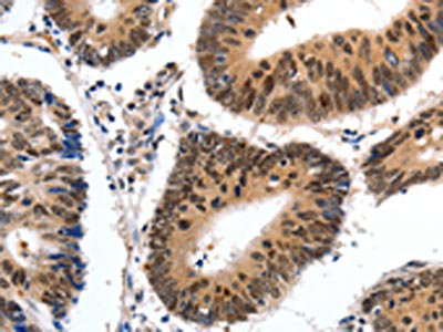
 IHC (Immunohiostchemistry)
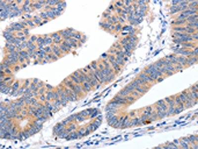
(The image on the left is immunohistochemistry of paraffin-embedded Human colorectal cancer tissue using AAA239598(USP45 Antibody) at dilution 1/25, on the right is treated with fusion protein. (Original magnification: ×200))
IHC (Immunohiostchemistry)
(The image on the left is immunohistochemistry of paraffin-embedded Human colorectal cancer tissue using AAA239598(USP45 Antibody) at dilution 1/25, on the right is treated with fusion protein. (Original magnification: ×200))
USP45, Polyclonal Antibody (Cat# AAA239598)
 SDS-PAGE
(Gel: 6%SDS-PAGE, Lysate: 40 ug, Lane: A172 cell, Primary antibody: AAA239600(USP47 Antibody) at dilution 1/400 dilution, Secondary antibody: Goat anti rabbit IgG at 1/8000 dilution, Exposure time: 30 seconds)
SDS-PAGE
(Gel: 6%SDS-PAGE, Lysate: 40 ug, Lane: A172 cell, Primary antibody: AAA239600(USP47 Antibody) at dilution 1/400 dilution, Secondary antibody: Goat anti rabbit IgG at 1/8000 dilution, Exposure time: 30 seconds)
USP47, Polyclonal Antibody (Cat# AAA239600)
 SDS-PAGE
(Gel: 12%SDS-PAGE, Lysate: 40 ug, Lane 1-4: PC3, TM4, hela and K562 cell, Primary antibody: AAA239602(VAMP4 Antibody) at dilution 1/650 dilution, Secondary antibody: Goat anti rabbit IgG at 1/8000 dilution, Exposure time: 10 seconds)
SDS-PAGE
(Gel: 12%SDS-PAGE, Lysate: 40 ug, Lane 1-4: PC3, TM4, hela and K562 cell, Primary antibody: AAA239602(VAMP4 Antibody) at dilution 1/650 dilution, Secondary antibody: Goat anti rabbit IgG at 1/8000 dilution, Exposure time: 10 seconds)
VAMP4, Polyclonal Antibody (Cat# AAA239602)
 SDS-PAGE
(Gel: 8%SDS-PAGE, Lysate: 40 ug, Lane: NIH/3T3 cell, Primary antibody: AAA239605(VAPA Antibody) at dilution 1/400 dilution, Secondary antibody: Goat anti rabbit IgG at 1/8000 dilution, Exposure time: 1 second)
SDS-PAGE
(Gel: 8%SDS-PAGE, Lysate: 40 ug, Lane: NIH/3T3 cell, Primary antibody: AAA239605(VAPA Antibody) at dilution 1/400 dilution, Secondary antibody: Goat anti rabbit IgG at 1/8000 dilution, Exposure time: 1 second)
VAPA, Polyclonal Antibody (Cat# AAA239605)
 SDS-PAGE
(Gel: 8%SDS-PAGE, Lysate: 40 ug, Lane: Mouse heart tissue, Primary antibody: AAA239608(VASH1 Antibody) at dilution 1/400 dilution, Secondary antibody: Goat anti rabbit IgG at 1/8000 dilution, Exposure time: 10 seconds)
SDS-PAGE
(Gel: 8%SDS-PAGE, Lysate: 40 ug, Lane: Mouse heart tissue, Primary antibody: AAA239608(VASH1 Antibody) at dilution 1/400 dilution, Secondary antibody: Goat anti rabbit IgG at 1/8000 dilution, Exposure time: 10 seconds)
VASH1, Polyclonal Antibody (Cat# AAA239608)
 SDS-PAGE
(Gel: 6%SDS-PAGE, Lysate: 40 ug, Lane: HT-29 cell, Primary antibody: AAA239611(VIL1 Antibody) at dilution 1/250 dilution, Secondary antibody: Goat anti rabbit IgG at 1/8000 dilution, Exposure time: 3 seconds)
SDS-PAGE
(Gel: 6%SDS-PAGE, Lysate: 40 ug, Lane: HT-29 cell, Primary antibody: AAA239611(VIL1 Antibody) at dilution 1/250 dilution, Secondary antibody: Goat anti rabbit IgG at 1/8000 dilution, Exposure time: 3 seconds)
VIL1, Polyclonal Antibody (Cat# AAA239611)

 SDS-PAGE
(Gel: 6%SDS-PAGE, Lysate: 40 ug, Lane: HT-29 cell, Primary antibody: AAA239612(VIL1 Antibody) at dilution 1/250 dilution, Secondary antibody: Goat anti rabbit IgG at 1/8000 dilution, Exposure time: 3 seconds)
SDS-PAGE
(Gel: 6%SDS-PAGE, Lysate: 40 ug, Lane: HT-29 cell, Primary antibody: AAA239612(VIL1 Antibody) at dilution 1/250 dilution, Secondary antibody: Goat anti rabbit IgG at 1/8000 dilution, Exposure time: 3 seconds)
VIL1, Polyclonal Antibody (Cat# AAA239612)
 IHC (Immunohiostchemistry)
(The image on the left is immunohistochemistry of paraffin-embedded Human esophagus cancer tissue using AAA239613(VIP Antibody) at dilution 1/20, on the right is treated with fusion protein. (Original magnification: ×200))
IHC (Immunohiostchemistry)
(The image on the left is immunohistochemistry of paraffin-embedded Human esophagus cancer tissue using AAA239613(VIP Antibody) at dilution 1/20, on the right is treated with fusion protein. (Original magnification: ×200))
VIP, Polyclonal Antibody (Cat# AAA239613)
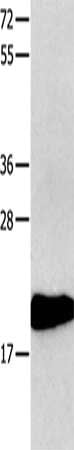
 SDS-PAGE
(Gel: 12%SDS-PAGE, Lysate: 40 ug, Lane: Human fetal liver tissue, Primary antibody: AAA239616(VPS28 Antibody) at dilution 1/200 dilution, Secondary antibody: Goat anti rabbit IgG at 1/8000 dilution, Exposure time: 5 seconds)
SDS-PAGE
(Gel: 12%SDS-PAGE, Lysate: 40 ug, Lane: Human fetal liver tissue, Primary antibody: AAA239616(VPS28 Antibody) at dilution 1/200 dilution, Secondary antibody: Goat anti rabbit IgG at 1/8000 dilution, Exposure time: 5 seconds)
VPS28, Polyclonal Antibody (Cat# AAA239616)
 SDS-PAGE
(Gel: 6%SDS-PAGE, Lysate: 40 ug, Lane: Human fetal brain tissue, Primary antibody: AAA239623(VPS35 Antibody) at dilution 1/250 dilution, Secondary antibody: Goat anti rabbit IgG at 1/8000 dilution, Exposure time: 20 seconds)
SDS-PAGE
(Gel: 6%SDS-PAGE, Lysate: 40 ug, Lane: Human fetal brain tissue, Primary antibody: AAA239623(VPS35 Antibody) at dilution 1/250 dilution, Secondary antibody: Goat anti rabbit IgG at 1/8000 dilution, Exposure time: 20 seconds)
VPS35, Polyclonal Antibody (Cat# AAA239623)
What are Polyclonal Antibodies?
Polyclonal antibodies are antibodies that come from multiple B cell clones of a host animal. The typical hosts used for the majority of polyclonal antibody production are rabbits, goats, sheep, and donkeys. These polyclonal antibodies, once having identified their target, will bind to different epitopes located at different regions or sequences on the same protein/antigen. As a result, they are ideal at locating and binding to the target, even if the target is in very low concentrations (due to many different antibodies being able to bind to the same target molecule, which allows for significant amplification of a downstream signal).
Polyclonal antibodies are typically produced by injecting an antigen into a host animal, which causes the animal’s immune system to attack the foreign antigen by mass generating antibodies against it. After a period of time, serum is collected from the animal and purified using physicochemical fractionation, class-specific affinity purification, and/or antigen-affinity purification.
Key Uses of Polyclonal Antibodies
- Western Blotting: This method is used to find specific proteins in biological samples after separating them by size.
- Immunohistochemistry: IHC helps visualize the location of proteins in tissue sections using various staining techniques.
- ELISA: (Enzyme-Linked Immunosorbent Assay) is typically used to identify specific protein quantities in a sample. ELISAs can be either “Quantitative” or “Qualitative”.
- Flow Cytometry: technique that identifies and measures the specific protein on the surface or inside the cells in a fluid suspension.
- Immunoprecipitation: IP isolates and studies a specific protein from a complex mixture using antibodies.
Why Buy Polyclonal Antibodies from AAA Biotech?
1. Ideal for Various Applications
Our antibodies are generally going to be validated for use in multiple types of assays, including ELISA, Western Blotting, Immunohistochemistry, Immunoprecipitation, amongst others. They are ideal for a wide range of research applications.
2. Rigorous Quality Control
All of the antibodies in our catalog undergo strict quality testing to ensure specificity, sensitivity, and consistent performance. We are confident in the ability of our antibodies to provide you with accurate results.
3. Wide Assortment of Antibodies
Antibodies in are catalog can be found for both common and exotic species, and these antibodies are also available in both conjugated and recombinant forms to suit many diverse experimental needs.
4. Highly Purified
Our antibodies are available in purified forms with over 85% purity, as confirmed by SDS-PAGE. They are also available with tags such as His, Flag, GST, or MBP. We cater to customers worldwide.
FAQ
1. How are polyclonal antibodies produced?
Traditionally, polyclonal antibodies are produced by injecting an antigen into a host animal (such as a rabbit or goat), which then triggers an immune response from the host animal. The animal’s B cells produce antibodies that will recognize different parts of the injected antigen. These antibodies are then collected from the animal’s blood and purified for use.
2. How do polyclonal antibodies differ from monoclonal antibodies?
Polyclonal antibodies are a mix of antibodies that bind to different locations (epitopes) of the same antigen, while monoclonal antibodies are identical and bind to just one specific epitope. This makes polyclonal antibodies more versatile and better at detecting proteins that may be present in low quantities or in altered/modified forms.
3. How should I store polyclonal antibodies?
Polyclonal antibodies should be stored at 4°C for short-term use (up to a few weeks) and at -20°C or -80°C for long-term storage. Avoid repeated freeze-thaw cycles by dividing them into small aliquots. Always check the datasheet for specific storage instructions.