Filters
▼Clonality
▼Type
▼Reactivity
▼Gene Name
▼Isotype
▼Host
▼Application
▼Clone
▼Polyclonal Antibodies
At AAA Biotech also known as AAA Bio or AAABio, we provide a broad range of purified polyclonal antibodies (pAbs) that are able to all be browsed online through our website. Due to their high specificity and strong binding affinity, these antibodies are ideal for wide swathes of research and experimental applications.
Our polyclonal antibodies can easily support your work, whether you use them for Western Blotting, Immunocytochemistry (with or without Immunofluorescence used in conjunction), Immunohistochemistry, Immunoprecipitation, and ELISA tests. We highly encourage you to browse our range of pAbs and choose the one that best suits your experimental model.
Viewing 900-950 of 96805 product results
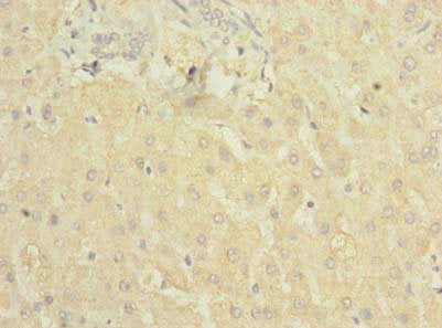
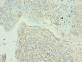
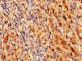
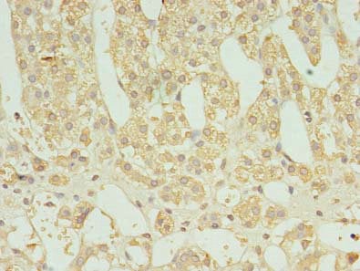
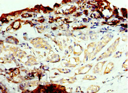
 IHC (Immunohiostchemistry)
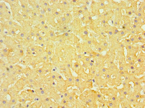
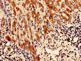
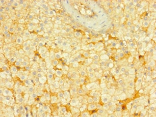
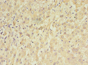
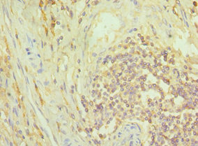
(Immunohistochemistry of paraffin-embedded human liver cancer using AAA118577 at dilution of 1:100)
IHC (Immunohiostchemistry)
(Immunohistochemistry of paraffin-embedded human liver cancer using AAA118577 at dilution of 1:100)
C5, Polyclonal Antibody (Cat# AAA118577)
parvovirus B19 Capsid protein VP2, Polyclonal Antibody (Cat# AAA118584)
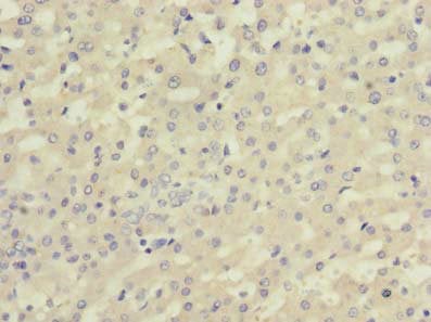
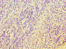
 IHC (Immunohiostchemistry)
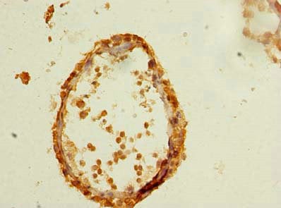
(Immunohistochemistry of paraffin-embedded human liver cancer using AAA118585 at dilution of 1:100)
IHC (Immunohiostchemistry)
(Immunohistochemistry of paraffin-embedded human liver cancer using AAA118585 at dilution of 1:100)
C10orf111, Polyclonal Antibody (Cat# AAA118585)
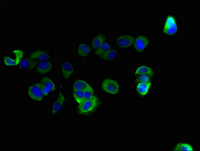
 IHC (Immunohistochemisry)
(Immunofluorescent analysis of A549 cells using AAA118586 at a dilution of 1:100 and Alexa Fluor 488-congugated AffiniPure Goat Anti-Rabbit IgG(H+L))
IHC (Immunohistochemisry)
(Immunofluorescent analysis of A549 cells using AAA118586 at a dilution of 1:100 and Alexa Fluor 488-congugated AffiniPure Goat Anti-Rabbit IgG(H+L))
Guanine nucleotide-binding protein subunit beta-5, Polyclonal Antibody (Cat# AAA118586)
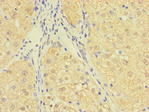
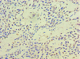
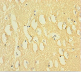
 IHC (Immunohiostchemistry)
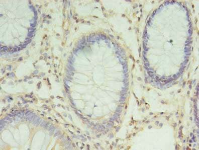
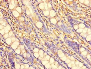
(Immunohistochemistry of paraffin-embedded human tonsil using AAA118590 at dilution of 1:100)
IHC (Immunohiostchemistry)
(Immunohistochemistry of paraffin-embedded human tonsil using AAA118590 at dilution of 1:100)
ASCC1, Polyclonal Antibody (Cat# AAA118590)
CXCR4, Polyclonal Antibody (Cat# AAA118599)
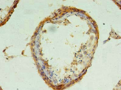
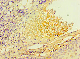
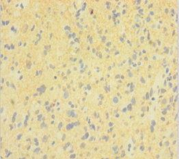
 IHC (Immunohiostchemistry)
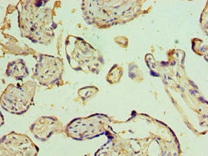
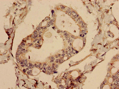
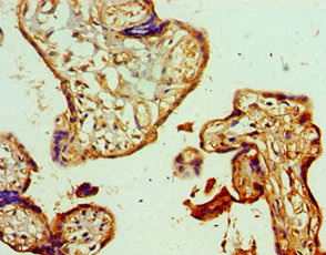
(Immunohistochemistry of paraffin-embedded human placenta using AAA118602 at dilution 1:100)
IHC (Immunohiostchemistry)
(Immunohistochemistry of paraffin-embedded human placenta using AAA118602 at dilution 1:100)
Ephrin-B1, Polyclonal Antibody (Cat# AAA118602)
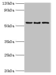
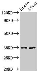
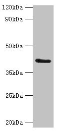
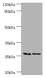
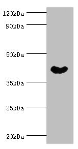
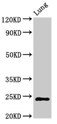 WB (Western Blot)
(Western BlotPositive WB detected in: Rat lung tissueAll lanes: CHMP6 antibody at 3ug/mlSecondaryGoat polyclonal to rabbit IgG at 1/50000 dilutionPredicted band size: 24 KDaObserved band size: 24 KDa)
WB (Western Blot)
(Western BlotPositive WB detected in: Rat lung tissueAll lanes: CHMP6 antibody at 3ug/mlSecondaryGoat polyclonal to rabbit IgG at 1/50000 dilutionPredicted band size: 24 KDaObserved band size: 24 KDa)
CHMP6, Polyclonal Antibody (Cat# AAA118603)
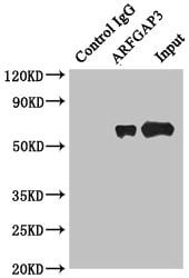 IP (Immunoprecipitation)
(Immunoprecipitating ARFGAP3 in HepG2 whole cell lysateLane 1: Rabbit monoclonal IgG(1ug)instead of AAA118604 in HepG2 whole cell lysate. For western blotting, a HRP-conjugated anti-rabbit IgG, specific to the non-reduced form of IgG was used as the Secondary antibody (1/50000)Lane 2:AAA118604(4ug)+ HepG2 whole cell lysate(500ug)Lane 3: HepG2 whole cell lysate (20ug))
IP (Immunoprecipitation)
(Immunoprecipitating ARFGAP3 in HepG2 whole cell lysateLane 1: Rabbit monoclonal IgG(1ug)instead of AAA118604 in HepG2 whole cell lysate. For western blotting, a HRP-conjugated anti-rabbit IgG, specific to the non-reduced form of IgG was used as the Secondary antibody (1/50000)Lane 2:AAA118604(4ug)+ HepG2 whole cell lysate(500ug)Lane 3: HepG2 whole cell lysate (20ug))
ARFGAP3, Polyclonal Antibody (Cat# AAA118604)
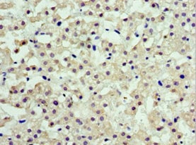
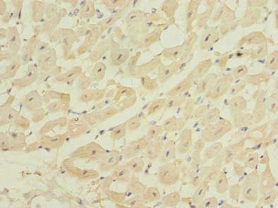 IHC (Immunohistochemisry)
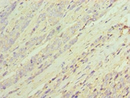
(Immunohistochemistry of paraffin-embedded human heart tissue using AAA118607 at dilution of 1:100)
IHC (Immunohistochemisry)
(Immunohistochemistry of paraffin-embedded human heart tissue using AAA118607 at dilution of 1:100)
ZBTB6, Polyclonal Antibody (Cat# AAA118607)
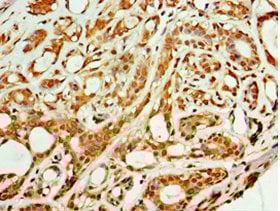
 IHC (Immunohiostchemistry)
(Immunohistochemistry of paraffin-embedded human pancreatic tissue using AAA118608 at dilution of 1:100)
IHC (Immunohiostchemistry)
(Immunohistochemistry of paraffin-embedded human pancreatic tissue using AAA118608 at dilution of 1:100)
CDC25C, Polyclonal Antibody (Cat# AAA118608)
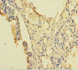
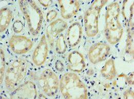
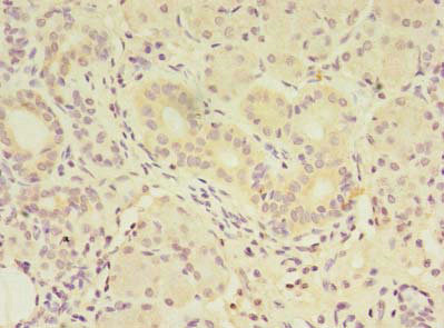
 IHC (Immunohiostchemistry)
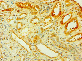
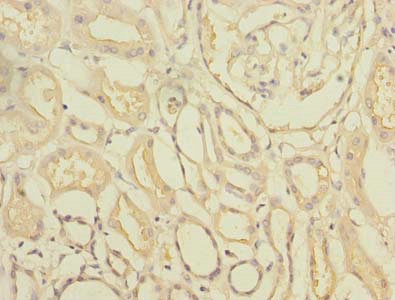
(Immunohistochemistry of paraffin-embedded human kidney using AAA118610 at dilution 1:100)
IHC (Immunohiostchemistry)
(Immunohistochemistry of paraffin-embedded human kidney using AAA118610 at dilution 1:100)
Cytokine-inducible SH2-containing protein, Polyclonal Antibody (Cat# AAA118610)
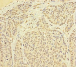
 IHC (Immunohistochemisry)
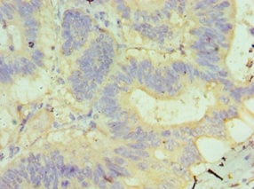
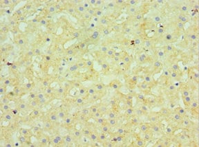
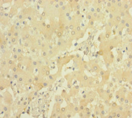
(Immunohistochemistry of paraffin-embedded human liver using AAA118615 at dilution 1:100)
IHC (Immunohistochemisry)
(Immunohistochemistry of paraffin-embedded human liver using AAA118615 at dilution 1:100)
GUSB, Polyclonal Antibody (Cat# AAA118615)
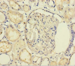
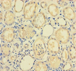
 IHC (Immunohistochemisry)
(Immunohistochemistry of paraffin-embedded human kidney using AAA118618 at dilution 1:100)
IHC (Immunohistochemisry)
(Immunohistochemistry of paraffin-embedded human kidney using AAA118618 at dilution 1:100)
MAPK4, Polyclonal Antibody (Cat# AAA118618)

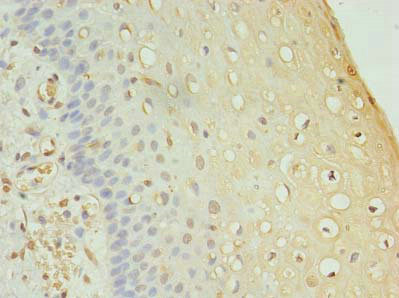
 IHC (Immunohiostchemistry)
(Immunohistochemistry of paraffin-embedded human kidney using AAA118619 at dilution 1:100)
IHC (Immunohiostchemistry)
(Immunohistochemistry of paraffin-embedded human kidney using AAA118619 at dilution 1:100)
Fibroblast growth factor 1, Polyclonal Antibody (Cat# AAA118619)
BDH2, Polyclonal Antibody (Cat# AAA118625)
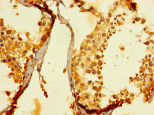
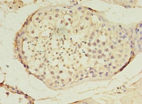
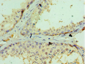
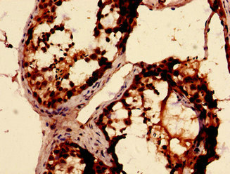 IHC (Immunohistochemistry)
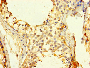
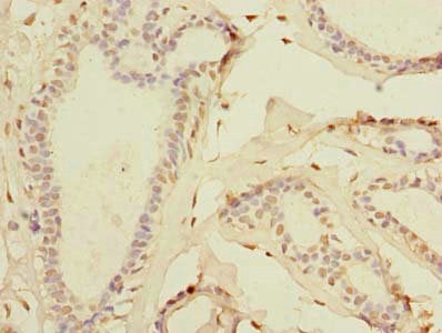
(Immunohistochemistry analysis of human testis tissue using AAA118627 at dilution of 1:100)
IHC (Immunohistochemistry)
(Immunohistochemistry analysis of human testis tissue using AAA118627 at dilution of 1:100)
RBKS, Polyclonal Antibody (Cat# AAA118627)
CFHR2, Polyclonal Antibody (Cat# AAA118632)
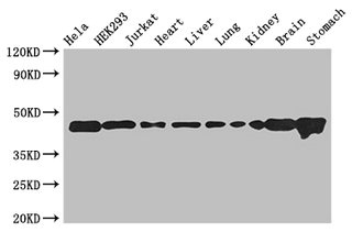
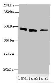
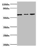
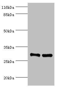
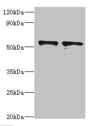
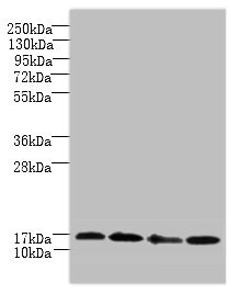
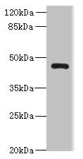
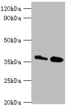
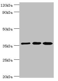
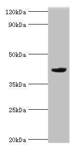
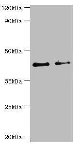
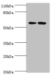 WB (Western Blot)
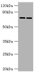
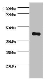
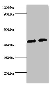
(Western blotAll lanes: DCAF4 antibody at 2ug/mlLane 1:293T whole cell lysateLane 2:Hela whole cell lysateSecondaryGoat polyclonal to rabbit at 1/10000 dilutionPredicted band size: 56,49,45,55 kDaObserved band size: 56 kDa)
WB (Western Blot)
(Western blotAll lanes: DCAF4 antibody at 2ug/mlLane 1:293T whole cell lysateLane 2:Hela whole cell lysateSecondaryGoat polyclonal to rabbit at 1/10000 dilutionPredicted band size: 56,49,45,55 kDaObserved band size: 56 kDa)
DCAF4, Polyclonal Antibody (Cat# AAA118633)
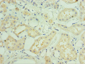
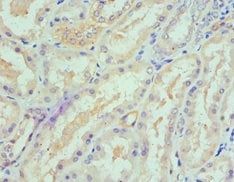 IHC (Immunohistochemisry)
(Immunohistochemistry of paraffin-embedded human kidney using AAA118634 at dilution 1:100)
IHC (Immunohistochemisry)
(Immunohistochemistry of paraffin-embedded human kidney using AAA118634 at dilution 1:100)
Serine palmitoyltransferase 1, Polyclonal Antibody (Cat# AAA118634)
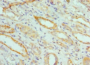
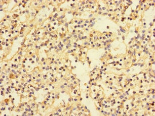
 IHC (Immunohiostchemistry)
(Immunohistochemistry of paraffin-embedded human kidney tissue using AAA118646 at dilution 1:100)
IHC (Immunohiostchemistry)
(Immunohistochemistry of paraffin-embedded human kidney tissue using AAA118646 at dilution 1:100)
Ribonuclease P protein subunit p29, Polyclonal Antibody (Cat# AAA118646)
 IHC (Immunohistochemisry)
(Immunohistochemistry of paraffin-embedded human adrenal gland tissue using AAA118649 at dilution of 1:100)
IHC (Immunohistochemisry)
(Immunohistochemistry of paraffin-embedded human adrenal gland tissue using AAA118649 at dilution of 1:100)
FAM136A, Polyclonal Antibody (Cat# AAA118649)
 IHC (Immunohistochemisry)
(Immunohistochemistry of paraffin-embedded human blood plasma using AAA118650 at dilution 1:100)
IHC (Immunohistochemisry)
(Immunohistochemistry of paraffin-embedded human blood plasma using AAA118650 at dilution 1:100)
Telomeric repeat-binding factor 2-interacting protein 1, Polyclonal Antibody (Cat# AAA118650)
Rotavirus A Outer capsid protein VP4, Polyclonal Antibody (Cat# AAA118656)
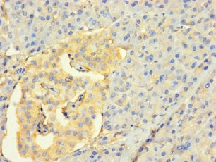
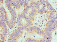
 IHC (Immunohistochemisry)
(Immunohistochemistry analysis of human pancreatic cancer using AAA118657 at dilution of 1:100)
IHC (Immunohistochemisry)
(Immunohistochemistry analysis of human pancreatic cancer using AAA118657 at dilution of 1:100)
CFAP61, Polyclonal Antibody (Cat# AAA118657)
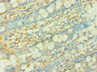
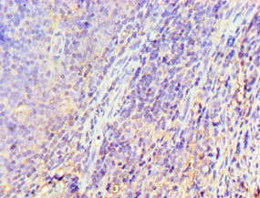
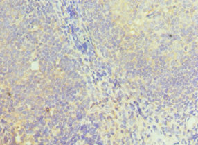
 IHC (Immunohiostchemistry)
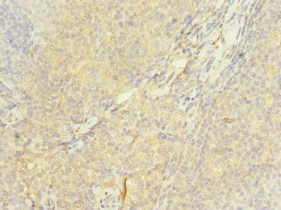
(Immunohistochemistry of paraffin-embedded human spleen tissue using AAA118658 at dilution of 1:100)
IHC (Immunohiostchemistry)
(Immunohistochemistry of paraffin-embedded human spleen tissue using AAA118658 at dilution of 1:100)
PDIK1L, Polyclonal Antibody (Cat# AAA118658)
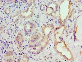 IHC (Immunohiostchemistry)
(Immunohistochemistry of paraffin-embedded human kidney using AAA118661 at dilution 1:100)
IHC (Immunohiostchemistry)
(Immunohistochemistry of paraffin-embedded human kidney using AAA118661 at dilution 1:100)
G protein-coupled receptor kinase 6, Polyclonal Antibody (Cat# AAA118661)
 IF (Immunofluorescence)
(Immunofluorescent analysis of U251 cells using AAA118662 at a dilution of 1:100 and Alexa Fluor 488-congugated AffiniPure Goat Anti-Rabbit IgG(H+L))
IF (Immunofluorescence)
(Immunofluorescent analysis of U251 cells using AAA118662 at a dilution of 1:100 and Alexa Fluor 488-congugated AffiniPure Goat Anti-Rabbit IgG(H+L))
TMEM61, Polyclonal Antibody (Cat# AAA118662)
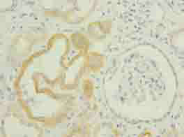
 IHC (Immunohistochemisry)
(Immunohistochemistry of paraffin-embedded human kidney using AAA118667 at dilution 1:100)
IHC (Immunohistochemisry)
(Immunohistochemistry of paraffin-embedded human kidney using AAA118667 at dilution 1:100)
FUT6, Polyclonal Antibody (Cat# AAA118667)
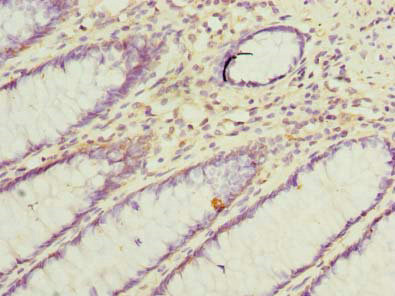
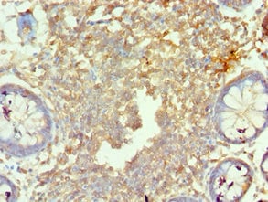
 IHC (Immunohiostchemistry)
(Immunohistochemistry of paraffin-embedded human rectal cancer using AAA118335 at dilution 1:100)
IHC (Immunohiostchemistry)
(Immunohistochemistry of paraffin-embedded human rectal cancer using AAA118335 at dilution 1:100)
Caspase recruitment domain-containing protein 8, Polyclonal Antibody (Cat# AAA118335)
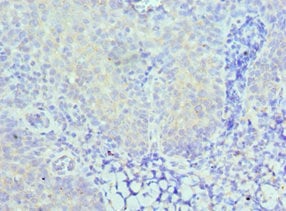
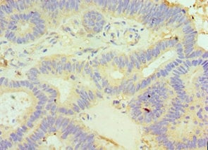
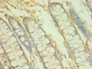
 IHC (Immunohiostchemistry)
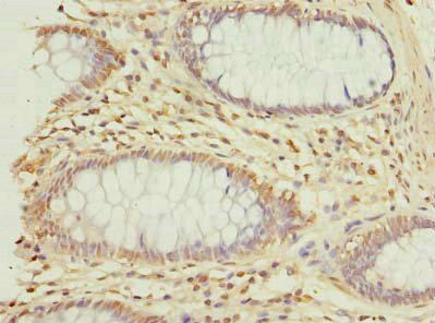
(Immunohistochemistry of paraffin-embedded human colon cancer using AAA118339 at dilution of 1:100)
IHC (Immunohiostchemistry)
(Immunohistochemistry of paraffin-embedded human colon cancer using AAA118339 at dilution of 1:100)
VAT1L, Polyclonal Antibody (Cat# AAA118339)
 IHC (Immunohiostchemistry)
(Immunohistochemistry of paraffin-embedded human testis tissue using AAA118340 at dilution of 1:100)
IHC (Immunohiostchemistry)
(Immunohistochemistry of paraffin-embedded human testis tissue using AAA118340 at dilution of 1:100)
FAM174A, Polyclonal Antibody (Cat# AAA118340)
 IHC (Immunohistochemisry)
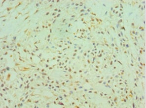
(Immunohistochemistry of paraffin-embedded human placenta tissue using AAA118347 at dilution 1:100)
IHC (Immunohistochemisry)
(Immunohistochemistry of paraffin-embedded human placenta tissue using AAA118347 at dilution 1:100)
GSTM4, Polyclonal Antibody (Cat# AAA118347)
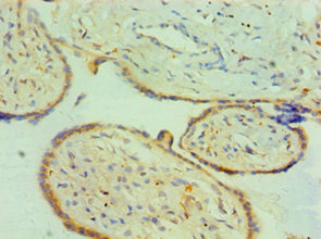 IHC (Immunohistochemisry)
(Immunohistochemistry of paraffin-embedded human placenta tissue using AAA118348 at dilution 1:100)
IHC (Immunohistochemisry)
(Immunohistochemistry of paraffin-embedded human placenta tissue using AAA118348 at dilution 1:100)
CTSK, Polyclonal Antibody (Cat# AAA118348)
 IHC (Immunohistochemisry)
(Immunohistochemistry of paraffin-embedded human adrenal gland tissue using AAA118350 at dilution of 1:100)
IHC (Immunohistochemisry)
(Immunohistochemistry of paraffin-embedded human adrenal gland tissue using AAA118350 at dilution of 1:100)
ENTPD3, Polyclonal Antibody (Cat# AAA118350)
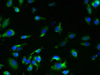
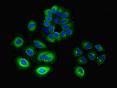 IF (Immunofluorescence)
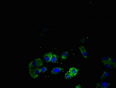
(Immunofluorescent analysis of A549 cells using AAA118353 at a dilution of 1:100 and Alexa Fluor 488-congugated AffiniPure Goat Anti-Rabbit IgG(H+L))
IF (Immunofluorescence)
(Immunofluorescent analysis of A549 cells using AAA118353 at a dilution of 1:100 and Alexa Fluor 488-congugated AffiniPure Goat Anti-Rabbit IgG(H+L))
GNG10, Polyclonal Antibody (Cat# AAA118353)
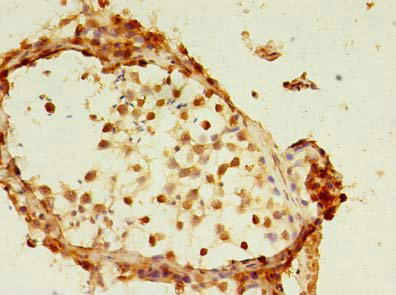
 IHC (Immunohiostchemistry)
(Immunohistochemistry of paraffin-embedded human testis using AAA118356 at dilution 1:100)
IHC (Immunohiostchemistry)
(Immunohistochemistry of paraffin-embedded human testis using AAA118356 at dilution 1:100)
TET3, Polyclonal Antibody (Cat# AAA118356)
Delta-like protein 3, Polyclonal Antibody (Cat# AAA118365)
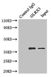 IP (Immunoprecipitation)
(Immunoprecipitating GLRX3 in HepG2 whole cell lysateLane 1: Rabbit monoclonal IgG(1ug)instead of AAA118368 in HepG2 whole cell lysate. For western blotting, a HRP-conjugated anti-rabbit IgG, specific to the non-reduced form of IgG was used as the Secondary antibody (1/50000)Lane 2: AAA118368(4ug)+ HepG2 whole cell lysate(500ug)Lane 3: HepG2 whole cell lysate (20ug))
IP (Immunoprecipitation)
(Immunoprecipitating GLRX3 in HepG2 whole cell lysateLane 1: Rabbit monoclonal IgG(1ug)instead of AAA118368 in HepG2 whole cell lysate. For western blotting, a HRP-conjugated anti-rabbit IgG, specific to the non-reduced form of IgG was used as the Secondary antibody (1/50000)Lane 2: AAA118368(4ug)+ HepG2 whole cell lysate(500ug)Lane 3: HepG2 whole cell lysate (20ug))
Glutaredoxin-3, Polyclonal Antibody (Cat# AAA118368)
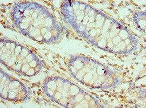
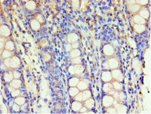
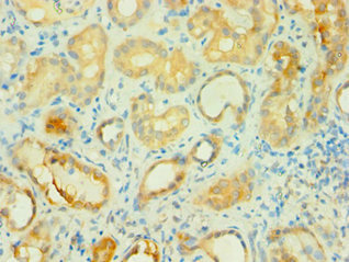
 IHC (Immunohiostchemistry)
(Immunohistochemistry of paraffin-embedded human rectum using AAA118369 at dilution 1:100)
IHC (Immunohiostchemistry)
(Immunohistochemistry of paraffin-embedded human rectum using AAA118369 at dilution 1:100)
Mortality factor 4-like protein 1, Polyclonal Antibody (Cat# AAA118369)
 IHC (Immunohistochemisry)
(Immunohistochemistry of paraffin-embedded human kidney using AAA118373 at dilution 1:100)
IHC (Immunohistochemisry)
(Immunohistochemistry of paraffin-embedded human kidney using AAA118373 at dilution 1:100)
Branched-chain-amino-acid aminotransferase, Polyclonal Antibody (Cat# AAA118373)
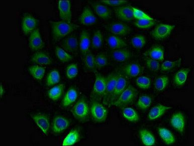
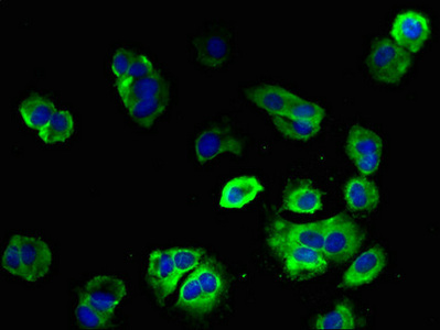
 IHC (Immunohistochemisry)
(Immunofluorescent analysis of A549 cells using AAA118376 at a dilution of 1:100 and Alexa Fluor 488-congugated AffiniPure Goat Anti-Rabbit IgG(H+L))
IHC (Immunohistochemisry)
(Immunofluorescent analysis of A549 cells using AAA118376 at a dilution of 1:100 and Alexa Fluor 488-congugated AffiniPure Goat Anti-Rabbit IgG(H+L))
Dynamin-1, Polyclonal Antibody (Cat# AAA118376)
rhinovirus A serotype 89 Genome polyprotein, Polyclonal Antibody (Cat# AAA118379)
 IHC (Immunohistochemisry)
(Immunohistochemistry of paraffin-embedded human rectal cancer using AAA118385 at dilution 1:100)
IHC (Immunohistochemisry)
(Immunohistochemistry of paraffin-embedded human rectal cancer using AAA118385 at dilution 1:100)
Cytoplasmic protein NCK1, Polyclonal Antibody (Cat# AAA118385)
Toxocara canis 26 kDa secreted antigen, Polyclonal Antibody (Cat# AAA118391)
 IF (Immunofluorescence)
(Immunofluorescent analysis of HepG2 cells using AAA118392 at a dilution of 1:100 and Alexa Fluor 488-congugated AffiniPure Goat Anti-Rabbit IgG(H+L))
IF (Immunofluorescence)
(Immunofluorescent analysis of HepG2 cells using AAA118392 at a dilution of 1:100 and Alexa Fluor 488-congugated AffiniPure Goat Anti-Rabbit IgG(H+L))
Somatostatin receptor type 4, Polyclonal Antibody (Cat# AAA118392)
Hepatitis E virus genotype 1 Non-structural polyprotein pORF1, Polyclonal Antibody (Cat# AAA118396)

 IHC (Immunohiostchemistry)
(Immunohistochemistry of paraffin-embedded human liver tissue using AAA118398 at dilution 1:100)
IHC (Immunohiostchemistry)
(Immunohistochemistry of paraffin-embedded human liver tissue using AAA118398 at dilution 1:100)
Serine protease 23, Polyclonal Antibody (Cat# AAA118398)
 IHC (Immunohiostchemistry)
(Immunohistochemistry of paraffin-embedded human breast cancer using AAA118399 at dilution of 1:100)
IHC (Immunohiostchemistry)
(Immunohistochemistry of paraffin-embedded human breast cancer using AAA118399 at dilution of 1:100)
TTC33, Polyclonal Antibody (Cat# AAA118399)
 IHC (Immunohistochemisry)
(Immunohistochemistry of paraffin-embedded human gallbladder using AAA118402 at dilution 1:100)
IHC (Immunohistochemisry)
(Immunohistochemistry of paraffin-embedded human gallbladder using AAA118402 at dilution 1:100)
Aldo-keto reductase family 1 member C3, Polyclonal Antibody (Cat# AAA118402)
What are Polyclonal Antibodies?
Polyclonal antibodies are antibodies that come from multiple B cell clones of a host animal. The typical hosts used for the majority of polyclonal antibody production are rabbits, goats, sheep, and donkeys. These polyclonal antibodies, once having identified their target, will bind to different epitopes located at different regions or sequences on the same protein/antigen. As a result, they are ideal at locating and binding to the target, even if the target is in very low concentrations (due to many different antibodies being able to bind to the same target molecule, which allows for significant amplification of a downstream signal).
Polyclonal antibodies are typically produced by injecting an antigen into a host animal, which causes the animal’s immune system to attack the foreign antigen by mass generating antibodies against it. After a period of time, serum is collected from the animal and purified using physicochemical fractionation, class-specific affinity purification, and/or antigen-affinity purification.
Key Uses of Polyclonal Antibodies
- Western Blotting: This method is used to find specific proteins in biological samples after separating them by size.
- Immunohistochemistry: IHC helps visualize the location of proteins in tissue sections using various staining techniques.
- ELISA: (Enzyme-Linked Immunosorbent Assay) is typically used to identify specific protein quantities in a sample. ELISAs can be either “Quantitative” or “Qualitative”.
- Flow Cytometry: technique that identifies and measures the specific protein on the surface or inside the cells in a fluid suspension.
- Immunoprecipitation: IP isolates and studies a specific protein from a complex mixture using antibodies.
Why Buy Polyclonal Antibodies from AAA Biotech?
1. Ideal for Various Applications
Our antibodies are generally going to be validated for use in multiple types of assays, including ELISA, Western Blotting, Immunohistochemistry, Immunoprecipitation, amongst others. They are ideal for a wide range of research applications.
2. Rigorous Quality Control
All of the antibodies in our catalog undergo strict quality testing to ensure specificity, sensitivity, and consistent performance. We are confident in the ability of our antibodies to provide you with accurate results.
3. Wide Assortment of Antibodies
Antibodies in are catalog can be found for both common and exotic species, and these antibodies are also available in both conjugated and recombinant forms to suit many diverse experimental needs.
4. Highly Purified
Our antibodies are available in purified forms with over 85% purity, as confirmed by SDS-PAGE. They are also available with tags such as His, Flag, GST, or MBP. We cater to customers worldwide.
FAQ
1. How are polyclonal antibodies produced?
Traditionally, polyclonal antibodies are produced by injecting an antigen into a host animal (such as a rabbit or goat), which then triggers an immune response from the host animal. The animal’s B cells produce antibodies that will recognize different parts of the injected antigen. These antibodies are then collected from the animal’s blood and purified for use.
2. How do polyclonal antibodies differ from monoclonal antibodies?
Polyclonal antibodies are a mix of antibodies that bind to different locations (epitopes) of the same antigen, while monoclonal antibodies are identical and bind to just one specific epitope. This makes polyclonal antibodies more versatile and better at detecting proteins that may be present in low quantities or in altered/modified forms.
3. How should I store polyclonal antibodies?
Polyclonal antibodies should be stored at 4°C for short-term use (up to a few weeks) and at -20°C or -80°C for long-term storage. Avoid repeated freeze-thaw cycles by dividing them into small aliquots. Always check the datasheet for specific storage instructions.