Filters
▼Clonality
▼Type
▼Reactivity
▼Gene Name
▼Isotype
▼Host
▼Application
▼Clone
▼Polyclonal Antibodies
At AAA Biotech also known as AAA Bio or AAABio, we provide a broad range of purified polyclonal antibodies (pAbs) that are able to all be browsed online through our website. Due to their high specificity and strong binding affinity, these antibodies are ideal for wide swathes of research and experimental applications.
Our polyclonal antibodies can easily support your work, whether you use them for Western Blotting, Immunocytochemistry (with or without Immunofluorescence used in conjunction), Immunohistochemistry, Immunoprecipitation, and ELISA tests. We highly encourage you to browse our range of pAbs and choose the one that best suits your experimental model.
Viewing 750-800 of 96805 product results

 IHC (Immunohiostchemistry)
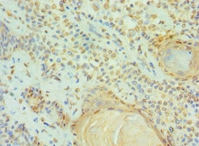
(Immunohistochemistry of paraffin-embedded Human thyroid cancer tissue using CYP11B1 Polyclonal Antibody at dilution 1:30)
IHC (Immunohiostchemistry)
(Immunohistochemistry of paraffin-embedded Human thyroid cancer tissue using CYP11B1 Polyclonal Antibody at dilution 1:30)
CYP11B1, Polyclonal Antibody (Cat# AAA169878)
 IHC (Immunohiostchemistry)
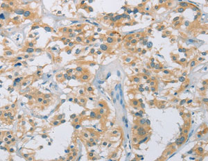
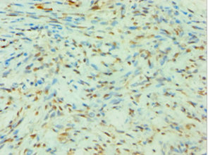
(Immunohistochemistry of paraffin-embedded Human liver cancer tissue using GPR171 Polyclonal Antibody at dilution 1:40)
IHC (Immunohiostchemistry)
(Immunohistochemistry of paraffin-embedded Human liver cancer tissue using GPR171 Polyclonal Antibody at dilution 1:40)
GPR171, Polyclonal Antibody (Cat# AAA169886)
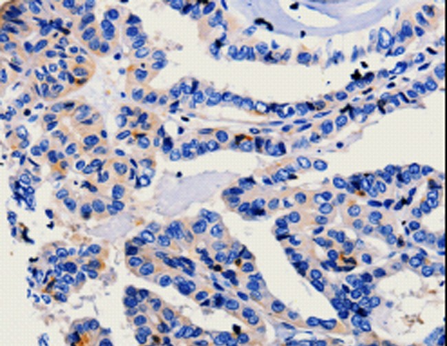 IHC (Immunohiostchemistry)
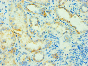
(Immunohistochemistry of paraffin-embedded Human thyroid cancer using CA3 Polyclonal Antibody at dilution of 1:25)
IHC (Immunohiostchemistry)
(Immunohistochemistry of paraffin-embedded Human thyroid cancer using CA3 Polyclonal Antibody at dilution of 1:25)
CA3, Polyclonal Antibody (Cat# AAA169889)
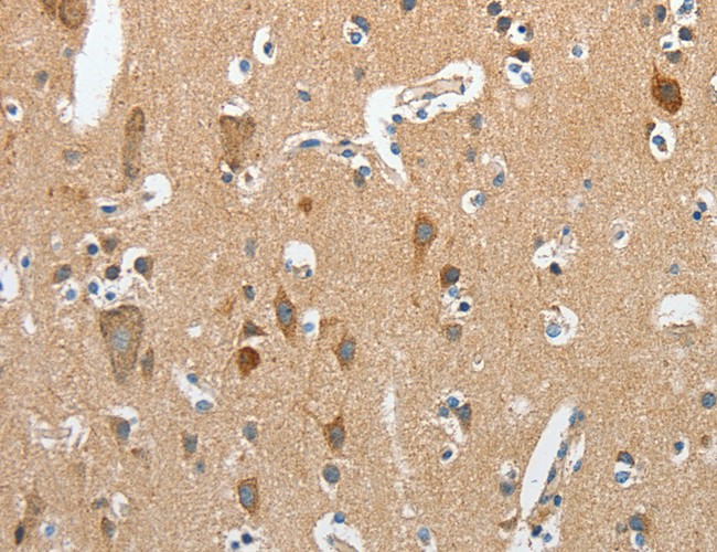 IHC (Immunohistochemisry)
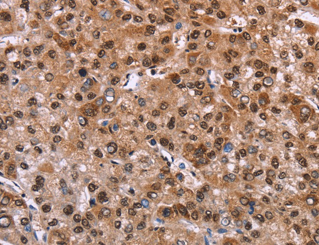
(Immunohistochemistry of paraffin-embedded Human brain using RCAN1 Polyclonal Antibody at dilution of 1:45)
IHC (Immunohistochemisry)
(Immunohistochemistry of paraffin-embedded Human brain using RCAN1 Polyclonal Antibody at dilution of 1:45)
RCAN1, Polyclonal Antibody (Cat# AAA169895)
 IHC (Immunohistochemisry)
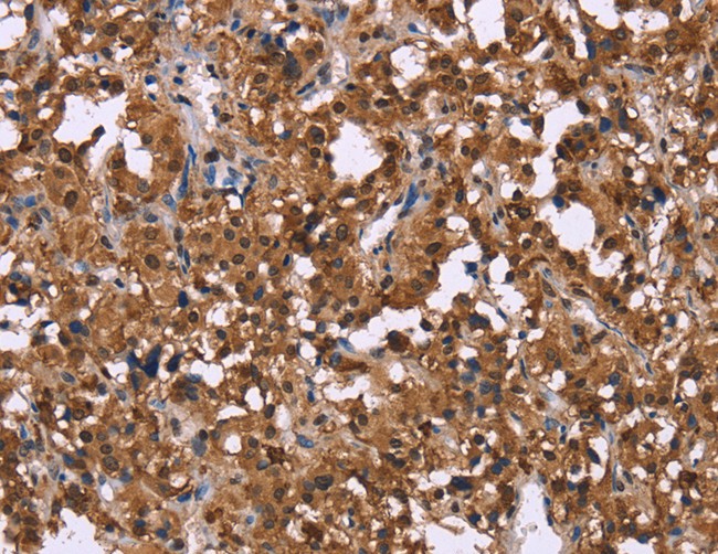
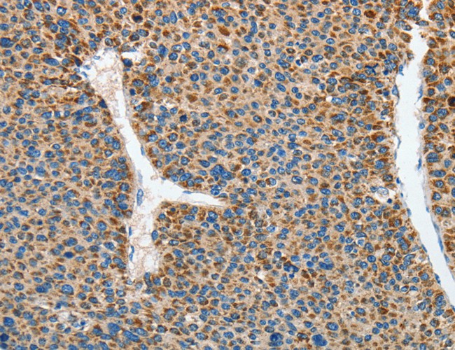
(Immunohistochemistry of paraffin-embedded Human liver cancer using AARS2 Polyclonal Antibody at dilution of 1:80)
IHC (Immunohistochemisry)
(Immunohistochemistry of paraffin-embedded Human liver cancer using AARS2 Polyclonal Antibody at dilution of 1:80)
AARS2, Polyclonal Antibody (Cat# AAA169897)

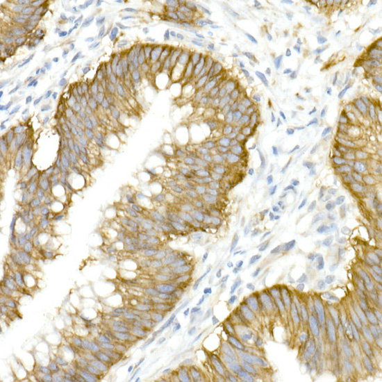
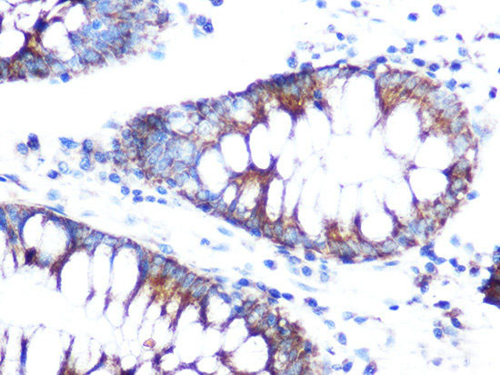
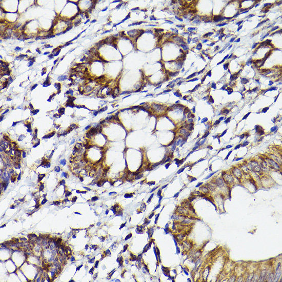 IHC (Immunohistochemistry)
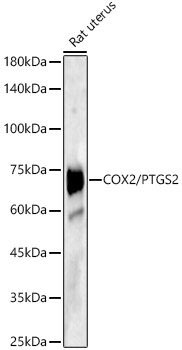
(Immunohistochemistry of paraffin-embedded human colon using COX2/PTGS2 Polyclonal antibody at dilution of 1:50 (40x lens).Perform high pressure antigen retrieval with 10 mM citrate buffer pH 6.0 before commencing with IHC staining protocol.)
IHC (Immunohistochemistry)
(Immunohistochemistry of paraffin-embedded human colon using COX2/PTGS2 Polyclonal antibody at dilution of 1:50 (40x lens).Perform high pressure antigen retrieval with 10 mM citrate buffer pH 6.0 before commencing with IHC staining protocol.)
Cox2, Polyclonal Antibody (Cat# AAA178691)
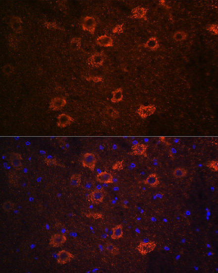 IF (Immunofluorescence)
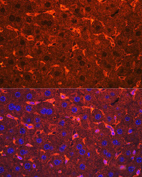
(Immunofluorescence analysis of mouse brain using SLC18A3 Polyclonal Antibody at dilution of 1:100 (40x lens). Blue: DAPI for nuclear staining.)
IF (Immunofluorescence)
(Immunofluorescence analysis of mouse brain using SLC18A3 Polyclonal Antibody at dilution of 1:100 (40x lens). Blue: DAPI for nuclear staining.)
SLC18A3, Polyclonal Antibody (Cat# AAA178703)
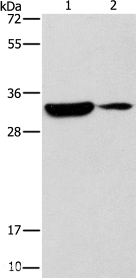
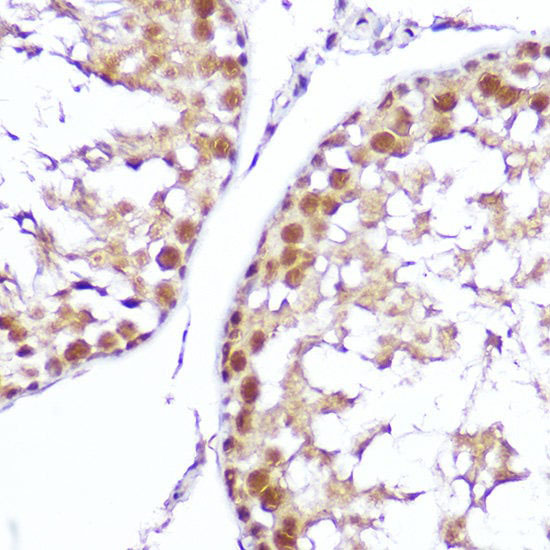
 WB (Western Blot)
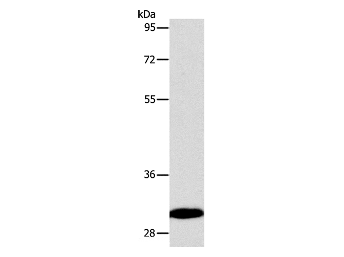
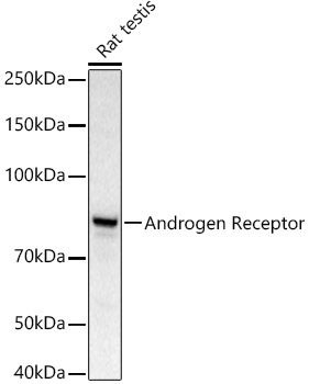
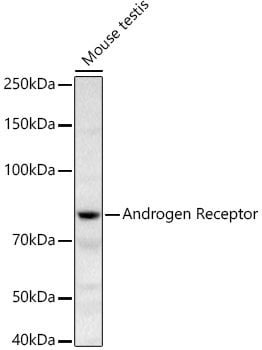
(Western blot analysis of extracts of Mouse testis using Androgen Receptor Polyclonal Antibody at 1:500 dilution.)
WB (Western Blot)
(Western blot analysis of extracts of Mouse testis using Androgen Receptor Polyclonal Antibody at 1:500 dilution.)
Androgen Receptor, Polyclonal Antibody (Cat# AAA178712)
 IHC (Immunohistochemistry)
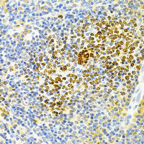
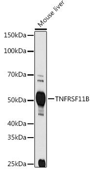
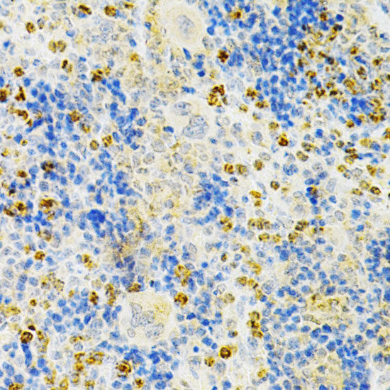
(Immunohistochemistry of paraffin-embedded mouse spleen using TNFRSF11B Polyclonal antibody at dilution of 1:200 (40x lens).Perform microwave antigen retrieval with 10 mM PBS buffer pH 7.2 before commencing with IHC staining protocol.)
IHC (Immunohistochemistry)
(Immunohistochemistry of paraffin-embedded mouse spleen using TNFRSF11B Polyclonal antibody at dilution of 1:200 (40x lens).Perform microwave antigen retrieval with 10 mM PBS buffer pH 7.2 before commencing with IHC staining protocol.)
TNFRSF11B, Polyclonal Antibody (Cat# AAA178713)
 IF (Immunofluorescence)
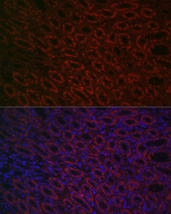
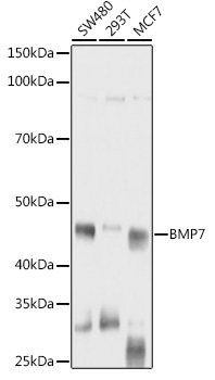
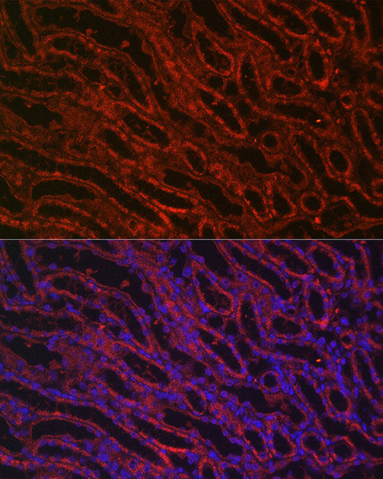
(Immunofluorescence analysis of mouse kidney cells using BMP7 Polyclonal Antibody at dilution of 1:100 (40x lens). Blue: DAPI for nuclear staining.)
IF (Immunofluorescence)
(Immunofluorescence analysis of mouse kidney cells using BMP7 Polyclonal Antibody at dilution of 1:100 (40x lens). Blue: DAPI for nuclear staining.)
BMP7, Polyclonal Antibody (Cat# AAA178898)
 IHC (Immunohiostchemistry)
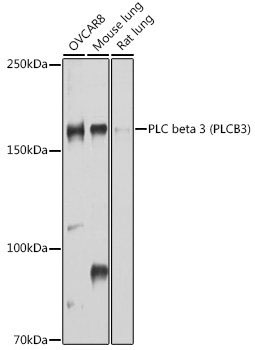
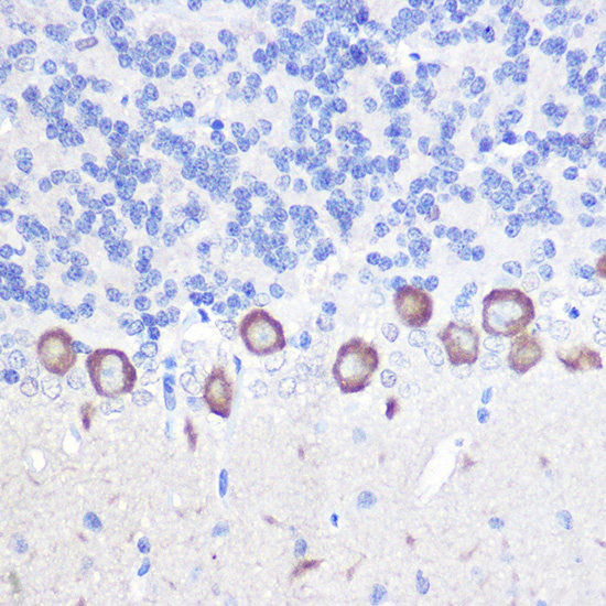
(Immunohistochemistry of paraffin-embedded rat brain using PLC beta 3 Polyclonal Antibody at dilution of 1:100 (40x lens).Perform high pressure antigen retrieval with 10 mM citrate buffer pH 6.0 before commencing with IHC staining protocol.)
IHC (Immunohiostchemistry)
(Immunohistochemistry of paraffin-embedded rat brain using PLC beta 3 Polyclonal Antibody at dilution of 1:100 (40x lens).Perform high pressure antigen retrieval with 10 mM citrate buffer pH 6.0 before commencing with IHC staining protocol.)
PLC beta 3, Polyclonal Antibody (Cat# AAA178904)
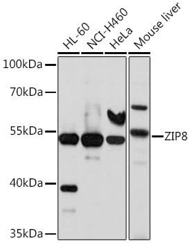
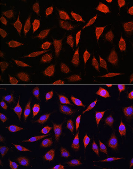 IF (Immunofluorescence)
(Immunofluorescence analysis of L929 cells using ZIP8 Polyclonal Antibody at dilution of 1:100. Blue: DAPI for nuclear staining.)
IF (Immunofluorescence)
(Immunofluorescence analysis of L929 cells using ZIP8 Polyclonal Antibody at dilution of 1:100. Blue: DAPI for nuclear staining.)
ZIP8, Polyclonal Antibody (Cat# AAA178908)
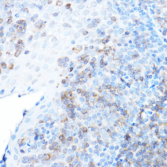
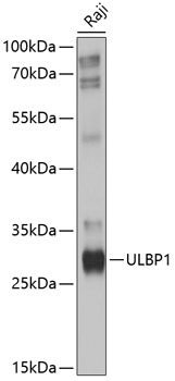
 IHC (Immunohistochemisry)
(Immunohistochemistry of paraffin-embedded mouse spleen using ULBP1 Polyclonal Antibody at dilution of 1:500 (40x lens).Perform high pressure antigen retrieval with 10 mM citrate buffer pH 6.0 before commencing with IHC staining protocol.)
IHC (Immunohistochemisry)
(Immunohistochemistry of paraffin-embedded mouse spleen using ULBP1 Polyclonal Antibody at dilution of 1:500 (40x lens).Perform high pressure antigen retrieval with 10 mM citrate buffer pH 6.0 before commencing with IHC staining protocol.)
ULBP1, Polyclonal Antibody (Cat# AAA178909)
 IHC (Immunohistochemisry)
(Immunohistochemistry of paraffin-embedded human esophageal cancer using MCT4/SLC16A3 Polyclonal Antibody at dilution of 1:100 (40x lens).Perform high pressure antigen retrieval with 10 mM citrate buffer pH 6.0 before commencing with IHC staining protocol.)
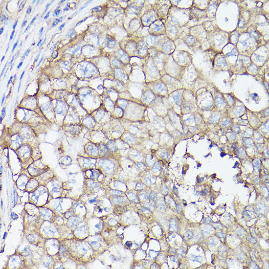
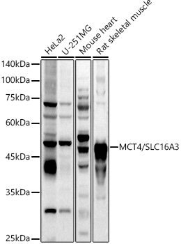
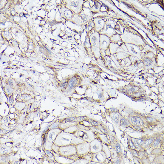
IHC (Immunohistochemisry)
(Immunohistochemistry of paraffin-embedded human esophageal cancer using MCT4/SLC16A3 Polyclonal Antibody at dilution of 1:100 (40x lens).Perform high pressure antigen retrieval with 10 mM citrate buffer pH 6.0 before commencing with IHC staining protocol.)
MCT4/SLC16A3, Polyclonal Antibody (Cat# AAA178911)
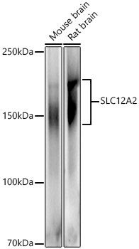
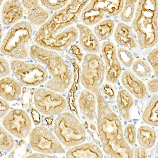 IHC (Immunohistochemistry)
(Immunohistochemistry of paraffin-embedded rat kidney using SLC12A2 Polyclonal Antibody at dilution of 1:50 (40x lens).Perform high pressure antigen retrieval with 10 mM citrate buffer pH 6.0 before commencing with IHC staining protocol.)
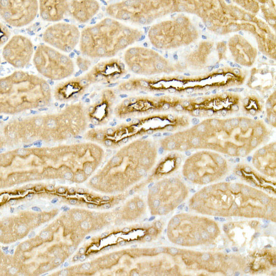
IHC (Immunohistochemistry)
(Immunohistochemistry of paraffin-embedded rat kidney using SLC12A2 Polyclonal Antibody at dilution of 1:50 (40x lens).Perform high pressure antigen retrieval with 10 mM citrate buffer pH 6.0 before commencing with IHC staining protocol.)
SLC12A2, Polyclonal Antibody (Cat# AAA178927)
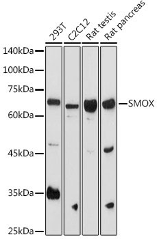
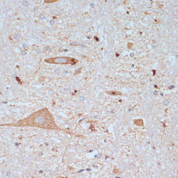
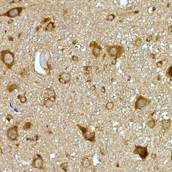
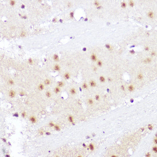 IHC (Immunohistochemisry)
(Immunohistochemistry of paraffin-embedded rat brain using SMOX Polyclonal Antibody at dilution of 1:100 (40x lens).Perform high pressure antigen retrieval with 10 mM citrate buffer pH 6.0 before commencing with IHC staining protocol.)
IHC (Immunohistochemisry)
(Immunohistochemistry of paraffin-embedded rat brain using SMOX Polyclonal Antibody at dilution of 1:100 (40x lens).Perform high pressure antigen retrieval with 10 mM citrate buffer pH 6.0 before commencing with IHC staining protocol.)
SMOX, Polyclonal Antibody (Cat# AAA178928)
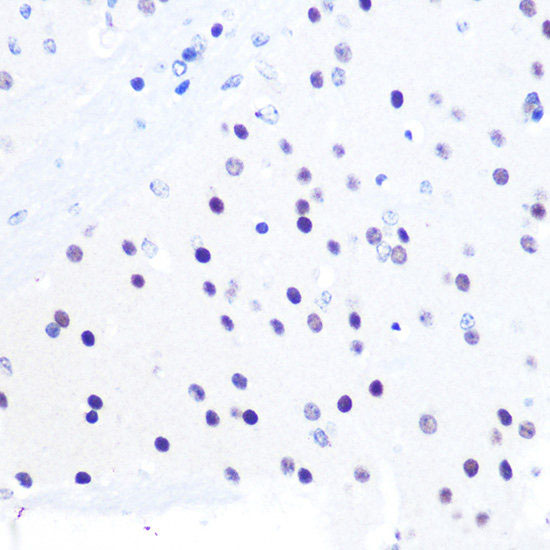
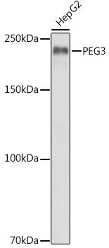
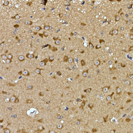
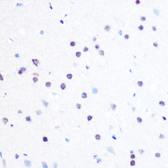 IHC (Immunohistochemisry)
(Immunohistochemistry of paraffin-embedded rat brain using PEG3 Polyclonal Antibody at dilution of 1:100 (40x lens).Perform microwave antigen retrieval with 10 mM PBS buffer pH 7.2 before commencing with IHC staining protocol.)
IHC (Immunohistochemisry)
(Immunohistochemistry of paraffin-embedded rat brain using PEG3 Polyclonal Antibody at dilution of 1:100 (40x lens).Perform microwave antigen retrieval with 10 mM PBS buffer pH 7.2 before commencing with IHC staining protocol.)
PEG3, Polyclonal Antibody (Cat# AAA178937)
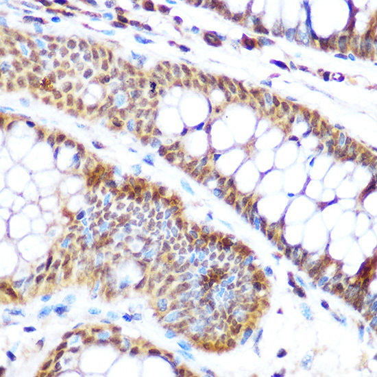
 IHC (Immunohistochemisry)
(Immunohistochemistry of paraffin-embedded rat ovary using SP3 Polyclonal Antibody at dilution of 1:100 (40x lens).Perform microwave antigen retrieval with 10 mM Tris/EDTA buffer pH 9.0 before commencing with IHC staining protocol.)
IHC (Immunohistochemisry)
(Immunohistochemistry of paraffin-embedded rat ovary using SP3 Polyclonal Antibody at dilution of 1:100 (40x lens).Perform microwave antigen retrieval with 10 mM Tris/EDTA buffer pH 9.0 before commencing with IHC staining protocol.)
SP3, Polyclonal Antibody (Cat# AAA178944)
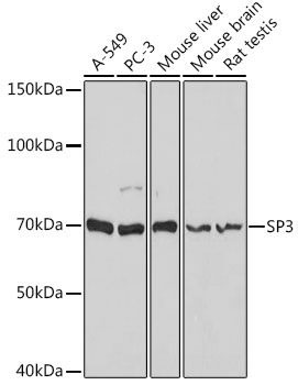
 WB (Western Blot)
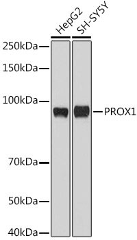
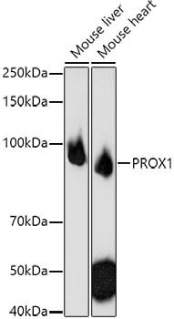
(Western blot analysis of extracts of various cell lines using PROX1 Polyclonal Antibody at 1:1000 dilution.)
WB (Western Blot)
(Western blot analysis of extracts of various cell lines using PROX1 Polyclonal Antibody at 1:1000 dilution.)
PROX1, Polyclonal Antibody (Cat# AAA178948)
 IP (Immunoprecipitation)
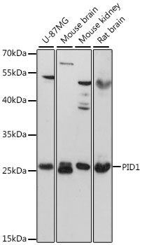
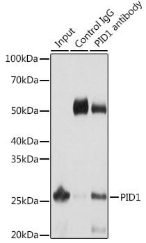
(Immunoprecipitation analysis of 25ug extracts of Mouse liver cells usingug PID1 Polyclonal Antibody.Western blot was performed from the immunoprecipitate using PID1 Polyclonal Antibody at a dilution of 1:1000.)
IP (Immunoprecipitation)
(Immunoprecipitation analysis of 25ug extracts of Mouse liver cells usingug PID1 Polyclonal Antibody.Western blot was performed from the immunoprecipitate using PID1 Polyclonal Antibody at a dilution of 1:1000.)
PID1, Polyclonal Antibody (Cat# AAA178951)
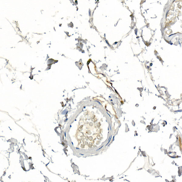
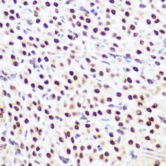
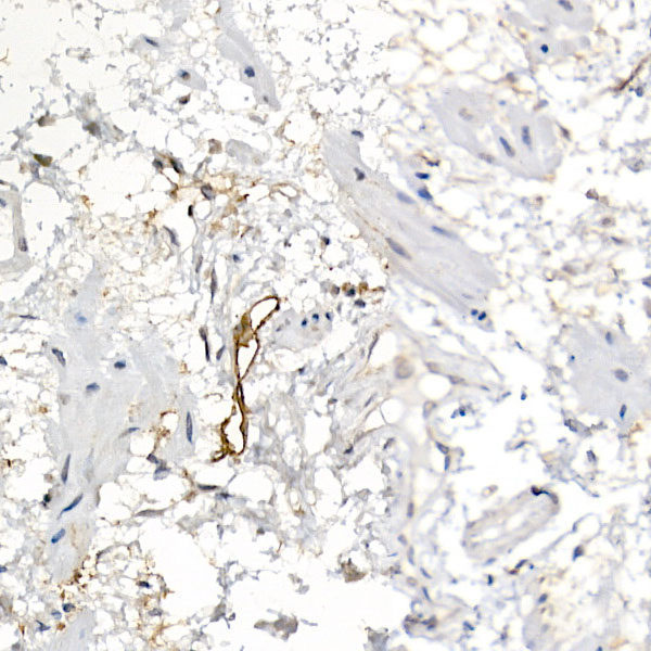 IHC (Immunohistochemisry)
(Immunohistochemistry of paraffin-embedded human esophagus using Podoplanin Polyclonal Antibody at dilution of 1:20 (40x lens).Perform high pressure antigen retrieval with 10 mM citrate buffer pH 6.0 before commencing with IHC staining protocol.)
IHC (Immunohistochemisry)
(Immunohistochemistry of paraffin-embedded human esophagus using Podoplanin Polyclonal Antibody at dilution of 1:20 (40x lens).Perform high pressure antigen retrieval with 10 mM citrate buffer pH 6.0 before commencing with IHC staining protocol.)
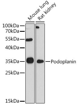
Podoplanin, Polyclonal Antibody (Cat# AAA178952)
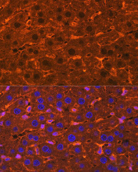
 IF (Immunofluorescence)
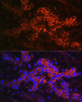
(Immunofluorescence analysis of Human liver cancer using Ceruloplasmin Polyclonal Antibody at dilution of 1:100. Blue: DAPI for nuclear staining.)
IF (Immunofluorescence)
(Immunofluorescence analysis of Human liver cancer using Ceruloplasmin Polyclonal Antibody at dilution of 1:100. Blue: DAPI for nuclear staining.)
Ceruloplasmin, Polyclonal Antibody (Cat# AAA178956)
 IF (Immunofluorescence)
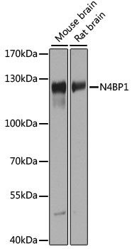
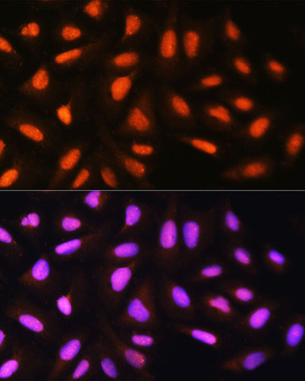
(Immunofluorescence analysis of U2OS cells using N4BP1 Polyclonal Antibody at dilution of 1:100. Blue: DAPI for nuclear staining.)
IF (Immunofluorescence)
(Immunofluorescence analysis of U2OS cells using N4BP1 Polyclonal Antibody at dilution of 1:100. Blue: DAPI for nuclear staining.)
N4BP1, Polyclonal Antibody (Cat# AAA178964)
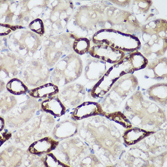
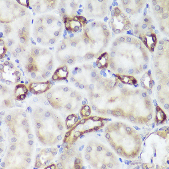 IHC (Immunohistochemisry)
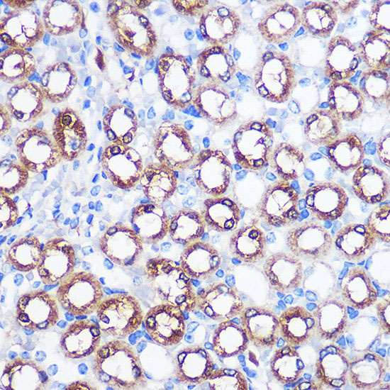
(Immunohistochemistry of paraffin-embedded rat kidney using CASR Polyclonal Antibody at dilution of 1:50 (40x lens).Perform high pressure antigen retrieval with 10 mM citrate buffer pH 6.0 before commencing with IHC staining protocol.)
IHC (Immunohistochemisry)
(Immunohistochemistry of paraffin-embedded rat kidney using CASR Polyclonal Antibody at dilution of 1:50 (40x lens).Perform high pressure antigen retrieval with 10 mM citrate buffer pH 6.0 before commencing with IHC staining protocol.)
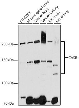
CASR, Polyclonal Antibody (Cat# AAA178967)
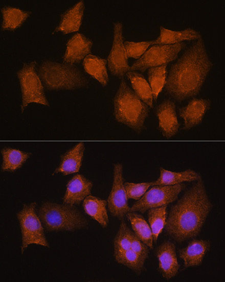
 IF (Immunofluorescence)
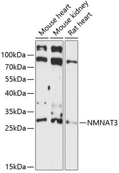
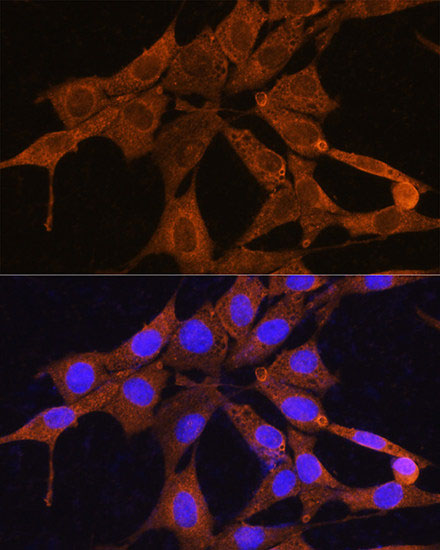
(Immunofluorescence analysis of NIH/3T3 cells using NMNAT3 Polyclonal Antibody at dilution of 1:100. Blue: DAPI for nuclear staining.)
IF (Immunofluorescence)
(Immunofluorescence analysis of NIH/3T3 cells using NMNAT3 Polyclonal Antibody at dilution of 1:100. Blue: DAPI for nuclear staining.)
NMNAT3, Polyclonal Antibody (Cat# AAA178970)
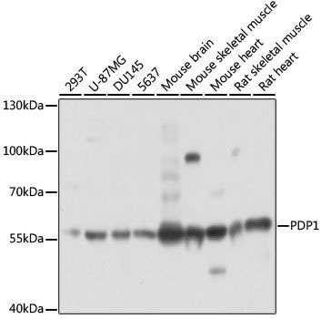
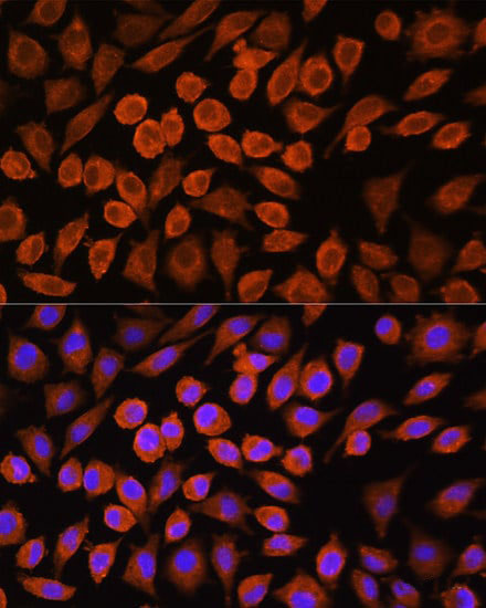 IF (Immunofluorescence)
(Immunofluorescence analysis of L929 cells using PDP1 Polyclonal Antibody at dilution of 1:100. Blue: DAPI for nuclear staining.)
IF (Immunofluorescence)
(Immunofluorescence analysis of L929 cells using PDP1 Polyclonal Antibody at dilution of 1:100. Blue: DAPI for nuclear staining.)
PDP1, Polyclonal Antibody (Cat# AAA178971)
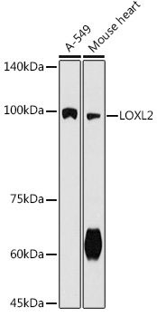
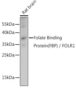
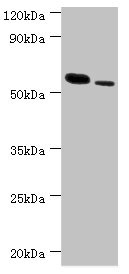
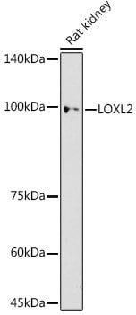 WB (Western Blot)
(Western blot analysis of extracts of Rat kidney using LOXL2 Polyclonal Antibody at 1:500 dilution.)
WB (Western Blot)
(Western blot analysis of extracts of Rat kidney using LOXL2 Polyclonal Antibody at 1:500 dilution.)
LOXL2, Polyclonal Antibody (Cat# AAA178975)
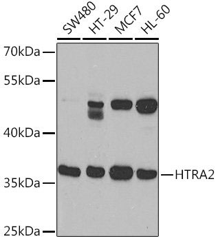
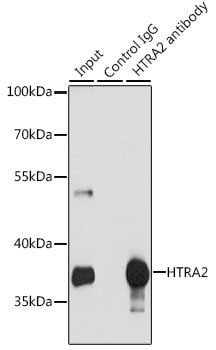 IP (Immunoprecipitation)
(Immunoprecipitation analysis of 200ug extracts of MCF-7 cells using 3 ug HTRA2 Polyclonal Antibody.Western blot was performed from the immunoprecipitate using HTRA2 Polyclonal Antibody at a dilution of 1:1000.)
IP (Immunoprecipitation)
(Immunoprecipitation analysis of 200ug extracts of MCF-7 cells using 3 ug HTRA2 Polyclonal Antibody.Western blot was performed from the immunoprecipitate using HTRA2 Polyclonal Antibody at a dilution of 1:1000.)
HTRA2, Polyclonal Antibody (Cat# AAA178985)
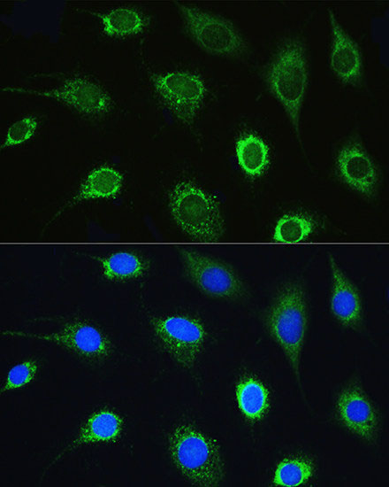
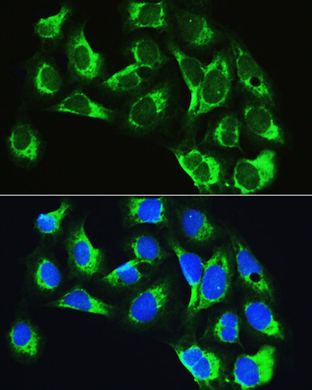 IF (Immunofluorescence)
(Immunofluorescence analysis of U2OS cells using LARS Polyclonal Antibody at dilution of 1:100. Blue: DAPI for nuclear staining.)
IF (Immunofluorescence)
(Immunofluorescence analysis of U2OS cells using LARS Polyclonal Antibody at dilution of 1:100. Blue: DAPI for nuclear staining.)
LARS, Polyclonal Antibody (Cat# AAA178987)
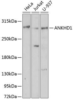
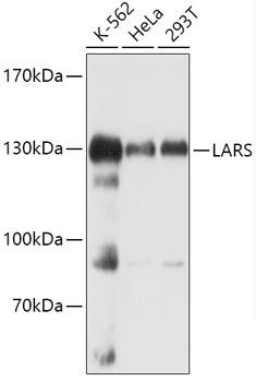
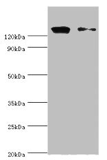
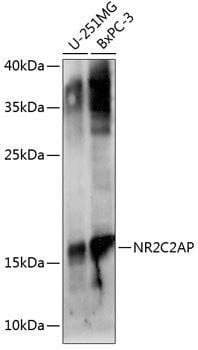 WB (Western Blot)
(Western blot analysis of extracts of various cell lines using NR2C2AP Polyclonal Antibody at 1:1000 dilution.)
WB (Western Blot)
(Western blot analysis of extracts of various cell lines using NR2C2AP Polyclonal Antibody at 1:1000 dilution.)
ANKHD1, Polyclonal Antibody (Cat# AAA178989)
 IHC (Immunohiostchemistry)
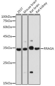
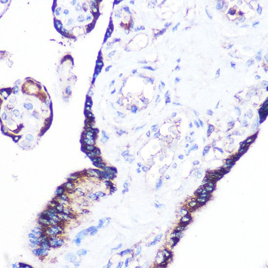
(Immunohistochemistry of paraffin-embedded human placenta using RRAGA Polyclonal Antibody at dilution of 1:100 (40x lens).Perform microwave antigen retrieval with 10 mM Tris/EDTA buffer pH 9.0 before commencing with IHC staining protocol.)
IHC (Immunohiostchemistry)
(Immunohistochemistry of paraffin-embedded human placenta using RRAGA Polyclonal Antibody at dilution of 1:100 (40x lens).Perform microwave antigen retrieval with 10 mM Tris/EDTA buffer pH 9.0 before commencing with IHC staining protocol.)
RRAGA, Polyclonal Antibody (Cat# AAA178993)
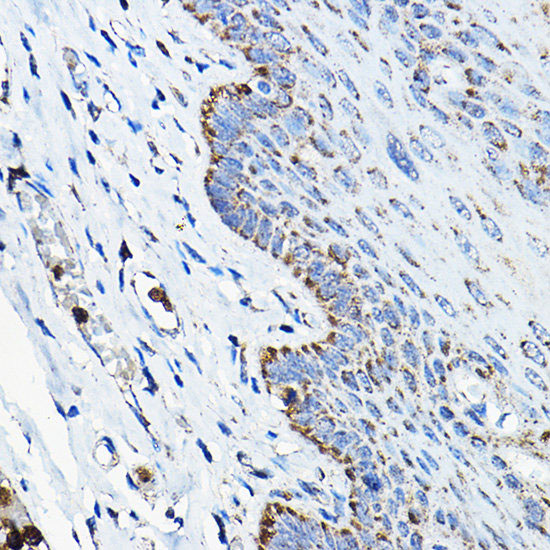
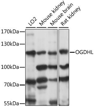
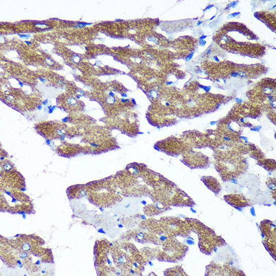
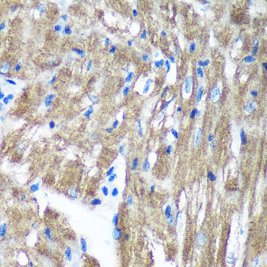
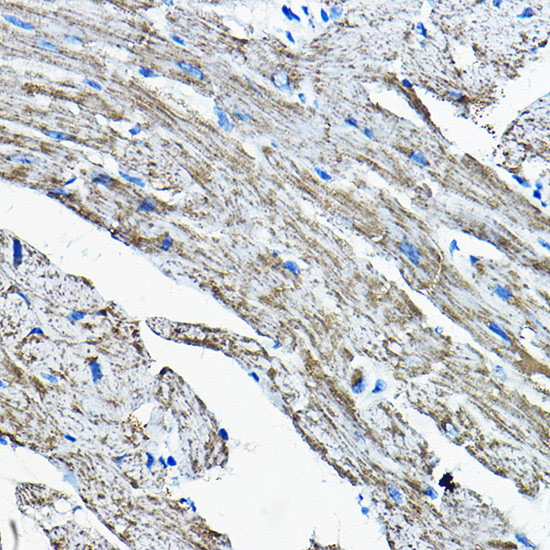 IHC (Immunohistochemisry)
(Immunohistochemistry of paraffin-embedded mouse heart using OGDHL Polyclonal Antibody at dilution of 1:200 (40x lens).Perform high pressure antigen retrieval with 10 mM citrate buffer pH 6.0 before commencing with IHC staining protocol.)
IHC (Immunohistochemisry)
(Immunohistochemistry of paraffin-embedded mouse heart using OGDHL Polyclonal Antibody at dilution of 1:200 (40x lens).Perform high pressure antigen retrieval with 10 mM citrate buffer pH 6.0 before commencing with IHC staining protocol.)
OGDHL, Polyclonal Antibody (Cat# AAA179004)
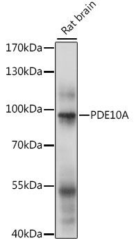
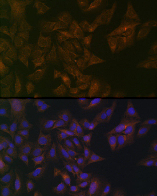
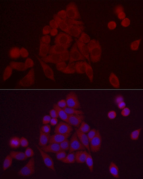 IF (Immunofluorescence)
(Immunofluorescence analysis of HeLa cells using PDE10A Polyclonal Antibody at dilution of 1:20 (40x lens). Blue: DAPI for nuclear staining.)
IF (Immunofluorescence)
(Immunofluorescence analysis of HeLa cells using PDE10A Polyclonal Antibody at dilution of 1:20 (40x lens). Blue: DAPI for nuclear staining.)
PDE10A, Polyclonal Antibody (Cat# AAA179005)
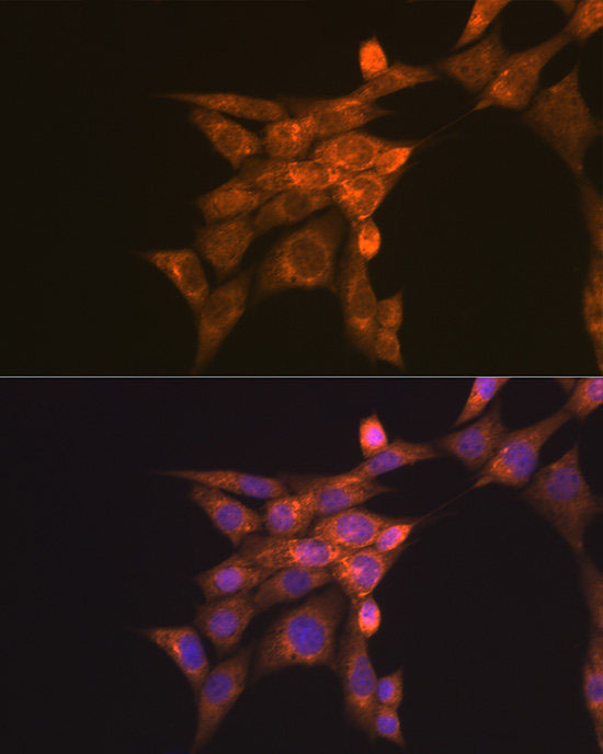

 IF (Immunofluorescence)
(Immunofluorescence analysis of U2OS cells using Folate Binding Protein Polyclonal Antibody at dilution of 1:100 (40x lens). Blue: DAPI for nuclear staining.)
IF (Immunofluorescence)
(Immunofluorescence analysis of U2OS cells using Folate Binding Protein Polyclonal Antibody at dilution of 1:100 (40x lens). Blue: DAPI for nuclear staining.)
Folate Binding Protein, Polyclonal Antibody (Cat# AAA179011)
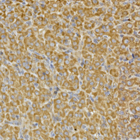
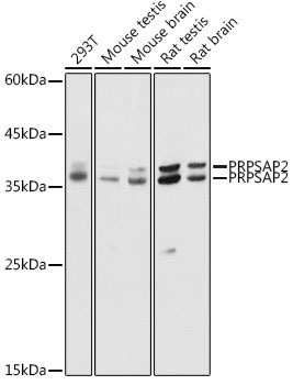
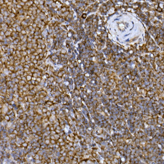 IHC (Immunohistochemistry)
(Immunohistochemistry of paraffin-embedded rat spleen using PRPSAP2 Polyclonal Antibody at dilution of 1:100 (40x lens).Perform high pressure antigen retrieval with 10 mM citrate buffer pH 6.0 before commencing with IHC staining protocol.)
IHC (Immunohistochemistry)
(Immunohistochemistry of paraffin-embedded rat spleen using PRPSAP2 Polyclonal Antibody at dilution of 1:100 (40x lens).Perform high pressure antigen retrieval with 10 mM citrate buffer pH 6.0 before commencing with IHC staining protocol.)
PRPSAP2, Polyclonal Antibody (Cat# AAA179012)

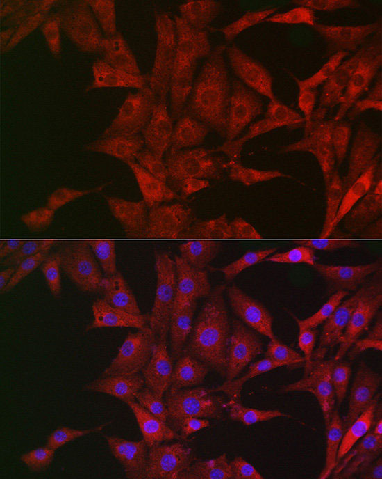
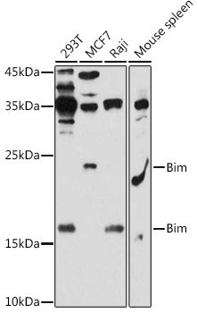
 IF (Immunofluorescence)
(Immunofluorescence analysis of U2OS cells using Bim Polyclonal Antibody at dilution of 1:200 (40x lens). Blue: DAPI for nuclear staining.)
IF (Immunofluorescence)
(Immunofluorescence analysis of U2OS cells using Bim Polyclonal Antibody at dilution of 1:200 (40x lens). Blue: DAPI for nuclear staining.)
Bim, Polyclonal Antibody (Cat# AAA179013)
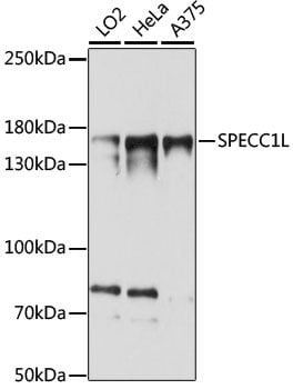
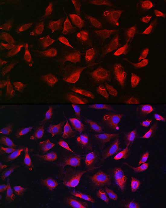
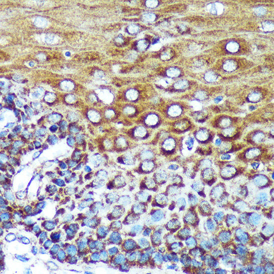 IHC (Immunohiostchemistry)
(Immunohistochemistry of paraffin-embedded Human esophageal using SPECC1L Polyclonal Antibody at dilution of 1:100 (40x lens).Perform microwave antigen retrieval with 10 mM PBS buffer pH 7.2 before commencing with IHC staining protocol.)
IHC (Immunohiostchemistry)
(Immunohistochemistry of paraffin-embedded Human esophageal using SPECC1L Polyclonal Antibody at dilution of 1:100 (40x lens).Perform microwave antigen retrieval with 10 mM PBS buffer pH 7.2 before commencing with IHC staining protocol.)
SPECC1L, Polyclonal Antibody (Cat# AAA179015)
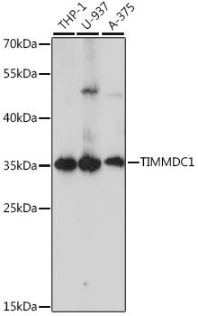
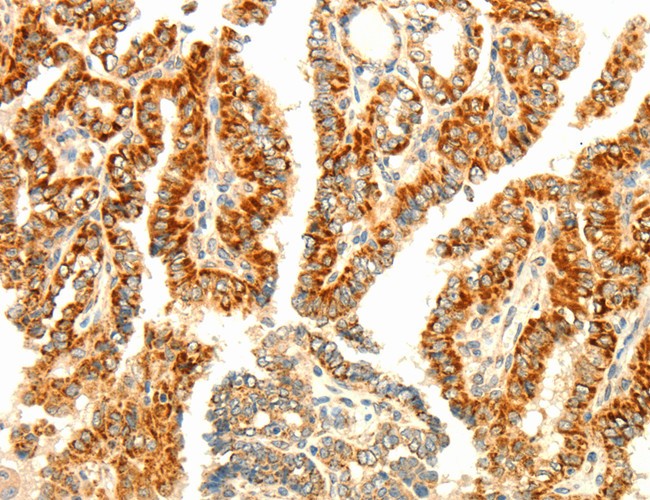
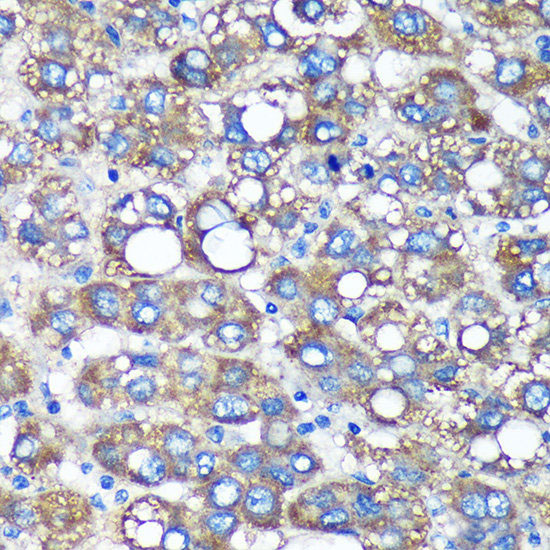 IHC (Immunohistochemistry)
(Immunohistochemistry of paraffin-embedded Human liver cancer using TIMMDC1 Polyclonal Antibody at dilution of 1:100 (40x lens).Perform microwave antigen retrieval with 10 mM PBS buffer pH 7.2 before commencing with IHC staining protocol.)
IHC (Immunohistochemistry)
(Immunohistochemistry of paraffin-embedded Human liver cancer using TIMMDC1 Polyclonal Antibody at dilution of 1:100 (40x lens).Perform microwave antigen retrieval with 10 mM PBS buffer pH 7.2 before commencing with IHC staining protocol.)
TIMMDC1, Polyclonal Antibody (Cat# AAA179016)
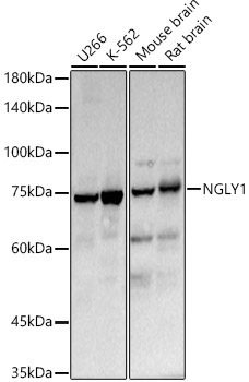
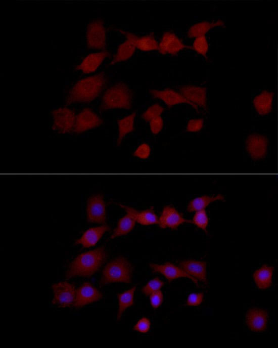 IF (Immunofluorescence)
(Immunofluorescence analysis of PC-12 cells using NGLY1 Polyclonal Antibody at dilution of 1:50 (40x lens). Blue: DAPI for nuclear staining.)
IF (Immunofluorescence)
(Immunofluorescence analysis of PC-12 cells using NGLY1 Polyclonal Antibody at dilution of 1:50 (40x lens). Blue: DAPI for nuclear staining.)
NGLY1, Polyclonal Antibody (Cat# AAA179019)
 IF (Immunofluorescence)
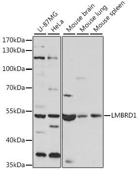
(Immunofluorescence analysis of L929 cells using LMBRD1 Polyclonal Antibody at dilution of 1:100. Blue: DAPI for nuclear staining.)
IF (Immunofluorescence)
(Immunofluorescence analysis of L929 cells using LMBRD1 Polyclonal Antibody at dilution of 1:100. Blue: DAPI for nuclear staining.)
LMBRD1, Polyclonal Antibody (Cat# AAA179020)
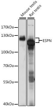
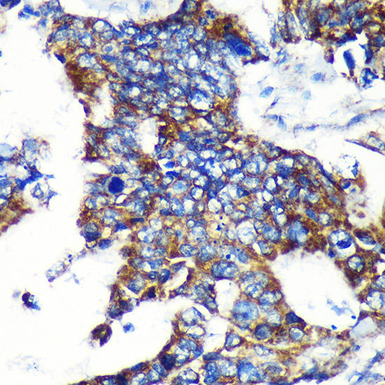 IHC (Immunohiostchemistry)
(Immunohistochemistry of paraffin-embedded Human colon carcinoma using ESPN Polyclonal Antibody at dilution of 1:100 (40x lens).Perform microwave antigen retrieval with 10 mM PBS buffer pH 7.2 before commencing with IHC staining protocol.)
IHC (Immunohiostchemistry)
(Immunohistochemistry of paraffin-embedded Human colon carcinoma using ESPN Polyclonal Antibody at dilution of 1:100 (40x lens).Perform microwave antigen retrieval with 10 mM PBS buffer pH 7.2 before commencing with IHC staining protocol.)
ESPN, Polyclonal Antibody (Cat# AAA179021)
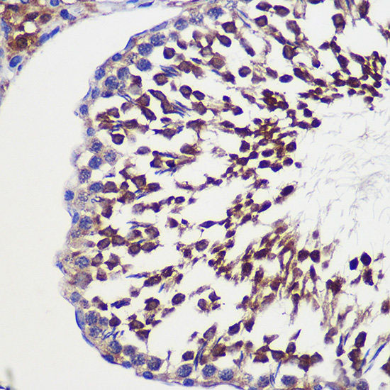
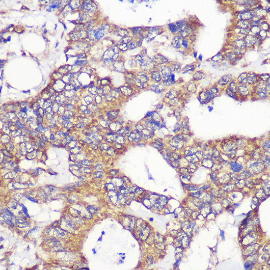
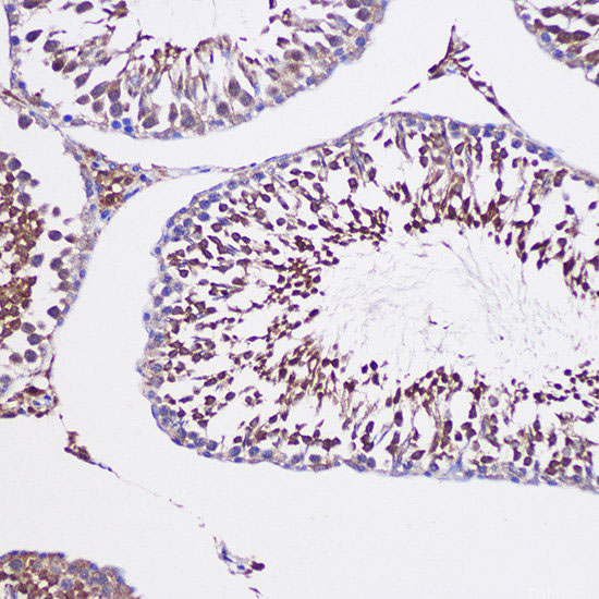 IHC (Immunohistochemistry)
(Immunohistochemistry of paraffin-embedded Rat testis using TUBGCP6 Polyclonal Antibody at dilution of 1:100 (20x lens).Perform microwave antigen retrieval with 10 mM PBS buffer pH 7.2 before commencing with IHC staining protocol.)
IHC (Immunohistochemistry)
(Immunohistochemistry of paraffin-embedded Rat testis using TUBGCP6 Polyclonal Antibody at dilution of 1:100 (20x lens).Perform microwave antigen retrieval with 10 mM PBS buffer pH 7.2 before commencing with IHC staining protocol.)
TUBGCP6, Polyclonal Antibody (Cat# AAA179022)
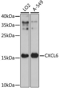
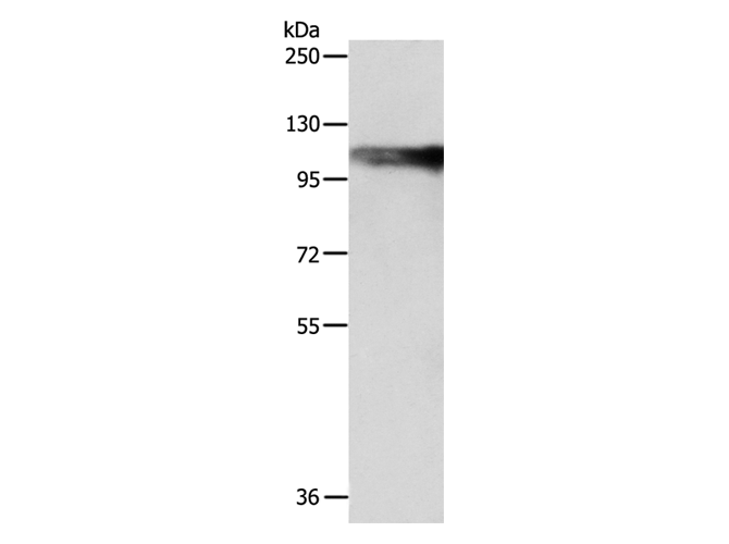
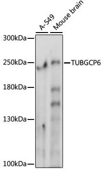
 WB (Western Blot)
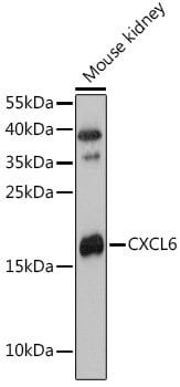
(Western blot analysis of extracts of Mouse kidney using CXCL6 Polyclonal Antibody at 1:3000 dilution.)
WB (Western Blot)
(Western blot analysis of extracts of Mouse kidney using CXCL6 Polyclonal Antibody at 1:3000 dilution.)
CXCL6, Polyclonal Antibody (Cat# AAA179039)
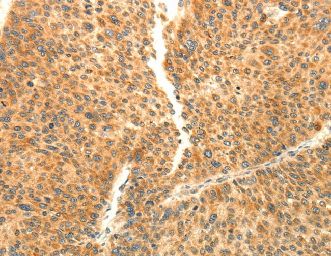
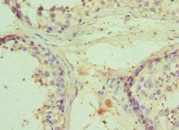 IHC (Immunohiostchemistry)
(Immunohistochemistry of paraffin-embedded human testicle using AAA118254 at dilution 1:100)
IHC (Immunohiostchemistry)
(Immunohistochemistry of paraffin-embedded human testicle using AAA118254 at dilution 1:100)
E3 ubiquitin-protein ligase RNF8, Polyclonal Antibody (Cat# AAA118254)
IGSF11, Polyclonal Antibody (Cat# AAA118255)
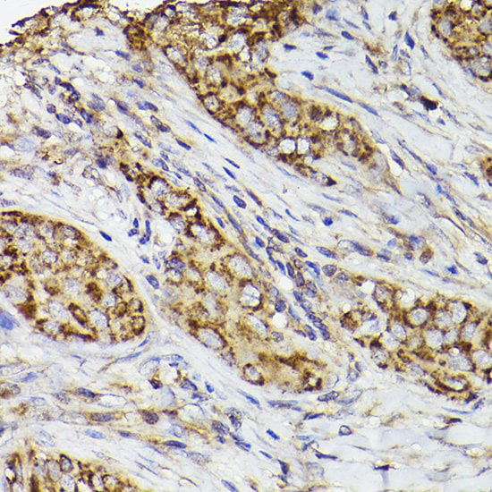
 IHC (Immunohiostchemistry)
(Immunohistochemistry of paraffin-embedded human breast cancer using AAA118258 at dilution 1:100)
IHC (Immunohiostchemistry)
(Immunohistochemistry of paraffin-embedded human breast cancer using AAA118258 at dilution 1:100)
Cell division cycle and apoptosis regulator protein 1, Polyclonal Antibody (Cat# AAA118258)
 IHC (Immunohiostchemistry)
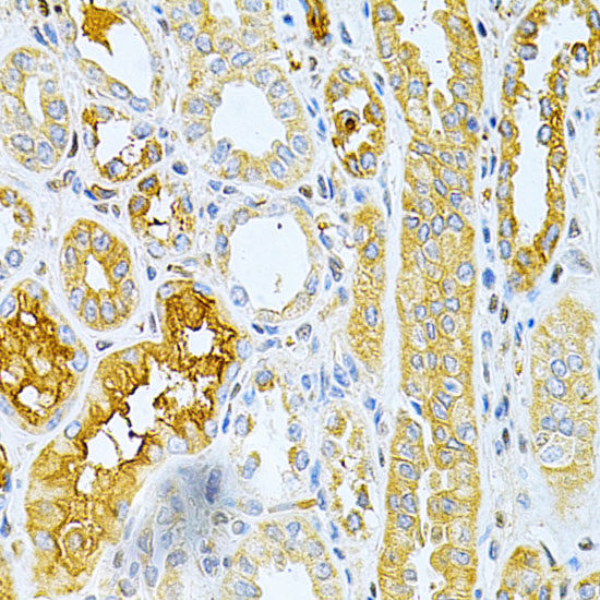
(Immunohistochemistry of paraffin-embedded human kidney using AAA118262 at dilution 1:100)
IHC (Immunohiostchemistry)
(Immunohistochemistry of paraffin-embedded human kidney using AAA118262 at dilution 1:100)
SLC4A2, Polyclonal Antibody (Cat# AAA118262)
distemper virus Fusion glycoprotein F0, Polyclonal Antibody (Cat# AAA118263)
Acinetobacter baumannii Elongation factor Tu, Polyclonal Antibody (Cat# AAA118268)
Vitamin K-dependent protein S, Polyclonal Antibody (Cat# AAA118272)
What are Polyclonal Antibodies?
Polyclonal antibodies are antibodies that come from multiple B cell clones of a host animal. The typical hosts used for the majority of polyclonal antibody production are rabbits, goats, sheep, and donkeys. These polyclonal antibodies, once having identified their target, will bind to different epitopes located at different regions or sequences on the same protein/antigen. As a result, they are ideal at locating and binding to the target, even if the target is in very low concentrations (due to many different antibodies being able to bind to the same target molecule, which allows for significant amplification of a downstream signal).
Polyclonal antibodies are typically produced by injecting an antigen into a host animal, which causes the animal’s immune system to attack the foreign antigen by mass generating antibodies against it. After a period of time, serum is collected from the animal and purified using physicochemical fractionation, class-specific affinity purification, and/or antigen-affinity purification.
Key Uses of Polyclonal Antibodies
- Western Blotting: This method is used to find specific proteins in biological samples after separating them by size.
- Immunohistochemistry: IHC helps visualize the location of proteins in tissue sections using various staining techniques.
- ELISA: (Enzyme-Linked Immunosorbent Assay) is typically used to identify specific protein quantities in a sample. ELISAs can be either “Quantitative” or “Qualitative”.
- Flow Cytometry: technique that identifies and measures the specific protein on the surface or inside the cells in a fluid suspension.
- Immunoprecipitation: IP isolates and studies a specific protein from a complex mixture using antibodies.
Why Buy Polyclonal Antibodies from AAA Biotech?
1. Ideal for Various Applications
Our antibodies are generally going to be validated for use in multiple types of assays, including ELISA, Western Blotting, Immunohistochemistry, Immunoprecipitation, amongst others. They are ideal for a wide range of research applications.
2. Rigorous Quality Control
All of the antibodies in our catalog undergo strict quality testing to ensure specificity, sensitivity, and consistent performance. We are confident in the ability of our antibodies to provide you with accurate results.
3. Wide Assortment of Antibodies
Antibodies in are catalog can be found for both common and exotic species, and these antibodies are also available in both conjugated and recombinant forms to suit many diverse experimental needs.
4. Highly Purified
Our antibodies are available in purified forms with over 85% purity, as confirmed by SDS-PAGE. They are also available with tags such as His, Flag, GST, or MBP. We cater to customers worldwide.
FAQ
1. How are polyclonal antibodies produced?
Traditionally, polyclonal antibodies are produced by injecting an antigen into a host animal (such as a rabbit or goat), which then triggers an immune response from the host animal. The animal’s B cells produce antibodies that will recognize different parts of the injected antigen. These antibodies are then collected from the animal’s blood and purified for use.
2. How do polyclonal antibodies differ from monoclonal antibodies?
Polyclonal antibodies are a mix of antibodies that bind to different locations (epitopes) of the same antigen, while monoclonal antibodies are identical and bind to just one specific epitope. This makes polyclonal antibodies more versatile and better at detecting proteins that may be present in low quantities or in altered/modified forms.
3. How should I store polyclonal antibodies?
Polyclonal antibodies should be stored at 4°C for short-term use (up to a few weeks) and at -20°C or -80°C for long-term storage. Avoid repeated freeze-thaw cycles by dividing them into small aliquots. Always check the datasheet for specific storage instructions.