Filters
▼Clonality
▼Type
▼Reactivity
▼Gene Name
▼Isotype
▼Host
▼Application
▼Clone
▼Polyclonal Antibodies
At AAA Biotech also known as AAA Bio or AAABio, we provide a broad range of purified polyclonal antibodies (pAbs) that are able to all be browsed online through our website. Due to their high specificity and strong binding affinity, these antibodies are ideal for wide swathes of research and experimental applications.
Our polyclonal antibodies can easily support your work, whether you use them for Western Blotting, Immunocytochemistry (with or without Immunofluorescence used in conjunction), Immunohistochemistry, Immunoprecipitation, and ELISA tests. We highly encourage you to browse our range of pAbs and choose the one that best suits your experimental model.
Viewing 550-600 of 96805 product results
 WB (Western Blot)
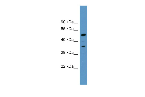
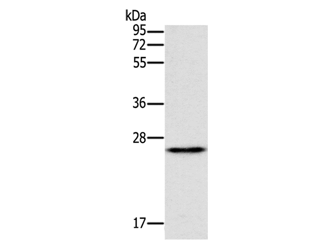
(WB Suggested Anti-Fabp5 AntibodyTitration: 1.0 ug/mlPositive Control: Mouse Thymus)
WB (Western Blot)
(WB Suggested Anti-Fabp5 AntibodyTitration: 1.0 ug/mlPositive Control: Mouse Thymus)
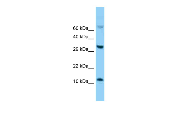
Fabp5, Polyclonal Antibody (Cat# AAA200986)
Predicted: Cow, Dog, Guinea Pig, Horse, Human, Mouse, Rabbit, Rat, Sheep, Zebrafish
 WB (Western Blot)
(WB Suggested Anti-CYP2J2 Antibody Titration: 0.2-1 ug/mlELISA Titer: 1:62500Positive Control: Human heart)
WB (Western Blot)
(WB Suggested Anti-CYP2J2 Antibody Titration: 0.2-1 ug/mlELISA Titer: 1:62500Positive Control: Human heart)
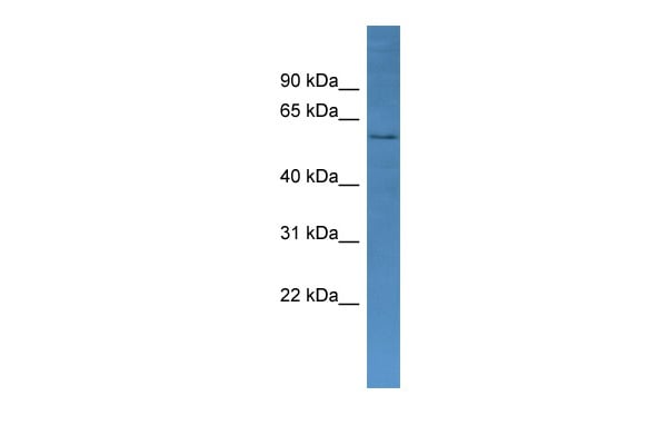
CYP2J2, Polyclonal Antibody (Cat# AAA200988)
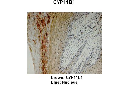
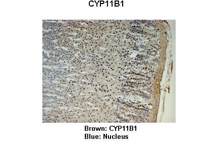
 WB (Western Blot)
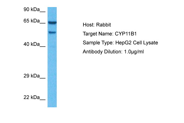
(WB Suggested Anti-CYP11B1 Antibody Titration: 0.2-1 ug/mlELISA Titer: 1:62500Positive Control: Human Liver)
WB (Western Blot)
(WB Suggested Anti-CYP11B1 Antibody Titration: 0.2-1 ug/mlELISA Titer: 1:62500Positive Control: Human Liver)
CYP11B1, Polyclonal Antibody (Cat# AAA200990)
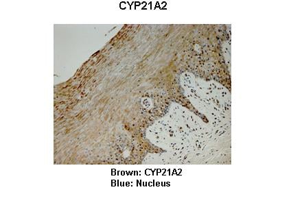
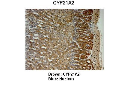
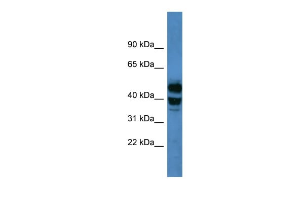 WB (Western Blot)
(WB Suggested Anti-CYP21A2 Antibody Titration: 0.2-1 ug/mlELISA Titer: 1:62500Positive Control: Human Muscle)
WB (Western Blot)
(WB Suggested Anti-CYP21A2 Antibody Titration: 0.2-1 ug/mlELISA Titer: 1:62500Positive Control: Human Muscle)
CYP21A2, Polyclonal Antibody (Cat# AAA200991)
 WB (Western Blot)
(WB Suggested Anti-AKR1C1 Antibody Titration: 0.2-1 ug/mlELISA Titer: 1:62500Positive Control: Human Liver)
WB (Western Blot)
(WB Suggested Anti-AKR1C1 Antibody Titration: 0.2-1 ug/mlELISA Titer: 1:62500Positive Control: Human Liver)
AKR1C1, Polyclonal Antibody (Cat# AAA200996)
 WB (Western Blot)
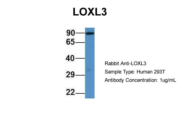
(WB Suggested Anti-LOXL3 Antibody Titration: 0.2-1 ug/mlELISA Titer: 1:1562500Positive Control: Human Placenta)
WB (Western Blot)
(WB Suggested Anti-LOXL3 Antibody Titration: 0.2-1 ug/mlELISA Titer: 1:1562500Positive Control: Human Placenta)
LOXL3, Polyclonal Antibody (Cat# AAA201003)
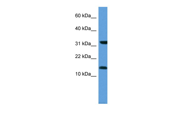
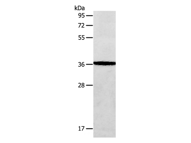
 WB (Western Blot)
(WB Suggested Anti-MARCKS AntibodyTitration: 1.0 ug/mlPositive Control: Fetal Liver)
WB (Western Blot)
(WB Suggested Anti-MARCKS AntibodyTitration: 1.0 ug/mlPositive Control: Fetal Liver)
MARCKS, Polyclonal Antibody (Cat# AAA201009)
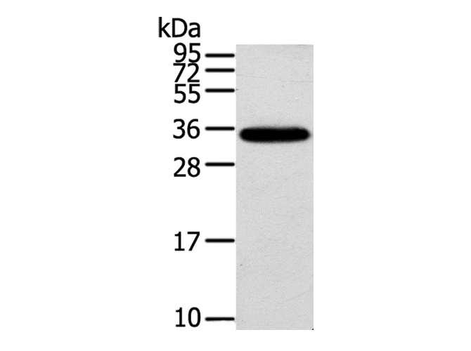
 WB (Western Blot)
(WB Suggested Anti-MGST3 Antibody Titration: 0.2-1 ug/mlELISA Titer: 1:312500Positive Control: Human heart)
WB (Western Blot)
(WB Suggested Anti-MGST3 Antibody Titration: 0.2-1 ug/mlELISA Titer: 1:312500Positive Control: Human heart)
MGST3, Polyclonal Antibody (Cat# AAA201013)
Predicted Species Reactivity: Human, Mouse, Rat, Cow, Dog, Goat, Guinea Pig, Horse, Rabbit, Zebrafish
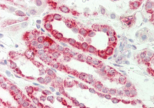
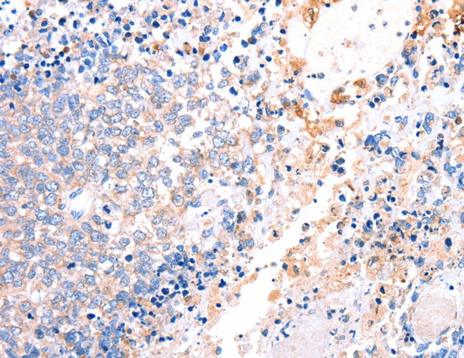
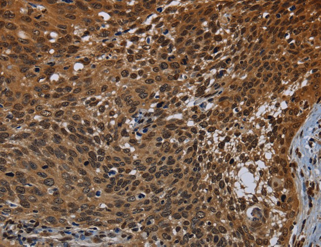
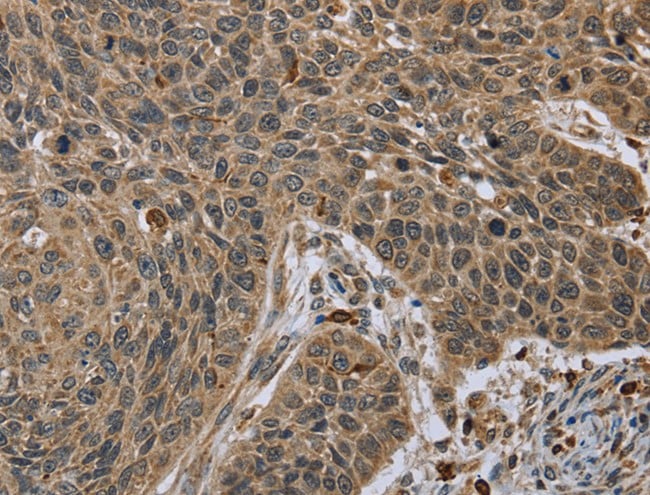
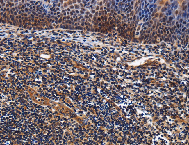
 IHC (Immunohistochemisry)
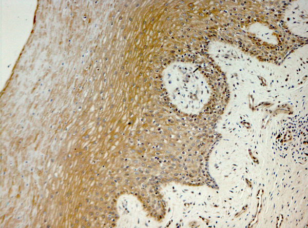
(Immunohistochemistry of paraffin-embedded Human gastric cancer using ERAS Polyclonal Antibody at dilution of 1:40)
IHC (Immunohistochemisry)
(Immunohistochemistry of paraffin-embedded Human gastric cancer using ERAS Polyclonal Antibody at dilution of 1:40)
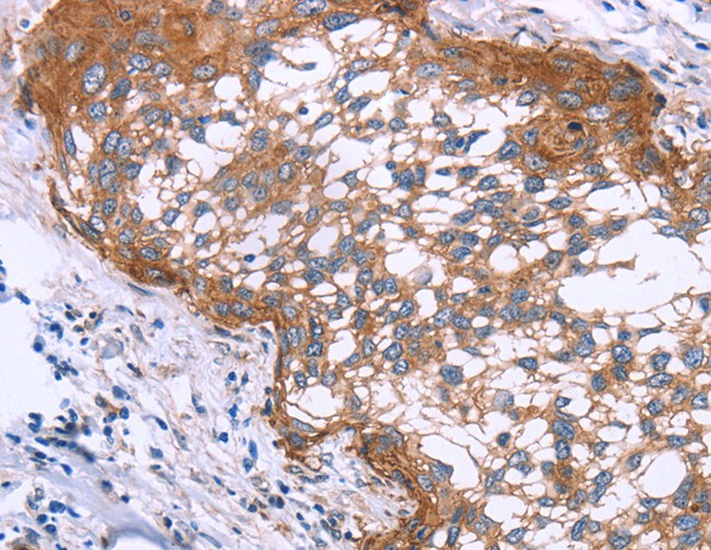
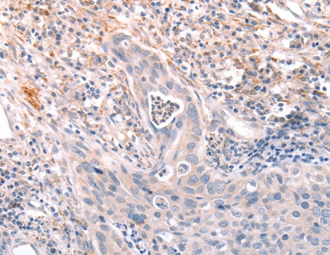
ERAS, Polyclonal Antibody (Cat# AAA170223)
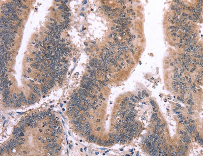
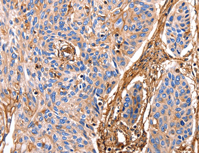
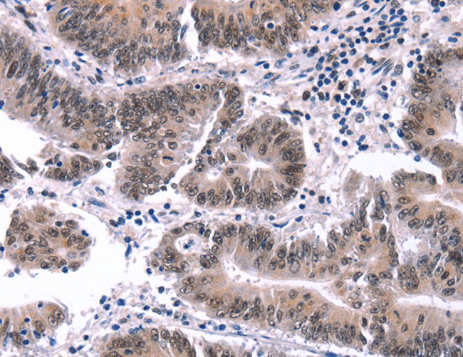
 IHC (Immunohiostchemistry)
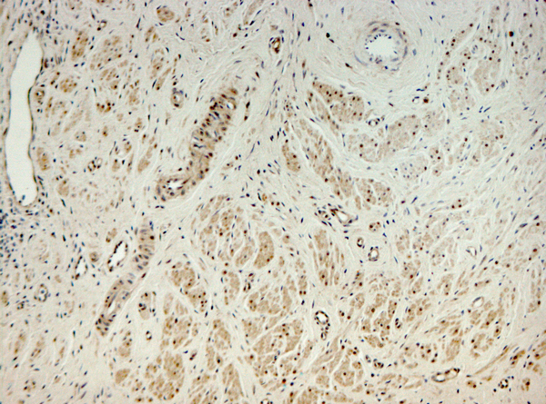
(Immunohistochemistry of paraffin-embedded Human breast cancer tissue using TPST2 Polyclonal Antibody at dilution 1:35)
IHC (Immunohiostchemistry)
(Immunohistochemistry of paraffin-embedded Human breast cancer tissue using TPST2 Polyclonal Antibody at dilution 1:35)
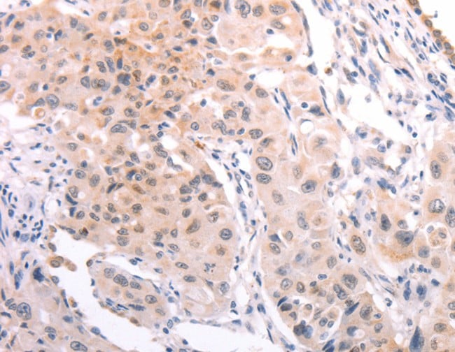
TPST2, Polyclonal Antibody (Cat# AAA170224)
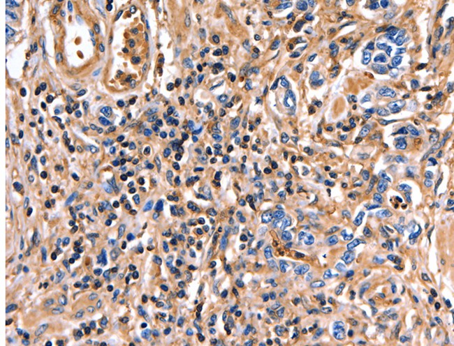
 IHC (Immunohiostchemistry)
(Immunohistochemistry of paraffin-embedded Human breast cancer tissue using IFT74 Polyclonal Antibody at dilution 1:60)
IHC (Immunohiostchemistry)
(Immunohistochemistry of paraffin-embedded Human breast cancer tissue using IFT74 Polyclonal Antibody at dilution 1:60)
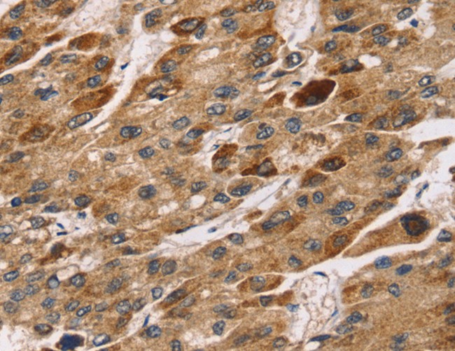
IFT74, Polyclonal Antibody (Cat# AAA170239)

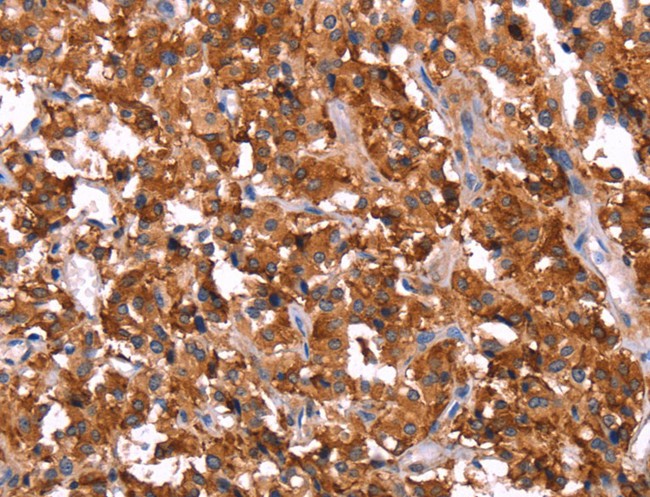
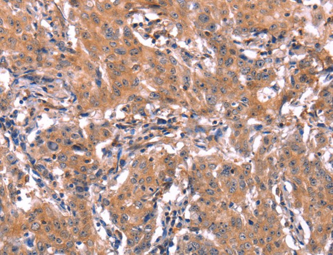
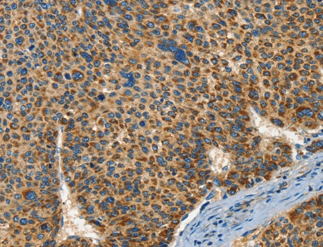
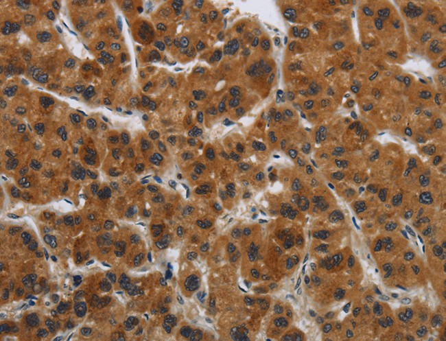
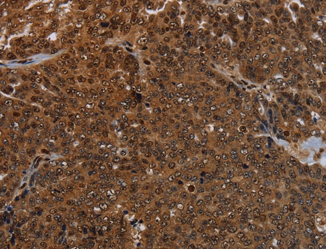
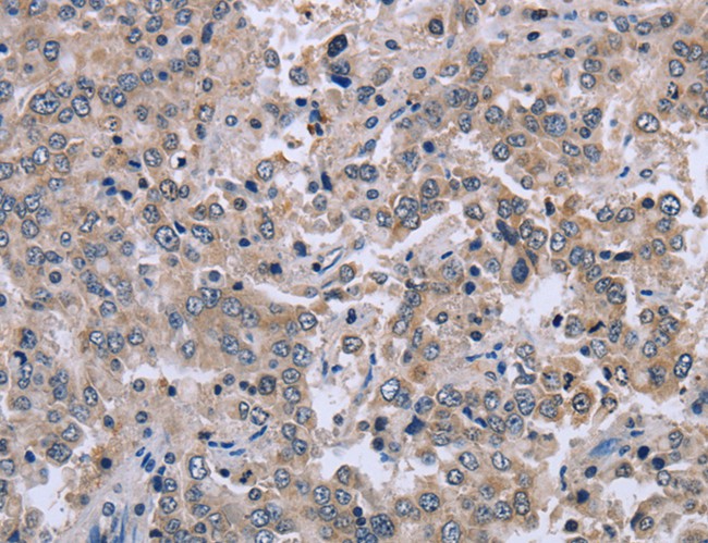
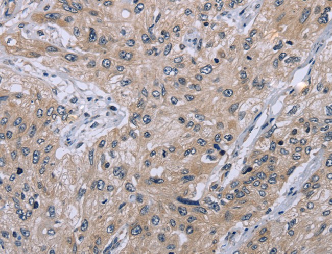
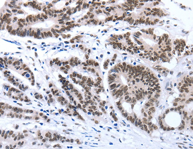
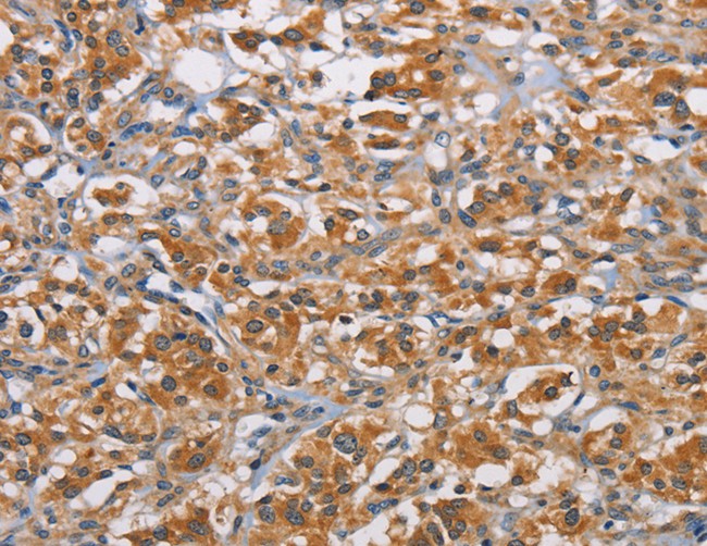
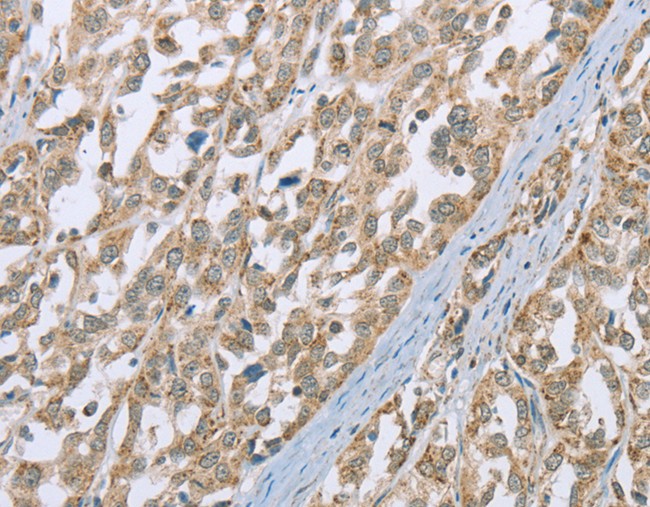
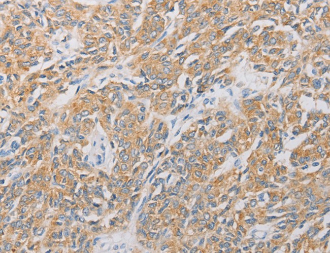
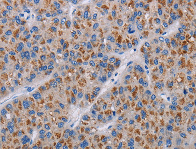
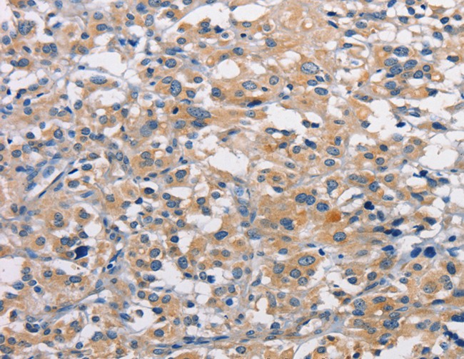
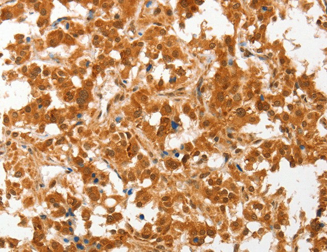
 IHC (Immunohistochemisry)
(Immunohistochemistry of paraffin-embedded Human liver cancer using CIAPIN1 Polyclonal Antibody at dilution of 1:60)
IHC (Immunohistochemisry)
(Immunohistochemistry of paraffin-embedded Human liver cancer using CIAPIN1 Polyclonal Antibody at dilution of 1:60)
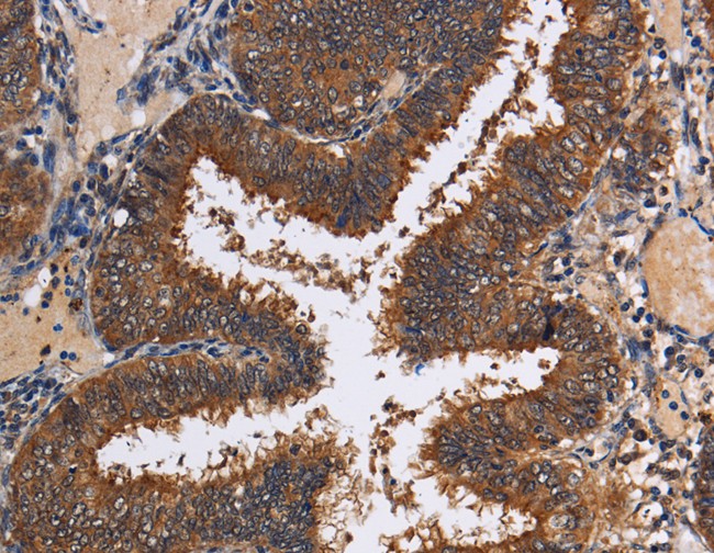
CIAPIN1, Polyclonal Antibody (Cat# AAA170246)
 IHC (Immunohiostchemistry)
(Immunohistochemistry of paraffin-embedded Human tonsil tissue using GYPC Polyclonal Antibody at dilution 1:60)
IHC (Immunohiostchemistry)
(Immunohistochemistry of paraffin-embedded Human tonsil tissue using GYPC Polyclonal Antibody at dilution 1:60)
GYPC, Polyclonal Antibody (Cat# AAA170251)

 IHC (Immunohiostchemistry)
(Immunohistochemistry of paraffin-embedded Human colon cancer tissue using SLC12A4 Polyclonal Antibody at dilution 1:30)
IHC (Immunohiostchemistry)
(Immunohistochemistry of paraffin-embedded Human colon cancer tissue using SLC12A4 Polyclonal Antibody at dilution 1:30)
SLC12A4, Polyclonal Antibody (Cat# AAA170258)
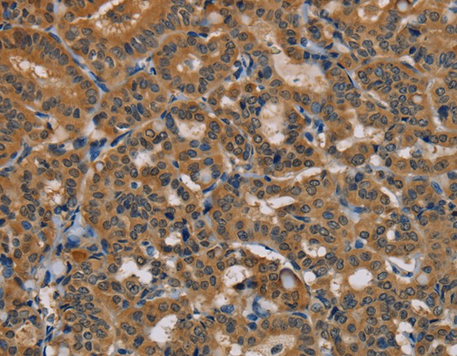
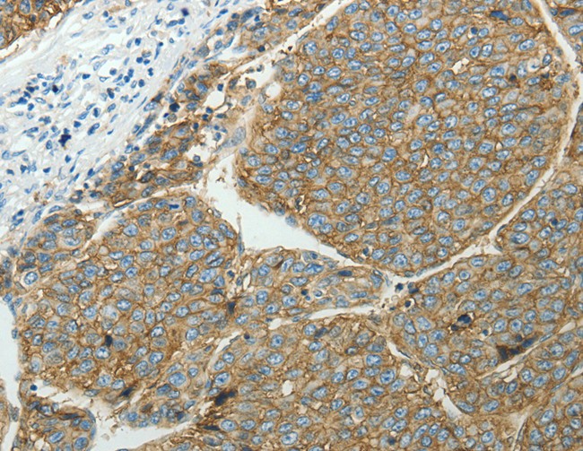
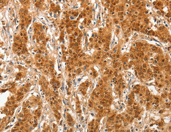
 IHC (Immunohiostchemistry)
(Immunohistochemistry of paraffin-embedded Human liver cancer tissue using GATA5 Polyclonal Antibody at dilution 1:50)
IHC (Immunohiostchemistry)
(Immunohistochemistry of paraffin-embedded Human liver cancer tissue using GATA5 Polyclonal Antibody at dilution 1:50)
GATA5, Polyclonal Antibody (Cat# AAA170259)
 IHC (Immunohiostchemistry)
(Immunohistochemistry of paraffin-embedded Human thyroid cancer tissue using ACAD9 Polyclonal Antibody at dilution 1:15)
IHC (Immunohiostchemistry)
(Immunohistochemistry of paraffin-embedded Human thyroid cancer tissue using ACAD9 Polyclonal Antibody at dilution 1:15)
ACAD9, Polyclonal Antibody (Cat# AAA170269)
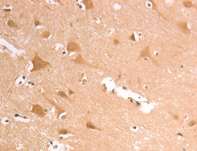
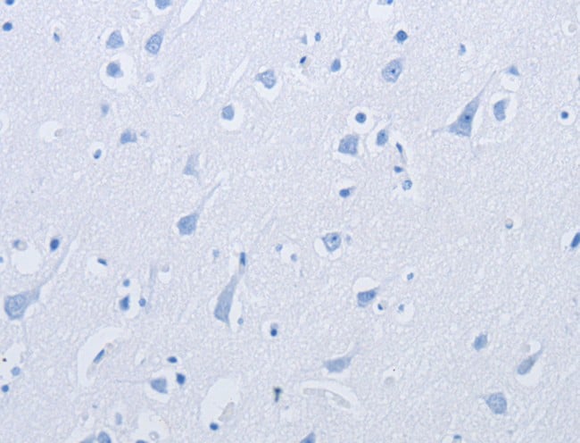
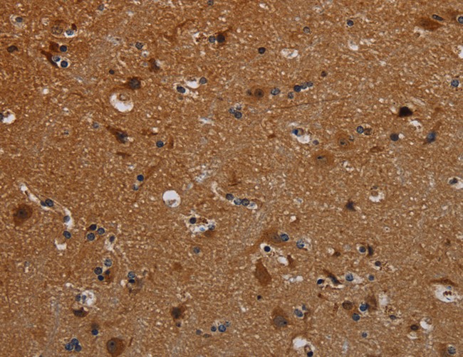
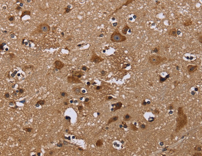 IHC (Immunohiostchemistry)
(Immunohistochemistry of paraffin-embedded Human brain tissue using NCSTN Polyclonal Antibody at dilution 1:50)
IHC (Immunohiostchemistry)
(Immunohistochemistry of paraffin-embedded Human brain tissue using NCSTN Polyclonal Antibody at dilution 1:50)
NCSTN, Polyclonal Antibody (Cat# AAA170274)
 IHC (Immunohiostchemistry)
(Immunohistochemistry of paraffin-embedded Human cervical cancer tissue using KDM4B Polyclonal Antibody at dilution 1:50)
IHC (Immunohiostchemistry)
(Immunohistochemistry of paraffin-embedded Human cervical cancer tissue using KDM4B Polyclonal Antibody at dilution 1:50)
KDM4B, Polyclonal Antibody (Cat# AAA170275)
 IHC (Immunohistochemisry)
(Immunohistochemistry of paraffin-embedded Human brain using AASDHPPT Polyclonal Antibody at dilution of 1:30)
IHC (Immunohistochemisry)
(Immunohistochemistry of paraffin-embedded Human brain using AASDHPPT Polyclonal Antibody at dilution of 1:30)
AASDHPPT, Polyclonal Antibody (Cat# AAA170281)
 IHC (Immunohiostchemistry)
(Immunohistochemistry of paraffin-embedded Human breast cancer tissue using MAPK13 Polyclonal Antibody at dilution 1:15)
IHC (Immunohiostchemistry)
(Immunohistochemistry of paraffin-embedded Human breast cancer tissue using MAPK13 Polyclonal Antibody at dilution 1:15)
MAPK13, Polyclonal Antibody (Cat# AAA170284)
 IHC (Immunohistochemisry)
(Immunohistochemistry of paraffin-embedded using KCNMB3 Polyclonal Antibody at dilution of 1:)
IHC (Immunohistochemisry)
(Immunohistochemistry of paraffin-embedded using KCNMB3 Polyclonal Antibody at dilution of 1:)
KCNMB3, Polyclonal Antibody (Cat# AAA170286)
 IHC (Immunohistochemisry)
(Immunohistochemistry of paraffin-embedded Human lung cancer using TNFRSF13C Polyclonal Antibody at dilution of 1:45)
IHC (Immunohistochemisry)
(Immunohistochemistry of paraffin-embedded Human lung cancer using TNFRSF13C Polyclonal Antibody at dilution of 1:45)
TNFRSF13C, Polyclonal Antibody (Cat# AAA170287)

 IHC (Immunohistochemisry)
(Immunohistochemistry of paraffin-embedded Human gastric cancer using ANXA6 Polyclonal Antibody at dilution of 1:17)
IHC (Immunohistochemisry)
(Immunohistochemistry of paraffin-embedded Human gastric cancer using ANXA6 Polyclonal Antibody at dilution of 1:17)
ANXA6, Polyclonal Antibody (Cat# AAA170291)
 IHC (Immunohiostchemistry)
(Immunohistochemistry of paraffin-embedded Human esophagus cancer tissue using EID1 Polyclonal Antibody at dilution 1:40)
IHC (Immunohiostchemistry)
(Immunohistochemistry of paraffin-embedded Human esophagus cancer tissue using EID1 Polyclonal Antibody at dilution 1:40)
EID1, Polyclonal Antibody (Cat# AAA170292)
 IHC (Immunohistochemisry)
(Immunohistochemistry of paraffin-embedded Human thyroid cancer using BRE Polyclonal Antibody at dilution of 1:40)
IHC (Immunohistochemisry)
(Immunohistochemistry of paraffin-embedded Human thyroid cancer using BRE Polyclonal Antibody at dilution of 1:40)
BRE, Polyclonal Antibody (Cat# AAA170294)
 IHC (Immunohiostchemistry)
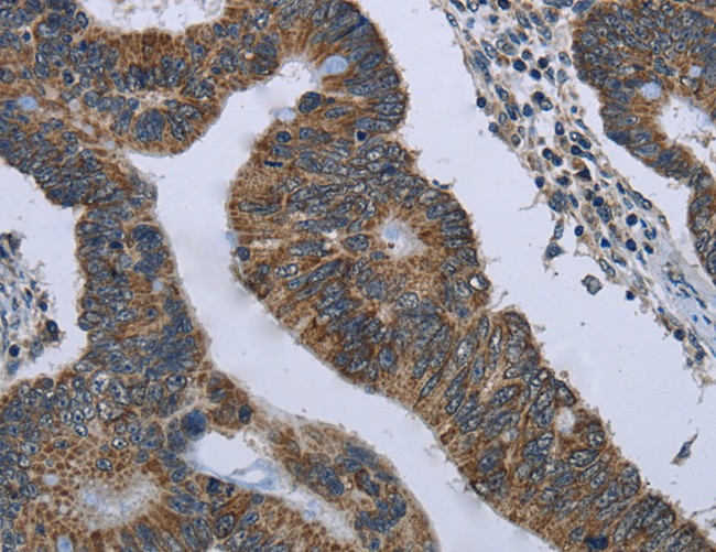
(Immunohistochemistry of paraffin-embedded Human colon cancer tissue using Map2 Polyclonal Antibody at dilution 1:25)
IHC (Immunohiostchemistry)
(Immunohistochemistry of paraffin-embedded Human colon cancer tissue using Map2 Polyclonal Antibody at dilution 1:25)
Map2, Polyclonal Antibody (Cat# AAA170295)
 IHC (Immunohiostchemistry)
(Immunohistochemistry of paraffin-embedded Human lung cancer tissue using IL22RA1 Polyclonal Antibody at dilution 1:15)
IHC (Immunohiostchemistry)
(Immunohistochemistry of paraffin-embedded Human lung cancer tissue using IL22RA1 Polyclonal Antibody at dilution 1:15)
IL22RA1, Polyclonal Antibody (Cat# AAA170296)
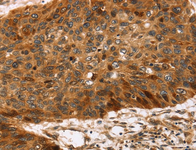
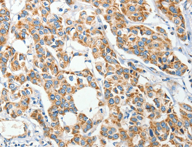
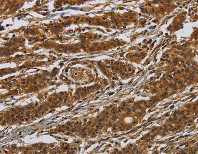
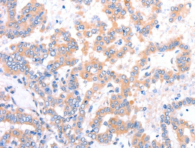
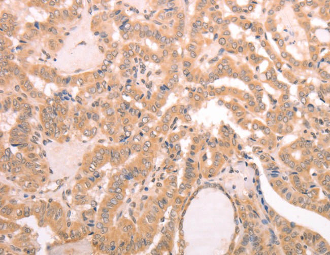
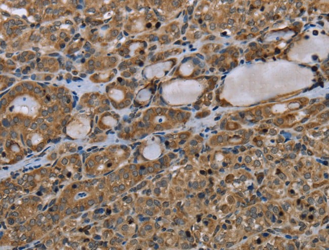
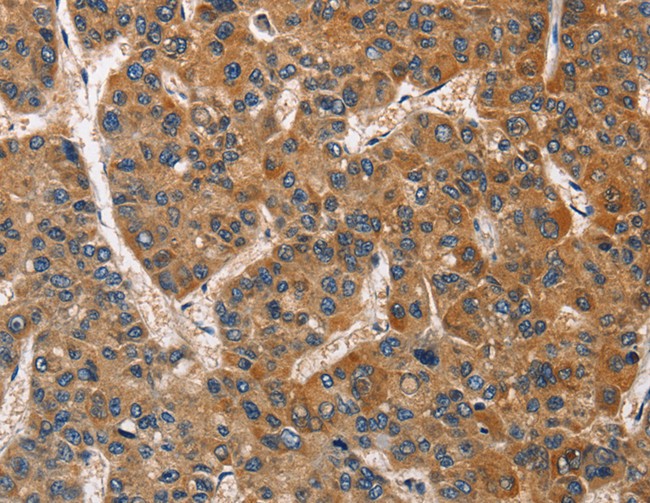
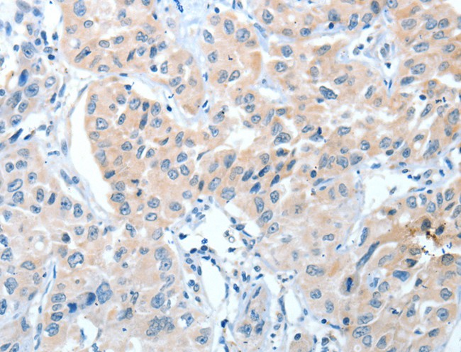
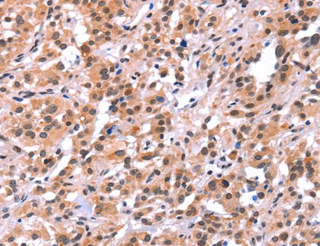
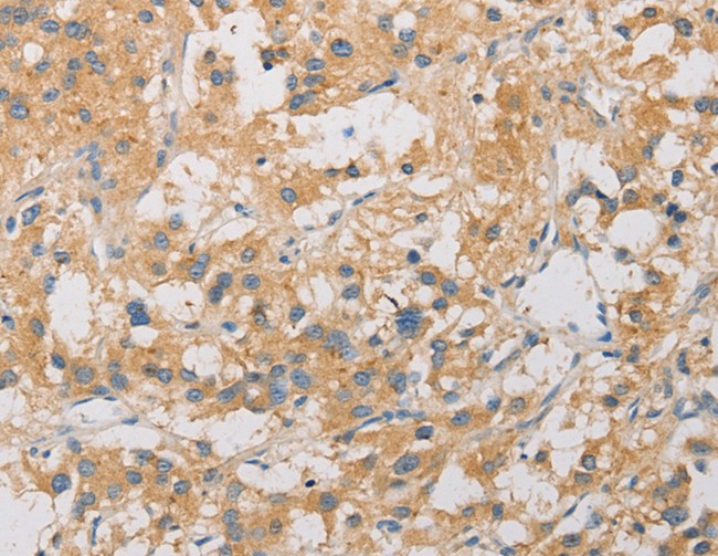
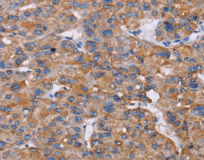
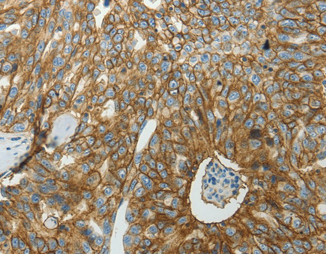
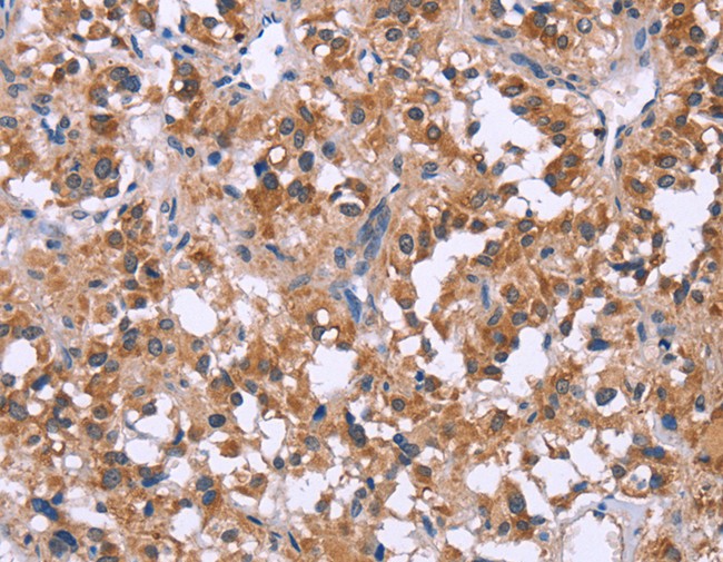
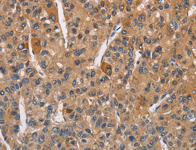 IHC (Immunohiostchemistry)
(Immunohistochemistry of paraffin-embedded Human liver cancer tissue using RICTOR Polyclonal Antibody at dilution 1:40)
IHC (Immunohiostchemistry)
(Immunohistochemistry of paraffin-embedded Human liver cancer tissue using RICTOR Polyclonal Antibody at dilution 1:40)
RICTOR, Polyclonal Antibody (Cat# AAA170299)
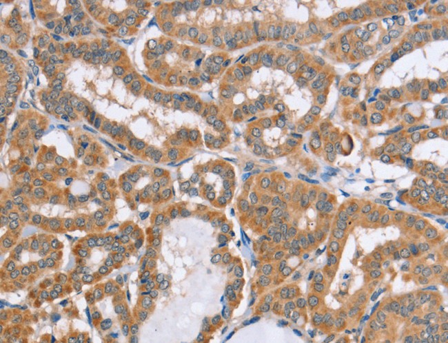
 IHC (Immunohistochemisry)
(Immunohistochemistry of paraffin-embedded Human brain using FABP2 Polyclonal Antibody at dilution of 1:40)
IHC (Immunohistochemisry)
(Immunohistochemistry of paraffin-embedded Human brain using FABP2 Polyclonal Antibody at dilution of 1:40)
FABP2, Polyclonal Antibody (Cat# AAA169990)
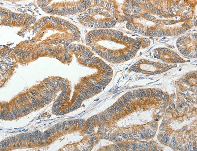
 IHC (Immunohiostchemistry)
(Immunohistochemistry of paraffin-embedded Human colon cancer tissue using HK1 Polyclonal Antibody at dilution 1:50)
IHC (Immunohiostchemistry)
(Immunohistochemistry of paraffin-embedded Human colon cancer tissue using HK1 Polyclonal Antibody at dilution 1:50)
HK1, Polyclonal Antibody (Cat# AAA169991)
 IHC (Immunohiostchemistry)
(Immunohistochemistry of paraffin-embedded Human brain tissue using PCDHB15 Polyclonal Antibody at dilution 1:50)
IHC (Immunohiostchemistry)
(Immunohistochemistry of paraffin-embedded Human brain tissue using PCDHB15 Polyclonal Antibody at dilution 1:50)
PCDHB15, Polyclonal Antibody (Cat# AAA169995)

 IHC (Immunohiostchemistry)
(Immunohistochemistry of paraffin-embedded Human thyroid cancer using COMP Polyclonal Antibody at dilution of 1:50)
IHC (Immunohiostchemistry)
(Immunohistochemistry of paraffin-embedded Human thyroid cancer using COMP Polyclonal Antibody at dilution of 1:50)
COMP, Polyclonal Antibody (Cat# AAA169999)
 IHC (Immunohiostchemistry)
(Immunohistochemistry of paraffin-embedded Human thyroid cancer tissue using MYF5 Polyclonal Antibody at dilution 1:15)
IHC (Immunohiostchemistry)
(Immunohistochemistry of paraffin-embedded Human thyroid cancer tissue using MYF5 Polyclonal Antibody at dilution 1:15)
MYF5, Polyclonal Antibody (Cat# AAA170001)
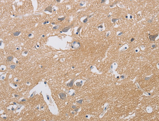
 IHC (Immunohistochemisry)
(Immunohistochemistry of paraffin-embedded Human brain using ECHS1 Polyclonal Antibody at dilution of 1:35)
IHC (Immunohistochemisry)
(Immunohistochemistry of paraffin-embedded Human brain using ECHS1 Polyclonal Antibody at dilution of 1:35)
ECHS1, Polyclonal Antibody (Cat# AAA170012)
 IHC (Immunohiostchemistry)
(Immunohistochemistry of paraffin-embedded Human prostate cancer tissue using ARSG Polyclonal Antibody at dilution 1:30)
IHC (Immunohiostchemistry)
(Immunohistochemistry of paraffin-embedded Human prostate cancer tissue using ARSG Polyclonal Antibody at dilution 1:30)
ARSG, Polyclonal Antibody (Cat# AAA170015)
 IHC (Immunohistochemisry)
(Immunohistochemistry of paraffin-embedded Human prostate cancer using NIT1 Polyclonal Antibody at dilution of 1:50)
IHC (Immunohistochemisry)
(Immunohistochemistry of paraffin-embedded Human prostate cancer using NIT1 Polyclonal Antibody at dilution of 1:50)
NIT1, Polyclonal Antibody (Cat# AAA170016)
 IHC (Immunohistochemisry)
(Immunohistochemistry of paraffin-embedded Human thyroid cancer using TAB3 Polyclonal Antibody at dilution of 1:35)
IHC (Immunohistochemisry)
(Immunohistochemistry of paraffin-embedded Human thyroid cancer using TAB3 Polyclonal Antibody at dilution of 1:35)
TAB3, Polyclonal Antibody (Cat# AAA170017)
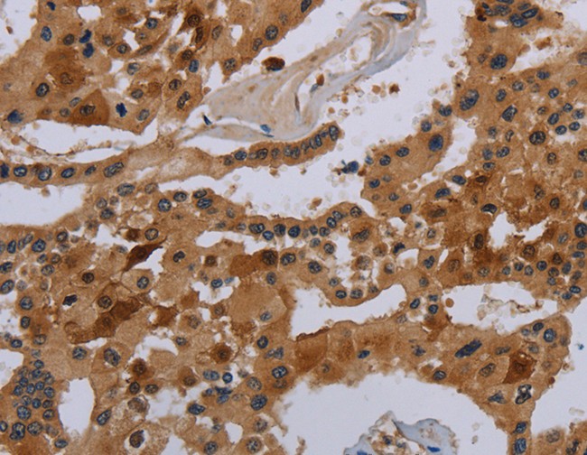
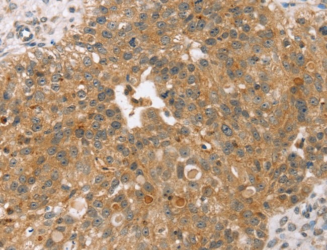
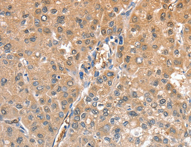
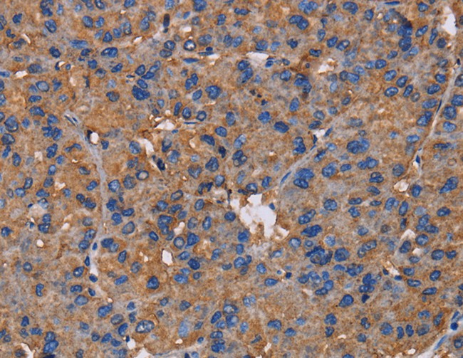
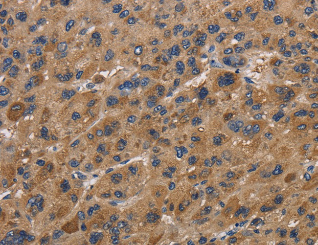
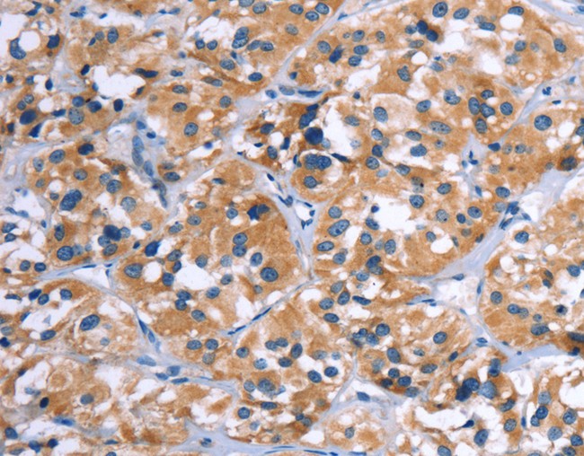
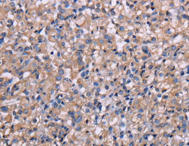
 IHC (Immunohiostchemistry)
(Immunohistochemistry of paraffin-embedded Human liver cancer tissue using COX18 Polyclonal Antibody at dilution 1:20)
IHC (Immunohiostchemistry)
(Immunohistochemistry of paraffin-embedded Human liver cancer tissue using COX18 Polyclonal Antibody at dilution 1:20)
COX18, Polyclonal Antibody (Cat# AAA170019)
 IHC (Immunohiostchemistry)
(Immunohistochemistry of paraffin-embedded Human ovarian cancer tissue using SLC16A1 Polyclonal Antibody at dilution 1:50)
IHC (Immunohiostchemistry)
(Immunohistochemistry of paraffin-embedded Human ovarian cancer tissue using SLC16A1 Polyclonal Antibody at dilution 1:50)
SLC16A1, Polyclonal Antibody (Cat# AAA170021)
 IHC (Immunohistochemisry)
(Immunohistochemistry of paraffin-embedded Human thyroid cancer using PRKCE Polyclonal Antibody at dilution of 1:50)
IHC (Immunohistochemisry)
(Immunohistochemistry of paraffin-embedded Human thyroid cancer using PRKCE Polyclonal Antibody at dilution of 1:50)
PRKCE, Polyclonal Antibody (Cat# AAA170022)
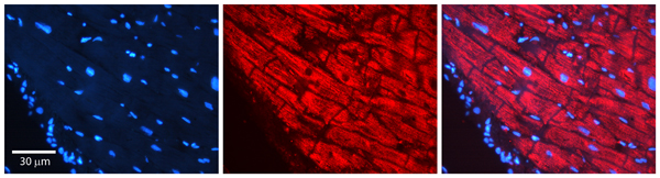
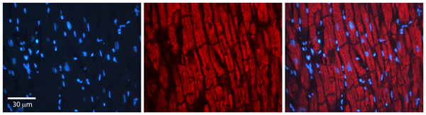
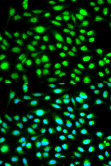 IF (Immunofluorescence)
(Immunofluorescence analysis of HeLa cell using CCNH antibody. Blue: DAPI for nuclear staining.)
IF (Immunofluorescence)
(Immunofluorescence analysis of HeLa cell using CCNH antibody. Blue: DAPI for nuclear staining.)
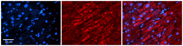
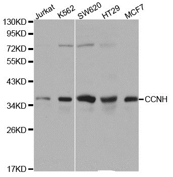
CCNH, Polyclonal Antibody (Cat# AAA170025)
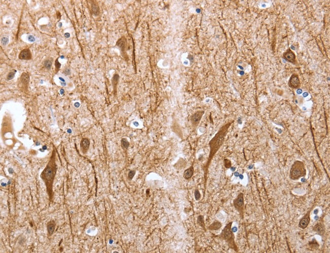
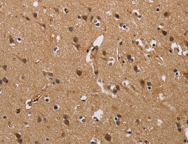
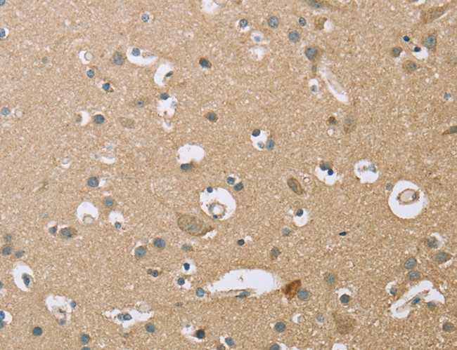
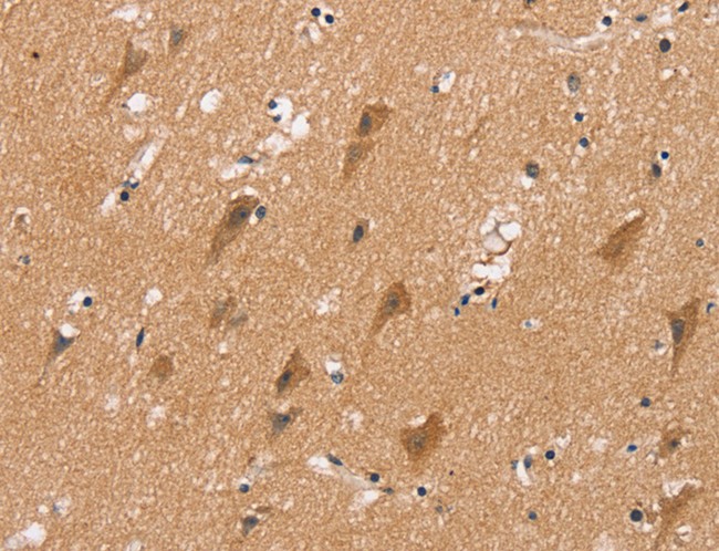 IHC (Immunohistochemisry)
(Immunohistochemistry of paraffin-embedded Human brain using MSMB Polyclonal Antibody at dilution of 1:20)
IHC (Immunohistochemisry)
(Immunohistochemistry of paraffin-embedded Human brain using MSMB Polyclonal Antibody at dilution of 1:20)
MSMB, Polyclonal Antibody (Cat# AAA170027)

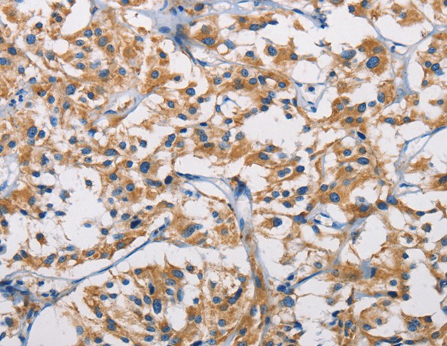
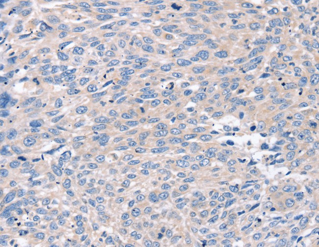
 IHC (Immunohiostchemistry)
(Immunohistochemistry of paraffin-embedded Human breast cancer using NMT2 Polyclonal Antibody at dilution of 1:20)
IHC (Immunohiostchemistry)
(Immunohistochemistry of paraffin-embedded Human breast cancer using NMT2 Polyclonal Antibody at dilution of 1:20)
NMT2, Polyclonal Antibody (Cat# AAA170031)
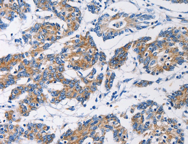
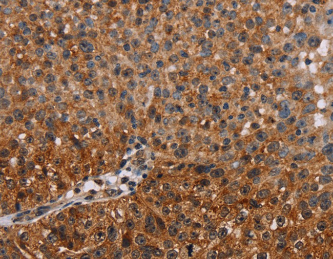
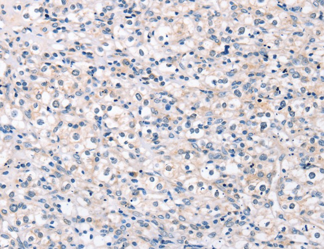
 IHC (Immunohiostchemistry)
(Immunohistochemistry of paraffin-embedded Human gastric cancer tissue using COL4A3BP Polyclonal Antibody at dilution 1:50)
IHC (Immunohiostchemistry)
(Immunohistochemistry of paraffin-embedded Human gastric cancer tissue using COL4A3BP Polyclonal Antibody at dilution 1:50)
COL4A3BP, Polyclonal Antibody (Cat# AAA170033)
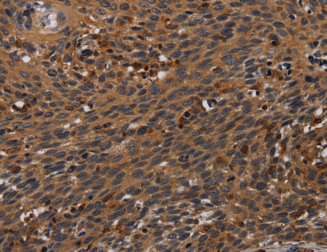
 IHC (Immunohistochemisry)
(Immunohistochemistry of paraffin-embedded Human cervical cancer using APAF1 Polyclonal Antibody at dilution of 1:20)
IHC (Immunohistochemisry)
(Immunohistochemistry of paraffin-embedded Human cervical cancer using APAF1 Polyclonal Antibody at dilution of 1:20)
APAF1, Polyclonal Antibody (Cat# AAA170034)
 IHC (Immunohiostchemistry)
(Immunohistochemistry of paraffin-embedded Human prostate cancer tissue using ROR1 Polyclonal Antibody at dilution 1:35)
IHC (Immunohiostchemistry)
(Immunohistochemistry of paraffin-embedded Human prostate cancer tissue using ROR1 Polyclonal Antibody at dilution 1:35)
ROR1, Polyclonal Antibody (Cat# AAA170037)
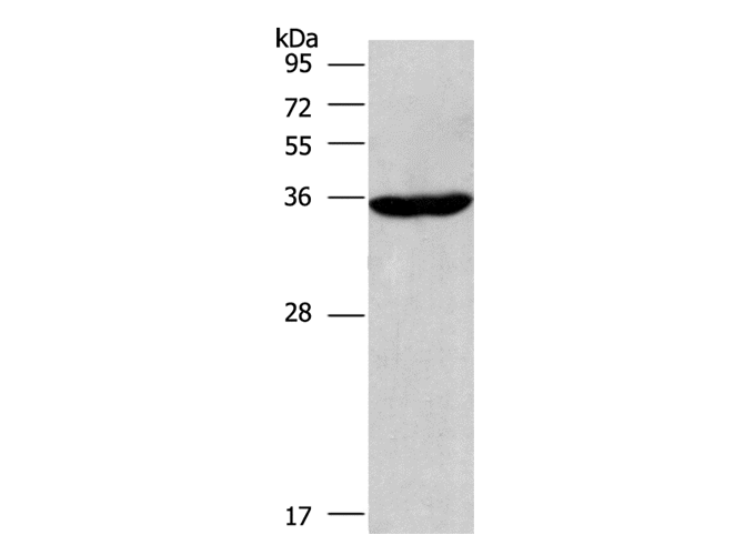
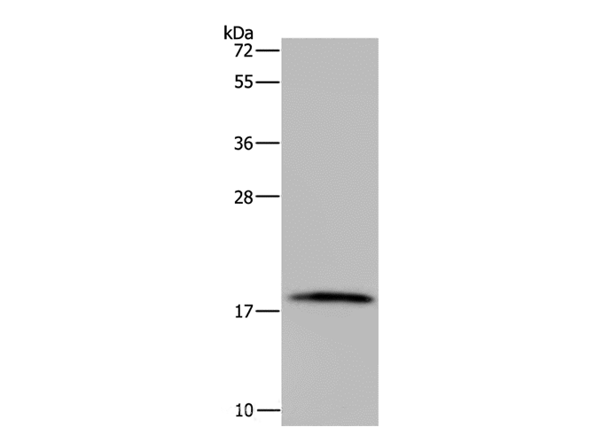
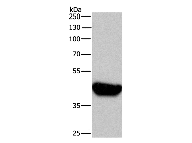
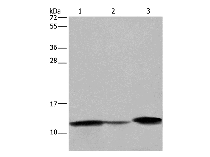
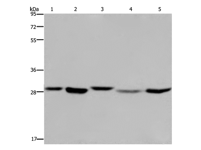
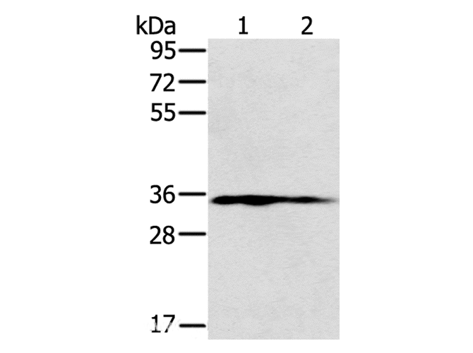
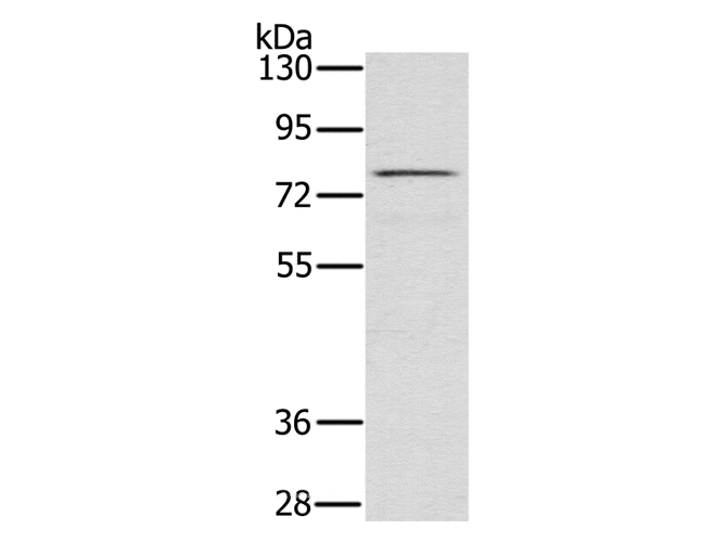
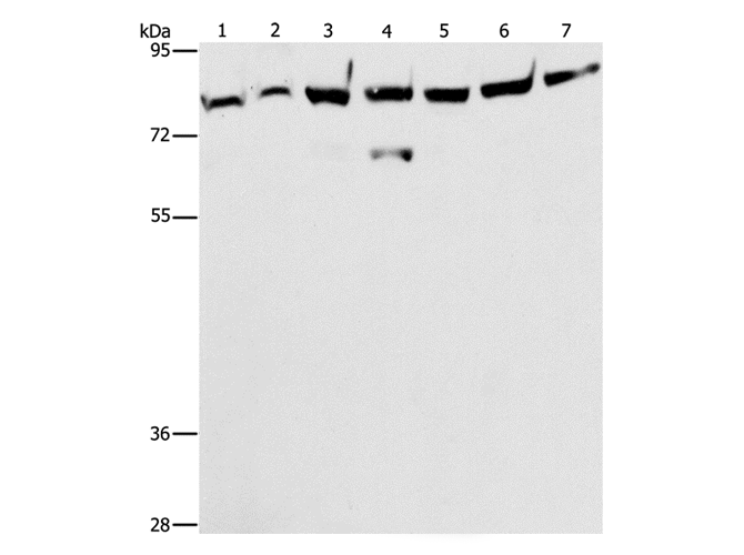
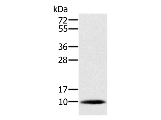
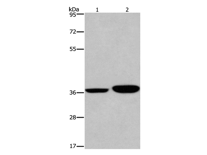
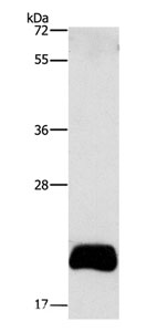 WB (Western Blot)
(Western Blot analysis of PC3 cell using APOBEC3C Polyclonal Antibody at dilution of 1:420)
WB (Western Blot)
(Western Blot analysis of PC3 cell using APOBEC3C Polyclonal Antibody at dilution of 1:420)
APOBEC3C, Polyclonal Antibody (Cat# AAA170038)
 IHC (Immunohiostchemistry)
(Immunohistochemistry of paraffin-embedded Human lung cancer tissue using A4GNT Polyclonal Antibody at dilution 1:30)
IHC (Immunohiostchemistry)
(Immunohistochemistry of paraffin-embedded Human lung cancer tissue using A4GNT Polyclonal Antibody at dilution 1:30)
A4GNT, Polyclonal Antibody (Cat# AAA170041)

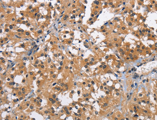
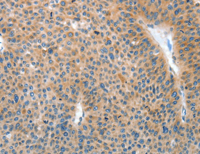
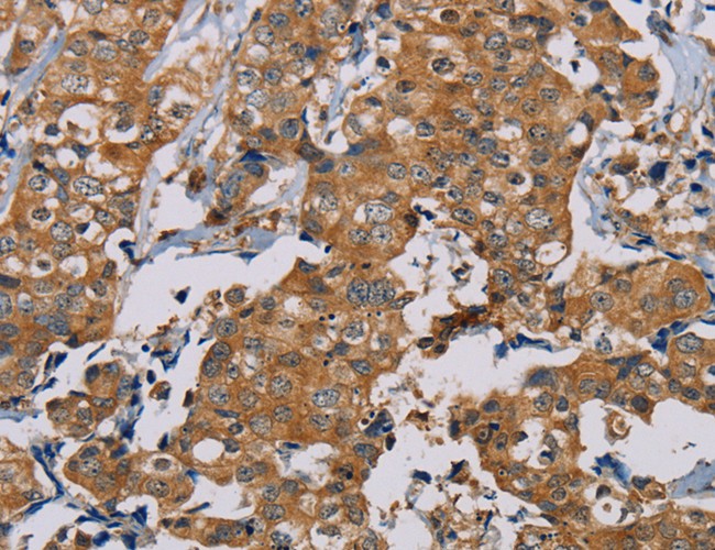
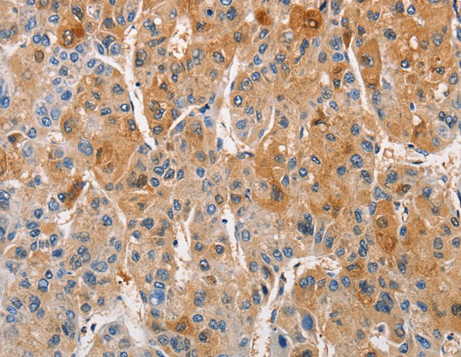
 IHC (Immunohiostchemistry)
(Immunohistochemistry of paraffin-embedded Human liver cancer tissue using TM4SF1 Polyclonal Antibody at dilution 1:30)
IHC (Immunohiostchemistry)
(Immunohistochemistry of paraffin-embedded Human liver cancer tissue using TM4SF1 Polyclonal Antibody at dilution 1:30)
TM4SF1, Polyclonal Antibody (Cat# AAA170044)
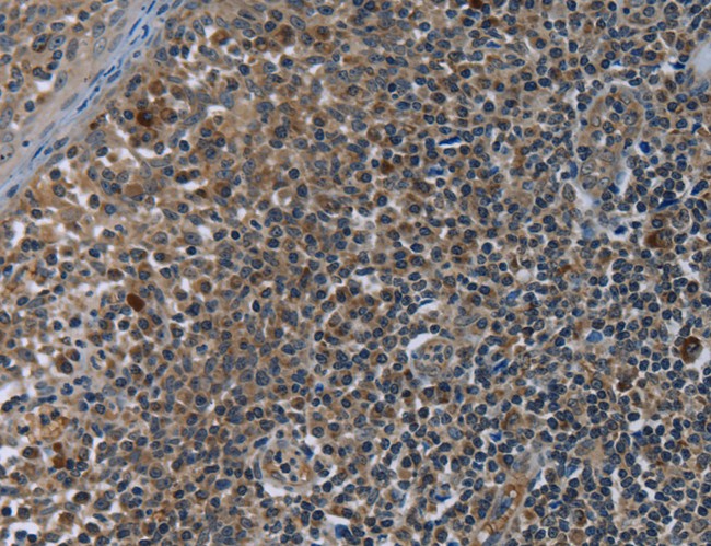
 IHC (Immunohiostchemistry)
(Immunohistochemistry of paraffin-embedded Human tonsil tissue using LRRC57 Polyclonal Antibody at dilution 1:40)
IHC (Immunohiostchemistry)
(Immunohistochemistry of paraffin-embedded Human tonsil tissue using LRRC57 Polyclonal Antibody at dilution 1:40)
LRRC57, Polyclonal Antibody (Cat# AAA170049)
What are Polyclonal Antibodies?
Polyclonal antibodies are antibodies that come from multiple B cell clones of a host animal. The typical hosts used for the majority of polyclonal antibody production are rabbits, goats, sheep, and donkeys. These polyclonal antibodies, once having identified their target, will bind to different epitopes located at different regions or sequences on the same protein/antigen. As a result, they are ideal at locating and binding to the target, even if the target is in very low concentrations (due to many different antibodies being able to bind to the same target molecule, which allows for significant amplification of a downstream signal).
Polyclonal antibodies are typically produced by injecting an antigen into a host animal, which causes the animal’s immune system to attack the foreign antigen by mass generating antibodies against it. After a period of time, serum is collected from the animal and purified using physicochemical fractionation, class-specific affinity purification, and/or antigen-affinity purification.
Key Uses of Polyclonal Antibodies
- Western Blotting: This method is used to find specific proteins in biological samples after separating them by size.
- Immunohistochemistry: IHC helps visualize the location of proteins in tissue sections using various staining techniques.
- ELISA: (Enzyme-Linked Immunosorbent Assay) is typically used to identify specific protein quantities in a sample. ELISAs can be either “Quantitative” or “Qualitative”.
- Flow Cytometry: technique that identifies and measures the specific protein on the surface or inside the cells in a fluid suspension.
- Immunoprecipitation: IP isolates and studies a specific protein from a complex mixture using antibodies.
Why Buy Polyclonal Antibodies from AAA Biotech?
1. Ideal for Various Applications
Our antibodies are generally going to be validated for use in multiple types of assays, including ELISA, Western Blotting, Immunohistochemistry, Immunoprecipitation, amongst others. They are ideal for a wide range of research applications.
2. Rigorous Quality Control
All of the antibodies in our catalog undergo strict quality testing to ensure specificity, sensitivity, and consistent performance. We are confident in the ability of our antibodies to provide you with accurate results.
3. Wide Assortment of Antibodies
Antibodies in are catalog can be found for both common and exotic species, and these antibodies are also available in both conjugated and recombinant forms to suit many diverse experimental needs.
4. Highly Purified
Our antibodies are available in purified forms with over 85% purity, as confirmed by SDS-PAGE. They are also available with tags such as His, Flag, GST, or MBP. We cater to customers worldwide.
FAQ
1. How are polyclonal antibodies produced?
Traditionally, polyclonal antibodies are produced by injecting an antigen into a host animal (such as a rabbit or goat), which then triggers an immune response from the host animal. The animal’s B cells produce antibodies that will recognize different parts of the injected antigen. These antibodies are then collected from the animal’s blood and purified for use.
2. How do polyclonal antibodies differ from monoclonal antibodies?
Polyclonal antibodies are a mix of antibodies that bind to different locations (epitopes) of the same antigen, while monoclonal antibodies are identical and bind to just one specific epitope. This makes polyclonal antibodies more versatile and better at detecting proteins that may be present in low quantities or in altered/modified forms.
3. How should I store polyclonal antibodies?
Polyclonal antibodies should be stored at 4°C for short-term use (up to a few weeks) and at -20°C or -80°C for long-term storage. Avoid repeated freeze-thaw cycles by dividing them into small aliquots. Always check the datasheet for specific storage instructions.