Filters
▼Clonality
▼Type
▼Reactivity
▼Gene Name
▼Isotype
▼Host
▼Application
▼Clone
▼Polyclonal Antibodies
At AAA Biotech also known as AAA Bio or AAABio, we provide a broad range of purified polyclonal antibodies (pAbs) that are able to all be browsed online through our website. Due to their high specificity and strong binding affinity, these antibodies are ideal for wide swathes of research and experimental applications.
Our polyclonal antibodies can easily support your work, whether you use them for Western Blotting, Immunocytochemistry (with or without Immunofluorescence used in conjunction), Immunohistochemistry, Immunoprecipitation, and ELISA tests. We highly encourage you to browse our range of pAbs and choose the one that best suits your experimental model.
Viewing 500-550 of 96805 product results
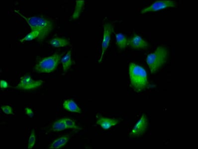
 IF (Immunofluorescence)
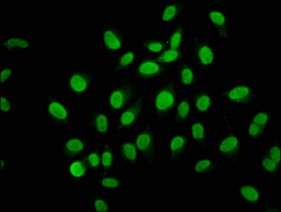
(Immunofluorescence staining of HepG2 cells with AAA234168 at 1:133, counter-stained with DAPI. The cells were fixed in 4% formaldehyde, permeabilized using 0.2% Triton X-100 and blocked in 10% normal Goat Serum. The cells were then incubated with the antibody overnight at 4 degree C.The secondary antibody was Alexa Fluor 488-congugated AffiniPure Goat Anti-Rabbit IgG (H+L).)
IF (Immunofluorescence)
(Immunofluorescence staining of HepG2 cells with AAA234168 at 1:133, counter-stained with DAPI. The cells were fixed in 4% formaldehyde, permeabilized using 0.2% Triton X-100 and blocked in 10% normal Goat Serum. The cells were then incubated with the antibody overnight at 4 degree C.The secondary antibody was Alexa Fluor 488-congugated AffiniPure Goat Anti-Rabbit IgG (H+L).)
ARID3A, Polyclonal Antibody (Cat# AAA234168)
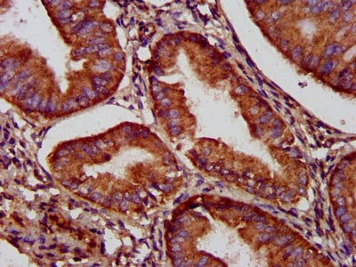
 IHC (Immunohistochemisry)
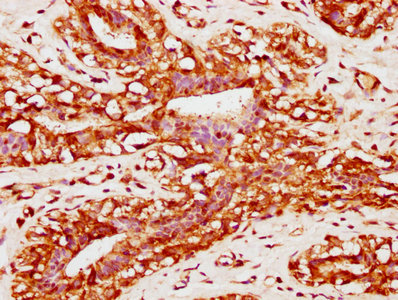
(IHC image of AAA234172 diluted at 1:200 and staining in paraffin-embedded human colon cancer performed on a Leica BondTM system. After dewaxing and hydration, antigen retrieval was mediated by high pressure in a citrate buffer (pH 6.0). Section was blocked with 10% normal goat serum 30min at RT. Then primary antibody (1% BSA) was incubated at 4 degree C overnight. The primary is detected by a biotinylated secondary antibody and visualized using an HRP conjugated SP system.)
IHC (Immunohistochemisry)
(IHC image of AAA234172 diluted at 1:200 and staining in paraffin-embedded human colon cancer performed on a Leica BondTM system. After dewaxing and hydration, antigen retrieval was mediated by high pressure in a citrate buffer (pH 6.0). Section was blocked with 10% normal goat serum 30min at RT. Then primary antibody (1% BSA) was incubated at 4 degree C overnight. The primary is detected by a biotinylated secondary antibody and visualized using an HRP conjugated SP system.)
MYOC, Polyclonal Antibody (Cat# AAA234172)
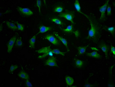
 IF (Immunofluorescence)
(Immunofluorescence staining of HepG2 cells with AAA234178 at 1:100, counter- stained with DAPI. The cells were fixed in 4% formaldehyde, permeabilized using 0.2% Triton X-100 and blocked in 10% normal Goat Serum. The cells were then incubated with the antibody overnight at 4°C. The secondary antibody was Alexa Fluor 488-congugated AffiniPure Goat Anti-Rabbit IgG(H+L).)
IF (Immunofluorescence)
(Immunofluorescence staining of HepG2 cells with AAA234178 at 1:100, counter- stained with DAPI. The cells were fixed in 4% formaldehyde, permeabilized using 0.2% Triton X-100 and blocked in 10% normal Goat Serum. The cells were then incubated with the antibody overnight at 4°C. The secondary antibody was Alexa Fluor 488-congugated AffiniPure Goat Anti-Rabbit IgG(H+L).)
SEMA4B, Polyclonal Antibody (Cat# AAA234178)
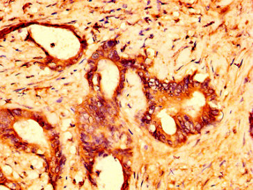
 IHC (Immunohiostchemistry)
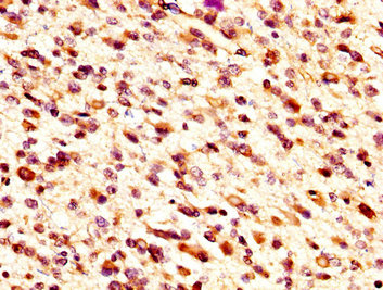
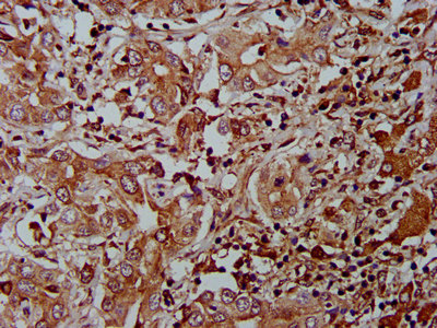
(IHC image of AAA234180 diluted at 1:300 and staining in paraffin-embedded human glioma cancer performed on a Leica BondTM system. After dewaxing and hydration, antigen retrieval was mediated by high pressure in a citrate buffer (pH 6.0). Section was blocked with 10% normal goat serum 30min at RT. Then primary antibody (1% BSA) was incubated at 4 degree C overnight. The primary is detected by a biotinylated secondary antibody and visualized using an HRP conjugated SP system.)
IHC (Immunohiostchemistry)
(IHC image of AAA234180 diluted at 1:300 and staining in paraffin-embedded human glioma cancer performed on a Leica BondTM system. After dewaxing and hydration, antigen retrieval was mediated by high pressure in a citrate buffer (pH 6.0). Section was blocked with 10% normal goat serum 30min at RT. Then primary antibody (1% BSA) was incubated at 4 degree C overnight. The primary is detected by a biotinylated secondary antibody and visualized using an HRP conjugated SP system.)
MID1IP1, Polyclonal Antibody (Cat# AAA234180)
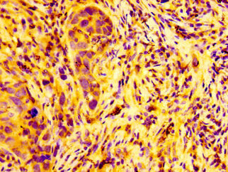
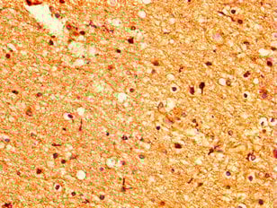
 IHC (Immunohiostchemistry)
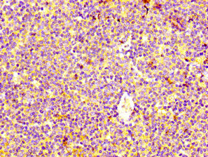
(IHC image of AAA234187 diluted at 1:400 and staining in paraffin-embedded human lymph node tissue performed on a Leica BondTM system. After dewaxing and hydration, antigen retrieval was mediated by high pressure in a citrate buffer (pH 6.0). Section was blocked with 10% normal goat serum 30min at RT. Then primary antibody (1% BSA) was incubated at 4 degree C overnight. The primary is detected by a biotinylated secondary antibody and visualized using an HRP conjugated SP system.)
IHC (Immunohiostchemistry)
(IHC image of AAA234187 diluted at 1:400 and staining in paraffin-embedded human lymph node tissue performed on a Leica BondTM system. After dewaxing and hydration, antigen retrieval was mediated by high pressure in a citrate buffer (pH 6.0). Section was blocked with 10% normal goat serum 30min at RT. Then primary antibody (1% BSA) was incubated at 4 degree C overnight. The primary is detected by a biotinylated secondary antibody and visualized using an HRP conjugated SP system.)
HRH4, Polyclonal Antibody (Cat# AAA234187)
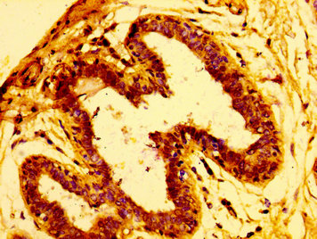
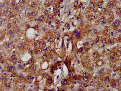
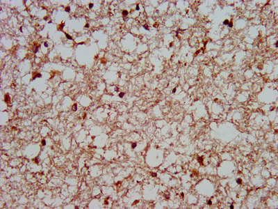
 IHC (Immunohiostchemistry)
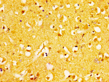
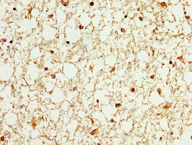
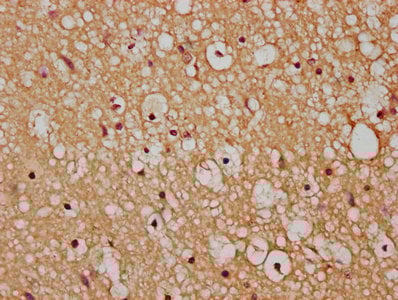
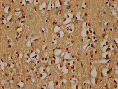
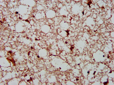
(IHC image of AAA234188 diluted at 1:200 and staining in paraffin-embedded human brain tissue performed on a Leica BondTM system. After dewaxing and hydration, antigen retrieval was mediated by high pressure in a citrate buffer (pH 6.0). Section was blocked with 10% normal goat serum 30min at RT. Then primary antibody (1% BSA) was incubated at 4 degree C overnight. The primary is detected by a biotinylated secondary antibody and visualized using an HRP conjugated SP system.)
IHC (Immunohiostchemistry)
(IHC image of AAA234188 diluted at 1:200 and staining in paraffin-embedded human brain tissue performed on a Leica BondTM system. After dewaxing and hydration, antigen retrieval was mediated by high pressure in a citrate buffer (pH 6.0). Section was blocked with 10% normal goat serum 30min at RT. Then primary antibody (1% BSA) was incubated at 4 degree C overnight. The primary is detected by a biotinylated secondary antibody and visualized using an HRP conjugated SP system.)
EPHA6, Polyclonal Antibody (Cat# AAA234188)
 IHC (Immunohistochemisry)
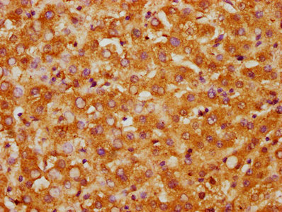
(IHC image of AAA234189 diluted at 1:100 and staining in paraffin-embedded human brain tissue performed on a Leica BondTM system. After dewaxing and hydration, antigen retrieval was mediated by high pressure in a citrate buffer (pH 6.0). Section was blocked with 10% normal goat serum 30min at RT. Then primary antibody (1% BSA) was incubated at 4 degree C overnight. The primary is detected by a biotinylated secondary antibody and visualized using an HRP conjugated SP system.)
IHC (Immunohistochemisry)
(IHC image of AAA234189 diluted at 1:100 and staining in paraffin-embedded human brain tissue performed on a Leica BondTM system. After dewaxing and hydration, antigen retrieval was mediated by high pressure in a citrate buffer (pH 6.0). Section was blocked with 10% normal goat serum 30min at RT. Then primary antibody (1% BSA) was incubated at 4 degree C overnight. The primary is detected by a biotinylated secondary antibody and visualized using an HRP conjugated SP system.)
WASF3, Polyclonal Antibody (Cat# AAA234189)
 IF (Immunofluorescence)
(Immunofluorescence staining of A549 cells with AAA234194 at 1:133, counter-stained with DAPI. The cells were fixed in 4% formaldehyde, permeabilized using 0.2% Triton X-100 and blocked in 10% normal Goat Serum. The cells were then incubated with the antibody overnight at 4 degree C.The secondary antibody was Alexa Fluor 488-congugated AffiniPure Goat Anti-Rabbit IgG (H+L).)
IF (Immunofluorescence)
(Immunofluorescence staining of A549 cells with AAA234194 at 1:133, counter-stained with DAPI. The cells were fixed in 4% formaldehyde, permeabilized using 0.2% Triton X-100 and blocked in 10% normal Goat Serum. The cells were then incubated with the antibody overnight at 4 degree C.The secondary antibody was Alexa Fluor 488-congugated AffiniPure Goat Anti-Rabbit IgG (H+L).)
ARFGEF2, Polyclonal Antibody (Cat# AAA234194)
 IF (Immunofluorescence)
(Immunofluorescence staining of Hela cells with AAA234195 at 1:133, counter-stained with DAPI. The cells were fixed in 4% formaldehyde, permeabilized using 0.2% Triton X-100 and blocked in 10% normal Goat Serum. The cells were then incubated with the antibody overnight at 4 degree C.The secondary antibody was Alexa Fluor 488-congugated AffiniPure Goat Anti-Rabbit IgG (H+L).)
IF (Immunofluorescence)
(Immunofluorescence staining of Hela cells with AAA234195 at 1:133, counter-stained with DAPI. The cells were fixed in 4% formaldehyde, permeabilized using 0.2% Triton X-100 and blocked in 10% normal Goat Serum. The cells were then incubated with the antibody overnight at 4 degree C.The secondary antibody was Alexa Fluor 488-congugated AffiniPure Goat Anti-Rabbit IgG (H+L).)
SSX2IP, Polyclonal Antibody (Cat# AAA234195)
 IHC (Immunohiostchemistry)
(IHC image of AAA234197 diluted at 1:100 and staining in paraffin-embedded human colon cancer performed on a Leica BondTM system. After dewaxing and hydration, antigen retrieval was mediated by high pressure in a citrate buffer (pH 6.0). Section was blocked with 10% normal goat serum 30min at RT. Then primary antibody (1% BSA) was incubated at 4 degree C overnight. The primary is detected by a biotinylated secondary antibody and visualized using an HRP conjugated SP system.)
IHC (Immunohiostchemistry)
(IHC image of AAA234197 diluted at 1:100 and staining in paraffin-embedded human colon cancer performed on a Leica BondTM system. After dewaxing and hydration, antigen retrieval was mediated by high pressure in a citrate buffer (pH 6.0). Section was blocked with 10% normal goat serum 30min at RT. Then primary antibody (1% BSA) was incubated at 4 degree C overnight. The primary is detected by a biotinylated secondary antibody and visualized using an HRP conjugated SP system.)
LIMS1, Polyclonal Antibody (Cat# AAA234197)
 IHC (Immunohiostchemistry)
(IHC image of AAA234198 diluted at 1:200 and staining in paraffin-embedded human breast cancer performed on a Leica BondTM system. After dewaxing and hydration, antigen retrieval was mediated by high pressure in a citrate buffer (pH 6.0). Section was blocked with 10% normal goat serum 30min at RT. Then primary antibody (1% BSA) was incubated at 4 degree C overnight. The primary is detected by a biotinylated secondary antibody and visualized using an HRP conjugated SP system.)
IHC (Immunohiostchemistry)
(IHC image of AAA234198 diluted at 1:200 and staining in paraffin-embedded human breast cancer performed on a Leica BondTM system. After dewaxing and hydration, antigen retrieval was mediated by high pressure in a citrate buffer (pH 6.0). Section was blocked with 10% normal goat serum 30min at RT. Then primary antibody (1% BSA) was incubated at 4 degree C overnight. The primary is detected by a biotinylated secondary antibody and visualized using an HRP conjugated SP system.)
CAPRIN1, Polyclonal Antibody (Cat# AAA234198)
 IHC (Immunohiostchemistry)
(IHC image of AAA234200 diluted at 1:600 and staining in paraffin-embedded human brain tissue performed on a Leica BondTM system. After dewaxing and hydration, antigen retrieval was mediated by high pressure in a citrate buffer (pH 6.0). Section was blocked with 10% normal goat serum 30min at RT. Then primary antibody (1% BSA) was incubated at 4 degree C overnight. The primary is detected by a biotinylated secondary antibody and visualized using an HRP conjugated SP system.)
IHC (Immunohiostchemistry)
(IHC image of AAA234200 diluted at 1:600 and staining in paraffin-embedded human brain tissue performed on a Leica BondTM system. After dewaxing and hydration, antigen retrieval was mediated by high pressure in a citrate buffer (pH 6.0). Section was blocked with 10% normal goat serum 30min at RT. Then primary antibody (1% BSA) was incubated at 4 degree C overnight. The primary is detected by a biotinylated secondary antibody and visualized using an HRP conjugated SP system.)
SORT1, Polyclonal Antibody (Cat# AAA234200)
 IF (Immunofluorescence)
(Immunofluorescence staining of A549 cells with AAA234207 at 1:166, counter-stained with DAPI. The cells were fixed in 4% formaldehyde, permeabilized using 0.2% Triton X-100 and blocked in 10% normal Goat Serum. The cells were then incubated with the antibody overnight at 4 degree C.The secondary antibody was Alexa Fluor 488-congugated AffiniPure Goat Anti-Rabbit IgG (H+L).)
IF (Immunofluorescence)
(Immunofluorescence staining of A549 cells with AAA234207 at 1:166, counter-stained with DAPI. The cells were fixed in 4% formaldehyde, permeabilized using 0.2% Triton X-100 and blocked in 10% normal Goat Serum. The cells were then incubated with the antibody overnight at 4 degree C.The secondary antibody was Alexa Fluor 488-congugated AffiniPure Goat Anti-Rabbit IgG (H+L).)
MSI1, Polyclonal Antibody (Cat# AAA234207)
 IF (Immunofluorescence)
(Immunofluorescence staining of A549 cells with AAA234209 at 1:100, counter-stained with DAPI. The cells were fixed in 4% formaldehyde, permeabilized using 0.2% Triton X-100 and blocked in 10% normal Goat Serum. The cells were then incubated with the antibody overnight at 4 degree C.The secondary antibody was Alexa Fluor 488-congugated AffiniPure Goat Anti-Rabbit IgG (H+L).)
IF (Immunofluorescence)
(Immunofluorescence staining of A549 cells with AAA234209 at 1:100, counter-stained with DAPI. The cells were fixed in 4% formaldehyde, permeabilized using 0.2% Triton X-100 and blocked in 10% normal Goat Serum. The cells were then incubated with the antibody overnight at 4 degree C.The secondary antibody was Alexa Fluor 488-congugated AffiniPure Goat Anti-Rabbit IgG (H+L).)
NUP98, Polyclonal Antibody (Cat# AAA234209)
 IHC (Immunohistochemisry)
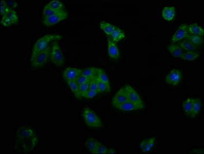
(Immunofluorescence staining of MCF-7 cells with AAA234210 at 1:133, counter-stained with DAPI. The cells were fixed in 4% formaldehyde, permeabilized using 0.2% Triton X-100 and blocked in 10% normal Goat Serum. The cells were then incubated with the antibody overnight at 4 degree C.The secondary antibody was Alexa Fluor 488-congugated AffiniPure Goat Anti-Rabbit IgG (H+L).)
IHC (Immunohistochemisry)
(Immunofluorescence staining of MCF-7 cells with AAA234210 at 1:133, counter-stained with DAPI. The cells were fixed in 4% formaldehyde, permeabilized using 0.2% Triton X-100 and blocked in 10% normal Goat Serum. The cells were then incubated with the antibody overnight at 4 degree C.The secondary antibody was Alexa Fluor 488-congugated AffiniPure Goat Anti-Rabbit IgG (H+L).)
OCLN, Polyclonal Antibody (Cat# AAA234210)

 IHC (Immunohistochemisry)
(Immunofluorescence staining of Hela cells with AAA234212 at 1:66, counter-stained with DAPI. The cells were fixed in 4% formaldehyde, permeabilized using 0.2% Triton X-100 and blocked in 10% normal Goat Serum. The cells were then incubated with the antibody overnight at 4 degree C.The secondary antibody was Alexa Fluor 488-congugated AffiniPure Goat Anti-Rabbit IgG (H+L).)
IHC (Immunohistochemisry)
(Immunofluorescence staining of Hela cells with AAA234212 at 1:66, counter-stained with DAPI. The cells were fixed in 4% formaldehyde, permeabilized using 0.2% Triton X-100 and blocked in 10% normal Goat Serum. The cells were then incubated with the antibody overnight at 4 degree C.The secondary antibody was Alexa Fluor 488-congugated AffiniPure Goat Anti-Rabbit IgG (H+L).)
PLXNB1, Polyclonal Antibody (Cat# AAA234212)
SELP, Polyclonal Antibody (Cat# AAA234213)
 IHC (Immunohiostchemistry)
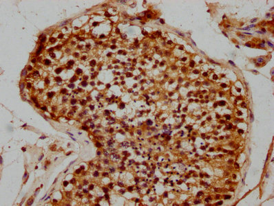
(IHC image of AAA234214 diluted at 1:300 and staining in paraffin-embedded human testis tissue performed on a Leica BondTM system. After dewaxing and hydration, antigen retrieval was mediated by high pressure in a citrate buffer (pH 6.0). Section was blocked with 10% normal goat serum 30min at RT. Then primary antibody (1% BSA) was incubated at 4 degree C overnight. The primary is detected by a biotinylated secondary antibody and visualized using an HRP conjugated SP system.)
IHC (Immunohiostchemistry)
(IHC image of AAA234214 diluted at 1:300 and staining in paraffin-embedded human testis tissue performed on a Leica BondTM system. After dewaxing and hydration, antigen retrieval was mediated by high pressure in a citrate buffer (pH 6.0). Section was blocked with 10% normal goat serum 30min at RT. Then primary antibody (1% BSA) was incubated at 4 degree C overnight. The primary is detected by a biotinylated secondary antibody and visualized using an HRP conjugated SP system.)
STAG3, Polyclonal Antibody (Cat# AAA234214)
 IHC (Immunohistochemisry)
(Immunofluorescence staining of PC3 cells with AAA234216 at 1:100, counter-stained with DAPI. The cells were fixed in 4% formaldehyde, permeabilized using 0.2% Triton X-100 and blocked in 10% normal Goat Serum. The cells were then incubated with the antibody overnight at 4 degree C.The secondary antibody was Alexa Fluor 488-congugated AffiniPure Goat Anti-Rabbit IgG (H+L).)
IHC (Immunohistochemisry)
(Immunofluorescence staining of PC3 cells with AAA234216 at 1:100, counter-stained with DAPI. The cells were fixed in 4% formaldehyde, permeabilized using 0.2% Triton X-100 and blocked in 10% normal Goat Serum. The cells were then incubated with the antibody overnight at 4 degree C.The secondary antibody was Alexa Fluor 488-congugated AffiniPure Goat Anti-Rabbit IgG (H+L).)
HEXA, Polyclonal Antibody (Cat# AAA234216)
 IF (Immunofluorescence)
(Immunofluorescence staining of U251 cells with AAA234217 at 1:166, counter-stained with DAPI. The cells were fixed in 4% formaldehyde, permeabilized using 0.2% Triton X-100 and blocked in 10% normal Goat Serum. The cells were then incubated with the antibody overnight at 4 degree C.The secondary antibody was Alexa Fluor 488-congugated AffiniPure Goat Anti-Rabbit IgG (H+L).)
IF (Immunofluorescence)
(Immunofluorescence staining of U251 cells with AAA234217 at 1:166, counter-stained with DAPI. The cells were fixed in 4% formaldehyde, permeabilized using 0.2% Triton X-100 and blocked in 10% normal Goat Serum. The cells were then incubated with the antibody overnight at 4 degree C.The secondary antibody was Alexa Fluor 488-congugated AffiniPure Goat Anti-Rabbit IgG (H+L).)
ABCC8, Polyclonal Antibody (Cat# AAA234217)
 IHC (Immunohistochemisry)
(Immunofluorescence staining of U251 cells with AAA234222 at 1:166, counter-stained with DAPI. The cells were fixed in 4% formaldehyde, permeabilized using 0.2% Triton X-100 and blocked in 10% normal Goat Serum. The cells were then incubated with the antibody overnight at 4 degree C.The secondary antibody was Alexa Fluor 488-congugated AffiniPure Goat Anti-Rabbit IgG (H+L).)
IHC (Immunohistochemisry)
(Immunofluorescence staining of U251 cells with AAA234222 at 1:166, counter-stained with DAPI. The cells were fixed in 4% formaldehyde, permeabilized using 0.2% Triton X-100 and blocked in 10% normal Goat Serum. The cells were then incubated with the antibody overnight at 4 degree C.The secondary antibody was Alexa Fluor 488-congugated AffiniPure Goat Anti-Rabbit IgG (H+L).)
GPR158, Polyclonal Antibody (Cat# AAA234222)
 IHC (Immunohiostchemistry)
(IHC image of AAA234228 diluted at 1:300 and staining in paraffin-embedded human breast cancer performed on a Leica BondTM system. After dewaxing and hydration, antigen retrieval was mediated by high pressure in a citrate buffer (pH 6.0). Section was blocked with 10% normal goat serum 30min at RT. Then primary antibody (1% BSA) was incubated at 4 degree C overnight. The primary is detected by a biotinylated secondary antibody and visualized using an HRP conjugated SP system.)
IHC (Immunohiostchemistry)
(IHC image of AAA234228 diluted at 1:300 and staining in paraffin-embedded human breast cancer performed on a Leica BondTM system. After dewaxing and hydration, antigen retrieval was mediated by high pressure in a citrate buffer (pH 6.0). Section was blocked with 10% normal goat serum 30min at RT. Then primary antibody (1% BSA) was incubated at 4 degree C overnight. The primary is detected by a biotinylated secondary antibody and visualized using an HRP conjugated SP system.)
FUT8, Polyclonal Antibody (Cat# AAA234228)
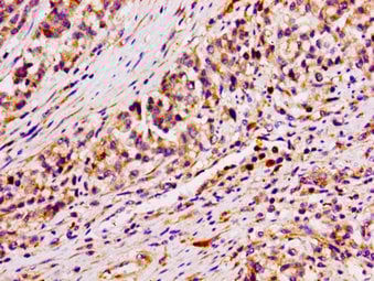
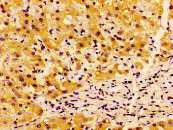
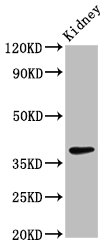
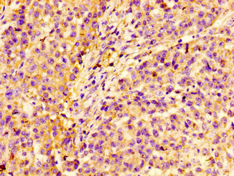
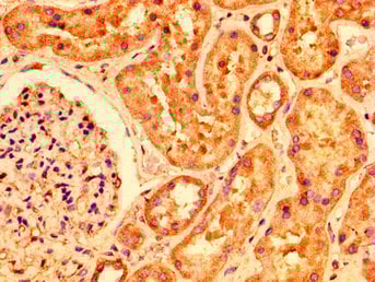
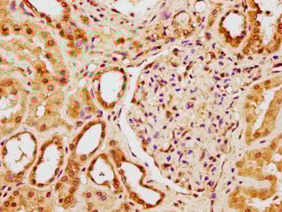
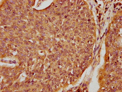
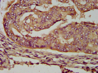
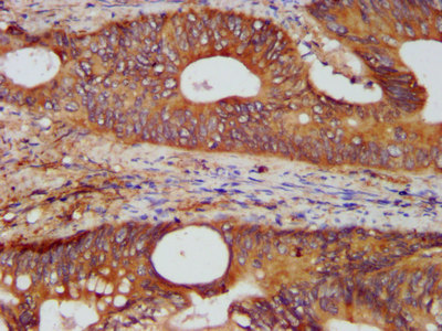
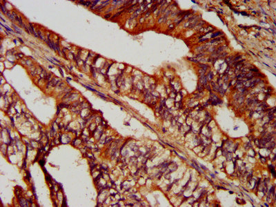
 IHC (Immunohiostchemistry)
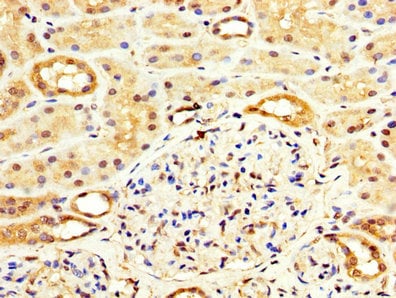
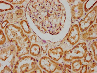
(IHC image of CSB-PA878945LA01HU diluted at 1:200 and staining in paraffin-embedded human kidney tissue performed on a Leica BondTM system. After dewaxing and hydration, antigen retrieval was mediated by high pressure in a citrate buffer (pH 6.0). Section was blocked with 10% normal goat serum 30min at RT. Then primary antibody (1% BSA) was incubated at 4 degree C overnight. The primary is detected by a biotinylated secondary antibody and visualized using an HRP conjugated SP system.)
IHC (Immunohiostchemistry)
(IHC image of CSB-PA878945LA01HU diluted at 1:200 and staining in paraffin-embedded human kidney tissue performed on a Leica BondTM system. After dewaxing and hydration, antigen retrieval was mediated by high pressure in a citrate buffer (pH 6.0). Section was blocked with 10% normal goat serum 30min at RT. Then primary antibody (1% BSA) was incubated at 4 degree C overnight. The primary is detected by a biotinylated secondary antibody and visualized using an HRP conjugated SP system.)
HACD3, Polyclonal Antibody (Cat# AAA235497)
Protein G Purified
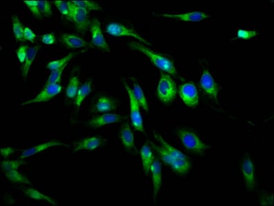
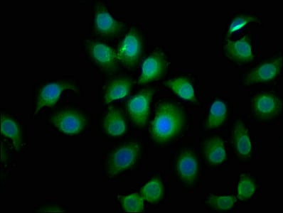
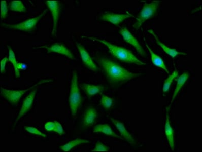
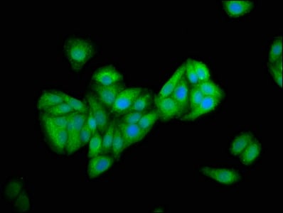
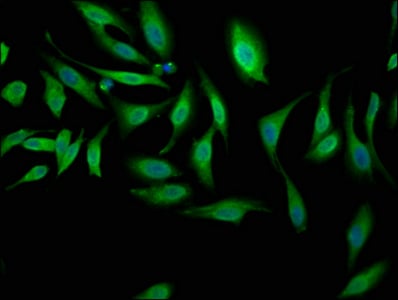
 IF (Immunofluorescence)
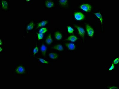
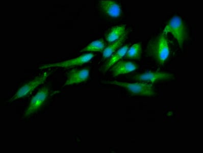
(Immunofluorescence staining of A549 cells with CSB-PA880107LA01HU at 1:166, counter-stained with DAPI. The cells were fixed in 4% formaldehyde, permeabilized using 0.2% Triton X-100 and blocked in 10% normal Goat Serum. The cells were then incubated with the antibody overnight at 4 degree C. The secondary antibody was Alexa Fluor 488-congugated AffiniPure Goat Anti-Rabbit IgG(H+L).)
IF (Immunofluorescence)
(Immunofluorescence staining of A549 cells with CSB-PA880107LA01HU at 1:166, counter-stained with DAPI. The cells were fixed in 4% formaldehyde, permeabilized using 0.2% Triton X-100 and blocked in 10% normal Goat Serum. The cells were then incubated with the antibody overnight at 4 degree C. The secondary antibody was Alexa Fluor 488-congugated AffiniPure Goat Anti-Rabbit IgG(H+L).)
EFHD1, Polyclonal Antibody (Cat# AAA235498)
Protein G Purified
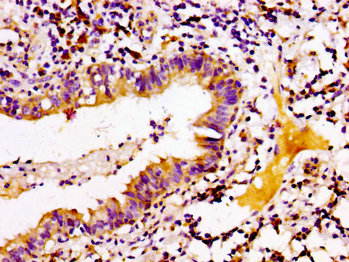
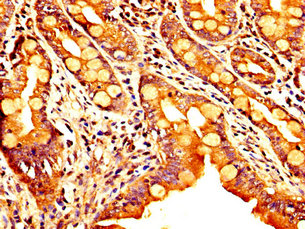
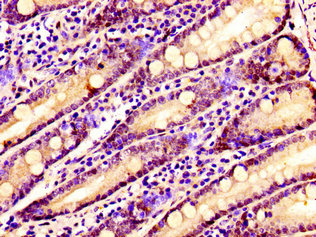
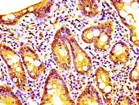
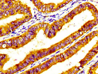
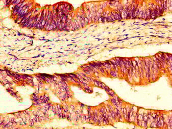
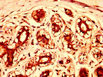
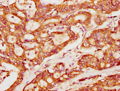
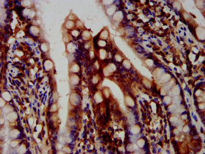
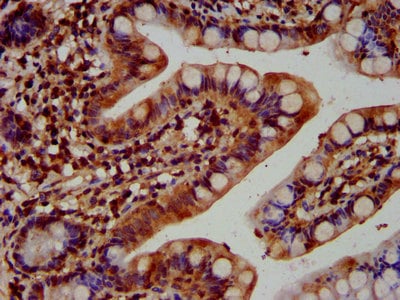
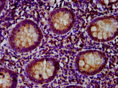
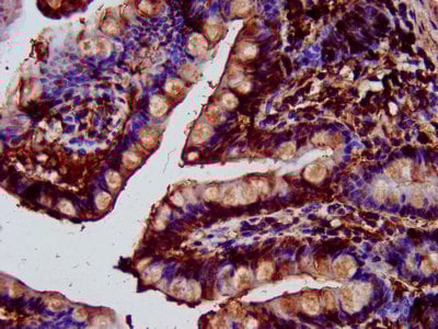
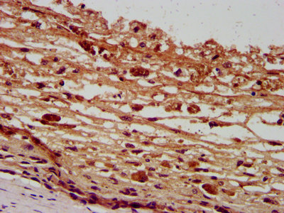
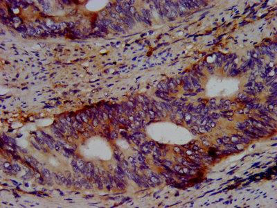
 IHC (Immunohiostchemistry)
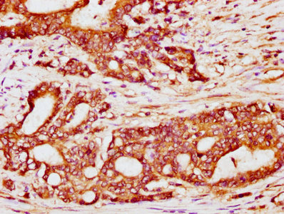
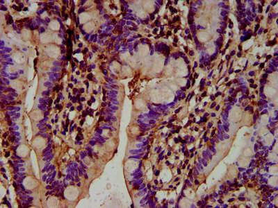
(IHC image of CSB-PA882156LA01HU diluted at 1:200 and staining in paraffin-embedded human small intestine tissue performed on a Leica BondTM system. After dewaxing and hydration, antigen retrieval was mediated by high pressure in a citrate buffer (pH 6.0). Section was blocked with 10% normal goat serum 30min at RT. Then primary antibody (1% BSA) was incubated at 4 degree C overnight. The primary is detected by a biotinylated secondary antibody and visualized using an HRP conjugated SP system.)
IHC (Immunohiostchemistry)
(IHC image of CSB-PA882156LA01HU diluted at 1:200 and staining in paraffin-embedded human small intestine tissue performed on a Leica BondTM system. After dewaxing and hydration, antigen retrieval was mediated by high pressure in a citrate buffer (pH 6.0). Section was blocked with 10% normal goat serum 30min at RT. Then primary antibody (1% BSA) was incubated at 4 degree C overnight. The primary is detected by a biotinylated secondary antibody and visualized using an HRP conjugated SP system.)
COQ3, Polyclonal Antibody (Cat# AAA235500)
Protein G Purified
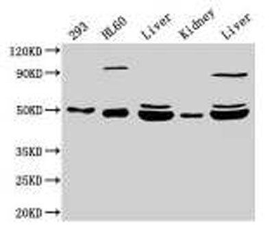
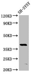
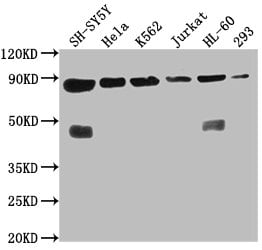 Application Data
(Western BlotPositive WB detected in: SH-SY5Y whole cell lysate, Hela whole cell lysate, K562 whole cell lysate, Jurkat whole cell lysate, HL60 whole cell lysate, 293 whole cell lysateAll lanes: YY1AP1 antibody at 1:2000SecondaryGoat polyclonal to rabbit IgG at 1/50000 dilutionPredicted band size: 88, 84, 82, 81, 76, 80, 51, 96, 98 kDaObserved band size: 88 kDa)
Application Data
(Western BlotPositive WB detected in: SH-SY5Y whole cell lysate, Hela whole cell lysate, K562 whole cell lysate, Jurkat whole cell lysate, HL60 whole cell lysate, 293 whole cell lysateAll lanes: YY1AP1 antibody at 1:2000SecondaryGoat polyclonal to rabbit IgG at 1/50000 dilutionPredicted band size: 88, 84, 82, 81, 76, 80, 51, 96, 98 kDaObserved band size: 88 kDa)
YY1AP1, Polyclonal Antibody (Cat# AAA235504)
Protein G Purified


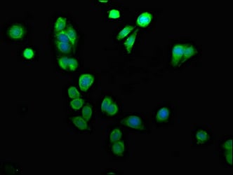
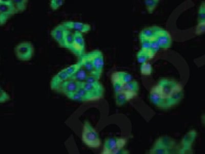
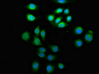
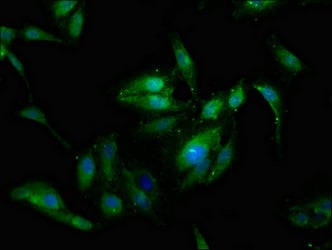
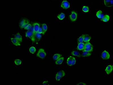
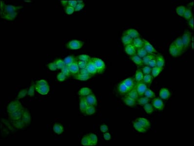
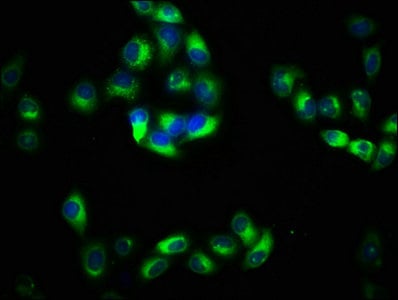
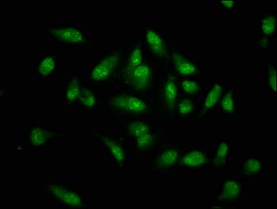
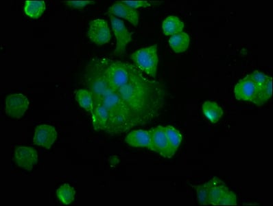
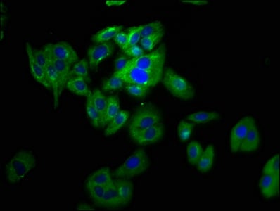
 IF (Immunofluorescence)
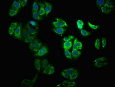
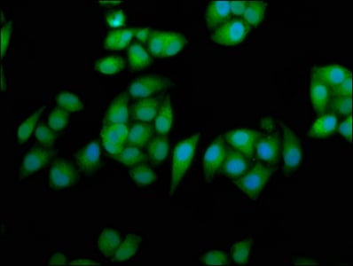
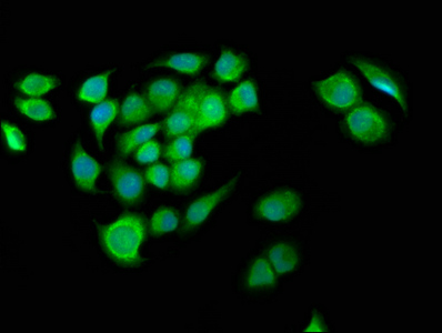
(Immunofluorescence staining of A549 cells with CSB-PA884627LA01HU at 1:50, counter-stained with DAPI. The cells were fixed in 4% formaldehyde, permeabilized using 0.2% Triton X-100 and blocked in 10% normal Goat Serum. The cells were then incubated with the antibody overnight at 4 degree C. The secondary antibody was Alexa Fluor 488-congugated AffiniPure Goat Anti-Rabbit IgG(H+L).)
IF (Immunofluorescence)
(Immunofluorescence staining of A549 cells with CSB-PA884627LA01HU at 1:50, counter-stained with DAPI. The cells were fixed in 4% formaldehyde, permeabilized using 0.2% Triton X-100 and blocked in 10% normal Goat Serum. The cells were then incubated with the antibody overnight at 4 degree C. The secondary antibody was Alexa Fluor 488-congugated AffiniPure Goat Anti-Rabbit IgG(H+L).)
BCL2L10, Polyclonal Antibody (Cat# AAA235505)
Protein G Purified
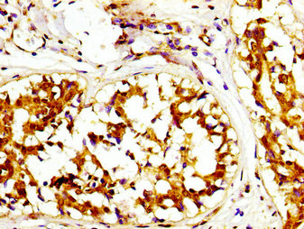
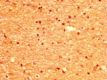
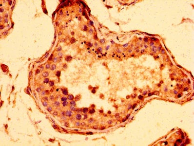
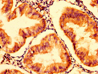
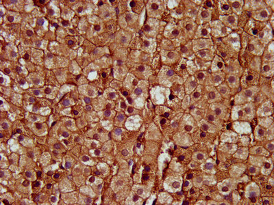
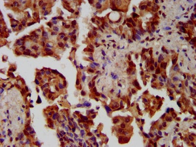
 IHC (Immunohiostchemistry)
(IHC image of CSB-PA885781LA01HU diluted at 1:300 and staining in paraffin-embedded human testis tissue performed on a Leica BondTM system. After dewaxing and hydration, antigen retrieval was mediated by high pressure in a citrate buffer (pH 6.0). Section was blocked with 10% normal goat serum 30min at RT. Then primary antibody (1% BSA) was incubated at 4 degree C overnight. The primary is detected by a biotinylated secondary antibody and visualized using an HRP conjugated SP system.)
IHC (Immunohiostchemistry)
(IHC image of CSB-PA885781LA01HU diluted at 1:300 and staining in paraffin-embedded human testis tissue performed on a Leica BondTM system. After dewaxing and hydration, antigen retrieval was mediated by high pressure in a citrate buffer (pH 6.0). Section was blocked with 10% normal goat serum 30min at RT. Then primary antibody (1% BSA) was incubated at 4 degree C overnight. The primary is detected by a biotinylated secondary antibody and visualized using an HRP conjugated SP system.)
ZIM2, Polyclonal Antibody (Cat# AAA235508)
Protein G Purified
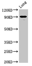
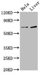
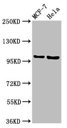
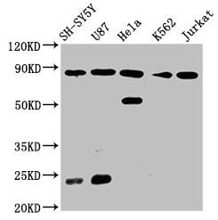
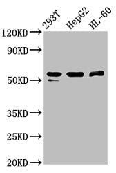
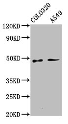
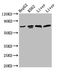
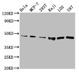
 WB (Western Blot)
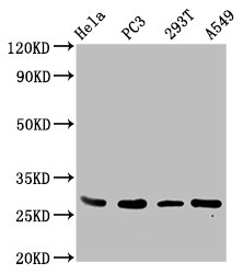
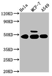
(Western BlotPositive WB detected in: Hela whole cell lysate, MCF-7 whole cell lysate, A549 whole cell lysateAll lanes: CSNK1G1 antibody at 3.4ug/mlSecondaryGoat polyclonal to rabbit IgG at 1/50000 dilutionPredicted band size: 49, 46 kDaObserved band size: 49 kDa)
WB (Western Blot)
(Western BlotPositive WB detected in: Hela whole cell lysate, MCF-7 whole cell lysate, A549 whole cell lysateAll lanes: CSNK1G1 antibody at 3.4ug/mlSecondaryGoat polyclonal to rabbit IgG at 1/50000 dilutionPredicted band size: 49, 46 kDaObserved band size: 49 kDa)
CSNK1G1, Polyclonal Antibody (Cat# AAA235510)
Protein G Purified
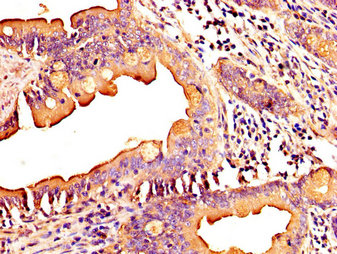
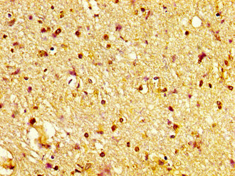
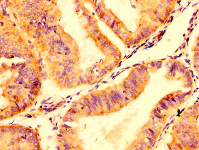
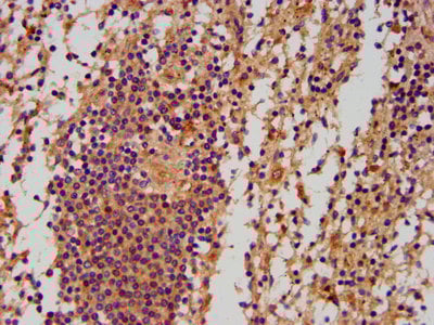
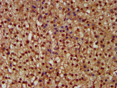
 IHC (Immunohiostchemistry)
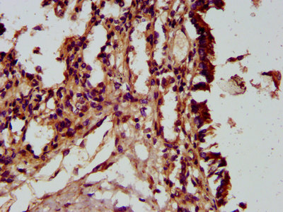
(IHC image of CSB-PA889077LA01HU diluted at 1:100 and staining in paraffin-embedded human tonsil tissue performed on a Leica BondTM system. After dewaxing and hydration, antigen retrieval was mediated by high pressure in a citrate buffer (pH 6.0). Section was blocked with 10% normal goat serum 30min at RT. Then primary antibody (1% BSA) was incubated at 4 degree C overnight. The primary is detected by a biotinylated secondary antibody and visualized using an HRP conjugated SP system.)
IHC (Immunohiostchemistry)
(IHC image of CSB-PA889077LA01HU diluted at 1:100 and staining in paraffin-embedded human tonsil tissue performed on a Leica BondTM system. After dewaxing and hydration, antigen retrieval was mediated by high pressure in a citrate buffer (pH 6.0). Section was blocked with 10% normal goat serum 30min at RT. Then primary antibody (1% BSA) was incubated at 4 degree C overnight. The primary is detected by a biotinylated secondary antibody and visualized using an HRP conjugated SP system.)
SPINK5, Polyclonal Antibody (Cat# AAA235511)
Protein G Purified
 IP (Immunoprecipitation)
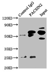
(Immunoprecipitating PACSIN2 in HepG2 whole cell lysateLane 1: Rabbit control IgG instead of AAA235513 in HepG2 whole cell lysate. For western blotting, a HRP-conjugated Protein G antibody was used as the secondary antibody (1/2000)Lane 2: AAA235513 (6ug) + HepG2 whole cell lysate (500ug)Lane 3: HepG2 whole cell lysate (20ug))
IP (Immunoprecipitation)
(Immunoprecipitating PACSIN2 in HepG2 whole cell lysateLane 1: Rabbit control IgG instead of AAA235513 in HepG2 whole cell lysate. For western blotting, a HRP-conjugated Protein G antibody was used as the secondary antibody (1/2000)Lane 2: AAA235513 (6ug) + HepG2 whole cell lysate (500ug)Lane 3: HepG2 whole cell lysate (20ug))
PACSIN2, Polyclonal Antibody (Cat# AAA235513)
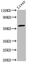
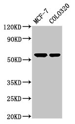
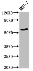
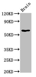
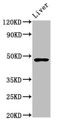
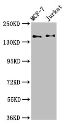
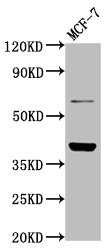
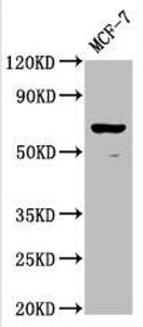 WB (Western Blot)
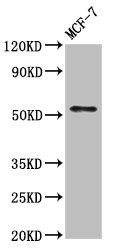
(Western BlotPositive WB detected in: MCF-7 whole cell lysateAll lanes: RPE65 antibody at 1:2000SecondaryGoat polyclonal to rabbit IgG at 1/50000 dilutionPredicted band size: 61 kDaObserved band size: 61 kDa)
WB (Western Blot)
(Western BlotPositive WB detected in: MCF-7 whole cell lysateAll lanes: RPE65 antibody at 1:2000SecondaryGoat polyclonal to rabbit IgG at 1/50000 dilutionPredicted band size: 61 kDaObserved band size: 61 kDa)
RPE65, Polyclonal Antibody (Cat# AAA234638)
RPE65, Polyclonal Antibody (Cat# AAA234639)
PAG2, Polyclonal Antibody (Cat# AAA234642)
 IHC (Immunohistochemisry)
(IHC image of AAA234643 diluted at 1:500 and staining in paraffin-embedded human endometrial cancer performed on a Leica BondTM system. After dewaxing and hydration, antigen retrieval was mediated by high pressure in a citrate buffer (pH 6.0). Section was blocked with 10% normal goat serum 30min at RT. Then primary antibody (1% BSA) was incubated at 4 degree C overnight. The primary is detected by a biotinylated secondary antibody and visualized using an HRP conjugated SP system.)
IHC (Immunohistochemisry)
(IHC image of AAA234643 diluted at 1:500 and staining in paraffin-embedded human endometrial cancer performed on a Leica BondTM system. After dewaxing and hydration, antigen retrieval was mediated by high pressure in a citrate buffer (pH 6.0). Section was blocked with 10% normal goat serum 30min at RT. Then primary antibody (1% BSA) was incubated at 4 degree C overnight. The primary is detected by a biotinylated secondary antibody and visualized using an HRP conjugated SP system.)
NADK2, Polyclonal Antibody (Cat# AAA234643)
 IHC (Immunohistochemisry)
(Immunofluorescence staining of Hela cells with AAA234644 at 1:100, counter-stained with DAPI. The cells were fixed in 4% formaldehyde, permeabilized using 0.2% Triton X-100 and blocked in 10% normal Goat Serum. The cells were then incubated with the antibody overnight at 4 degree C.The secondary antibody was Alexa Fluor 488-congugated AffiniPure Goat Anti-Rabbit IgG (H+L).)
IHC (Immunohistochemisry)
(Immunofluorescence staining of Hela cells with AAA234644 at 1:100, counter-stained with DAPI. The cells were fixed in 4% formaldehyde, permeabilized using 0.2% Triton X-100 and blocked in 10% normal Goat Serum. The cells were then incubated with the antibody overnight at 4 degree C.The secondary antibody was Alexa Fluor 488-congugated AffiniPure Goat Anti-Rabbit IgG (H+L).)
HECTD2, Polyclonal Antibody (Cat# AAA234644)
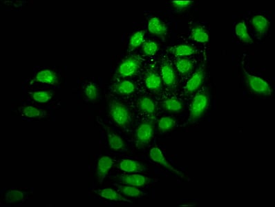
 IF (Immunofluorescence)
(Immunofluorescence staining of A549 cells with AAA234645 at 1:133, counter-stained with DAPI. The cells were fixed in 4% formaldehyde, permeabilized using 0.2% Triton X-100 and blocked in 10% normal Goat Serum. The cells were then incubated with the antibody overnight at 4 degree C.The secondary antibody was Alexa Fluor 488-congugated AffiniPure Goat Anti-Rabbit IgG (H+L).)
IF (Immunofluorescence)
(Immunofluorescence staining of A549 cells with AAA234645 at 1:133, counter-stained with DAPI. The cells were fixed in 4% formaldehyde, permeabilized using 0.2% Triton X-100 and blocked in 10% normal Goat Serum. The cells were then incubated with the antibody overnight at 4 degree C.The secondary antibody was Alexa Fluor 488-congugated AffiniPure Goat Anti-Rabbit IgG (H+L).)
G2E3, Polyclonal Antibody (Cat# AAA234645)
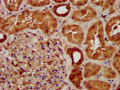
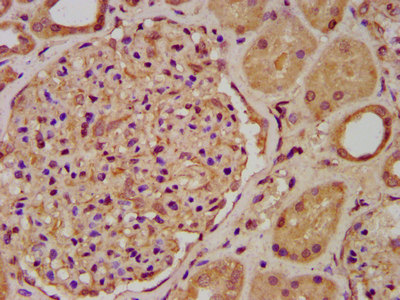 IHC (Immunohiostchemistry)
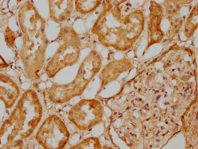
(IHC image of AAA234646 diluted at 1:200 and staining in paraffin-embedded human kidney tissue performed on a Leica BondTM system. After dewaxing and hydration, antigen retrieval was mediated by high pressure in a citrate buffer (pH 6.0). Section was blocked with 10% normal goat serum 30min at RT. Then primary antibody (1% BSA) was incubated at 4 degree C overnight. The primary is detected by a biotinylated secondary antibody and visualized using an HRP conjugated SP system.)
IHC (Immunohiostchemistry)
(IHC image of AAA234646 diluted at 1:200 and staining in paraffin-embedded human kidney tissue performed on a Leica BondTM system. After dewaxing and hydration, antigen retrieval was mediated by high pressure in a citrate buffer (pH 6.0). Section was blocked with 10% normal goat serum 30min at RT. Then primary antibody (1% BSA) was incubated at 4 degree C overnight. The primary is detected by a biotinylated secondary antibody and visualized using an HRP conjugated SP system.)
ENOSF1, Polyclonal Antibody (Cat# AAA234646)
 IHC (Immunohistochemisry)
(Immunofluorescence staining of Hela cells with AAA234647 at 1:133, counter-stained with DAPI. The cells were fixed in 4% formaldehyde, permeabilized using 0.2% Triton X-100 and blocked in 10% normal Goat Serum. The cells were then incubated with the antibody overnight at 4 degree C.The secondary antibody was Alexa Fluor 488-congugated AffiniPure Goat Anti-Rabbit IgG (H+L).)
IHC (Immunohistochemisry)
(Immunofluorescence staining of Hela cells with AAA234647 at 1:133, counter-stained with DAPI. The cells were fixed in 4% formaldehyde, permeabilized using 0.2% Triton X-100 and blocked in 10% normal Goat Serum. The cells were then incubated with the antibody overnight at 4 degree C.The secondary antibody was Alexa Fluor 488-congugated AffiniPure Goat Anti-Rabbit IgG (H+L).)
OTUD7B, Polyclonal Antibody (Cat# AAA234647)
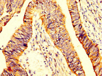
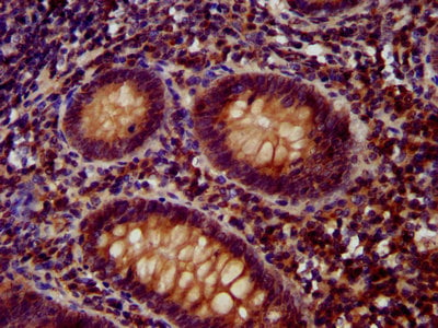
 IHC (Immunohistochemisry)
(IHC image of AAA234652 diluted at 1:400 and staining in paraffin-embedded human small intestine tissue performed on a Leica BondTM system. After dewaxing and hydration, antigen retrieval was mediated by high pressure in a citrate buffer (pH 6.0). Section was blocked with 10% normal goat serum 30min at RT. Then primary antibody (1% BSA) was incubated at 4 degree C overnight. The primary is detected by a biotinylated secondary antibody and visualized using an HRP conjugated SP system.)
IHC (Immunohistochemisry)
(IHC image of AAA234652 diluted at 1:400 and staining in paraffin-embedded human small intestine tissue performed on a Leica BondTM system. After dewaxing and hydration, antigen retrieval was mediated by high pressure in a citrate buffer (pH 6.0). Section was blocked with 10% normal goat serum 30min at RT. Then primary antibody (1% BSA) was incubated at 4 degree C overnight. The primary is detected by a biotinylated secondary antibody and visualized using an HRP conjugated SP system.)
KTN1, Polyclonal Antibody (Cat# AAA234652)
 IF (Immunofluorescence)
(Immunofluorescence staining of HepG2 cells with AAA234654 at 1:166, counter-stained with DAPI. The cells were fixed in 4% formaldehyde, permeabilized using 0.2% Triton X-100 and blocked in 10% normal Goat Serum. The cells were then incubated with the antibody overnight at 4 degree C.The secondary antibody was Alexa Fluor 488-congugated AffiniPure Goat Anti-Rabbit IgG (H+L).)
IF (Immunofluorescence)
(Immunofluorescence staining of HepG2 cells with AAA234654 at 1:166, counter-stained with DAPI. The cells were fixed in 4% formaldehyde, permeabilized using 0.2% Triton X-100 and blocked in 10% normal Goat Serum. The cells were then incubated with the antibody overnight at 4 degree C.The secondary antibody was Alexa Fluor 488-congugated AffiniPure Goat Anti-Rabbit IgG (H+L).)
PNPLA5, Polyclonal Antibody (Cat# AAA234654)
 IF (Immunofluorescence)
(Immunofluorescence staining of SH-SY5Y cells with AAA234655 at 1:200, counter-stained with DAPI. The cells were fixed in 4% formaldehyde, permeabilized using 0.2% Triton X-100 and blocked in 10% normal Goat Serum. The cells were then incubated with the antibody overnight at 4 degree C.The secondary antibody was Alexa Fluor 488-congugated AffiniPure Goat Anti-Rabbit IgG (H+L).)
IF (Immunofluorescence)
(Immunofluorescence staining of SH-SY5Y cells with AAA234655 at 1:200, counter-stained with DAPI. The cells were fixed in 4% formaldehyde, permeabilized using 0.2% Triton X-100 and blocked in 10% normal Goat Serum. The cells were then incubated with the antibody overnight at 4 degree C.The secondary antibody was Alexa Fluor 488-congugated AffiniPure Goat Anti-Rabbit IgG (H+L).)
LAPTM4B, Polyclonal Antibody (Cat# AAA234655)
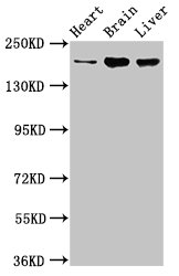
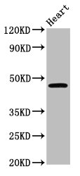 WB (Western Blot)
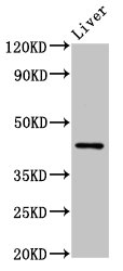
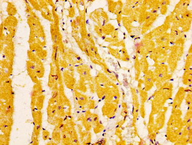
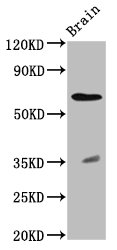
(Western BlotPositive WB detected in: Mouse heart tissueAll lanes: CERS3 antibody at 3.6ug/mlSecondaryGoat polyclonal to rabbit IgG at 1/50000 dilutionPredicted band size: 47 KDaObserved band size: 47 KDa)
WB (Western Blot)
(Western BlotPositive WB detected in: Mouse heart tissueAll lanes: CERS3 antibody at 3.6ug/mlSecondaryGoat polyclonal to rabbit IgG at 1/50000 dilutionPredicted band size: 47 KDaObserved band size: 47 KDa)
CERS3, Polyclonal Antibody (Cat# AAA234658)
 IHC (Immunohistochemisry)
(Immunofluorescence staining of A549 cells with AAA234659 at 1:133, counter-stained with DAPI. The cells were fixed in 4% formaldehyde, permeabilized using 0.2% Triton X-100 and blocked in 10% normal Goat Serum. The cells were then incubated with the antibody overnight at 4 degree C.The secondary antibody was Alexa Fluor 488-congugated AffiniPure Goat Anti-Rabbit IgG (H+L).)
IHC (Immunohistochemisry)
(Immunofluorescence staining of A549 cells with AAA234659 at 1:133, counter-stained with DAPI. The cells were fixed in 4% formaldehyde, permeabilized using 0.2% Triton X-100 and blocked in 10% normal Goat Serum. The cells were then incubated with the antibody overnight at 4 degree C.The secondary antibody was Alexa Fluor 488-congugated AffiniPure Goat Anti-Rabbit IgG (H+L).)
AHNAK2, Polyclonal Antibody (Cat# AAA234659)
 IHC (Immunohistochemisry)
(Immunofluorescence staining of Hela cells with AAA234662 at 1:133, counter-stained with DAPI. The cells were fixed in 4% formaldehyde, permeabilized using 0.2% Triton X-100 and blocked in 10% normal Goat Serum. The cells were then incubated with the antibody overnight at 4 degree C.The secondary antibody was Alexa Fluor 488-congugated AffiniPure Goat Anti-Rabbit IgG (H+L).)
IHC (Immunohistochemisry)
(Immunofluorescence staining of Hela cells with AAA234662 at 1:133, counter-stained with DAPI. The cells were fixed in 4% formaldehyde, permeabilized using 0.2% Triton X-100 and blocked in 10% normal Goat Serum. The cells were then incubated with the antibody overnight at 4 degree C.The secondary antibody was Alexa Fluor 488-congugated AffiniPure Goat Anti-Rabbit IgG (H+L).)
MKX, Polyclonal Antibody (Cat# AAA234662)
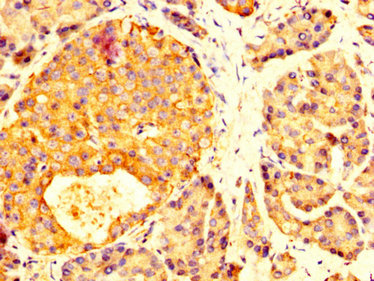
 IHC (Immunohistochemisry)
(Immunofluorescence staining of MCF-7 cells with AAA234666 at 1:133, counter-stained with DAPI. The cells were fixed in 4% formaldehyde, permeabilized using 0.2% Triton X-100 and blocked in 10% normal Goat Serum. The cells were then incubated with the antibody overnight at 4 degree C.The secondary antibody was Alexa Fluor 488-congugated AffiniPure Goat Anti-Rabbit IgG (H+L).)
IHC (Immunohistochemisry)
(Immunofluorescence staining of MCF-7 cells with AAA234666 at 1:133, counter-stained with DAPI. The cells were fixed in 4% formaldehyde, permeabilized using 0.2% Triton X-100 and blocked in 10% normal Goat Serum. The cells were then incubated with the antibody overnight at 4 degree C.The secondary antibody was Alexa Fluor 488-congugated AffiniPure Goat Anti-Rabbit IgG (H+L).)
SAT2, Polyclonal Antibody (Cat# AAA234666)
 IHC (Immunohistochemisry)
(Immunofluorescence staining of Hela cells with AAA234668 at 1:100, counter-stained with DAPI. The cells were fixed in 4% formaldehyde, permeabilized using 0.2% Triton X-100 and blocked in 10% normal Goat Serum. The cells were then incubated with the antibody overnight at 4 degree C.The secondary antibody was Alexa Fluor 488-congugated AffiniPure Goat Anti-Rabbit IgG (H+L).)
IHC (Immunohistochemisry)
(Immunofluorescence staining of Hela cells with AAA234668 at 1:100, counter-stained with DAPI. The cells were fixed in 4% formaldehyde, permeabilized using 0.2% Triton X-100 and blocked in 10% normal Goat Serum. The cells were then incubated with the antibody overnight at 4 degree C.The secondary antibody was Alexa Fluor 488-congugated AffiniPure Goat Anti-Rabbit IgG (H+L).)
NEK1, Polyclonal Antibody (Cat# AAA234668)
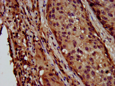
 IHC (Immunohistochemisry)
(Immunofluorescence staining of HepG2 cells with AAA234670 at 1:133, counter-stained with DAPI. The cells were fixed in 4% formaldehyde, permeabilized using 0.2% Triton X-100 and blocked in 10% normal Goat Serum. The cells were then incubated with the antibody overnight at 4 degree C.The secondary antibody was Alexa Fluor 488-congugated AffiniPure Goat Anti-Rabbit IgG (H+L).)
IHC (Immunohistochemisry)
(Immunofluorescence staining of HepG2 cells with AAA234670 at 1:133, counter-stained with DAPI. The cells were fixed in 4% formaldehyde, permeabilized using 0.2% Triton X-100 and blocked in 10% normal Goat Serum. The cells were then incubated with the antibody overnight at 4 degree C.The secondary antibody was Alexa Fluor 488-congugated AffiniPure Goat Anti-Rabbit IgG (H+L).)
PANX2, Polyclonal Antibody (Cat# AAA234670)
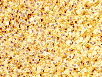
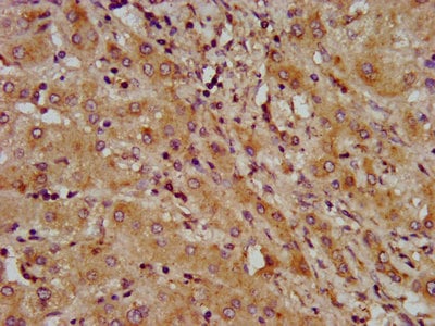
 IHC (Immunohistochemisry)
(IHC image of AAA234675 diluted at 1:300 and staining in paraffin-embedded human kidney tissue performed on a Leica BondTM system. After dewaxing and hydration, antigen retrieval was mediated by high pressure in a citrate buffer (pH 6.0). Section was blocked with 10% normal goat serum 30min at RT. Then primary antibody (1% BSA) was incubated at 4 degree C overnight. The primary is detected by a biotinylated secondary antibody and visualized using an HRP conjugated SP system.)
IHC (Immunohistochemisry)
(IHC image of AAA234675 diluted at 1:300 and staining in paraffin-embedded human kidney tissue performed on a Leica BondTM system. After dewaxing and hydration, antigen retrieval was mediated by high pressure in a citrate buffer (pH 6.0). Section was blocked with 10% normal goat serum 30min at RT. Then primary antibody (1% BSA) was incubated at 4 degree C overnight. The primary is detected by a biotinylated secondary antibody and visualized using an HRP conjugated SP system.)
SUCLG2, Polyclonal Antibody (Cat# AAA234675)
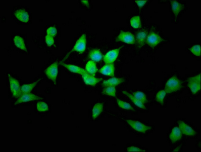
 IF (Immunofluorescence)
(Immunofluorescence staining of HepG2 cells with AAA234679 at 1:166, counter-stained with DAPI. The cells were fixed in 4% formaldehyde, permeabilized using 0.2% Triton X-100 and blocked in 10% normal Goat Serum. The cells were then incubated with the antibody overnight at 4 degree C.The secondary antibody was Alexa Fluor 488-congugated AffiniPure Goat Anti-Rabbit IgG (H+L).)
IF (Immunofluorescence)
(Immunofluorescence staining of HepG2 cells with AAA234679 at 1:166, counter-stained with DAPI. The cells were fixed in 4% formaldehyde, permeabilized using 0.2% Triton X-100 and blocked in 10% normal Goat Serum. The cells were then incubated with the antibody overnight at 4 degree C.The secondary antibody was Alexa Fluor 488-congugated AffiniPure Goat Anti-Rabbit IgG (H+L).)
KCNV2, Polyclonal Antibody (Cat# AAA234679)
What are Polyclonal Antibodies?
Polyclonal antibodies are antibodies that come from multiple B cell clones of a host animal. The typical hosts used for the majority of polyclonal antibody production are rabbits, goats, sheep, and donkeys. These polyclonal antibodies, once having identified their target, will bind to different epitopes located at different regions or sequences on the same protein/antigen. As a result, they are ideal at locating and binding to the target, even if the target is in very low concentrations (due to many different antibodies being able to bind to the same target molecule, which allows for significant amplification of a downstream signal).
Polyclonal antibodies are typically produced by injecting an antigen into a host animal, which causes the animal’s immune system to attack the foreign antigen by mass generating antibodies against it. After a period of time, serum is collected from the animal and purified using physicochemical fractionation, class-specific affinity purification, and/or antigen-affinity purification.
Key Uses of Polyclonal Antibodies
- Western Blotting: This method is used to find specific proteins in biological samples after separating them by size.
- Immunohistochemistry: IHC helps visualize the location of proteins in tissue sections using various staining techniques.
- ELISA: (Enzyme-Linked Immunosorbent Assay) is typically used to identify specific protein quantities in a sample. ELISAs can be either “Quantitative” or “Qualitative”.
- Flow Cytometry: technique that identifies and measures the specific protein on the surface or inside the cells in a fluid suspension.
- Immunoprecipitation: IP isolates and studies a specific protein from a complex mixture using antibodies.
Why Buy Polyclonal Antibodies from AAA Biotech?
1. Ideal for Various Applications
Our antibodies are generally going to be validated for use in multiple types of assays, including ELISA, Western Blotting, Immunohistochemistry, Immunoprecipitation, amongst others. They are ideal for a wide range of research applications.
2. Rigorous Quality Control
All of the antibodies in our catalog undergo strict quality testing to ensure specificity, sensitivity, and consistent performance. We are confident in the ability of our antibodies to provide you with accurate results.
3. Wide Assortment of Antibodies
Antibodies in are catalog can be found for both common and exotic species, and these antibodies are also available in both conjugated and recombinant forms to suit many diverse experimental needs.
4. Highly Purified
Our antibodies are available in purified forms with over 85% purity, as confirmed by SDS-PAGE. They are also available with tags such as His, Flag, GST, or MBP. We cater to customers worldwide.
FAQ
1. How are polyclonal antibodies produced?
Traditionally, polyclonal antibodies are produced by injecting an antigen into a host animal (such as a rabbit or goat), which then triggers an immune response from the host animal. The animal’s B cells produce antibodies that will recognize different parts of the injected antigen. These antibodies are then collected from the animal’s blood and purified for use.
2. How do polyclonal antibodies differ from monoclonal antibodies?
Polyclonal antibodies are a mix of antibodies that bind to different locations (epitopes) of the same antigen, while monoclonal antibodies are identical and bind to just one specific epitope. This makes polyclonal antibodies more versatile and better at detecting proteins that may be present in low quantities or in altered/modified forms.
3. How should I store polyclonal antibodies?
Polyclonal antibodies should be stored at 4°C for short-term use (up to a few weeks) and at -20°C or -80°C for long-term storage. Avoid repeated freeze-thaw cycles by dividing them into small aliquots. Always check the datasheet for specific storage instructions.