Filters
▼Clonality
▼Type
▼Reactivity
▼Gene Name
▼Isotype
▼Host
▼Application
▼Clone
▼Polyclonal Antibodies
At AAA Biotech also known as AAA Bio or AAABio, we provide a broad range of purified polyclonal antibodies (pAbs) that are able to all be browsed online through our website. Due to their high specificity and strong binding affinity, these antibodies are ideal for wide swathes of research and experimental applications.
Our polyclonal antibodies can easily support your work, whether you use them for Western Blotting, Immunocytochemistry (with or without Immunofluorescence used in conjunction), Immunohistochemistry, Immunoprecipitation, and ELISA tests. We highly encourage you to browse our range of pAbs and choose the one that best suits your experimental model.
Viewing 400-450 of 96805 product results
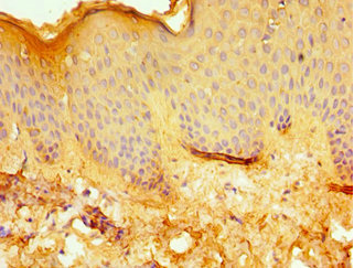

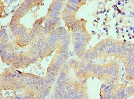
 IHC (Immunohistochemisry)
(Immunohistochemistry of paraffin-embedded human lung cancer using AAA119529 at dilution 1:100)
IHC (Immunohistochemisry)
(Immunohistochemistry of paraffin-embedded human lung cancer using AAA119529 at dilution 1:100)
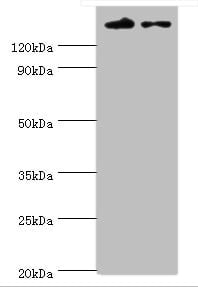
TFIIH basal transcription factor complex helicase XPD, Polyclonal Antibody (Cat# AAA119529)
 IHC (Immunohistochemisry)
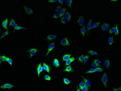
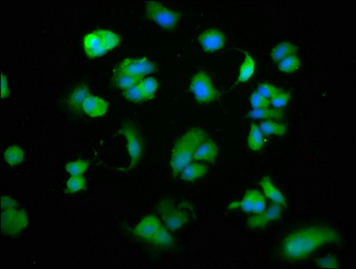
(Immunofluorescent analysis of HepG2 cells using AAA119530 at a dilution of 1:100 and Alexa Fluor 488-congugated AffiniPure Goat Anti-Rabbit IgG(H+L))
IHC (Immunohistochemisry)
(Immunofluorescent analysis of HepG2 cells using AAA119530 at a dilution of 1:100 and Alexa Fluor 488-congugated AffiniPure Goat Anti-Rabbit IgG(H+L))
SDHC, Polyclonal Antibody (Cat# AAA119530)
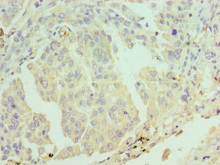
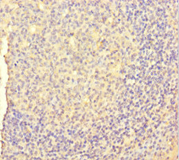
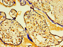
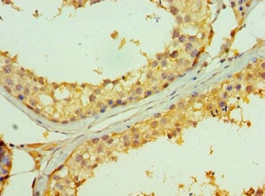
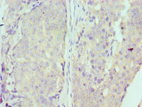
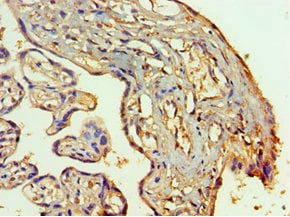
 IHC (Immunohiostchemistry)
(Immunohistochemistry of paraffin-embedded human pancreatic tissue using AAA119531 at dilution of 1:100)
IHC (Immunohiostchemistry)
(Immunohistochemistry of paraffin-embedded human pancreatic tissue using AAA119531 at dilution of 1:100)
TBCA, Polyclonal Antibody (Cat# AAA119531)
Peptide deformylase, Polyclonal Antibody (Cat# AAA119535)
Mycoplasma hyopneumoniae 46 kDa surface antigen, Polyclonal Antibody (Cat# AAA119537)
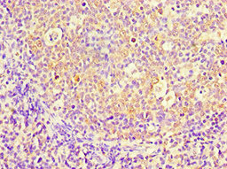
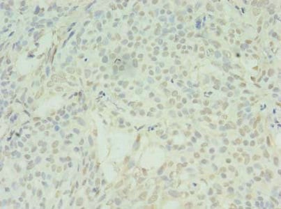
 IHC (Immunohistochemistry)
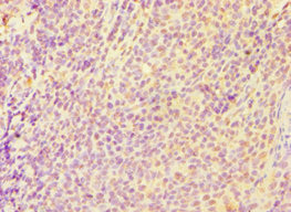
(Immunohistochemistry of paraffin-embedded human thymus using AAA119539 at dilution 1:100)
IHC (Immunohistochemistry)
(Immunohistochemistry of paraffin-embedded human thymus using AAA119539 at dilution 1:100)
PBK, Polyclonal Antibody (Cat# AAA119539)
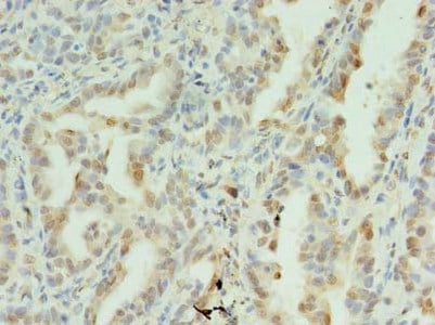
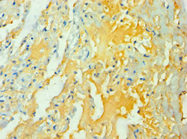
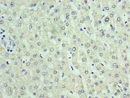
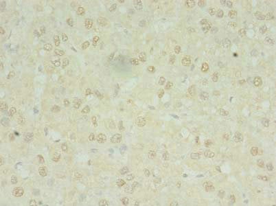
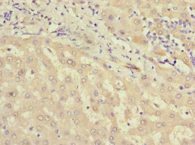
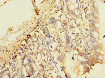
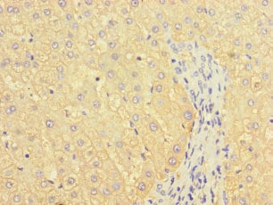 IHC (Immunohiostchemistry)
(Immunohistochemistry of paraffin-embedded human liver tissue using AAA119541 at dilution of 1:100)
IHC (Immunohiostchemistry)
(Immunohistochemistry of paraffin-embedded human liver tissue using AAA119541 at dilution of 1:100)
B3GALT2, Polyclonal Antibody (Cat# AAA119541)
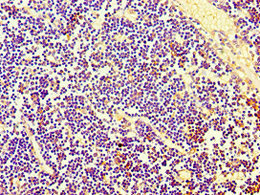
 IHC (Immunohiostchemistry)
(Immunohistochemistry of paraffin-embedded human tonsil using AAA119542 at dilution 1:100)
IHC (Immunohiostchemistry)
(Immunohistochemistry of paraffin-embedded human tonsil using AAA119542 at dilution 1:100)
EIF2AK4, Polyclonal Antibody (Cat# AAA119542)
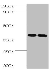
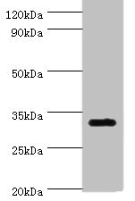
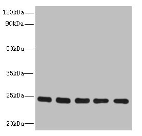
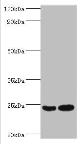
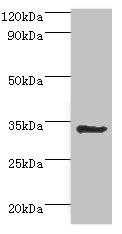
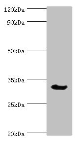
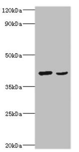
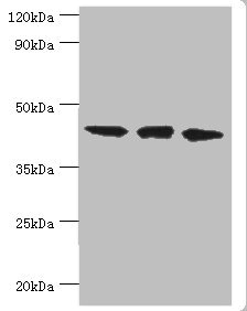
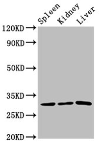
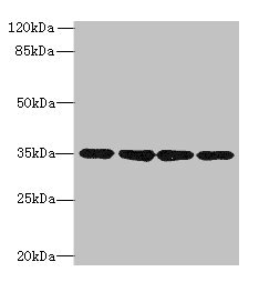
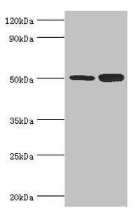
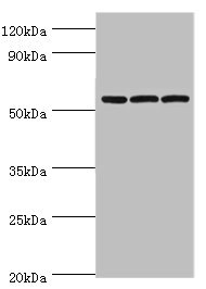
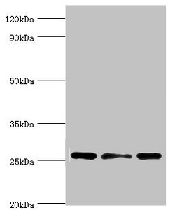
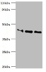
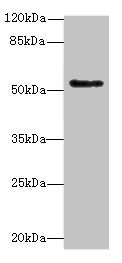
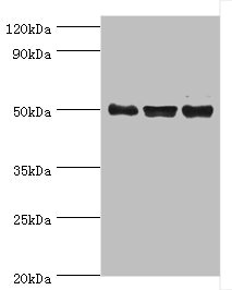
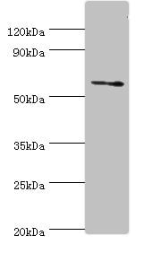
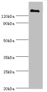
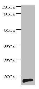 WB (Western Blot)
(Western blotAll lanes: TNNC2 antibody at 10ug/ml+mouse skeletal muscle tissueSecondaryGoat polyclonal to rabbit at 1/10000 dilutionPredicted band size: 18kDaObserved band size: 18kDa)
WB (Western Blot)
(Western blotAll lanes: TNNC2 antibody at 10ug/ml+mouse skeletal muscle tissueSecondaryGoat polyclonal to rabbit at 1/10000 dilutionPredicted band size: 18kDaObserved band size: 18kDa)
TNNC2, Polyclonal Antibody (Cat# AAA119543)
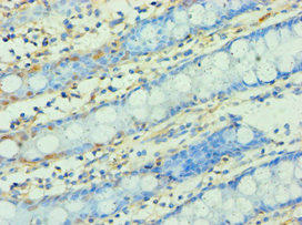
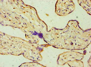
 IHC (Immunohiostchemistry)
(Immunohistochemistry of paraffin-embedded human placenta tissue using AAA119547 at dilution 1:100)
IHC (Immunohiostchemistry)
(Immunohistochemistry of paraffin-embedded human placenta tissue using AAA119547 at dilution 1:100)
SLC25A15, Polyclonal Antibody (Cat# AAA119547)
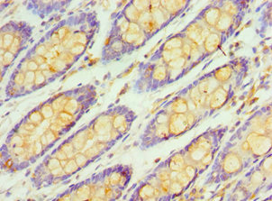
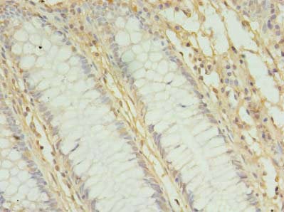
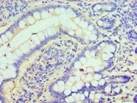
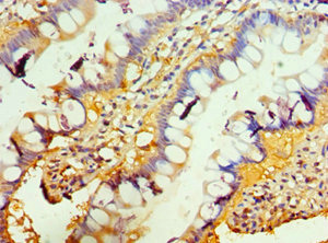
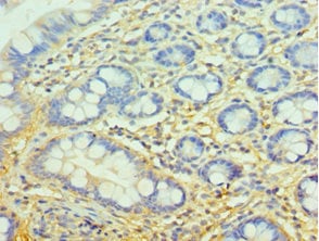
 IHC (Immunohistochemisry)
(Immunohistochemistry of paraffin-embedded human small intestine tissue using AAA119549 at dilution of 1:100)
IHC (Immunohistochemisry)
(Immunohistochemistry of paraffin-embedded human small intestine tissue using AAA119549 at dilution of 1:100)
RAB6B, Polyclonal Antibody (Cat# AAA119549)
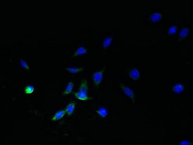
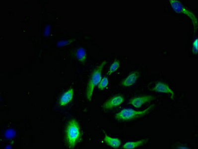 IF (Immunofluorescence)
(Immunofluorescent analysis of Hela cells using AAA119552 at a dilution of 1:100 and Alexa Fluor 488-congugated AffiniPure Goat Anti-Rabbit IgG(H+L))
IF (Immunofluorescence)
(Immunofluorescent analysis of Hela cells using AAA119552 at a dilution of 1:100 and Alexa Fluor 488-congugated AffiniPure Goat Anti-Rabbit IgG(H+L))
NKG7, Polyclonal Antibody (Cat# AAA119552)
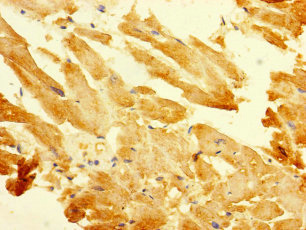
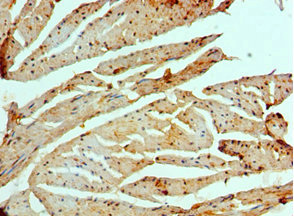
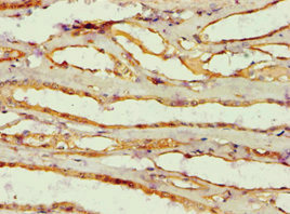
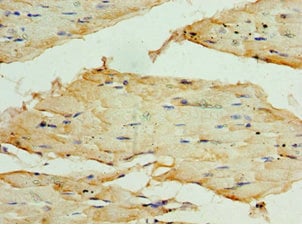
 IHC (Immunohiostchemistry)
(Immunohistochemistry of paraffin-embedded human skeletal muscle using AAA119557 at dilution 1:100)
IHC (Immunohiostchemistry)
(Immunohistochemistry of paraffin-embedded human skeletal muscle using AAA119557 at dilution 1:100)
Ubiquinone biosynthesis protein COQ7 homolog, Polyclonal Antibody (Cat# AAA119557)
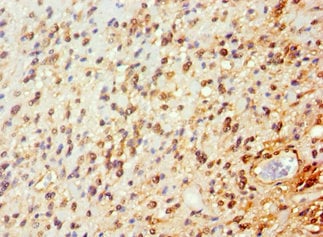
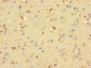 IHC (Immunohiostchemistry)
(Immunohistochemistry of paraffin-embedded human brain tissue using AAA119325 at dilution of 1:100)
IHC (Immunohiostchemistry)
(Immunohistochemistry of paraffin-embedded human brain tissue using AAA119325 at dilution of 1:100)
SMIM19, Polyclonal Antibody (Cat# AAA119325)
Clostridium botulinum Botulinum neurotoxin type C1, Polyclonal Antibody (Cat# AAA119329)
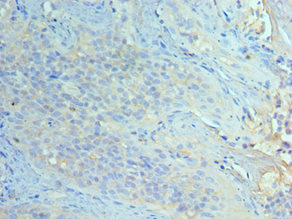
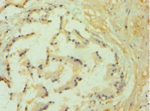
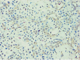
 IHC (Immunohiostchemistry)
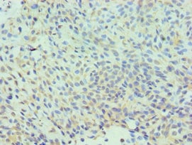
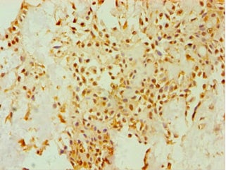
(Immunohistochemistry of paraffin-embedded human breast cancer using AAA119333 at dilution 1:100)
IHC (Immunohiostchemistry)
(Immunohistochemistry of paraffin-embedded human breast cancer using AAA119333 at dilution 1:100)
GTP-binding nuclear protein Ran, Polyclonal Antibody (Cat# AAA119333)
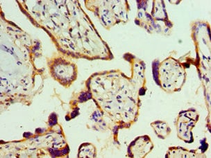
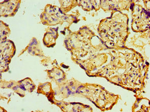
 IHC (Immunohiostchemistry)
(Immunohistochemistry of paraffin-embedded human placenta using AAA119334 at dilution 1:100)
IHC (Immunohiostchemistry)
(Immunohistochemistry of paraffin-embedded human placenta using AAA119334 at dilution 1:100)
Gap junction alpha-1 protein, Polyclonal Antibody (Cat# AAA119334)

 IHC (Immunohiostchemistry)
(Immunohistochemistry of paraffin-embedded human prostate tissue using AAA119336 at dilution 1:100)
IHC (Immunohiostchemistry)
(Immunohistochemistry of paraffin-embedded human prostate tissue using AAA119336 at dilution 1:100)
GLO1, Polyclonal Antibody (Cat# AAA119336)
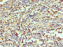
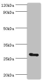
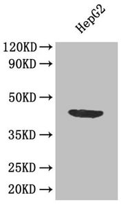
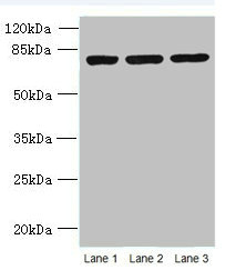
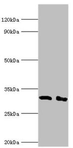
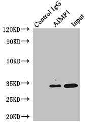 IP (Immunoprecipitation)
(Immunoprecipitating AIMP1 in HepG2 whole cell lysateLane 1: Rabbit monoclonal IgG(1ug)instead of AAA119338 in HepG2 whole cell lysate. For western blotting, a HRP-conjugated anti-rabbit IgG, specific to the non-reduced form of IgG was used as the Secondary antibody (1/50000)Lane 2: AAA119338(4ug)+ HepG2 whole cell lysate(500ug)Lane 3: HepG2 whole cell lysate (20ug))
IP (Immunoprecipitation)
(Immunoprecipitating AIMP1 in HepG2 whole cell lysateLane 1: Rabbit monoclonal IgG(1ug)instead of AAA119338 in HepG2 whole cell lysate. For western blotting, a HRP-conjugated anti-rabbit IgG, specific to the non-reduced form of IgG was used as the Secondary antibody (1/50000)Lane 2: AAA119338(4ug)+ HepG2 whole cell lysate(500ug)Lane 3: HepG2 whole cell lysate (20ug))
Aminoacyl tRNA synthase complex-interacting multifunctional protein 1, Polyclonal Antibody (Cat# AAA119338)
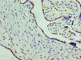
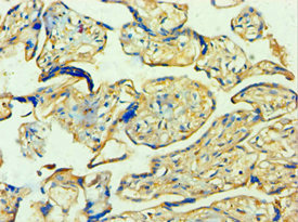
 IHC (Immunohiostchemistry)
(Immunohistochemistry of paraffin-embedded human placenta using AAA119344 at dilution 1:100)
IHC (Immunohiostchemistry)
(Immunohistochemistry of paraffin-embedded human placenta using AAA119344 at dilution 1:100)
E3 ubiquitin-protein ligase RBX1, Polyclonal Antibody (Cat# AAA119344)
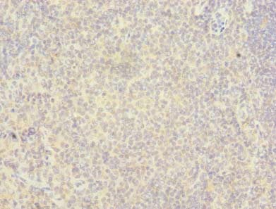
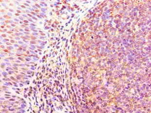
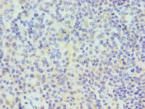
 IHC (Immunohistochemisry)
(Immunohistochemistry of paraffin-embedded human tonsil tissuetissue using AAA119354 at dilution 1:100)
IHC (Immunohistochemisry)
(Immunohistochemistry of paraffin-embedded human tonsil tissuetissue using AAA119354 at dilution 1:100)
Caspase-6, Polyclonal Antibody (Cat# AAA119354)

 IF (Immunofluorescence)
(Immunofluorescent analysis of A375 cells using AAA119360 at a dilution of 1:100 and Alexa Fluor 488-congugated AffiniPure Goat Anti-Rabbit IgG(H+L))
IF (Immunofluorescence)
(Immunofluorescent analysis of A375 cells using AAA119360 at a dilution of 1:100 and Alexa Fluor 488-congugated AffiniPure Goat Anti-Rabbit IgG(H+L))
HLA-DRB1, Polyclonal Antibody (Cat# AAA119360)
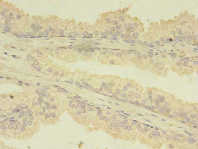
 IHC (Immunohistochemistry)
(Immunohistochemistry of paraffin-embedded human placenta using AAA119362 at dilution 1:100)
IHC (Immunohistochemistry)
(Immunohistochemistry of paraffin-embedded human placenta using AAA119362 at dilution 1:100)
Protein Wnt-2, Polyclonal Antibody (Cat# AAA119362)
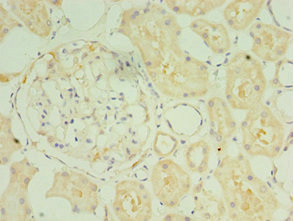
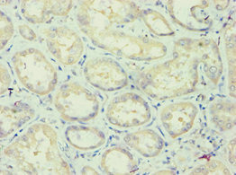
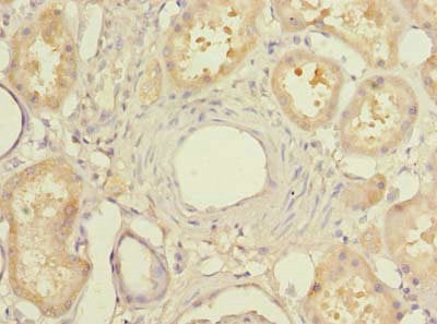
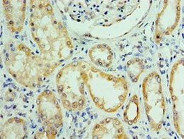 IHC (Immunohistochemisry)
(Immunohistochemistry of paraffin-embedded human kidney using AAA119366 at dilution 1:100)
IHC (Immunohistochemisry)
(Immunohistochemistry of paraffin-embedded human kidney using AAA119366 at dilution 1:100)
C14orf166, Polyclonal Antibody (Cat# AAA119366)
 IHC (Immunohiostchemistry)
(Immunohistochemistry of paraffin-embedded human rectal cancer using AAA119368 at dilution 1:100)
IHC (Immunohiostchemistry)
(Immunohistochemistry of paraffin-embedded human rectal cancer using AAA119368 at dilution 1:100)
Splicing factor 3B subunit 2, Polyclonal Antibody (Cat# AAA119368)
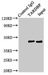 IP (Immunoprecipitation)
(Immunoprecipitating TARDBP in Hela whole cell lysateLane 1: Rabbit control IgG (1ug)instead of AAA119370 in Hela whole cell lysate. For western blotting,a HRP-conjugated Protein G antibody was used as the secondary antibody (1/2000)Lane 2: AAA119370 (8ug)+ Hela whole cell lysate (500ug)Lane 3: Hela whole cell lysate (10ug))
IP (Immunoprecipitation)
(Immunoprecipitating TARDBP in Hela whole cell lysateLane 1: Rabbit control IgG (1ug)instead of AAA119370 in Hela whole cell lysate. For western blotting,a HRP-conjugated Protein G antibody was used as the secondary antibody (1/2000)Lane 2: AAA119370 (8ug)+ Hela whole cell lysate (500ug)Lane 3: Hela whole cell lysate (10ug))
TAR DNA-binding protein 43, Polyclonal Antibody (Cat# AAA119370)
 IHC (Immunohiostchemistry)
(Immunohistochemistry of paraffin-embedded human kidney tissue using AAA119372 at dilution 1:100)
IHC (Immunohiostchemistry)
(Immunohistochemistry of paraffin-embedded human kidney tissue using AAA119372 at dilution 1:100)
SULT1C2, Polyclonal Antibody (Cat# AAA119372)
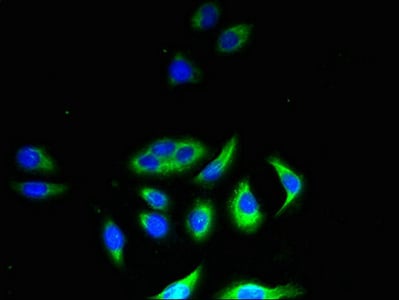
 IF (Immunofluorescence)
(Immunofluorescence staining of MCF-7 cells with AAA119376 at 1:266, counterstained with DAPI. The cells were fixed in 4% formaldehyde, permeabilized using 0.2% Triton X-100 and blocked in 10% normal Goat Serum. The cells were then incubated with the antibody overnight at 4°C. The secondary antibody was Alexa Fluor 488-congugated AffiniPure Goat Anti-Rabbit IgG(H+L).)
IF (Immunofluorescence)
(Immunofluorescence staining of MCF-7 cells with AAA119376 at 1:266, counterstained with DAPI. The cells were fixed in 4% formaldehyde, permeabilized using 0.2% Triton X-100 and blocked in 10% normal Goat Serum. The cells were then incubated with the antibody overnight at 4°C. The secondary antibody was Alexa Fluor 488-congugated AffiniPure Goat Anti-Rabbit IgG(H+L).)
CD27, Polyclonal Antibody (Cat# AAA119376)
Plasmodium falciparum L-lactate dehydrogenase, Polyclonal Antibody (Cat# AAA119379)
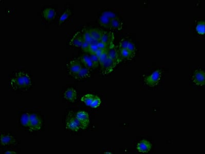
 IF (Immunofluorescence)
(Immunofluorescent analysis of A549 cells using AAA119382 at a dilution of 1:100 and Alexa Fluor 488-congugated AffiniPure Goat Anti-Rabbit IgG(H+L))
IF (Immunofluorescence)
(Immunofluorescent analysis of A549 cells using AAA119382 at a dilution of 1:100 and Alexa Fluor 488-congugated AffiniPure Goat Anti-Rabbit IgG(H+L))
Macrophage colony-stimulating factor 1, Polyclonal Antibody (Cat# AAA119382)
 IHC (Immunohiostchemistry)
(Immunohistochemistry of paraffin-embedded human placenta using AAA119383 at dilution 1:100)
IHC (Immunohiostchemistry)
(Immunohistochemistry of paraffin-embedded human placenta using AAA119383 at dilution 1:100)
HAUS augmin-like complex subunit 6, Polyclonal Antibody (Cat# AAA119383)
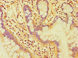
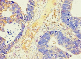
 IHC (Immunohistochemisry)
(Immunohistochemistry of paraffin-embedded human colon cancer using AAA119387 at dilution of 1:100)
IHC (Immunohistochemisry)
(Immunohistochemistry of paraffin-embedded human colon cancer using AAA119387 at dilution of 1:100)
MRPL3, Polyclonal Antibody (Cat# AAA119387)
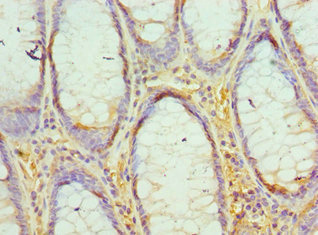 IHC (Immunohiostchemistry)
(Immunohistochemistry of paraffin-embedded human colon cancer using AAA119389at dilution 1:100)
IHC (Immunohiostchemistry)
(Immunohistochemistry of paraffin-embedded human colon cancer using AAA119389at dilution 1:100)
Polycomb protein EED, Polyclonal Antibody (Cat# AAA119389)
 IHC (Immunohistochemisry)
(Immunohistochemistry of paraffin-embedded human kidney using AAA119392 at dilution 1:100)
IHC (Immunohistochemisry)
(Immunohistochemistry of paraffin-embedded human kidney using AAA119392 at dilution 1:100)
Alpha-amylase 2B, Polyclonal Antibody (Cat# AAA119392)
Salmonella heidelberg D-serine dehydratase, Polyclonal Antibody (Cat# AAA119396)
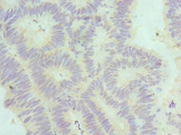
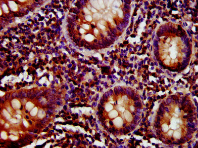
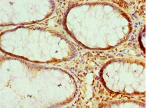
 IHC (Immunohistochemisry)
(Immunohistochemistry of paraffin-embedded human small intestine using AAA119398 at dilution 1:100)
IHC (Immunohistochemisry)
(Immunohistochemistry of paraffin-embedded human small intestine using AAA119398 at dilution 1:100)
Chloride intracellular channel protein 1, Polyclonal Antibody (Cat# AAA119398)
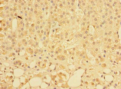
 IHC (Immunohiostchemistry)
(Immunohistochemistry of paraffin-embedded human testicle using AAA119399 at dilution 1:100)
IHC (Immunohiostchemistry)
(Immunohistochemistry of paraffin-embedded human testicle using AAA119399 at dilution 1:100)
Tumor necrosis factor receptor superfamily member 13B, Polyclonal Antibody (Cat# AAA119399)
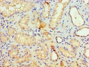
 IHC (Immunohistochemistry)
(Immunohistochemistry of paraffin-embedded human kidney using AAA119402 at dilution 1:100)
IHC (Immunohistochemistry)
(Immunohistochemistry of paraffin-embedded human kidney using AAA119402 at dilution 1:100)
Apolipoprotein L1, Polyclonal Antibody (Cat# AAA119402)
Alpha-L-iduronidase, Polyclonal Antibody (Cat# AAA119244)
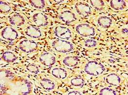
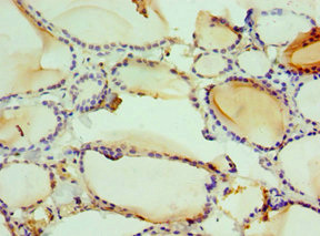
 IHC (Immunohistochemisry)
(Immunohistochemistry of paraffin-embedded human kidney tissue using AAA119245 at dilution of 1:100)
IHC (Immunohistochemisry)
(Immunohistochemistry of paraffin-embedded human kidney tissue using AAA119245 at dilution of 1:100)
MIER2, Polyclonal Antibody (Cat# AAA119245)
 IHC (Immunohiostchemistry)
(Immunohistochemistry of paraffin-embedded human colon cancer using AAA119248 at dilution 1:100)
IHC (Immunohiostchemistry)
(Immunohistochemistry of paraffin-embedded human colon cancer using AAA119248 at dilution 1:100)
Non-histone chromosomal protein HMG-17, Polyclonal Antibody (Cat# AAA119248)
 IHC (Immunohiostchemistry)
(Immunohistochemistry of paraffin-embedded human rectal cancer using AAA119251 at dilution 1:100)
IHC (Immunohiostchemistry)
(Immunohistochemistry of paraffin-embedded human rectal cancer using AAA119251 at dilution 1:100)
DNA replication licensing factor MCM7, Polyclonal Antibody (Cat# AAA119251)
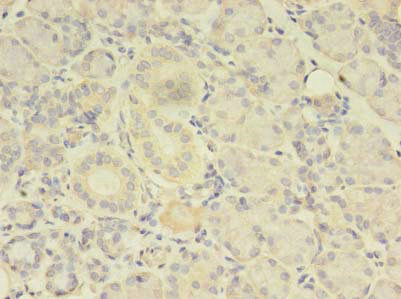
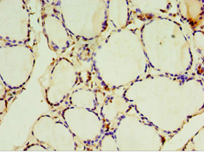
 IHC (Immunohistochemisry)
(Immunohistochemistry of paraffin-embedded human adrenal gland tissue using AAA119253 at dilution of 1:100)
IHC (Immunohistochemisry)
(Immunohistochemistry of paraffin-embedded human adrenal gland tissue using AAA119253 at dilution of 1:100)
PLEKHS1, Polyclonal Antibody (Cat# AAA119253)
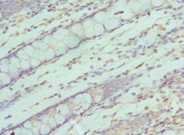
 IHC (Immunohiostchemistry)
(Immunohistochemistry of paraffin-embedded human skeletal muscle tissue using AAA119257 at dilution 1:100)
IHC (Immunohiostchemistry)
(Immunohistochemistry of paraffin-embedded human skeletal muscle tissue using AAA119257 at dilution 1:100)
TERF1, Polyclonal Antibody (Cat# AAA119257)
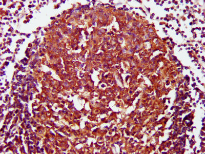
 IHC (Immunohiostchemistry)
(Immunohistochemistry of paraffin-embedded human thymus tissue using AAA119260 at dilution 1:100)
IHC (Immunohiostchemistry)
(Immunohistochemistry of paraffin-embedded human thymus tissue using AAA119260 at dilution 1:100)
Lymphocyte function-associated antigen 3, Polyclonal Antibody (Cat# AAA119260)
PQLC3, Polyclonal Antibody (Cat# AAA119262)
 IHC (Immunohistochemisry)
(Immunohistochemistry of paraffin-embedded human placenta using AAA119263 at dilution 1:100)
IHC (Immunohistochemisry)
(Immunohistochemistry of paraffin-embedded human placenta using AAA119263 at dilution 1:100)
Delta (24)-sterol reductase, Polyclonal Antibody (Cat# AAA119263)
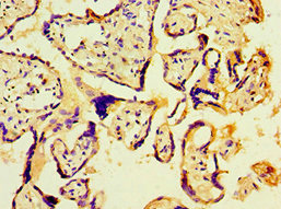
 IHC (Immunohistochemisry)
(Immunohistochemistry of paraffin-embedded human placenta using AAA119267 at dilution 1:100)
IHC (Immunohistochemisry)
(Immunohistochemistry of paraffin-embedded human placenta using AAA119267 at dilution 1:100)
SETDB1, Polyclonal Antibody (Cat# AAA119267)
 IHC (Immunohiostchemistry)
(Immunohistochemistry of paraffin-embedded human small intestine tissue using AAA119277 at dilution 1:100)
IHC (Immunohiostchemistry)
(Immunohistochemistry of paraffin-embedded human small intestine tissue using AAA119277 at dilution 1:100)
CFH, Polyclonal Antibody (Cat# AAA119277)

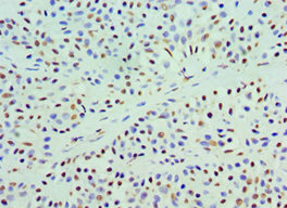
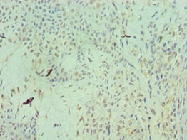
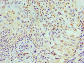
 IHC (Immunohiostchemistry)
(Immunohistochemistry of paraffin-embedded human glioma using AAA119281 at dilution 1:100)
IHC (Immunohiostchemistry)
(Immunohistochemistry of paraffin-embedded human glioma using AAA119281 at dilution 1:100)
Aryl hydrocarbon receptor nuclear translocator 2, Polyclonal Antibody (Cat# AAA119281)
What are Polyclonal Antibodies?
Polyclonal antibodies are antibodies that come from multiple B cell clones of a host animal. The typical hosts used for the majority of polyclonal antibody production are rabbits, goats, sheep, and donkeys. These polyclonal antibodies, once having identified their target, will bind to different epitopes located at different regions or sequences on the same protein/antigen. As a result, they are ideal at locating and binding to the target, even if the target is in very low concentrations (due to many different antibodies being able to bind to the same target molecule, which allows for significant amplification of a downstream signal).
Polyclonal antibodies are typically produced by injecting an antigen into a host animal, which causes the animal’s immune system to attack the foreign antigen by mass generating antibodies against it. After a period of time, serum is collected from the animal and purified using physicochemical fractionation, class-specific affinity purification, and/or antigen-affinity purification.
Key Uses of Polyclonal Antibodies
- Western Blotting: This method is used to find specific proteins in biological samples after separating them by size.
- Immunohistochemistry: IHC helps visualize the location of proteins in tissue sections using various staining techniques.
- ELISA: (Enzyme-Linked Immunosorbent Assay) is typically used to identify specific protein quantities in a sample. ELISAs can be either “Quantitative” or “Qualitative”.
- Flow Cytometry: technique that identifies and measures the specific protein on the surface or inside the cells in a fluid suspension.
- Immunoprecipitation: IP isolates and studies a specific protein from a complex mixture using antibodies.
Why Buy Polyclonal Antibodies from AAA Biotech?
1. Ideal for Various Applications
Our antibodies are generally going to be validated for use in multiple types of assays, including ELISA, Western Blotting, Immunohistochemistry, Immunoprecipitation, amongst others. They are ideal for a wide range of research applications.
2. Rigorous Quality Control
All of the antibodies in our catalog undergo strict quality testing to ensure specificity, sensitivity, and consistent performance. We are confident in the ability of our antibodies to provide you with accurate results.
3. Wide Assortment of Antibodies
Antibodies in are catalog can be found for both common and exotic species, and these antibodies are also available in both conjugated and recombinant forms to suit many diverse experimental needs.
4. Highly Purified
Our antibodies are available in purified forms with over 85% purity, as confirmed by SDS-PAGE. They are also available with tags such as His, Flag, GST, or MBP. We cater to customers worldwide.
FAQ
1. How are polyclonal antibodies produced?
Traditionally, polyclonal antibodies are produced by injecting an antigen into a host animal (such as a rabbit or goat), which then triggers an immune response from the host animal. The animal’s B cells produce antibodies that will recognize different parts of the injected antigen. These antibodies are then collected from the animal’s blood and purified for use.
2. How do polyclonal antibodies differ from monoclonal antibodies?
Polyclonal antibodies are a mix of antibodies that bind to different locations (epitopes) of the same antigen, while monoclonal antibodies are identical and bind to just one specific epitope. This makes polyclonal antibodies more versatile and better at detecting proteins that may be present in low quantities or in altered/modified forms.
3. How should I store polyclonal antibodies?
Polyclonal antibodies should be stored at 4°C for short-term use (up to a few weeks) and at -20°C or -80°C for long-term storage. Avoid repeated freeze-thaw cycles by dividing them into small aliquots. Always check the datasheet for specific storage instructions.