Filters
▼Clonality
▼Type
▼Reactivity
▼Gene Name
▼Isotype
▼Host
▼Application
▼Clone
▼Polyclonal Antibodies
At AAA Biotech also known as AAA Bio or AAABio, we provide a broad range of purified polyclonal antibodies (pAbs) that are able to all be browsed online through our website. Due to their high specificity and strong binding affinity, these antibodies are ideal for wide swathes of research and experimental applications.
Our polyclonal antibodies can easily support your work, whether you use them for Western Blotting, Immunocytochemistry (with or without Immunofluorescence used in conjunction), Immunohistochemistry, Immunoprecipitation, and ELISA tests. We highly encourage you to browse our range of pAbs and choose the one that best suits your experimental model.
Viewing 800-850 of 96805 product results
 SDS-PAGE
(Gel: 15%SDS-PAGE, Lysate: 40 ug, Lane: 293T cells, Primary antibody: AAA238262(CDCA8 Antibody) at dilution 1/800, Secondary antibody: Goat anti rabbit IgG at 1/8000 dilution, Exposure time: 1 second)
SDS-PAGE
(Gel: 15%SDS-PAGE, Lysate: 40 ug, Lane: 293T cells, Primary antibody: AAA238262(CDCA8 Antibody) at dilution 1/800, Secondary antibody: Goat anti rabbit IgG at 1/8000 dilution, Exposure time: 1 second)
CDCA8, Polyclonal Antibody (Cat# AAA238262)
 IHC (Immunohiostchemistry)
(The image on the left is immunohistochemistry of paraffin-embedded Human cervical cancer tissue using AAA238263(ALPI Antibody) at dilution 1/25, on the right is treated with fusion protein. (Original magnification: ×200))
IHC (Immunohiostchemistry)
(The image on the left is immunohistochemistry of paraffin-embedded Human cervical cancer tissue using AAA238263(ALPI Antibody) at dilution 1/25, on the right is treated with fusion protein. (Original magnification: ×200))
ALPI, Polyclonal Antibody (Cat# AAA238263)

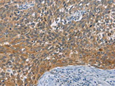
 IHC (Immunohiostchemistry)
(The image on the left is immunohistochemistry of paraffin-embedded Human cervical cancer tissue using AAA238268(TLR8 Antibody) at dilution 1/25, on the right is treated with fusion protein. (Original magnification: ×200))
IHC (Immunohiostchemistry)
(The image on the left is immunohistochemistry of paraffin-embedded Human cervical cancer tissue using AAA238268(TLR8 Antibody) at dilution 1/25, on the right is treated with fusion protein. (Original magnification: ×200))
TLR8, Polyclonal Antibody (Cat# AAA238268)
 IHC (Immunohiostchemistry)
(The image on the left is immunohistochemistry of paraffin-embedded Human breast cancer tissue using AAA238269(TLR6 Antibody) at dilution 1/25, on the right is treated with fusion protein. (Original magnification: ×200))
IHC (Immunohiostchemistry)
(The image on the left is immunohistochemistry of paraffin-embedded Human breast cancer tissue using AAA238269(TLR6 Antibody) at dilution 1/25, on the right is treated with fusion protein. (Original magnification: ×200))
TLR6, Polyclonal Antibody (Cat# AAA238269)
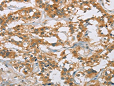
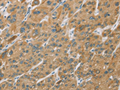
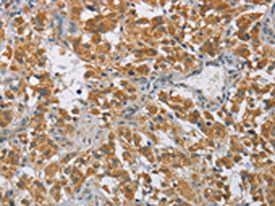
 IHC (Immunohiostchemistry)
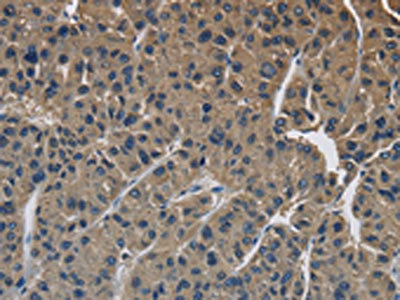
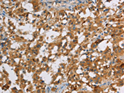
(The image on the left is immunohistochemistry of paraffin-embedded Human liver cancer tissue using AAA238272(TLR3 Antibody) at dilution 1/40, on the right is treated with fusion protein. (Original magnification: ×200))
IHC (Immunohiostchemistry)
(The image on the left is immunohistochemistry of paraffin-embedded Human liver cancer tissue using AAA238272(TLR3 Antibody) at dilution 1/40, on the right is treated with fusion protein. (Original magnification: ×200))
TLR3, Polyclonal Antibody (Cat# AAA238272)
 IHC (Immunohiostchemistry)
(The image on the left is immunohistochemistry of paraffin-embedded Human colon cancer tissue using AAA238276(UCHL5 Antibody) at dilution 1/20, on the right is treated with fusion protein. (Original magnification: ×200))
IHC (Immunohiostchemistry)
(The image on the left is immunohistochemistry of paraffin-embedded Human colon cancer tissue using AAA238276(UCHL5 Antibody) at dilution 1/20, on the right is treated with fusion protein. (Original magnification: ×200))
UCHL5, Polyclonal Antibody (Cat# AAA238276)
 IHC (Immunohiostchemistry)
(The image on the left is immunohistochemistry of paraffin-embedded Human esophagus cancer tissue using AAA238280(EPS15L1 Antibody) at dilution 1/20, on the right is treated with fusion protein. (Original magnification: ×200))
IHC (Immunohiostchemistry)
(The image on the left is immunohistochemistry of paraffin-embedded Human esophagus cancer tissue using AAA238280(EPS15L1 Antibody) at dilution 1/20, on the right is treated with fusion protein. (Original magnification: ×200))
EPS15L1, Polyclonal Antibody (Cat# AAA238280)
 IHC (Immunohiostchemistry)
(The image on the left is immunohistochemistry of paraffin-embedded Human prostate cancer tissue using AAA238283(STAM Antibody) at dilution 1/40, on the right is treated with fusion protein. (Original magnification: ×200))
IHC (Immunohiostchemistry)
(The image on the left is immunohistochemistry of paraffin-embedded Human prostate cancer tissue using AAA238283(STAM Antibody) at dilution 1/40, on the right is treated with fusion protein. (Original magnification: ×200))
STAM, Polyclonal Antibody (Cat# AAA238283)
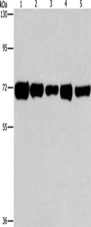 SDS-PAGE
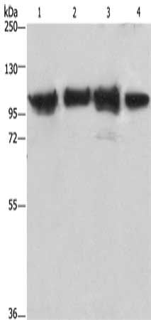
(Gel: 8%SDS-PAGE, Lysate: 40 ug, Lane 1-5: Hela cells, A549 cells, MCF7 cells, human liver cancer tissue, Human lung cancer tissue, Primary antibody: AAA238286(RPN1 Antibody) at dilution 1/550, Secondary antibody: Goat anti rabbit IgG at 1/8000 dilution, Exposure time: 1 minute)
SDS-PAGE
(Gel: 8%SDS-PAGE, Lysate: 40 ug, Lane 1-5: Hela cells, A549 cells, MCF7 cells, human liver cancer tissue, Human lung cancer tissue, Primary antibody: AAA238286(RPN1 Antibody) at dilution 1/550, Secondary antibody: Goat anti rabbit IgG at 1/8000 dilution, Exposure time: 1 minute)
RPN1, Polyclonal Antibody (Cat# AAA238286)
 SDS-PAGE
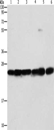
(Gel: 10%SDS-PAGE, Lysate: 40 ug, Lane 1-6: Mouse liver tissue, Mouse brain tissue, 293T cells, A549 cells, A431 cells, PC3 cells, Primary antibody: AAA238294(SIGMAR1 Antibody) at dilution 1/310, Secondary antibody: Goat anti rabbit IgG at 1/8000 dilution, Exposure time: 1 second)
SDS-PAGE
(Gel: 10%SDS-PAGE, Lysate: 40 ug, Lane 1-6: Mouse liver tissue, Mouse brain tissue, 293T cells, A549 cells, A431 cells, PC3 cells, Primary antibody: AAA238294(SIGMAR1 Antibody) at dilution 1/310, Secondary antibody: Goat anti rabbit IgG at 1/8000 dilution, Exposure time: 1 second)
SIGMAR1, Polyclonal Antibody (Cat# AAA238294)
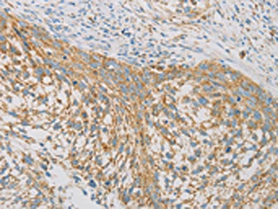
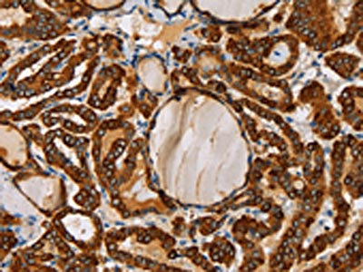
 IHC (Immunohiostchemistry)
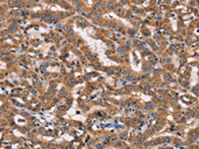
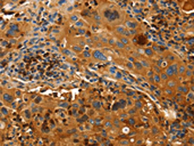
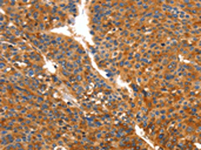
(The image on the left is immunohistochemistry of paraffin-embedded Human thyroid cancer tissue using AAA238300(REN Antibody) at dilution 1/40, on the right is treated with fusion protein. (Original magnification: ×200))
IHC (Immunohiostchemistry)
(The image on the left is immunohistochemistry of paraffin-embedded Human thyroid cancer tissue using AAA238300(REN Antibody) at dilution 1/40, on the right is treated with fusion protein. (Original magnification: ×200))
REN, Polyclonal Antibody (Cat# AAA238300)
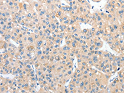
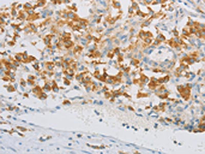
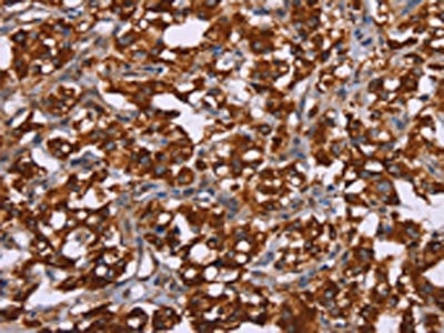
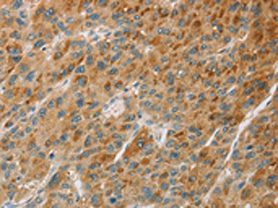
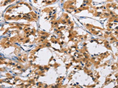
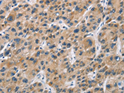
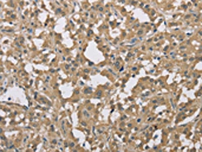
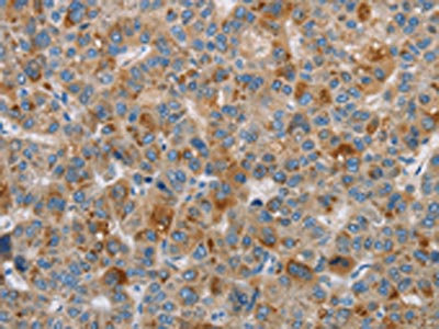
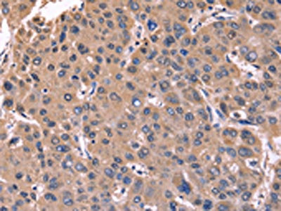 IHC (Immunohiostchemistry)
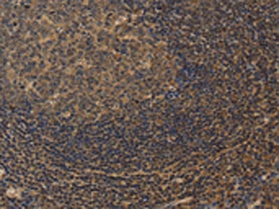
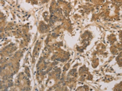
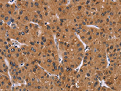
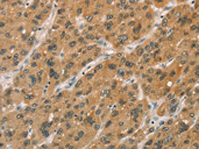
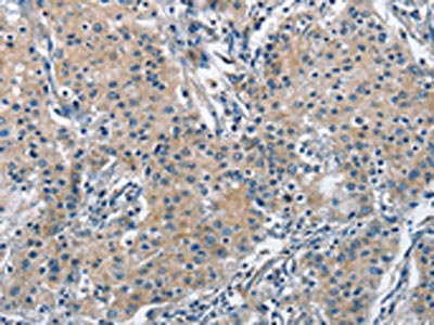
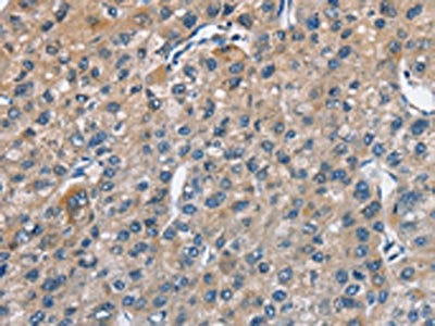
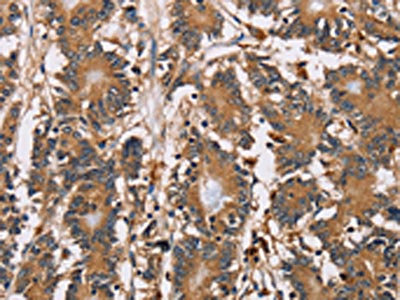
(The image on the left is immunohistochemistry of paraffin-embedded Human liver cancer tissue using AAA238667(CSNK1D Antibody) at dilution 1/50, on the right is treated with fusion protein. (Original magnification: ×200))
IHC (Immunohiostchemistry)
(The image on the left is immunohistochemistry of paraffin-embedded Human liver cancer tissue using AAA238667(CSNK1D Antibody) at dilution 1/50, on the right is treated with fusion protein. (Original magnification: ×200))
CSNK1D, Polyclonal Antibody (Cat# AAA238667)
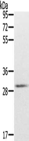
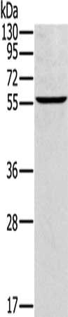
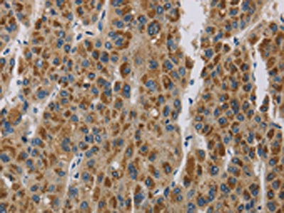
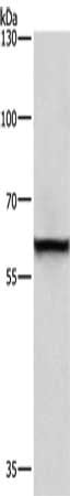
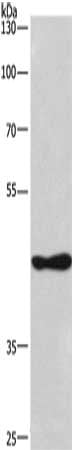 SDS-PAGE
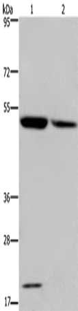
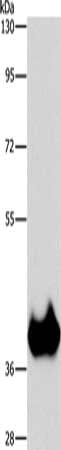
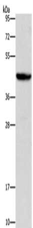
(Gel: 8%SDS-PAGE, Lysate: 40 ug, Lane: 293T cells, Primary antibody: AAA238668(CSNK1D Antibody) at dilution 1/270, Secondary antibody: Goat anti rabbit IgG at 1/8000 dilution, Exposure time: 5 minutes)
SDS-PAGE
(Gel: 8%SDS-PAGE, Lysate: 40 ug, Lane: 293T cells, Primary antibody: AAA238668(CSNK1D Antibody) at dilution 1/270, Secondary antibody: Goat anti rabbit IgG at 1/8000 dilution, Exposure time: 5 minutes)
CSNK1D, Polyclonal Antibody (Cat# AAA238668)
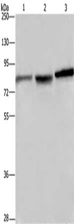
 SDS-PAGE
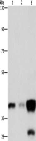
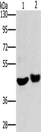
(Gel: 10%SDS-PAGE, Lysate: 40 ug, Lane 1-3: PC3 cells, human brain malignant glioma tissue, A172 cells, Primary antibody: AAA238669(CTAGE5 Antibody) at dilution 1/200, Secondary antibody: Goat anti rabbit IgG at 1/8000 dilution, Exposure time: 1 second)
SDS-PAGE
(Gel: 10%SDS-PAGE, Lysate: 40 ug, Lane 1-3: PC3 cells, human brain malignant glioma tissue, A172 cells, Primary antibody: AAA238669(CTAGE5 Antibody) at dilution 1/200, Secondary antibody: Goat anti rabbit IgG at 1/8000 dilution, Exposure time: 1 second)
CTAGE5, Polyclonal Antibody (Cat# AAA238669)
 IHC (Immunohiostchemistry)
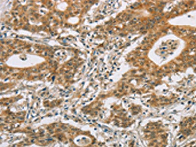
(The image on the left is immunohistochemistry of paraffin-embedded Human gastric cancer tissue using AAA238673(CTNNBIP1 Antibody) at dilution 1/20, on the right is treated with fusion protein. (Original magnification: ×200))
IHC (Immunohiostchemistry)
(The image on the left is immunohistochemistry of paraffin-embedded Human gastric cancer tissue using AAA238673(CTNNBIP1 Antibody) at dilution 1/20, on the right is treated with fusion protein. (Original magnification: ×200))
CTNNBIP1, Polyclonal Antibody (Cat# AAA238673)
 IHC (Immunohiostchemistry)
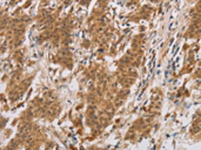
(The image on the left is immunohistochemistry of paraffin-embedded Human thyroid cancer tissue using AAA238676(CUEDC2 Antibody) at dilution 1/30, on the right is treated with fusion protein. (Original magnification: ×200))
IHC (Immunohiostchemistry)
(The image on the left is immunohistochemistry of paraffin-embedded Human thyroid cancer tissue using AAA238676(CUEDC2 Antibody) at dilution 1/30, on the right is treated with fusion protein. (Original magnification: ×200))
CUEDC2, Polyclonal Antibody (Cat# AAA238676)
 SDS-PAGE
(Gel: 8%SDS-PAGE, Lysate: 40 ug, Lane 1-2: Human fetal brain tissue, mouse brain tissue, Primary antibody: AAA238678(CXCR6 Antibody) at dilution 1/350, Secondary antibody: Goat anti rabbit IgG at 1/8000 dilution, Exposure time: 2 minutes)
SDS-PAGE
(Gel: 8%SDS-PAGE, Lysate: 40 ug, Lane 1-2: Human fetal brain tissue, mouse brain tissue, Primary antibody: AAA238678(CXCR6 Antibody) at dilution 1/350, Secondary antibody: Goat anti rabbit IgG at 1/8000 dilution, Exposure time: 2 minutes)
CXCR6, Polyclonal Antibody (Cat# AAA238678)
 SDS-PAGE
(Gel: 8%SDS-PAGE, Lysate: 40 ug, Lane: Mouse brain tissue, Primary antibody: AAA238680(CXXC5 Antibody) at dilution 1/200, Secondary antibody: Goat anti rabbit IgG at 1/8000 dilution, Exposure time: 1 minute)
SDS-PAGE
(Gel: 8%SDS-PAGE, Lysate: 40 ug, Lane: Mouse brain tissue, Primary antibody: AAA238680(CXXC5 Antibody) at dilution 1/200, Secondary antibody: Goat anti rabbit IgG at 1/8000 dilution, Exposure time: 1 minute)
CXXC5, Polyclonal Antibody (Cat# AAA238680)
 IHC (Immunohiostchemistry)
(The image on the left is immunohistochemistry of paraffin-embedded Human colon cancer tissue using AAA238681(CXXC5 Antibody) at dilution 1/25, on the right is treated with fusion protein. (Original magnification: ×200))
IHC (Immunohiostchemistry)
(The image on the left is immunohistochemistry of paraffin-embedded Human colon cancer tissue using AAA238681(CXXC5 Antibody) at dilution 1/25, on the right is treated with fusion protein. (Original magnification: ×200))
CXXC5, Polyclonal Antibody (Cat# AAA238681)
 SDS-PAGE
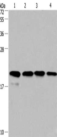
(Gel: 10%SDS-PAGE, Lysate: 40 ug, Lane 1-4: Human fetal liver tissue, 293T cells, human liver cancer tissue, hela cells, Primary antibody: AAA238683(PPIB Antibody) at dilution 1/450, Secondary antibody: Goat anti rabbit IgG at 1/8000 dilution, Exposure time: 20 seconds)
SDS-PAGE
(Gel: 10%SDS-PAGE, Lysate: 40 ug, Lane 1-4: Human fetal liver tissue, 293T cells, human liver cancer tissue, hela cells, Primary antibody: AAA238683(PPIB Antibody) at dilution 1/450, Secondary antibody: Goat anti rabbit IgG at 1/8000 dilution, Exposure time: 20 seconds)
PPIB, Polyclonal Antibody (Cat# AAA238683)
 SDS-PAGE
(Gel: 10%SDS-PAGE, Lysate: 40 ug, Lane 1-4: PC3 cells, 293T cells, human liver cancer tissue, hela cells, Primary antibody: AAA238684(PPIF Antibody) at dilution 1/500, Secondary antibody: Goat anti rabbit IgG at 1/8000 dilution, Exposure time: 20 seconds)
SDS-PAGE
(Gel: 10%SDS-PAGE, Lysate: 40 ug, Lane 1-4: PC3 cells, 293T cells, human liver cancer tissue, hela cells, Primary antibody: AAA238684(PPIF Antibody) at dilution 1/500, Secondary antibody: Goat anti rabbit IgG at 1/8000 dilution, Exposure time: 20 seconds)
PPIF, Polyclonal Antibody (Cat# AAA238684)
 SDS-PAGE
(Gel: 8%SDS-PAGE, Lysate: 40 ug, Lane: Mouse liver tissue, Primary antibody: AAA238689(CYP2E1 Antibody) at dilution 1/300, Secondary antibody: Goat anti rabbit IgG at 1/8000 dilution, Exposure time: 20 seconds)
SDS-PAGE
(Gel: 8%SDS-PAGE, Lysate: 40 ug, Lane: Mouse liver tissue, Primary antibody: AAA238689(CYP2E1 Antibody) at dilution 1/300, Secondary antibody: Goat anti rabbit IgG at 1/8000 dilution, Exposure time: 20 seconds)
CYP2E1, Polyclonal Antibody (Cat# AAA238689)
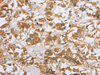
 IHC (Immunohiostchemistry)
(The image on the left is immunohistochemistry of paraffin-embedded Human thyroid cancer tissue using AAA238691(CYP2W1 Antibody) at dilution 1/60, on the right is treated with fusion protein. (Original magnification: ×200))
IHC (Immunohiostchemistry)
(The image on the left is immunohistochemistry of paraffin-embedded Human thyroid cancer tissue using AAA238691(CYP2W1 Antibody) at dilution 1/60, on the right is treated with fusion protein. (Original magnification: ×200))
CYP2W1, Polyclonal Antibody (Cat# AAA238691)
 IHC (Immunohiostchemistry)
(The image on the left is immunohistochemistry of paraffin-embedded Human thyroid cancer tissue using AAA238694(CYP11B1 Antibody) at dilution 1/30, on the right is treated with fusion protein. (Original magnification: ×200))
IHC (Immunohiostchemistry)
(The image on the left is immunohistochemistry of paraffin-embedded Human thyroid cancer tissue using AAA238694(CYP11B1 Antibody) at dilution 1/30, on the right is treated with fusion protein. (Original magnification: ×200))
CYP11B1, Polyclonal Antibody (Cat# AAA238694)
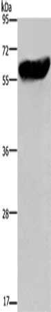
 SDS-PAGE
(Gel: 6%SDS-PAGE, Lysate: 40 ug, Lane 1-2: Mouse liver tissue, human fetal liver tissue, Primary antibody: AAA238696(CYP27A1 Antibody) at dilution 1/200, Secondary antibody: Goat anti rabbit IgG at 1/8000 dilution, Exposure time: 7 minutes)
SDS-PAGE
(Gel: 6%SDS-PAGE, Lysate: 40 ug, Lane 1-2: Mouse liver tissue, human fetal liver tissue, Primary antibody: AAA238696(CYP27A1 Antibody) at dilution 1/200, Secondary antibody: Goat anti rabbit IgG at 1/8000 dilution, Exposure time: 7 minutes)
CYP27A1, Polyclonal Antibody (Cat# AAA238696)
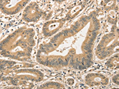
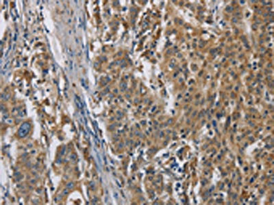
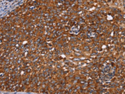
 IHC (Immunohiostchemistry)
(The image on the left is immunohistochemistry of paraffin-embedded Human gastric cancer tissue using AAA238698(CYP11A1 Antibody) at dilution 1/50, on the right is treated with fusion protein. (Original magnification: ×200))
IHC (Immunohiostchemistry)
(The image on the left is immunohistochemistry of paraffin-embedded Human gastric cancer tissue using AAA238698(CYP11A1 Antibody) at dilution 1/50, on the right is treated with fusion protein. (Original magnification: ×200))
CYP11A1, Polyclonal Antibody (Cat# AAA238698)

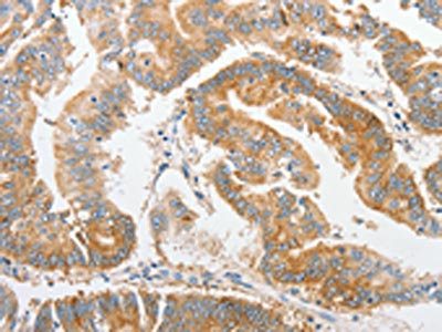
 IHC (Immunohiostchemistry)
(The image on the left is immunohistochemistry of paraffin-embedded Human gastric cancer tissue using AAA238700(CYP17A1 Antibody) at dilution 1/20, on the right is treated with fusion protein. (Original magnification: ×200))
IHC (Immunohiostchemistry)
(The image on the left is immunohistochemistry of paraffin-embedded Human gastric cancer tissue using AAA238700(CYP17A1 Antibody) at dilution 1/20, on the right is treated with fusion protein. (Original magnification: ×200))
CYP17A1, Polyclonal Antibody (Cat# AAA238700)
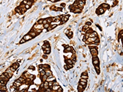
 SDS-PAGE
(Gel: 8%SDS-PAGE, Lysate: 40 ug, Lane: Human breast infiltrative duct tissue, Primary antibody: AAA238702(CYP21A2 Antibody) at dilution 1/350, Secondary antibody: Goat anti rabbit IgG at 1/8000 dilution, Exposure time: 2 minutes)
SDS-PAGE
(Gel: 8%SDS-PAGE, Lysate: 40 ug, Lane: Human breast infiltrative duct tissue, Primary antibody: AAA238702(CYP21A2 Antibody) at dilution 1/350, Secondary antibody: Goat anti rabbit IgG at 1/8000 dilution, Exposure time: 2 minutes)
CYP21A2, Polyclonal Antibody (Cat# AAA238702)
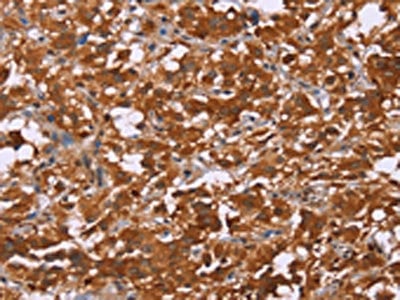
 IHC (Immunohiostchemistry)
(The image is immunohistochemistry of paraffin-embedded Human thyroid cancer tissue using AAA238703(LOXL4 Antibody) at dilution 1/20. (Original magnification: ×200))
IHC (Immunohiostchemistry)
(The image is immunohistochemistry of paraffin-embedded Human thyroid cancer tissue using AAA238703(LOXL4 Antibody) at dilution 1/20. (Original magnification: ×200))
LOXL4, Polyclonal Antibody (Cat# AAA238703)
 SDS-PAGE
(Gel: 8%SDS-PAGE, Lysate: 40 ug, Lane: Mouse liver tissue, Primary antibody: AAA238707(CYP4A11 Antibody) at dilution 1/250, Secondary antibody: Goat anti rabbit IgG at 1/8000 dilution, Exposure time: 2 minutes)
SDS-PAGE
(Gel: 8%SDS-PAGE, Lysate: 40 ug, Lane: Mouse liver tissue, Primary antibody: AAA238707(CYP4A11 Antibody) at dilution 1/250, Secondary antibody: Goat anti rabbit IgG at 1/8000 dilution, Exposure time: 2 minutes)
CYP4A11, Polyclonal Antibody (Cat# AAA238707)
 IHC (Immunohiostchemistry)
(The image on the left is immunohistochemistry of paraffin-embedded Human colon cancer tissue using AAA238710(CST6 Antibody) at dilution 1/70, on the right is treated with fusion protein. (Original magnification: ×200))
IHC (Immunohiostchemistry)
(The image on the left is immunohistochemistry of paraffin-embedded Human colon cancer tissue using AAA238710(CST6 Antibody) at dilution 1/70, on the right is treated with fusion protein. (Original magnification: ×200))
CST6, Polyclonal Antibody (Cat# AAA238710)
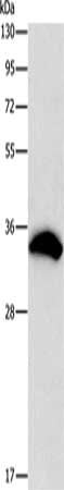
 SDS-PAGE
(Gel: 12%SDS-PAGE, Lysate: 40 ug, Lane: Human liver cancer tissue, Primary antibody: AAA238712(CST2 Antibody) at dilution 1/250, Secondary antibody: Goat anti rabbit IgG at 1/8000 dilution, Exposure time: 5 minutes)
SDS-PAGE
(Gel: 12%SDS-PAGE, Lysate: 40 ug, Lane: Human liver cancer tissue, Primary antibody: AAA238712(CST2 Antibody) at dilution 1/250, Secondary antibody: Goat anti rabbit IgG at 1/8000 dilution, Exposure time: 5 minutes)
CST2, Polyclonal Antibody (Cat# AAA238712)
 IHC (Immunohiostchemistry)
(The image on the left is immunohistochemistry of paraffin-embedded Human thyroid cancer tissue using AAA238717(DACH1 Antibody) at dilution 1/50, on the right is treated with fusion protein. (Original magnification: ×200))
IHC (Immunohiostchemistry)
(The image on the left is immunohistochemistry of paraffin-embedded Human thyroid cancer tissue using AAA238717(DACH1 Antibody) at dilution 1/50, on the right is treated with fusion protein. (Original magnification: ×200))
DACH1, Polyclonal Antibody (Cat# AAA238717)
 IHC (Immunohiostchemistry)
(The image on the left is immunohistochemistry of paraffin-embedded Human colon cancer tissue using AAA238718(DACH1 Antibody) at dilution 1/50, on the right is treated with fusion protein. (Original magnification: ×200))
IHC (Immunohiostchemistry)
(The image on the left is immunohistochemistry of paraffin-embedded Human colon cancer tissue using AAA238718(DACH1 Antibody) at dilution 1/50, on the right is treated with fusion protein. (Original magnification: ×200))
DACH1, Polyclonal Antibody (Cat# AAA238718)
 IHC (Immunohiostchemistry)
(The image on the left is immunohistochemistry of paraffin-embedded Human thyroid cancer tissue using AAA238720(DACH2 Antibody) at dilution 1/40, on the right is treated with fusion protein. (Original magnification: ×200))
IHC (Immunohiostchemistry)
(The image on the left is immunohistochemistry of paraffin-embedded Human thyroid cancer tissue using AAA238720(DACH2 Antibody) at dilution 1/40, on the right is treated with fusion protein. (Original magnification: ×200))
DACH2, Polyclonal Antibody (Cat# AAA238720)
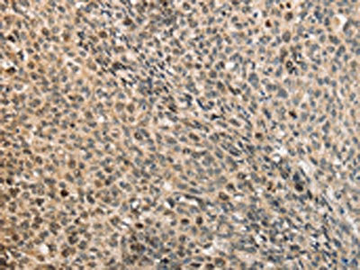
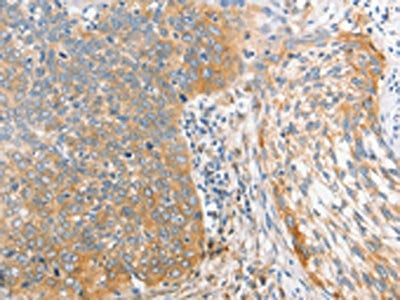
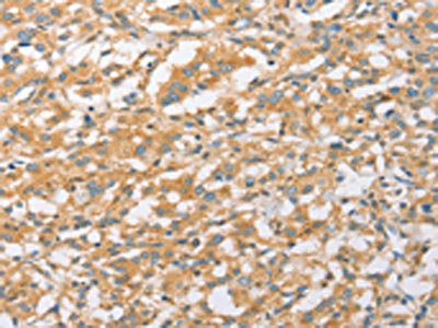
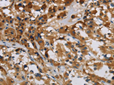
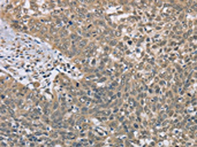
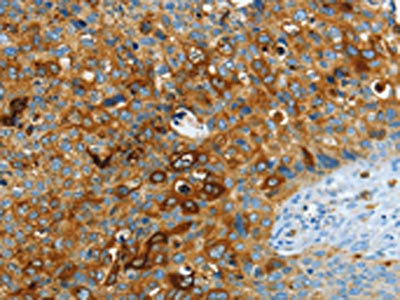
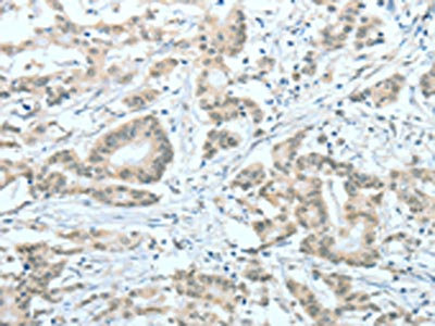
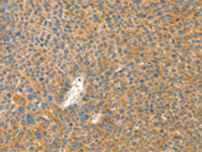
 IHC (Immunohiostchemistry)
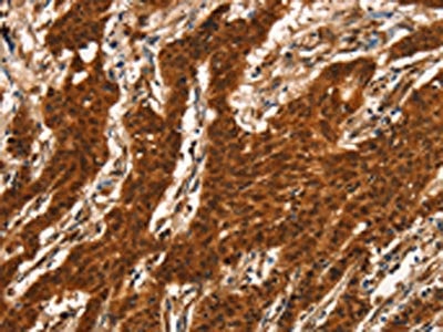
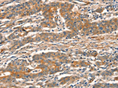
(The image on the left is immunohistochemistry of paraffin-embedded Human cervical cancer tissue using AAA238721(DIDO1 Antibody) at dilution 1/60, on the right is treated with fusion protein. (Original magnification: ×200))
IHC (Immunohiostchemistry)
(The image on the left is immunohistochemistry of paraffin-embedded Human cervical cancer tissue using AAA238721(DIDO1 Antibody) at dilution 1/60, on the right is treated with fusion protein. (Original magnification: ×200))
DIDO1, Polyclonal Antibody (Cat# AAA238721)
 SDS-PAGE
(Gel: 8%SDS-PAGE, Lysate: 40 ug, Lane 1-3: Mouse brain tissue, A172 cells, Hela cells, Primary antibody: AAA238724(DBH Antibody) at dilution 1/200, Secondary antibody: Goat anti rabbit IgG at 1/8000 dilution, Exposure time: 10 seconds)
SDS-PAGE
(Gel: 8%SDS-PAGE, Lysate: 40 ug, Lane 1-3: Mouse brain tissue, A172 cells, Hela cells, Primary antibody: AAA238724(DBH Antibody) at dilution 1/200, Secondary antibody: Goat anti rabbit IgG at 1/8000 dilution, Exposure time: 10 seconds)
DBH, Polyclonal Antibody (Cat# AAA238724)
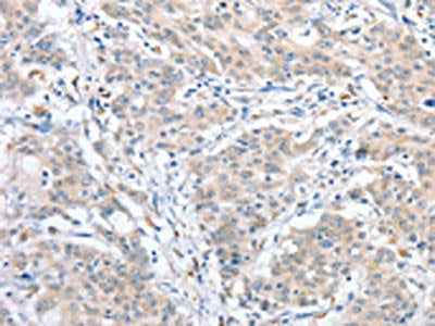
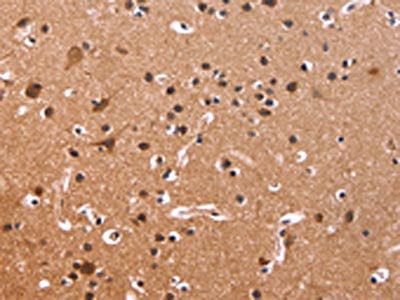
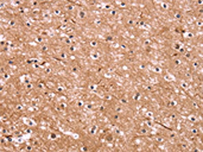
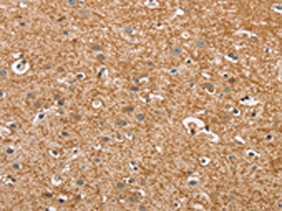
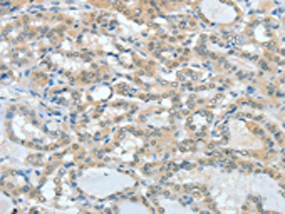
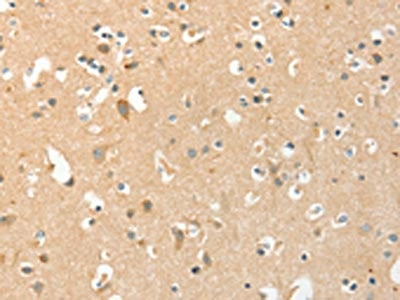
 IHC (Immunohiostchemistry)
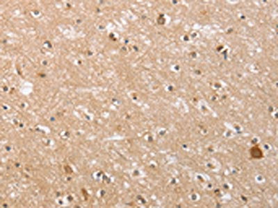
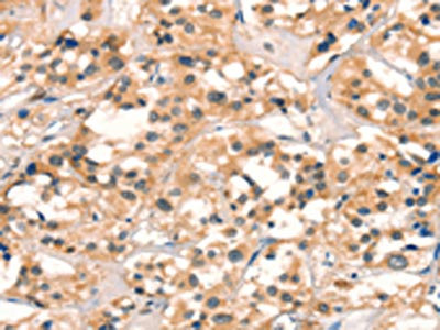
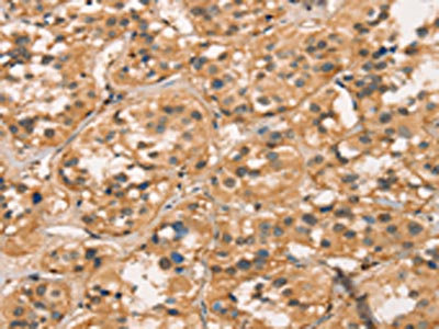
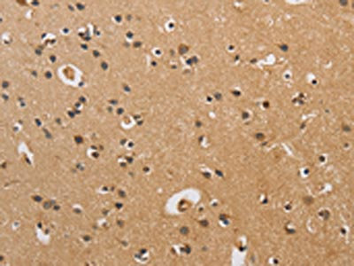
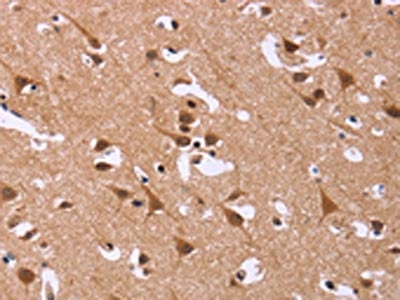
(The image on the left is immunohistochemistry of paraffin-embedded Human brain tissue using AAA238726(DCBLD2 Antibody) at dilution 1/50, on the right is treated with fusion protein. (Original magnification: ×200))
IHC (Immunohiostchemistry)
(The image on the left is immunohistochemistry of paraffin-embedded Human brain tissue using AAA238726(DCBLD2 Antibody) at dilution 1/50, on the right is treated with fusion protein. (Original magnification: ×200))
DCBLD2, Polyclonal Antibody (Cat# AAA238726)
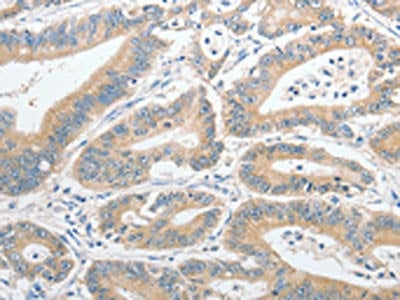
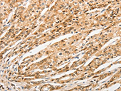
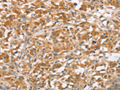
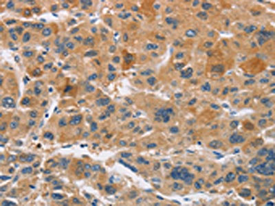
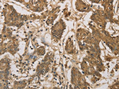
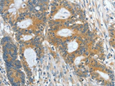
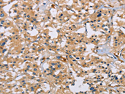
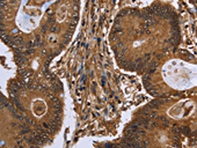 IHC (Immunohiostchemistry)
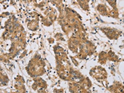
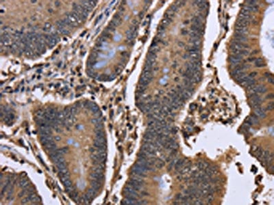
(The image on the left is immunohistochemistry of paraffin-embedded Human colon cancer tissue using AAA238728(DCP1A Antibody) at dilution 1/50, on the right is treated with fusion protein. (Original magnification: ×200))
IHC (Immunohiostchemistry)
(The image on the left is immunohistochemistry of paraffin-embedded Human colon cancer tissue using AAA238728(DCP1A Antibody) at dilution 1/50, on the right is treated with fusion protein. (Original magnification: ×200))
DCP1A, Polyclonal Antibody (Cat# AAA238728)
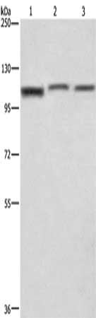
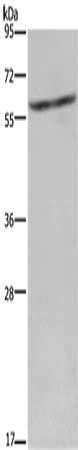
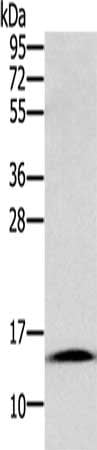
 SDS-PAGE
(Gel: 6%SDS-PAGE, Lysate: 40 ug, Lane: Hela cells, Primary antibody: AAA238729(DCP1A Antibody) at dilution 1/1100, Secondary antibody: Goat anti rabbit IgG at 1/8000 dilution, Exposure time: 10 seconds)
SDS-PAGE
(Gel: 6%SDS-PAGE, Lysate: 40 ug, Lane: Hela cells, Primary antibody: AAA238729(DCP1A Antibody) at dilution 1/1100, Secondary antibody: Goat anti rabbit IgG at 1/8000 dilution, Exposure time: 10 seconds)
DCP1A, Polyclonal Antibody (Cat# AAA238729)
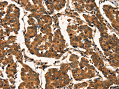
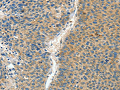
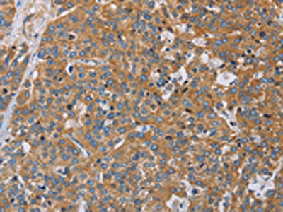
 IHC (Immunohiostchemistry)
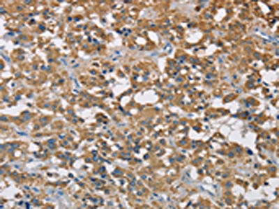
(The image on the left is immunohistochemistry of paraffin-embedded Human thyroid cancer tissue using AAA238730(TNFRSF10D Antibody) at dilution 1/60, on the right is treated with fusion protein. (Original magnification: ×200))
IHC (Immunohiostchemistry)
(The image on the left is immunohistochemistry of paraffin-embedded Human thyroid cancer tissue using AAA238730(TNFRSF10D Antibody) at dilution 1/60, on the right is treated with fusion protein. (Original magnification: ×200))
TNFRSF10D, Polyclonal Antibody (Cat# AAA238730)
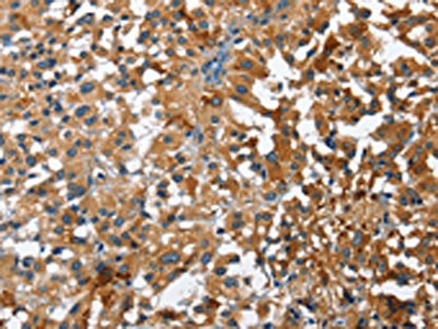
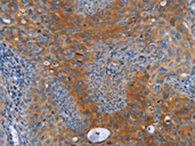
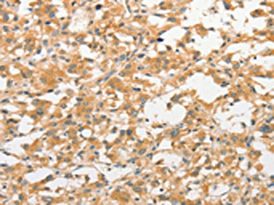
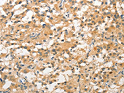
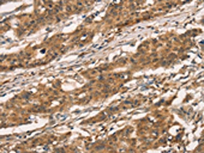
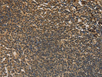
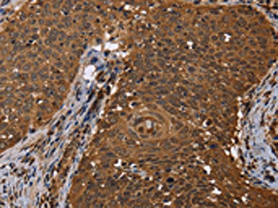
 IHC (Immunohiostchemistry)
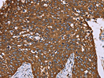
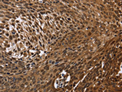
(The image on the left is immunohistochemistry of paraffin-embedded Human cervical cancer tissue using AAA238412(STK17B Antibody) at dilution 1/25, on the right is treated with fusion protein. (Original magnification: ×200))
IHC (Immunohiostchemistry)
(The image on the left is immunohistochemistry of paraffin-embedded Human cervical cancer tissue using AAA238412(STK17B Antibody) at dilution 1/25, on the right is treated with fusion protein. (Original magnification: ×200))
STK17B, Polyclonal Antibody (Cat# AAA238412)
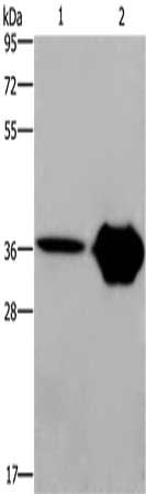
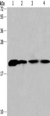
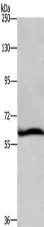
 SDS-PAGE
(Gel: 8%SDS-PAGE, Lysate: 40 ug, Lane 1-3: Human colon cancer tissue, human fetal brain tissue, Human fetal lung tissue, Primary antibody: AAA238417(ASAH1 Antibody) at dilution 1/800, Secondary antibody: Goat anti rabbit IgG at 1/8000 dilution, Exposure time: 30 seconds)
SDS-PAGE
(Gel: 8%SDS-PAGE, Lysate: 40 ug, Lane 1-3: Human colon cancer tissue, human fetal brain tissue, Human fetal lung tissue, Primary antibody: AAA238417(ASAH1 Antibody) at dilution 1/800, Secondary antibody: Goat anti rabbit IgG at 1/8000 dilution, Exposure time: 30 seconds)
ASAH1, Polyclonal Antibody (Cat# AAA238417)
 SDS-PAGE
(Gel: 6%SDS-PAGE, Lysate: 40 ug, Lane: A431 cells, Primary antibody: AAA238422(TRIP4 Antibody) at dilution 1/400, Secondary antibody: Goat anti rabbit IgG at 1/8000 dilution, Exposure time: 10 seconds)
SDS-PAGE
(Gel: 6%SDS-PAGE, Lysate: 40 ug, Lane: A431 cells, Primary antibody: AAA238422(TRIP4 Antibody) at dilution 1/400, Secondary antibody: Goat anti rabbit IgG at 1/8000 dilution, Exposure time: 10 seconds)
TRIP4, Polyclonal Antibody (Cat# AAA238422)
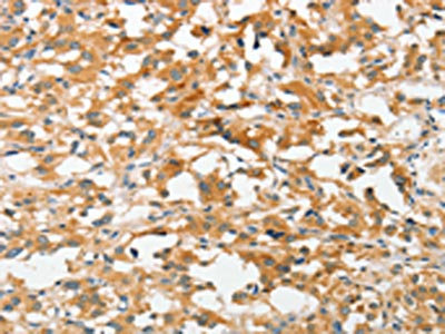
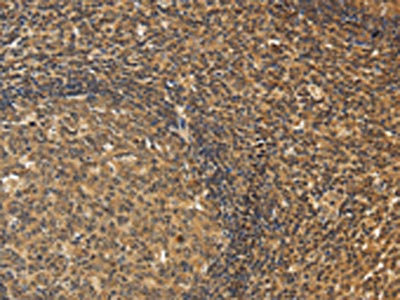
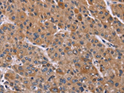
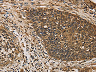
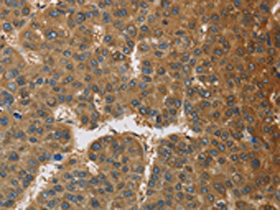
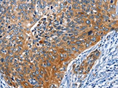
 IHC (Immunohiostchemistry)
(The image on the left is immunohistochemistry of paraffin-embedded Human thyroid cancer tissue using AAA238425(ASH2L Antibody) at dilution 1/60, on the right is treated with fusion protein. (Original magnification: ×200))
IHC (Immunohiostchemistry)
(The image on the left is immunohistochemistry of paraffin-embedded Human thyroid cancer tissue using AAA238425(ASH2L Antibody) at dilution 1/60, on the right is treated with fusion protein. (Original magnification: ×200))
ASH2L, Polyclonal Antibody (Cat# AAA238425)
 SDS-PAGE
(Gel: 8%SDS-PAGE, Lysate: 40 ug, Lane: Mouse brain tissue, Primary antibody: AAA238429(ASPA Antibody) at dilution 1/1150, Secondary antibody: Goat anti rabbit IgG at 1/8000 dilution, Exposure time: 5 seconds)
SDS-PAGE
(Gel: 8%SDS-PAGE, Lysate: 40 ug, Lane: Mouse brain tissue, Primary antibody: AAA238429(ASPA Antibody) at dilution 1/1150, Secondary antibody: Goat anti rabbit IgG at 1/8000 dilution, Exposure time: 5 seconds)
ASPA, Polyclonal Antibody (Cat# AAA238429)
 SDS-PAGE
(Gel: 8%SDS-PAGE, Lysate: 40 ug, Lane 1-2: Mouse muscle tissue, HT29 cells, Primary antibody: AAA238433(HTR3C Antibody) at dilution 1/650, Secondary antibody: Goat anti rabbit IgG at 1/8000 dilution, Exposure time: 30 seconds)
SDS-PAGE
(Gel: 8%SDS-PAGE, Lysate: 40 ug, Lane 1-2: Mouse muscle tissue, HT29 cells, Primary antibody: AAA238433(HTR3C Antibody) at dilution 1/650, Secondary antibody: Goat anti rabbit IgG at 1/8000 dilution, Exposure time: 30 seconds)
HTR3C, Polyclonal Antibody (Cat# AAA238433)
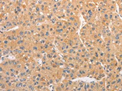
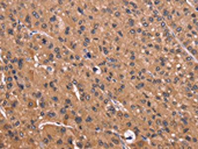
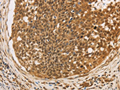
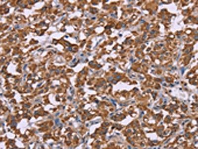
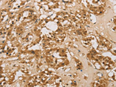
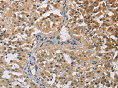
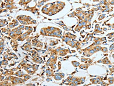
 IHC (Immunohiostchemistry)
(The image on the left is immunohistochemistry of paraffin-embedded Human brain tissue using AAA238434(AANAT Antibody) at dilution 1/40, on the right is treated with fusion protein. (Original magnification: ×200))
IHC (Immunohiostchemistry)
(The image on the left is immunohistochemistry of paraffin-embedded Human brain tissue using AAA238434(AANAT Antibody) at dilution 1/40, on the right is treated with fusion protein. (Original magnification: ×200))
AANAT, Polyclonal Antibody (Cat# AAA238434)
 SDS-PAGE
(Gel: 6%SDS-PAGE, Lysate: 40 ug, Lane 1-4: Lovo cells, mouse kidney tissue, 231 cells, hepG2 cells, Primary antibody: AAA238438(ACTN4 Antibody) at dilution 1/485, Secondary antibody: Goat anti rabbit IgG at 1/8000 dilution, Exposure time: 10 seconds)
SDS-PAGE
(Gel: 6%SDS-PAGE, Lysate: 40 ug, Lane 1-4: Lovo cells, mouse kidney tissue, 231 cells, hepG2 cells, Primary antibody: AAA238438(ACTN4 Antibody) at dilution 1/485, Secondary antibody: Goat anti rabbit IgG at 1/8000 dilution, Exposure time: 10 seconds)
ACTN4, Polyclonal Antibody (Cat# AAA238438)
 SDS-PAGE
(Gel: 10%SDS-PAGE, Lysate: 40 ug, Lane: Mouse liver tissue, Primary antibody: AAA238442(ORM2 Antibody) at dilution 1/700, Secondary antibody: Goat anti rabbit IgG at 1/8000 dilution, Exposure time: 10 seconds)
SDS-PAGE
(Gel: 10%SDS-PAGE, Lysate: 40 ug, Lane: Mouse liver tissue, Primary antibody: AAA238442(ORM2 Antibody) at dilution 1/700, Secondary antibody: Goat anti rabbit IgG at 1/8000 dilution, Exposure time: 10 seconds)
ORM2, Polyclonal Antibody (Cat# AAA238442)
What are Polyclonal Antibodies?
Polyclonal antibodies are antibodies that come from multiple B cell clones of a host animal. The typical hosts used for the majority of polyclonal antibody production are rabbits, goats, sheep, and donkeys. These polyclonal antibodies, once having identified their target, will bind to different epitopes located at different regions or sequences on the same protein/antigen. As a result, they are ideal at locating and binding to the target, even if the target is in very low concentrations (due to many different antibodies being able to bind to the same target molecule, which allows for significant amplification of a downstream signal).
Polyclonal antibodies are typically produced by injecting an antigen into a host animal, which causes the animal’s immune system to attack the foreign antigen by mass generating antibodies against it. After a period of time, serum is collected from the animal and purified using physicochemical fractionation, class-specific affinity purification, and/or antigen-affinity purification.
Key Uses of Polyclonal Antibodies
- Western Blotting: This method is used to find specific proteins in biological samples after separating them by size.
- Immunohistochemistry: IHC helps visualize the location of proteins in tissue sections using various staining techniques.
- ELISA: (Enzyme-Linked Immunosorbent Assay) is typically used to identify specific protein quantities in a sample. ELISAs can be either “Quantitative” or “Qualitative”.
- Flow Cytometry: technique that identifies and measures the specific protein on the surface or inside the cells in a fluid suspension.
- Immunoprecipitation: IP isolates and studies a specific protein from a complex mixture using antibodies.
Why Buy Polyclonal Antibodies from AAA Biotech?
1. Ideal for Various Applications
Our antibodies are generally going to be validated for use in multiple types of assays, including ELISA, Western Blotting, Immunohistochemistry, Immunoprecipitation, amongst others. They are ideal for a wide range of research applications.
2. Rigorous Quality Control
All of the antibodies in our catalog undergo strict quality testing to ensure specificity, sensitivity, and consistent performance. We are confident in the ability of our antibodies to provide you with accurate results.
3. Wide Assortment of Antibodies
Antibodies in are catalog can be found for both common and exotic species, and these antibodies are also available in both conjugated and recombinant forms to suit many diverse experimental needs.
4. Highly Purified
Our antibodies are available in purified forms with over 85% purity, as confirmed by SDS-PAGE. They are also available with tags such as His, Flag, GST, or MBP. We cater to customers worldwide.
FAQ
1. How are polyclonal antibodies produced?
Traditionally, polyclonal antibodies are produced by injecting an antigen into a host animal (such as a rabbit or goat), which then triggers an immune response from the host animal. The animal’s B cells produce antibodies that will recognize different parts of the injected antigen. These antibodies are then collected from the animal’s blood and purified for use.
2. How do polyclonal antibodies differ from monoclonal antibodies?
Polyclonal antibodies are a mix of antibodies that bind to different locations (epitopes) of the same antigen, while monoclonal antibodies are identical and bind to just one specific epitope. This makes polyclonal antibodies more versatile and better at detecting proteins that may be present in low quantities or in altered/modified forms.
3. How should I store polyclonal antibodies?
Polyclonal antibodies should be stored at 4°C for short-term use (up to a few weeks) and at -20°C or -80°C for long-term storage. Avoid repeated freeze-thaw cycles by dividing them into small aliquots. Always check the datasheet for specific storage instructions.