Filters
▼Clonality
▼Type
▼Reactivity
▼Gene Name
▼Isotype
▼Host
▼Application
▼Clone
▼Polyclonal Antibodies
At AAA Biotech also known as AAA Bio or AAABio, we provide a broad range of purified polyclonal antibodies (pAbs) that are able to all be browsed online through our website. Due to their high specificity and strong binding affinity, these antibodies are ideal for wide swathes of research and experimental applications.
Our polyclonal antibodies can easily support your work, whether you use them for Western Blotting, Immunocytochemistry (with or without Immunofluorescence used in conjunction), Immunohistochemistry, Immunoprecipitation, and ELISA tests. We highly encourage you to browse our range of pAbs and choose the one that best suits your experimental model.
Viewing 950-1000 of 96805 product results
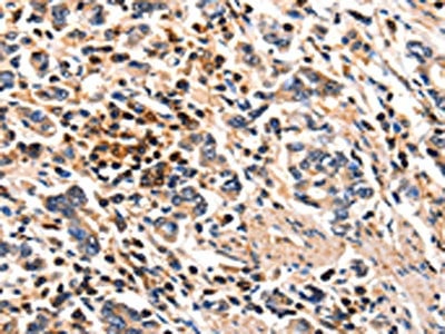
 IHC (Immunohiostchemistry)
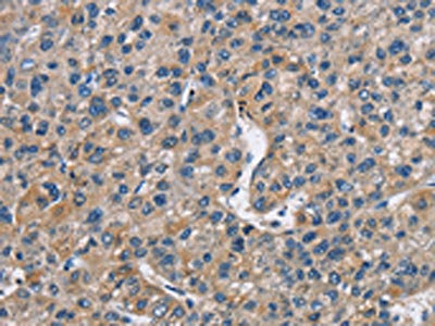
(The image on the left is immunohistochemistry of paraffin-embedded Human liver cancer tissue using AAA238192(ARHGAP15 Antibody) at dilution 1/30, on the right is treated with fusion protein. (Original magnification: ×200))
IHC (Immunohiostchemistry)
(The image on the left is immunohistochemistry of paraffin-embedded Human liver cancer tissue using AAA238192(ARHGAP15 Antibody) at dilution 1/30, on the right is treated with fusion protein. (Original magnification: ×200))
ARHGAP15, Polyclonal Antibody (Cat# AAA238192)
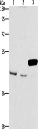
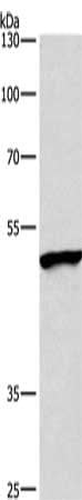
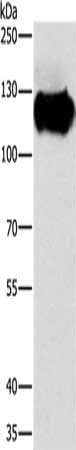
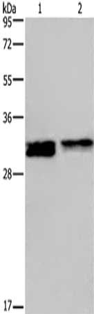
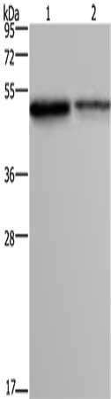
 SDS-PAGE
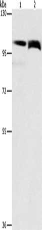
(Gel: 8%SDS-PAGE, Lysate: 40 ug, Lane 1-2: Hela cells, K562 cells, Primary antibody: AAA238196(ARHGEF5 Antibody) at dilution 1/1150, Secondary antibody: Goat anti rabbit IgG at 1/8000 dilution, Exposure time: 1 minute)
SDS-PAGE
(Gel: 8%SDS-PAGE, Lysate: 40 ug, Lane 1-2: Hela cells, K562 cells, Primary antibody: AAA238196(ARHGEF5 Antibody) at dilution 1/1150, Secondary antibody: Goat anti rabbit IgG at 1/8000 dilution, Exposure time: 1 minute)
ARHGEF5, Polyclonal Antibody (Cat# AAA238196)
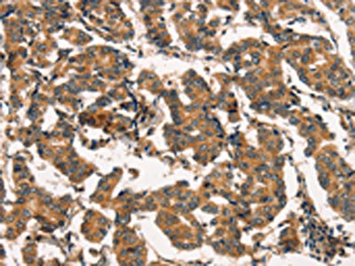
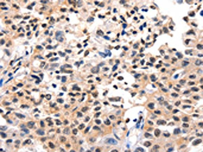
 IHC (Immunohiostchemistry)
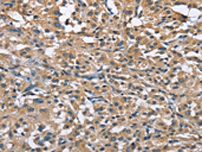
(The image on the left is immunohistochemistry of paraffin-embedded Human thyroid cancer tissue using AAA238206(DIRAS3 Antibody) at dilution 1/25, on the right is treated with fusion protein. (Original magnification: ×200))
IHC (Immunohiostchemistry)
(The image on the left is immunohistochemistry of paraffin-embedded Human thyroid cancer tissue using AAA238206(DIRAS3 Antibody) at dilution 1/25, on the right is treated with fusion protein. (Original magnification: ×200))
DIRAS3, Polyclonal Antibody (Cat# AAA238206)
 SDS-PAGE
(Gel: 8%SDS-PAGE, Lysate: 40 ug, Lane 1-3: A172 cells, K562 cells, mouse pancreas tissue, Primary antibody: AAA238216(ARMCX3 Antibody) at dilution 1/900, Secondary antibody: Goat anti rabbit IgG at 1/8000 dilution, Exposure time: 30 seconds)
SDS-PAGE
(Gel: 8%SDS-PAGE, Lysate: 40 ug, Lane 1-3: A172 cells, K562 cells, mouse pancreas tissue, Primary antibody: AAA238216(ARMCX3 Antibody) at dilution 1/900, Secondary antibody: Goat anti rabbit IgG at 1/8000 dilution, Exposure time: 30 seconds)
ARMCX3, Polyclonal Antibody (Cat# AAA238216)
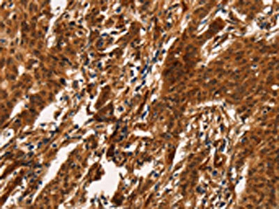 IHC (Immunohiostchemistry)
(The image on the left is immunohistochemistry of paraffin-embedded Human gastric cancer tissue using AAA238217(ARL6IP1 Antibody) at dilution 1/20, on the right is treated with fusion protein. (Original magnification: ×200))
IHC (Immunohiostchemistry)
(The image on the left is immunohistochemistry of paraffin-embedded Human gastric cancer tissue using AAA238217(ARL6IP1 Antibody) at dilution 1/20, on the right is treated with fusion protein. (Original magnification: ×200))
ARL6IP1, Polyclonal Antibody (Cat# AAA238217)
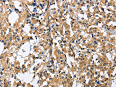
 IHC (Immunohiostchemistry)
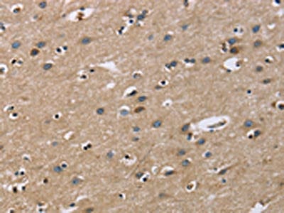
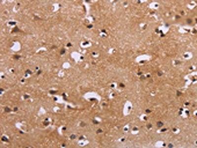
(The image on the left is immunohistochemistry of paraffin-embedded Human brain tissue using AAA238219(GAB1 Antibody) at dilution 1/40, on the right is treated with fusion protein. (Original magnification: ×200))
IHC (Immunohiostchemistry)
(The image on the left is immunohistochemistry of paraffin-embedded Human brain tissue using AAA238219(GAB1 Antibody) at dilution 1/40, on the right is treated with fusion protein. (Original magnification: ×200))
GAB1, Polyclonal Antibody (Cat# AAA238219)
 SDS-PAGE
(Gel: 6%SDS-PAGE, Lysate: 40 ug, Lane 1-2: Lovo cells, 231 cells, Primary antibody: AAA238223(FLT3 Antibody) at dilution 1/240, Secondary antibody: Goat anti rabbit IgG at 1/8000 dilution, Exposure time: 15 seconds)
SDS-PAGE
(Gel: 6%SDS-PAGE, Lysate: 40 ug, Lane 1-2: Lovo cells, 231 cells, Primary antibody: AAA238223(FLT3 Antibody) at dilution 1/240, Secondary antibody: Goat anti rabbit IgG at 1/8000 dilution, Exposure time: 15 seconds)
FLT3, Polyclonal Antibody (Cat# AAA238223)
 IHC (Immunohiostchemistry)
(The image on the left is immunohistochemistry of paraffin-embedded Human thyroid cancer tissue using AAA238226(SPIRE2 Antibody) at dilution 1/30, on the right is treated with fusion protein. (Original magnification: ×200))
IHC (Immunohiostchemistry)
(The image on the left is immunohistochemistry of paraffin-embedded Human thyroid cancer tissue using AAA238226(SPIRE2 Antibody) at dilution 1/30, on the right is treated with fusion protein. (Original magnification: ×200))
SPIRE2, Polyclonal Antibody (Cat# AAA238226)
 IHC (Immunohiostchemistry)
(The image on the left is immunohistochemistry of paraffin-embedded Human colon cancer tissue using AAA238232(PRDM14 Antibody) at dilution 1/30, on the right is treated with fusion protein. (Original magnification: ×200))
IHC (Immunohiostchemistry)
(The image on the left is immunohistochemistry of paraffin-embedded Human colon cancer tissue using AAA238232(PRDM14 Antibody) at dilution 1/30, on the right is treated with fusion protein. (Original magnification: ×200))
PRDM14, Polyclonal Antibody (Cat# AAA238232)
 SDS-PAGE
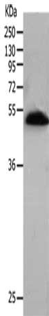
(Gel: 10%SDS-PAGE, Lysate: 60 ug, Lane: A549 cells, Primary antibody: AAA238498(BMP15 Antibody) at dilution 1/266.7, Secondary antibody: Goat anti rabbit IgG at 1/8000 dilution, Exposure time: 5 minutes)
SDS-PAGE
(Gel: 10%SDS-PAGE, Lysate: 60 ug, Lane: A549 cells, Primary antibody: AAA238498(BMP15 Antibody) at dilution 1/266.7, Secondary antibody: Goat anti rabbit IgG at 1/8000 dilution, Exposure time: 5 minutes)
BMP15, Polyclonal Antibody (Cat# AAA238498)
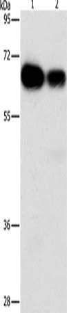
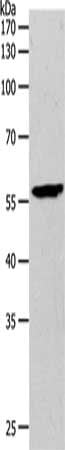
 SDS-PAGE
(Gel: 8%SDS-PAGE, Lysate: 40 ug, Lane: A549 cells, Primary antibody: AAA238499(BMP15 Antibody) at dilution 1/400, Secondary antibody: Goat anti rabbit IgG at 1/8000 dilution, Exposure time: 40 seconds)
SDS-PAGE
(Gel: 8%SDS-PAGE, Lysate: 40 ug, Lane: A549 cells, Primary antibody: AAA238499(BMP15 Antibody) at dilution 1/400, Secondary antibody: Goat anti rabbit IgG at 1/8000 dilution, Exposure time: 40 seconds)
BMP15, Polyclonal Antibody (Cat# AAA238499)
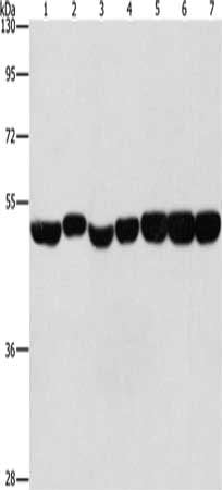 SDS-PAGE
(Gel: 8%SDS-PAGE, Lysate: 40 ug, Lane 1-7: Hela cells, A431 cells, hepG2 cells, mouse brain tissue, Mouse liver tissue, mouse pancreas tissue, human fetal brain tissue, Primary antibody: AAA238505(ENO1 Antibody) at dilution 1/700, Secondary antibody: Goat anti rabbit IgG at 1/8000 dilution, Exposure time: 10 seconds)
SDS-PAGE
(Gel: 8%SDS-PAGE, Lysate: 40 ug, Lane 1-7: Hela cells, A431 cells, hepG2 cells, mouse brain tissue, Mouse liver tissue, mouse pancreas tissue, human fetal brain tissue, Primary antibody: AAA238505(ENO1 Antibody) at dilution 1/700, Secondary antibody: Goat anti rabbit IgG at 1/8000 dilution, Exposure time: 10 seconds)
ENO1, Polyclonal Antibody (Cat# AAA238505)
 IHC (Immunohiostchemistry)
(The image on the left is immunohistochemistry of paraffin-embedded Human thyroid cancer tissue using AAA238508(ZMYND11 Antibody) at dilution 1/70, on the right is treated with fusion protein. (Original magnification: ×200))
IHC (Immunohiostchemistry)
(The image on the left is immunohistochemistry of paraffin-embedded Human thyroid cancer tissue using AAA238508(ZMYND11 Antibody) at dilution 1/70, on the right is treated with fusion protein. (Original magnification: ×200))
ZMYND11, Polyclonal Antibody (Cat# AAA238508)
 SDS-PAGE
(Gel: 6%SDS-PAGE, Lysate: 40 ug, Lane 1-3: Mouse brain tissue, Mouse heart tissue, mouse liver tissue, Primary antibody: AAA238516(CABLES1 Antibody) at dilution 1/350, Secondary antibody: Goat anti rabbit IgG at 1/8000 dilution, Exposure time: 1 minute)
SDS-PAGE
(Gel: 6%SDS-PAGE, Lysate: 40 ug, Lane 1-3: Mouse brain tissue, Mouse heart tissue, mouse liver tissue, Primary antibody: AAA238516(CABLES1 Antibody) at dilution 1/350, Secondary antibody: Goat anti rabbit IgG at 1/8000 dilution, Exposure time: 1 minute)
CABLES1, Polyclonal Antibody (Cat# AAA238516)
 IHC (Immunohiostchemistry)
(The image on the left is immunohistochemistry of paraffin-embedded Human colon cancer tissue using AAA238518(CADM3 Antibody) at dilution 1/40, on the right is treated with fusion protein. (Original magnification: ×200))
IHC (Immunohiostchemistry)
(The image on the left is immunohistochemistry of paraffin-embedded Human colon cancer tissue using AAA238518(CADM3 Antibody) at dilution 1/40, on the right is treated with fusion protein. (Original magnification: ×200))
CADM3, Polyclonal Antibody (Cat# AAA238518)
 SDS-PAGE
(Gel: 8%SDS-PAGE, Lysate: 40 ug, Lane: Lovo cells, Primary antibody: AAA238524(CAST Antibody) at dilution 1/750, Secondary antibody: Goat anti rabbit IgG at 1/8000 dilution, Exposure time: 7 seconds)
SDS-PAGE
(Gel: 8%SDS-PAGE, Lysate: 40 ug, Lane: Lovo cells, Primary antibody: AAA238524(CAST Antibody) at dilution 1/750, Secondary antibody: Goat anti rabbit IgG at 1/8000 dilution, Exposure time: 7 seconds)
CAST, Polyclonal Antibody (Cat# AAA238524)
 SDS-PAGE
(Gel: 8%SDS-PAGE, Lysate: 40 ug, Lane: Lovo cells, Primary antibody: AAA238525(CAST Antibody) at dilution 1/600, Secondary antibody: Goat anti rabbit IgG at 1/8000 dilution, Exposure time: 7 seconds)
SDS-PAGE
(Gel: 8%SDS-PAGE, Lysate: 40 ug, Lane: Lovo cells, Primary antibody: AAA238525(CAST Antibody) at dilution 1/600, Secondary antibody: Goat anti rabbit IgG at 1/8000 dilution, Exposure time: 7 seconds)
CAST, Polyclonal Antibody (Cat# AAA238525)
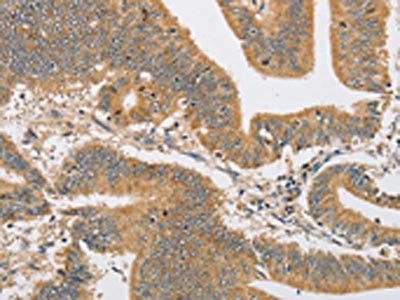
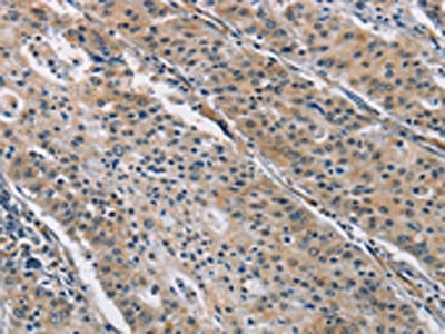
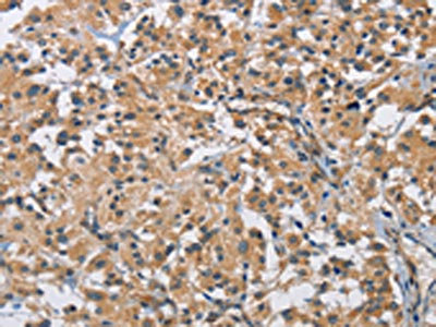
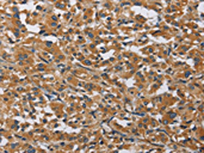
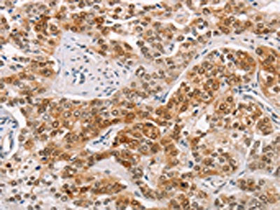
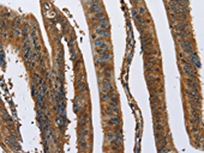
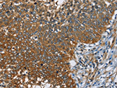
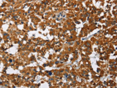
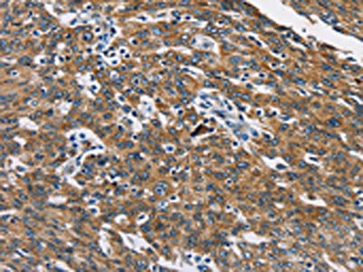
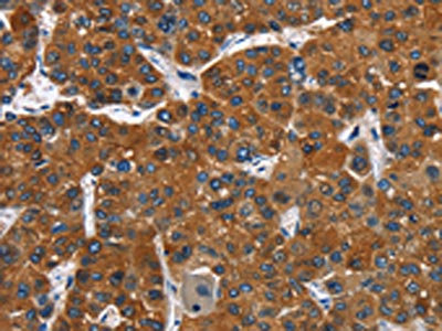
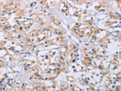
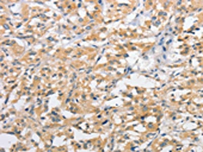
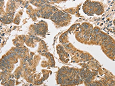
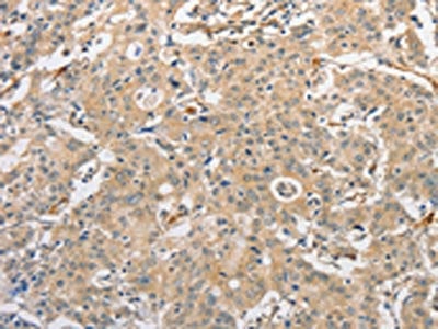
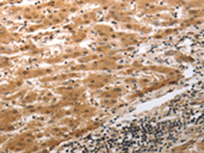
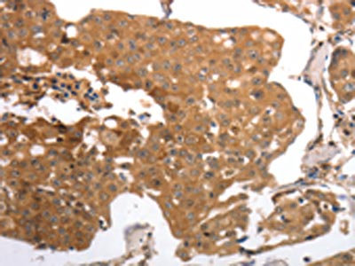
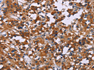
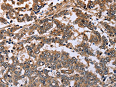
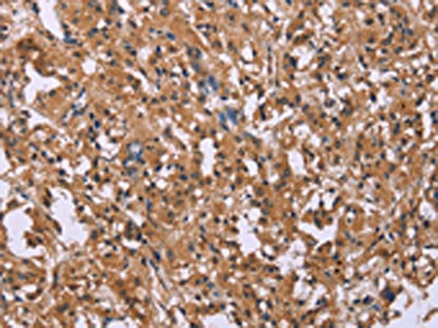
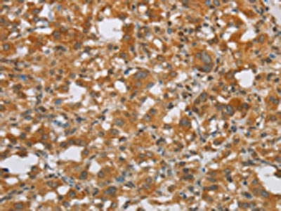
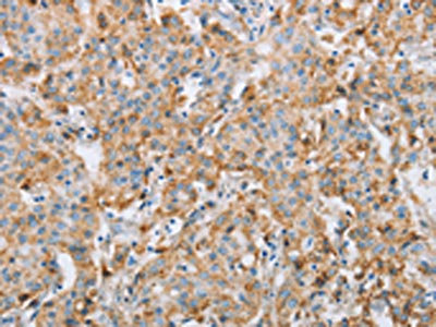
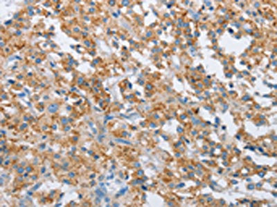
 IHC (Immunohiostchemistry)
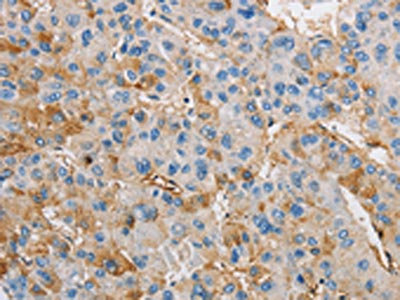
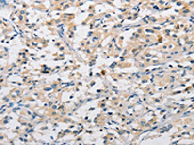
(The image is immunohistochemistry of paraffin-embedded Human liver cancer tissue using AAA238527(CALU Antibody) at dilution 1/40. (Original magnification: ×200))
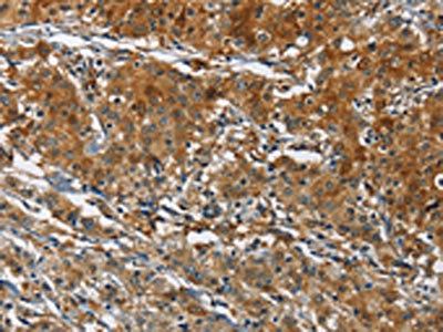
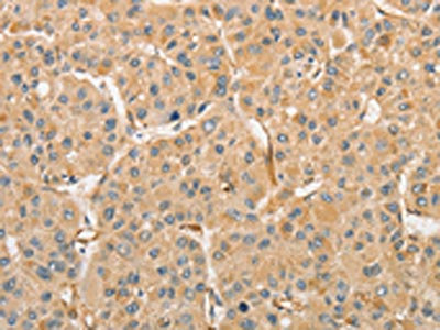
IHC (Immunohiostchemistry)
(The image is immunohistochemistry of paraffin-embedded Human liver cancer tissue using AAA238527(CALU Antibody) at dilution 1/40. (Original magnification: ×200))
CALU, Polyclonal Antibody (Cat# AAA238527)
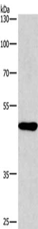
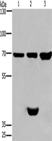
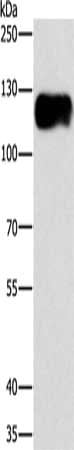
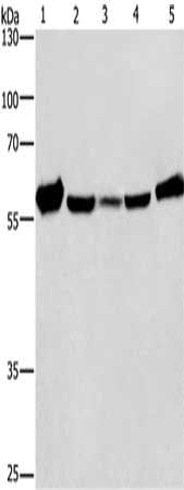
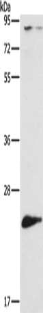
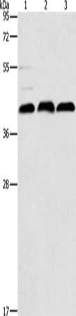
 SDS-PAGE
(Gel: 8%SDS-PAGE, Lysate: 40 ug, Lane 1-5: Hela cells, mouse liver tissue, 231 cells, human placenta tissue, human carcinoma of sigmoid tissue, Primary antibody: AAA238533(CANX Antibody) at dilution 1/420, Secondary antibody: Goat anti rabbit IgG at 1/8000 dilution, Exposure time: 20 seconds)
SDS-PAGE
(Gel: 8%SDS-PAGE, Lysate: 40 ug, Lane 1-5: Hela cells, mouse liver tissue, 231 cells, human placenta tissue, human carcinoma of sigmoid tissue, Primary antibody: AAA238533(CANX Antibody) at dilution 1/420, Secondary antibody: Goat anti rabbit IgG at 1/8000 dilution, Exposure time: 20 seconds)
CANX, Polyclonal Antibody (Cat# AAA238533)
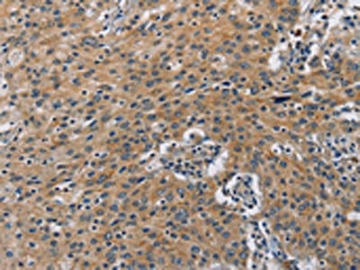
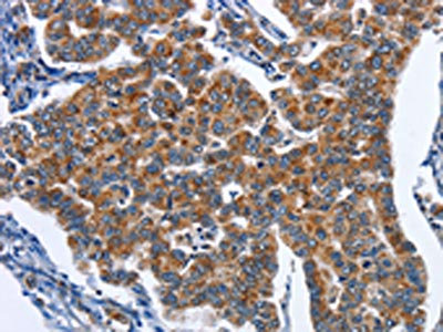
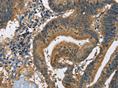
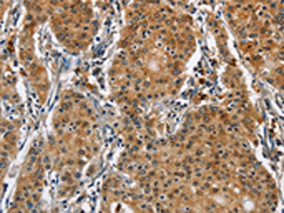
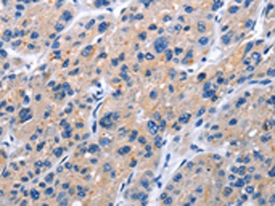
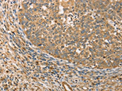
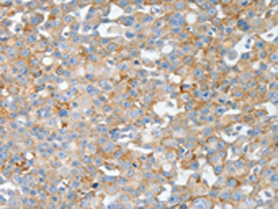
 IHC (Immunohiostchemistry)
(The image on the left is immunohistochemistry of paraffin-embedded Human thyroid cancer tissue using AAA238542(CARD9 Antibody) at dilution 1/70, on the right is treated with fusion protein. (Original magnification: ×200))
IHC (Immunohiostchemistry)
(The image on the left is immunohistochemistry of paraffin-embedded Human thyroid cancer tissue using AAA238542(CARD9 Antibody) at dilution 1/70, on the right is treated with fusion protein. (Original magnification: ×200))
CARD9, Polyclonal Antibody (Cat# AAA238542)
 SDS-PAGE
(Gel: 6%SDS-PAGE, Lysate: 40 ug, Lane: Human fetal brain tissue, Primary antibody: AAA238544(CARD14 Antibody) at dilution 1/650, Secondary antibody: Goat anti rabbit IgG at 1/8000 dilution, Exposure time: 10 minutes)
SDS-PAGE
(Gel: 6%SDS-PAGE, Lysate: 40 ug, Lane: Human fetal brain tissue, Primary antibody: AAA238544(CARD14 Antibody) at dilution 1/650, Secondary antibody: Goat anti rabbit IgG at 1/8000 dilution, Exposure time: 10 minutes)
CARD14, Polyclonal Antibody (Cat# AAA238544)
 SDS-PAGE
(Gel: 6%SDS-PAGE, Lysate: 40 ug, Lane 1-2: 293T cells, A549 cells, Primary antibody: AAA238548(CASC3 Antibody) at dilution 1/400, Secondary antibody: Goat anti rabbit IgG at 1/8000 dilution, Exposure time: 10 seconds)
SDS-PAGE
(Gel: 6%SDS-PAGE, Lysate: 40 ug, Lane 1-2: 293T cells, A549 cells, Primary antibody: AAA238548(CASC3 Antibody) at dilution 1/400, Secondary antibody: Goat anti rabbit IgG at 1/8000 dilution, Exposure time: 10 seconds)
CASC3, Polyclonal Antibody (Cat# AAA238548)
 SDS-PAGE
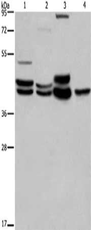
(Gel: 8%SDS-PAGE, Lysate: 40 ug, Lane 1-5: Mouse liver tissue, Raji cells, hela cells, hepG2 cells, A549 cells, Primary antibody: AAA238549(CAT Antibody) at dilution 1/266.6, Secondary antibody: Goat anti rabbit IgG at 1/8000 dilution, Exposure time: 20 seconds)
SDS-PAGE
(Gel: 8%SDS-PAGE, Lysate: 40 ug, Lane 1-5: Mouse liver tissue, Raji cells, hela cells, hepG2 cells, A549 cells, Primary antibody: AAA238549(CAT Antibody) at dilution 1/266.6, Secondary antibody: Goat anti rabbit IgG at 1/8000 dilution, Exposure time: 20 seconds)
CAT, Polyclonal Antibody (Cat# AAA238549)
 SDS-PAGE
(Gel: 8%SDS-PAGE, Lysate: 40 ug, Lane: Hela cells, Primary antibody: AAA238550(CAT Antibody) at dilution 1/300, Secondary antibody: Goat anti rabbit IgG at 1/8000 dilution, Exposure time: 3 minutes)
SDS-PAGE
(Gel: 8%SDS-PAGE, Lysate: 40 ug, Lane: Hela cells, Primary antibody: AAA238550(CAT Antibody) at dilution 1/300, Secondary antibody: Goat anti rabbit IgG at 1/8000 dilution, Exposure time: 3 minutes)
CAT, Polyclonal Antibody (Cat# AAA238550)
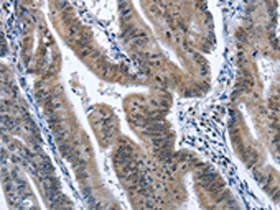
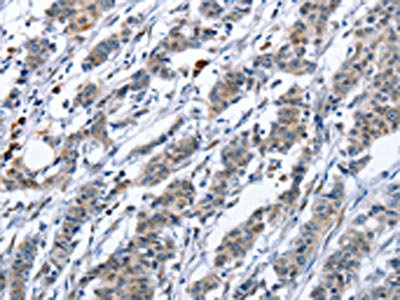
 IHC (Immunohiostchemistry)
(The image on the left is immunohistochemistry of paraffin-embedded Human thyroid cancer tissue using AAA238553(CATSPER3 Antibody) at dilution 1/60, on the right is treated with fusion protein. (Original magnification: ×200))
IHC (Immunohiostchemistry)
(The image on the left is immunohistochemistry of paraffin-embedded Human thyroid cancer tissue using AAA238553(CATSPER3 Antibody) at dilution 1/60, on the right is treated with fusion protein. (Original magnification: ×200))
CATSPER3, Polyclonal Antibody (Cat# AAA238553)
 IHC (Immunohiostchemistry)
(The image on the left is immunohistochemistry of paraffin-embedded Human thyroid cancer tissue using AAA238556(CCR9 Antibody) at dilution 1/60, on the right is treated with fusion protein. (Original magnification: ×200))
IHC (Immunohiostchemistry)
(The image on the left is immunohistochemistry of paraffin-embedded Human thyroid cancer tissue using AAA238556(CCR9 Antibody) at dilution 1/60, on the right is treated with fusion protein. (Original magnification: ×200))
CCR9, Polyclonal Antibody (Cat# AAA238556)
 IHC (Immunohiostchemistry)
(The image on the left is immunohistochemistry of paraffin-embedded Human gastric cancer tissue using AAA238560(TNFRSF9 Antibody) at dilution 1/40, on the right is treated with fusion protein. (Original magnification: ×200))
IHC (Immunohiostchemistry)
(The image on the left is immunohistochemistry of paraffin-embedded Human gastric cancer tissue using AAA238560(TNFRSF9 Antibody) at dilution 1/40, on the right is treated with fusion protein. (Original magnification: ×200))
TNFRSF9, Polyclonal Antibody (Cat# AAA238560)
 IHC (Immunohiostchemistry)
(The image on the left is immunohistochemistry of paraffin-embedded Human thyroid cancer tissue using AAA238564(KIR2DL3/KIR2DL1/KIR2DL4/KIR2DS4 Antibody) at dilution 1/30, on the right is treated with fusion protein. (Original magnification: ×200))
IHC (Immunohiostchemistry)
(The image on the left is immunohistochemistry of paraffin-embedded Human thyroid cancer tissue using AAA238564(KIR2DL3/KIR2DL1/KIR2DL4/KIR2DS4 Antibody) at dilution 1/30, on the right is treated with fusion protein. (Original magnification: ×200))
KIR2DL3/KIR2DL1/KIR2DL4/KIR2DS4, Polyclonal Antibody (Cat# AAA238564)
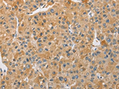
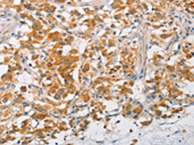
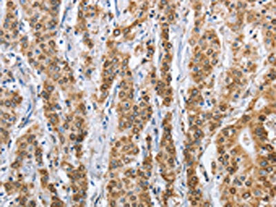
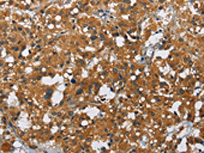
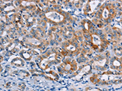
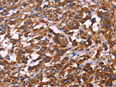
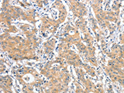
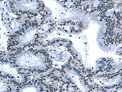
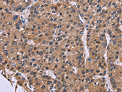
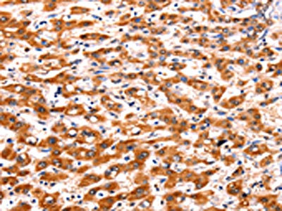
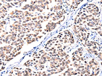
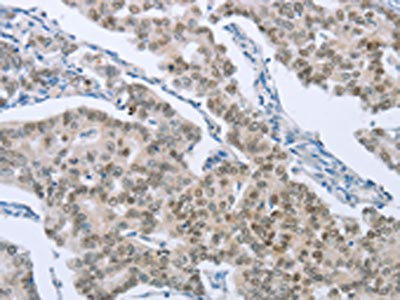
 IHC (Immunohiostchemistry)
(The image on the left is immunohistochemistry of paraffin-embedded Human liver cancer tissue using AAA238135(ANGPTL7 Antibody) at dilution 1/30, on the right is treated with fusion protein. (Original magnification: ×200))
IHC (Immunohiostchemistry)
(The image on the left is immunohistochemistry of paraffin-embedded Human liver cancer tissue using AAA238135(ANGPTL7 Antibody) at dilution 1/30, on the right is treated with fusion protein. (Original magnification: ×200))
ANGPTL7, Polyclonal Antibody (Cat# AAA238135)
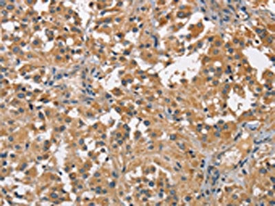
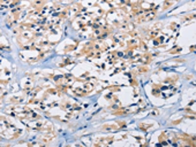
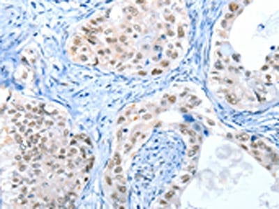
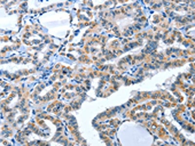 IHC (Immunohiostchemistry)
(The image on the left is immunohistochemistry of paraffin-embedded Human thyroid cancer tissue using AAA238136(ACE2 Antibody) at dilution 1/30, on the right is treated with fusion protein. (Original magnification: ×200))
IHC (Immunohiostchemistry)
(The image on the left is immunohistochemistry of paraffin-embedded Human thyroid cancer tissue using AAA238136(ACE2 Antibody) at dilution 1/30, on the right is treated with fusion protein. (Original magnification: ×200))
ACE2, Polyclonal Antibody (Cat# AAA238136)
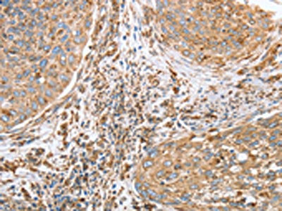
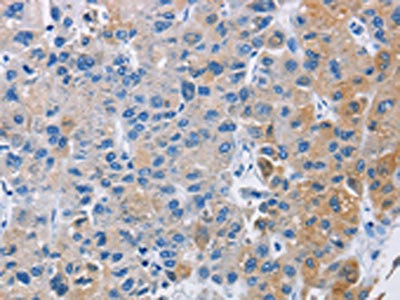
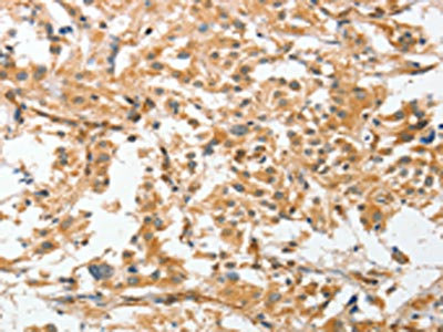
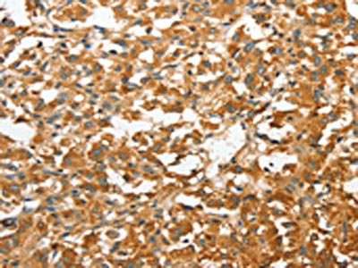
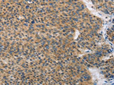
 IHC (Immunohiostchemistry)
(The image on the left is immunohistochemistry of paraffin-embedded Human gastric cancer tissue using AAA238139(ANKMY2 Antibody) at dilution 1/30, on the right is treated with fusion protein. (Original magnification: ×200))
IHC (Immunohiostchemistry)
(The image on the left is immunohistochemistry of paraffin-embedded Human gastric cancer tissue using AAA238139(ANKMY2 Antibody) at dilution 1/30, on the right is treated with fusion protein. (Original magnification: ×200))
ANKMY2, Polyclonal Antibody (Cat# AAA238139)
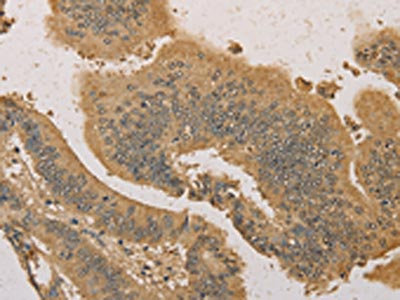
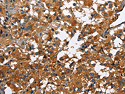
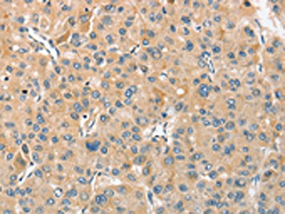
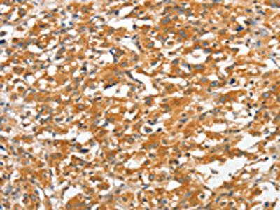
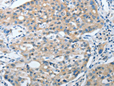
 IHC (Immunohiostchemistry)
(The image on the left is immunohistochemistry of paraffin-embedded Human liver cancer tissue using AAA238145(ANXA9 Antibody) at dilution 1/30, on the right is treated with fusion protein. (Original magnification: ×200))
IHC (Immunohiostchemistry)
(The image on the left is immunohistochemistry of paraffin-embedded Human liver cancer tissue using AAA238145(ANXA9 Antibody) at dilution 1/30, on the right is treated with fusion protein. (Original magnification: ×200))
ANXA9, Polyclonal Antibody (Cat# AAA238145)
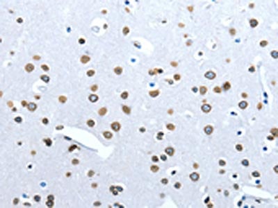
 IHC (Immunohiostchemistry)
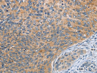
(The image on the left is immunohistochemistry of paraffin-embedded Human cervical cancer tissue using AAA238150(RPL15 Antibody) at dilution 1/25, on the right is treated with fusion protein. (Original magnification: ×200))
IHC (Immunohiostchemistry)
(The image on the left is immunohistochemistry of paraffin-embedded Human cervical cancer tissue using AAA238150(RPL15 Antibody) at dilution 1/25, on the right is treated with fusion protein. (Original magnification: ×200))
RPL15, Polyclonal Antibody (Cat# AAA238150)
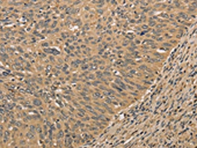
 IHC (Immunohiostchemistry)
(The image on the left is immunohistochemistry of paraffin-embedded Human lung cancer tissue using AAA238157(ANAPC13 Antibody) at dilution 1/25, on the right is treated with fusion protein. (Original magnification: ×200))
IHC (Immunohiostchemistry)
(The image on the left is immunohistochemistry of paraffin-embedded Human lung cancer tissue using AAA238157(ANAPC13 Antibody) at dilution 1/25, on the right is treated with fusion protein. (Original magnification: ×200))
ANAPC13, Polyclonal Antibody (Cat# AAA238157)
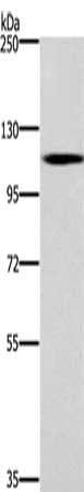
 SDS-PAGE
(Gel: 10%SDS-PAGE, Lysate: 50 ug, Lane: Mouse liver tissue, Primary antibody: AAA238158(ATG10 Antibody) at dilution 1/500, Secondary antibody: Goat anti rabbit IgG at 1/8000 dilution, Exposure time: 1 minute)
SDS-PAGE
(Gel: 10%SDS-PAGE, Lysate: 50 ug, Lane: Mouse liver tissue, Primary antibody: AAA238158(ATG10 Antibody) at dilution 1/500, Secondary antibody: Goat anti rabbit IgG at 1/8000 dilution, Exposure time: 1 minute)
ATG10, Polyclonal Antibody (Cat# AAA238158)
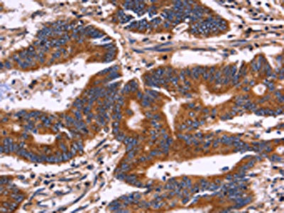
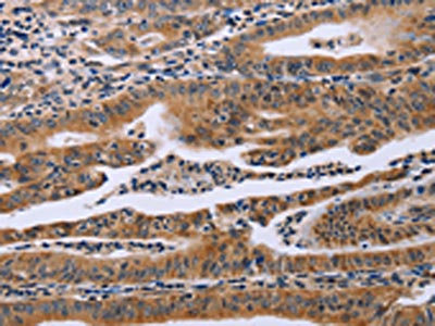
 IHC (Immunohiostchemistry)
(The image on the left is immunohistochemistry of paraffin-embedded Human colon cancer tissue using AAA238160(ATG3 Antibody) at dilution 1/40, on the right is treated with fusion protein. (Original magnification: ×200))
IHC (Immunohiostchemistry)
(The image on the left is immunohistochemistry of paraffin-embedded Human colon cancer tissue using AAA238160(ATG3 Antibody) at dilution 1/40, on the right is treated with fusion protein. (Original magnification: ×200))
ATG3, Polyclonal Antibody (Cat# AAA238160)
 SDS-PAGE
(Gel: 12%SDS-PAGE, Lysate: 40 ug, Lane 1-3: K562 cells, mouse kidney tissue, Raji cells, Primary antibody: AAA238161(ATG3 Antibody) at dilution 1/325, Secondary antibody: Goat anti rabbit IgG at 1/8000 dilution, Exposure time: 10 seconds)
SDS-PAGE
(Gel: 12%SDS-PAGE, Lysate: 40 ug, Lane 1-3: K562 cells, mouse kidney tissue, Raji cells, Primary antibody: AAA238161(ATG3 Antibody) at dilution 1/325, Secondary antibody: Goat anti rabbit IgG at 1/8000 dilution, Exposure time: 10 seconds)
ATG3, Polyclonal Antibody (Cat# AAA238161)
 SDS-PAGE
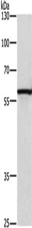
(Gel: 10%SDS-PAGE, Lysate: 80 ug, Lane: Human lung tissue, Primary antibody: AAA240035(BPIFB2 Antibody) at dilution 1/530, Secondary antibody: Goat anti rabbit IgG at 1/8000 dilution, Exposure time: 10 minutes)
SDS-PAGE
(Gel: 10%SDS-PAGE, Lysate: 80 ug, Lane: Human lung tissue, Primary antibody: AAA240035(BPIFB2 Antibody) at dilution 1/530, Secondary antibody: Goat anti rabbit IgG at 1/8000 dilution, Exposure time: 10 minutes)
BPIFB2, Polyclonal Antibody (Cat# AAA240035)
 SDS-PAGE
(Gel: 10%SDS-PAGE, Lysate: 80 ug, Lane: Mouse brain tissue, Primary antibody: AAA240080(MMP27 Antibody) at dilution 1/500, Secondary antibody: Goat anti rabbit IgG at 1/8000 dilution, Exposure time: 2 minutes)
SDS-PAGE
(Gel: 10%SDS-PAGE, Lysate: 80 ug, Lane: Mouse brain tissue, Primary antibody: AAA240080(MMP27 Antibody) at dilution 1/500, Secondary antibody: Goat anti rabbit IgG at 1/8000 dilution, Exposure time: 2 minutes)
MMP27, Polyclonal Antibody (Cat# AAA240080)
 SDS-PAGE
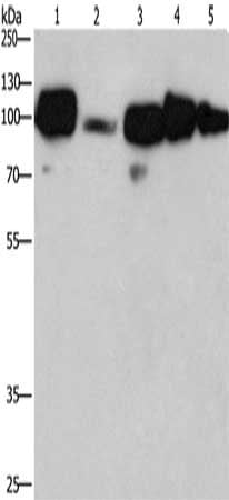
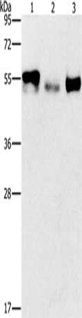
(Gel: 8%SDS-PAGE,Lysate: 40 ug,Lane 1-4: 293T cells, Human brain malignant glioma tissue, Mouse brain tissue, Human hepatocellsular carcinoma tissue,Primary antibody: AAA239855(MAPK1/MAPK3 Antibody) at dilution 1/550 dilution,Secondary antibody: Goat anti rabbit IgG at 1/8000 dilution,Exposure time: 20 seconds)
SDS-PAGE
(Gel: 8%SDS-PAGE,Lysate: 40 ug,Lane 1-4: 293T cells, Human brain malignant glioma tissue, Mouse brain tissue, Human hepatocellsular carcinoma tissue,Primary antibody: AAA239855(MAPK1/MAPK3 Antibody) at dilution 1/550 dilution,Secondary antibody: Goat anti rabbit IgG at 1/8000 dilution,Exposure time: 20 seconds)
MAPK1/MAPK3, Polyclonal Antibody (Cat# AAA239855)
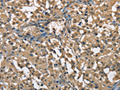
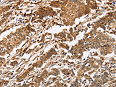
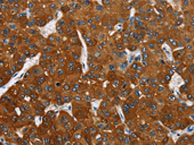
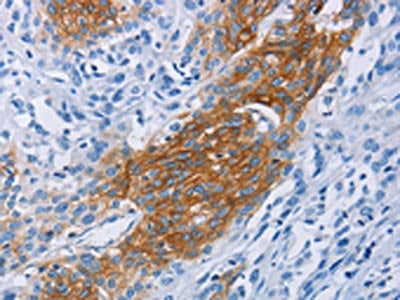
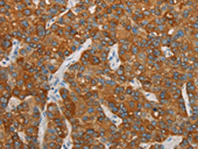
 IHC (Immunohiostchemistry)
(The image on the left is immunohistochemistry of paraffin-embedded Human liver cancer tissue using AAA239860(FAF1 Antibody) at dilution 1/40, on the right is treated with synthetic peptide. (Original magnification: ×200))
IHC (Immunohiostchemistry)
(The image on the left is immunohistochemistry of paraffin-embedded Human liver cancer tissue using AAA239860(FAF1 Antibody) at dilution 1/40, on the right is treated with synthetic peptide. (Original magnification: ×200))
FAF1, Polyclonal Antibody (Cat# AAA239860)
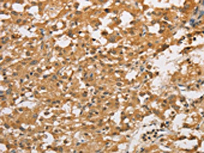
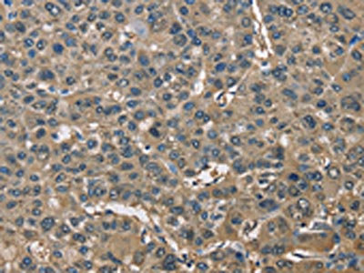
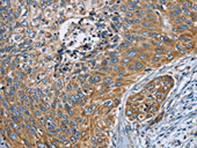
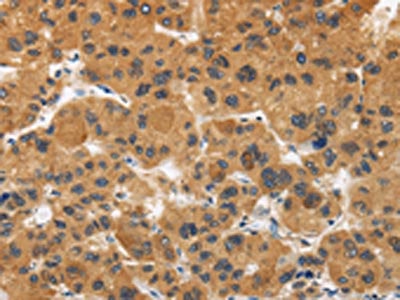
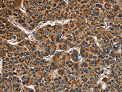
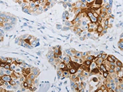
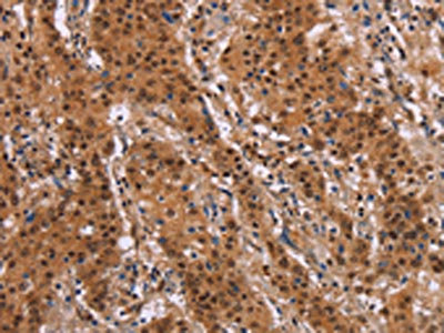
 IHC (Immunohiostchemistry)
(The image on the left is immunohistochemistry of paraffin-embedded Human liver cancer tissue using AAA239864(FGFR2 Antibody) at dilution 1/20, on the right is treated with synthetic peptide. (Original magnification: ×200))
IHC (Immunohiostchemistry)
(The image on the left is immunohistochemistry of paraffin-embedded Human liver cancer tissue using AAA239864(FGFR2 Antibody) at dilution 1/20, on the right is treated with synthetic peptide. (Original magnification: ×200))
FGFR2, Polyclonal Antibody (Cat# AAA239864)

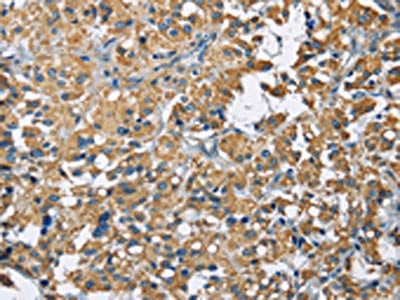
 IHC (Immunohiostchemistry)
(The image on the left is immunohistochemistry of paraffin-embedded Human liver cancer tissue using AAA239865(FBLN1 Antibody) at dilution 1/47, on the right is treated with synthetic peptide. (Original magnification: ×200))
IHC (Immunohiostchemistry)
(The image on the left is immunohistochemistry of paraffin-embedded Human liver cancer tissue using AAA239865(FBLN1 Antibody) at dilution 1/47, on the right is treated with synthetic peptide. (Original magnification: ×200))
FBLN1, Polyclonal Antibody (Cat# AAA239865)
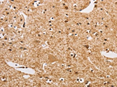
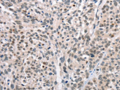
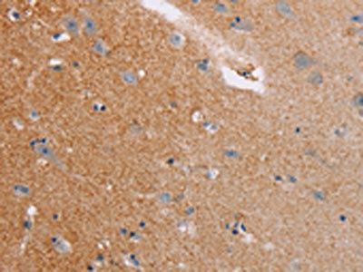
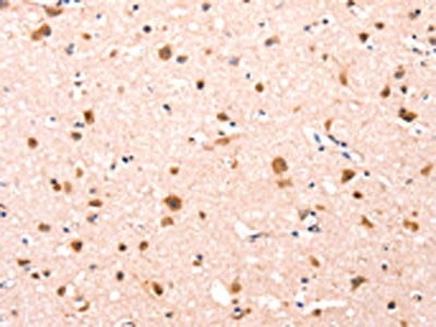 IHC (Immunohiostchemistry)
(The image on the left is immunohistochemistry of paraffin-embedded Human brain tissue using AAA239866(FBLN1 Antibody) at dilution 1/12, on the right is treated with synthetic peptide. (Original magnification: ×200))
IHC (Immunohiostchemistry)
(The image on the left is immunohistochemistry of paraffin-embedded Human brain tissue using AAA239866(FBLN1 Antibody) at dilution 1/12, on the right is treated with synthetic peptide. (Original magnification: ×200))
FBLN1, Polyclonal Antibody (Cat# AAA239866)
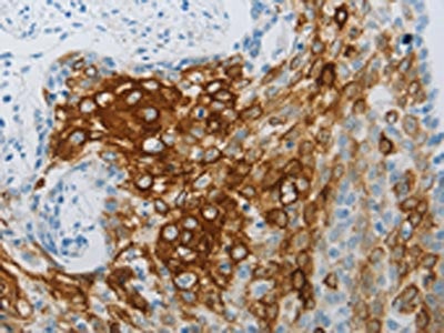
 IHC (Immunohiostchemistry)
(The image on the left is immunohistochemistry of paraffin-embedded Human esophagus cancer tissue using AAA239867(FLG Antibody) at dilution 1/30, on the right is treated with synthetic peptide. (Original magnification: ×200))
IHC (Immunohiostchemistry)
(The image on the left is immunohistochemistry of paraffin-embedded Human esophagus cancer tissue using AAA239867(FLG Antibody) at dilution 1/30, on the right is treated with synthetic peptide. (Original magnification: ×200))
FLG, Polyclonal Antibody (Cat# AAA239867)
 SDS-PAGE
(Gel: 8%SDS-PAGE, Lysate: 40 ug, Lane 1-3: Human kidney cancer tissue, Human lung cancer tissue, human placenta tissue, Primary antibody: AAA239868(FOSB Antibody) at dilution 1/200, Secondary antibody: Goat anti rabbit IgG at 1/8000 dilution, Exposure time: 10 minutes)
SDS-PAGE
(Gel: 8%SDS-PAGE, Lysate: 40 ug, Lane 1-3: Human kidney cancer tissue, Human lung cancer tissue, human placenta tissue, Primary antibody: AAA239868(FOSB Antibody) at dilution 1/200, Secondary antibody: Goat anti rabbit IgG at 1/8000 dilution, Exposure time: 10 minutes)
FOSB, Polyclonal Antibody (Cat# AAA239868)
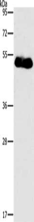
 SDS-PAGE
(Gel: 10%SDS-PAGE,Lysate: 40 ug,Lane 1-2: Hela cells, Jurkat cells,Primary antibody: AAA239869(FOSL1 Antibody) at dilution 1/615 dilution,Secondary antibody: Goat anti rabbit IgG at 1/8000 dilution,Exposure time: 20 seconds)
SDS-PAGE
(Gel: 10%SDS-PAGE,Lysate: 40 ug,Lane 1-2: Hela cells, Jurkat cells,Primary antibody: AAA239869(FOSL1 Antibody) at dilution 1/615 dilution,Secondary antibody: Goat anti rabbit IgG at 1/8000 dilution,Exposure time: 20 seconds)
FOSL1, Polyclonal Antibody (Cat# AAA239869)
 SDS-PAGE
(Gel: 8%SDS-PAGE, Lysate: 40 ug, Lane: Human placenta tissue, Primary antibody: AAA239872(FOXF2 Antibody) at dilution 1/733, Secondary antibody: Goat anti rabbit IgG at 1/8000 dilution, Exposure time: 5 minutes)
SDS-PAGE
(Gel: 8%SDS-PAGE, Lysate: 40 ug, Lane: Human placenta tissue, Primary antibody: AAA239872(FOXF2 Antibody) at dilution 1/733, Secondary antibody: Goat anti rabbit IgG at 1/8000 dilution, Exposure time: 5 minutes)
FOXF2, Polyclonal Antibody (Cat# AAA239872)
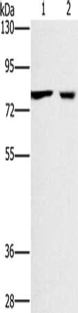
 SDS-PAGE
(Gel: 8%SDS-PAGE, Lysate: 40 ug, Lane 1-2: Human prostate tissue, MCF7 cells, Primary antibody: AAA239873(FOXA1 Antibody) at dilution 1/300, Secondary antibody: Goat anti rabbit IgG at 1/8000 dilution, Exposure time: 5 minutes)
SDS-PAGE
(Gel: 8%SDS-PAGE, Lysate: 40 ug, Lane 1-2: Human prostate tissue, MCF7 cells, Primary antibody: AAA239873(FOXA1 Antibody) at dilution 1/300, Secondary antibody: Goat anti rabbit IgG at 1/8000 dilution, Exposure time: 5 minutes)
FOXA1, Polyclonal Antibody (Cat# AAA239873)
 IHC (Immunohiostchemistry)
(The image on the left is immunohistochemistry of paraffin-embedded Human lung cancer tissue using AAA239875(FOXB2 Antibody) at dilution 1/5, on the right is treated with synthetic peptide. (Original magnification: ×200))
IHC (Immunohiostchemistry)
(The image on the left is immunohistochemistry of paraffin-embedded Human lung cancer tissue using AAA239875(FOXB2 Antibody) at dilution 1/5, on the right is treated with synthetic peptide. (Original magnification: ×200))
FOXB2, Polyclonal Antibody (Cat# AAA239875)
What are Polyclonal Antibodies?
Polyclonal antibodies are antibodies that come from multiple B cell clones of a host animal. The typical hosts used for the majority of polyclonal antibody production are rabbits, goats, sheep, and donkeys. These polyclonal antibodies, once having identified their target, will bind to different epitopes located at different regions or sequences on the same protein/antigen. As a result, they are ideal at locating and binding to the target, even if the target is in very low concentrations (due to many different antibodies being able to bind to the same target molecule, which allows for significant amplification of a downstream signal).
Polyclonal antibodies are typically produced by injecting an antigen into a host animal, which causes the animal’s immune system to attack the foreign antigen by mass generating antibodies against it. After a period of time, serum is collected from the animal and purified using physicochemical fractionation, class-specific affinity purification, and/or antigen-affinity purification.
Key Uses of Polyclonal Antibodies
- Western Blotting: This method is used to find specific proteins in biological samples after separating them by size.
- Immunohistochemistry: IHC helps visualize the location of proteins in tissue sections using various staining techniques.
- ELISA: (Enzyme-Linked Immunosorbent Assay) is typically used to identify specific protein quantities in a sample. ELISAs can be either “Quantitative” or “Qualitative”.
- Flow Cytometry: technique that identifies and measures the specific protein on the surface or inside the cells in a fluid suspension.
- Immunoprecipitation: IP isolates and studies a specific protein from a complex mixture using antibodies.
Why Buy Polyclonal Antibodies from AAA Biotech?
1. Ideal for Various Applications
Our antibodies are generally going to be validated for use in multiple types of assays, including ELISA, Western Blotting, Immunohistochemistry, Immunoprecipitation, amongst others. They are ideal for a wide range of research applications.
2. Rigorous Quality Control
All of the antibodies in our catalog undergo strict quality testing to ensure specificity, sensitivity, and consistent performance. We are confident in the ability of our antibodies to provide you with accurate results.
3. Wide Assortment of Antibodies
Antibodies in are catalog can be found for both common and exotic species, and these antibodies are also available in both conjugated and recombinant forms to suit many diverse experimental needs.
4. Highly Purified
Our antibodies are available in purified forms with over 85% purity, as confirmed by SDS-PAGE. They are also available with tags such as His, Flag, GST, or MBP. We cater to customers worldwide.
FAQ
1. How are polyclonal antibodies produced?
Traditionally, polyclonal antibodies are produced by injecting an antigen into a host animal (such as a rabbit or goat), which then triggers an immune response from the host animal. The animal’s B cells produce antibodies that will recognize different parts of the injected antigen. These antibodies are then collected from the animal’s blood and purified for use.
2. How do polyclonal antibodies differ from monoclonal antibodies?
Polyclonal antibodies are a mix of antibodies that bind to different locations (epitopes) of the same antigen, while monoclonal antibodies are identical and bind to just one specific epitope. This makes polyclonal antibodies more versatile and better at detecting proteins that may be present in low quantities or in altered/modified forms.
3. How should I store polyclonal antibodies?
Polyclonal antibodies should be stored at 4°C for short-term use (up to a few weeks) and at -20°C or -80°C for long-term storage. Avoid repeated freeze-thaw cycles by dividing them into small aliquots. Always check the datasheet for specific storage instructions.