Filters
▼Clonality
▼Type
▼Reactivity
▼Gene Name
▼Isotype
▼Host
▼Application
▼Clone
▼Polyclonal Antibodies
At AAA Biotech also known as AAA Bio or AAABio, we provide a broad range of purified polyclonal antibodies (pAbs) that are able to all be browsed online through our website. Due to their high specificity and strong binding affinity, these antibodies are ideal for wide swathes of research and experimental applications.
Our polyclonal antibodies can easily support your work, whether you use them for Western Blotting, Immunocytochemistry (with or without Immunofluorescence used in conjunction), Immunohistochemistry, Immunoprecipitation, and ELISA tests. We highly encourage you to browse our range of pAbs and choose the one that best suits your experimental model.
Viewing 1700-1750 of 96805 product results
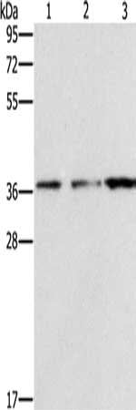
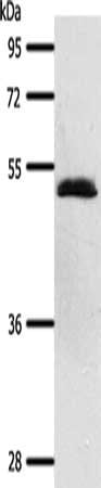
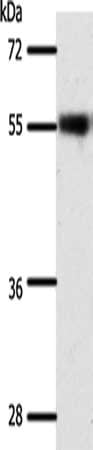
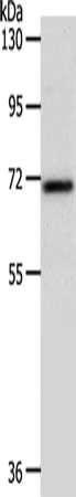
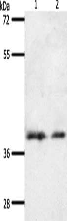
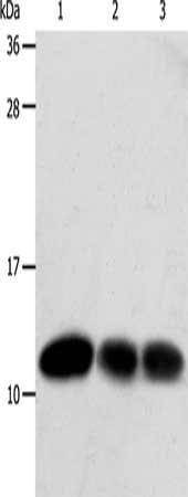
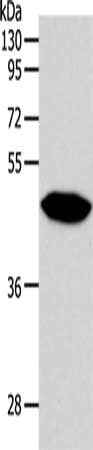
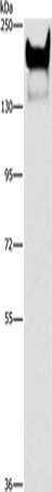
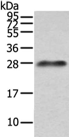
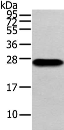
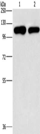 SDS-PAGE
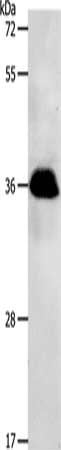
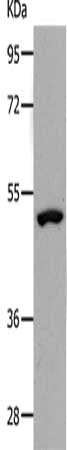
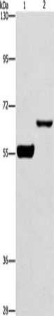
(Gel: 10%SDS-PAGE, Lysate: 40 ug, Lane 1-2: A431 cells, hela cells, Primary antibody: AAA240764(PIP5K1C Antibody) at dilution 1/500, Secondary antibody: Goat anti rabbit IgG at 1/8000 dilution, Exposure time: 2 minutes)
SDS-PAGE
(Gel: 10%SDS-PAGE, Lysate: 40 ug, Lane 1-2: A431 cells, hela cells, Primary antibody: AAA240764(PIP5K1C Antibody) at dilution 1/500, Secondary antibody: Goat anti rabbit IgG at 1/8000 dilution, Exposure time: 2 minutes)
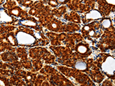
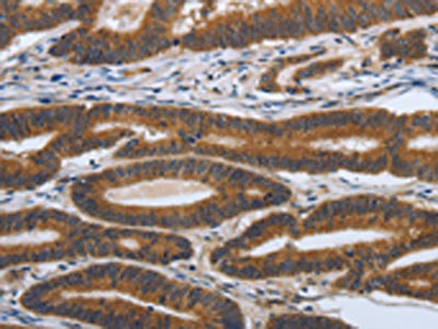
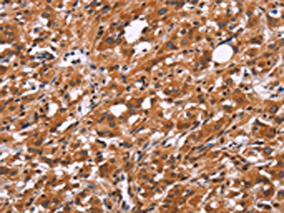
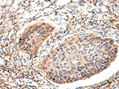
PIP5K1C, Polyclonal Antibody (Cat# AAA240764)
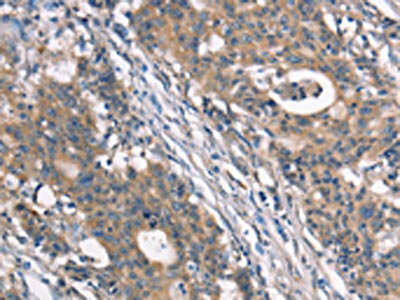
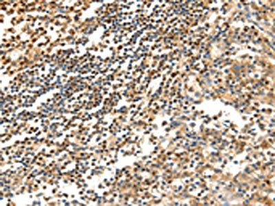
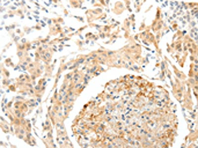
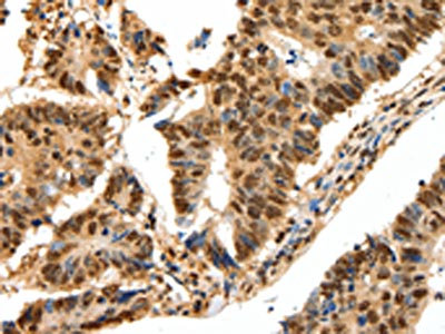
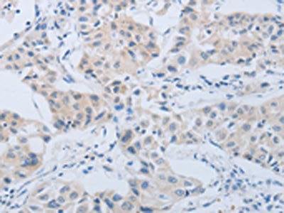
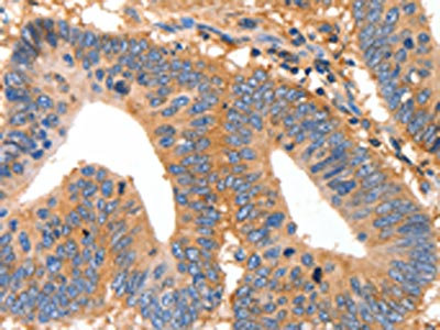
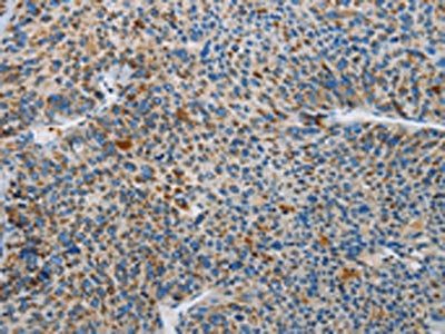 IHC (Immunohiostchemistry)
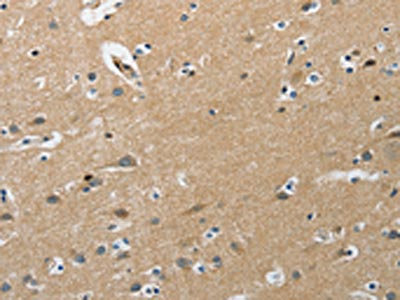
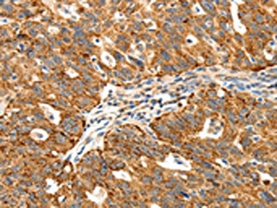
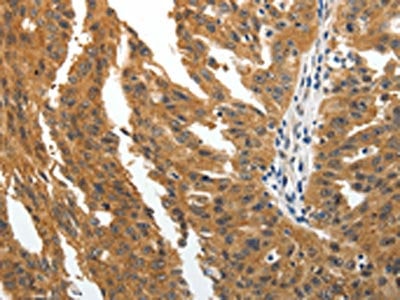
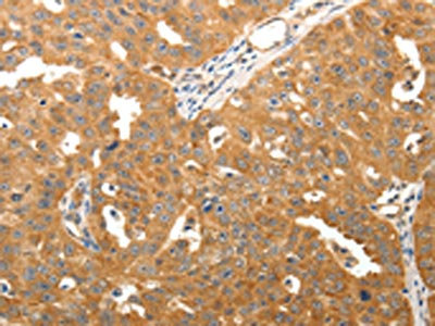
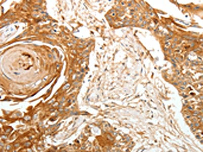
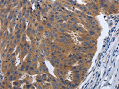
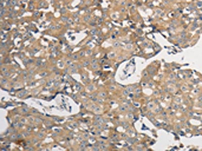
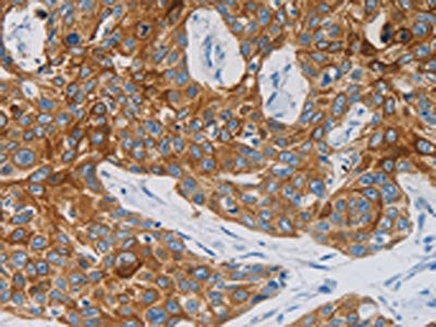
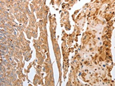
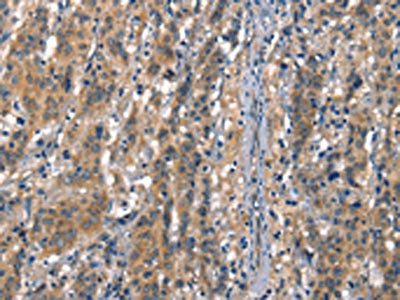
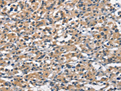
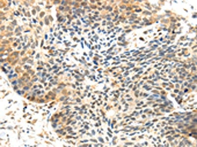
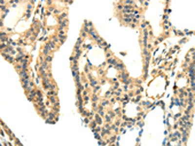
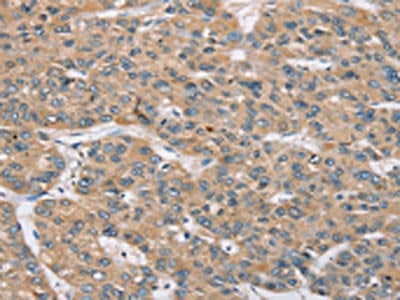
(The image on the left is immunohistochemistry of paraffin-embedded Human liver cancer tissue using AAA240770(NEK5 Antibody) at dilution 1/25, on the right is treated with synthetic peptide. (Original magnification: ×200))
IHC (Immunohiostchemistry)
(The image on the left is immunohistochemistry of paraffin-embedded Human liver cancer tissue using AAA240770(NEK5 Antibody) at dilution 1/25, on the right is treated with synthetic peptide. (Original magnification: ×200))
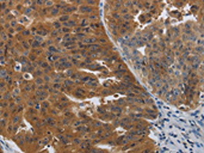
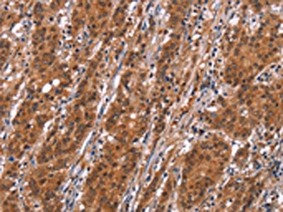
NEK5, Polyclonal Antibody (Cat# AAA240770)
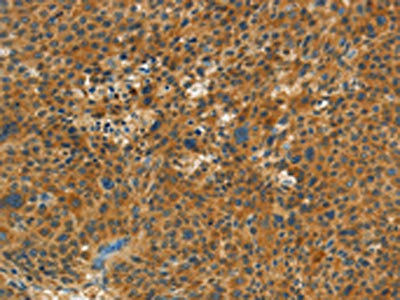
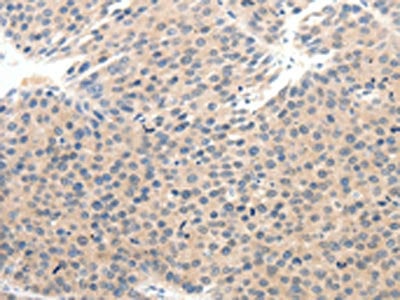
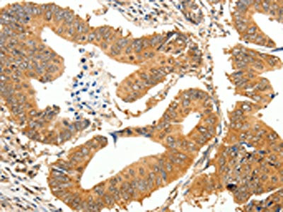
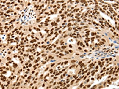
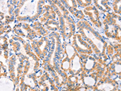
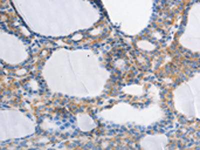
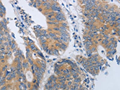
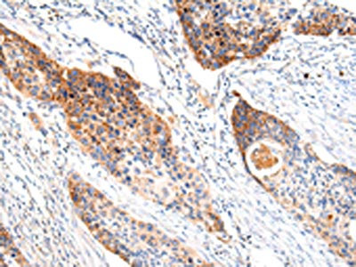
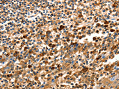
 IHC (Immunohiostchemistry)
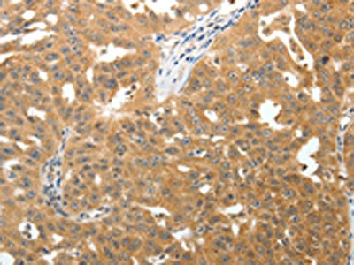
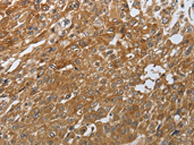
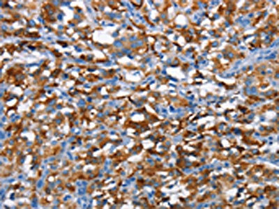
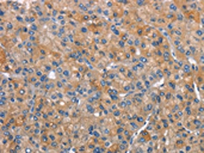
(The image on the left is immunohistochemistry of paraffin-embedded Human thyroid cancer tissue using AAA240777(NPAP1 Antibody) at dilution 1/60, on the right is treated with synthetic peptide. (Original magnification: ×200))
IHC (Immunohiostchemistry)
(The image on the left is immunohistochemistry of paraffin-embedded Human thyroid cancer tissue using AAA240777(NPAP1 Antibody) at dilution 1/60, on the right is treated with synthetic peptide. (Original magnification: ×200))
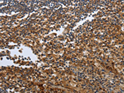
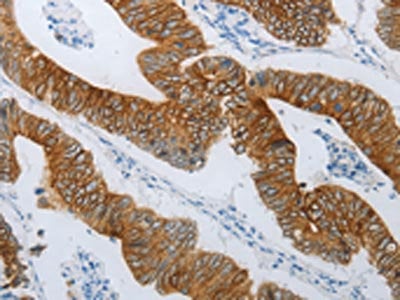
NPAP1, Polyclonal Antibody (Cat# AAA240777)
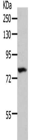
 SDS-PAGE
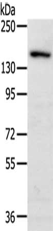
(Gel: 10%SDS-PAGE, Lysate: 40 ug, Lane 1-3: Mouse liver tissue, A172 cells, human prostate tissue, Primary antibody: AAA240780(NDNL2 Antibody) at dilution 1/1500, Secondary antibody: Goat anti rabbit IgG at 1/8000 dilution, Exposure time: 1 minute)
SDS-PAGE
(Gel: 10%SDS-PAGE, Lysate: 40 ug, Lane 1-3: Mouse liver tissue, A172 cells, human prostate tissue, Primary antibody: AAA240780(NDNL2 Antibody) at dilution 1/1500, Secondary antibody: Goat anti rabbit IgG at 1/8000 dilution, Exposure time: 1 minute)
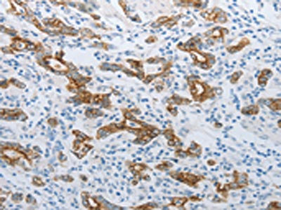
NDNL2, Polyclonal Antibody (Cat# AAA240780)
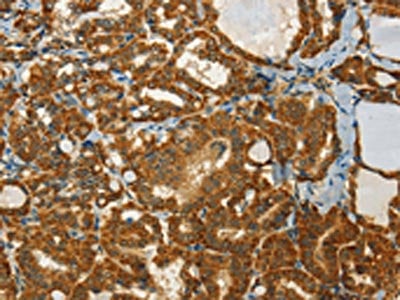
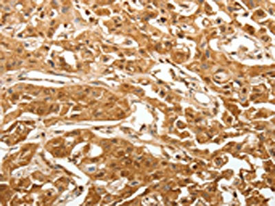
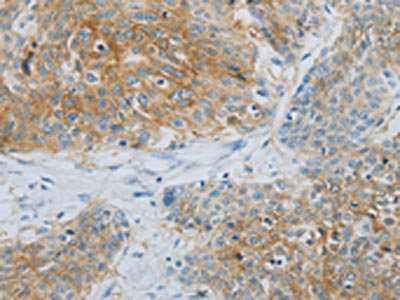
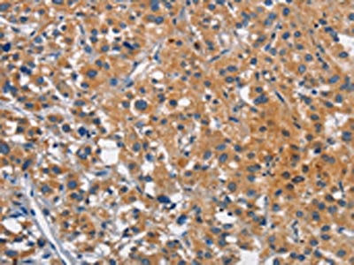
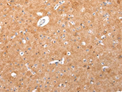
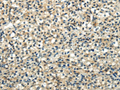
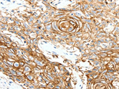 IHC (Immunohiostchemistry)
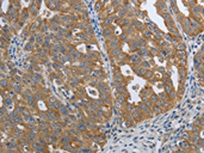
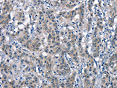
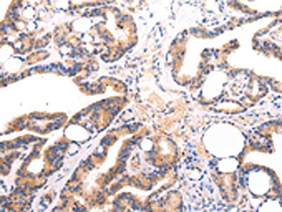
(The image on the left is immunohistochemistry of paraffin-embedded Human esophagus cancer tissue using AAA240538(SCN10A Antibody) at dilution 1/25, on the right is treated with synthetic peptide. (Original magnification: ×200))
IHC (Immunohiostchemistry)
(The image on the left is immunohistochemistry of paraffin-embedded Human esophagus cancer tissue using AAA240538(SCN10A Antibody) at dilution 1/25, on the right is treated with synthetic peptide. (Original magnification: ×200))
SCN10A, Polyclonal Antibody (Cat# AAA240538)
 SDS-PAGE
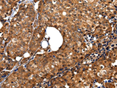
(Gel: 10%SDS-PAGE, Lysate: 40 ug, Lane: Human liver cancer tissue, Primary antibody: AAA240546(SCTR Antibody) at dilution 1/600, Secondary antibody: Goat anti rabbit IgG at 1/8000 dilution, Exposure time: 20 seconds)
SDS-PAGE
(Gel: 10%SDS-PAGE, Lysate: 40 ug, Lane: Human liver cancer tissue, Primary antibody: AAA240546(SCTR Antibody) at dilution 1/600, Secondary antibody: Goat anti rabbit IgG at 1/8000 dilution, Exposure time: 20 seconds)
SCTR, Polyclonal Antibody (Cat# AAA240546)
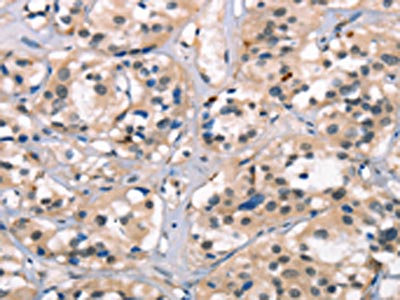
 IHC (Immunohiostchemistry)
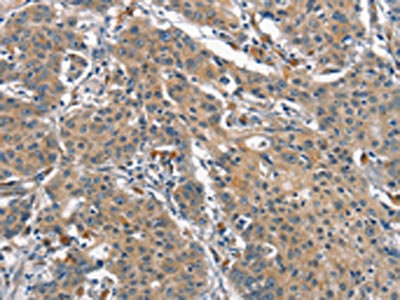
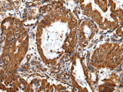
(The image on the left is immunohistochemistry of paraffin-embedded Human cervical cancer tissue using AAA240548(SEMA3D Antibody) at dilution 1/50, on the right is treated with synthetic peptide. (Original magnification: ×200))
IHC (Immunohiostchemistry)
(The image on the left is immunohistochemistry of paraffin-embedded Human cervical cancer tissue using AAA240548(SEMA3D Antibody) at dilution 1/50, on the right is treated with synthetic peptide. (Original magnification: ×200))
SEMA3D, Polyclonal Antibody (Cat# AAA240548)
 SDS-PAGE
(Gel: 10%SDS-PAGE, Lysate: 40 ug, Lane: Mouse eyes tissue, Primary antibody: AAA240556(SLC32A1 Antibody) at dilution 1/500, Secondary antibody: Goat anti rabbit IgG at 1/8000 dilution, Exposure time: 20 seconds)
SDS-PAGE
(Gel: 10%SDS-PAGE, Lysate: 40 ug, Lane: Mouse eyes tissue, Primary antibody: AAA240556(SLC32A1 Antibody) at dilution 1/500, Secondary antibody: Goat anti rabbit IgG at 1/8000 dilution, Exposure time: 20 seconds)
SLC32A1, Polyclonal Antibody (Cat# AAA240556)
 SDS-PAGE
(Gel: 10%SDS-PAGE, Lysate: 40 ug, Lane: Mouse liver tissue, Primary antibody: AAA240557(SOX13 Antibody) at dilution 1/800, Secondary antibody: Goat anti rabbit IgG at 1/8000 dilution, Exposure time: 1 minute)
SDS-PAGE
(Gel: 10%SDS-PAGE, Lysate: 40 ug, Lane: Mouse liver tissue, Primary antibody: AAA240557(SOX13 Antibody) at dilution 1/800, Secondary antibody: Goat anti rabbit IgG at 1/8000 dilution, Exposure time: 1 minute)
SOX13, Polyclonal Antibody (Cat# AAA240557)
 IHC (Immunohiostchemistry)
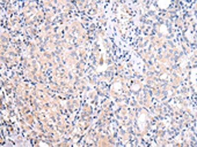
(The image on the left is immunohistochemistry of paraffin-embedded Human ovarian cancer tissue using AAA240559(SP1 Antibody) at dilution 1/30, on the right is treated with synthetic peptide. (Original magnification: ×200))
IHC (Immunohiostchemistry)
(The image on the left is immunohistochemistry of paraffin-embedded Human ovarian cancer tissue using AAA240559(SP1 Antibody) at dilution 1/30, on the right is treated with synthetic peptide. (Original magnification: ×200))
SP1, Polyclonal Antibody (Cat# AAA240559)
 IHC (Immunohiostchemistry)
(The image on the left is immunohistochemistry of paraffin-embedded Human ovarian cancer tissue using AAA240567(SSB Antibody) at dilution 1/20, on the right is treated with synthetic peptide. (Original magnification: ×200))
IHC (Immunohiostchemistry)
(The image on the left is immunohistochemistry of paraffin-embedded Human ovarian cancer tissue using AAA240567(SSB Antibody) at dilution 1/20, on the right is treated with synthetic peptide. (Original magnification: ×200))
SSB, Polyclonal Antibody (Cat# AAA240567)
 IHC (Immunohiostchemistry)
(The image on the left is immunohistochemistry of paraffin-embedded Human thyroid cancer tissue using AAA240571(SSTR1 Antibody) at dilution 1/50, on the right is treated with synthetic peptide. (Original magnification: ×200))
IHC (Immunohiostchemistry)
(The image on the left is immunohistochemistry of paraffin-embedded Human thyroid cancer tissue using AAA240571(SSTR1 Antibody) at dilution 1/50, on the right is treated with synthetic peptide. (Original magnification: ×200))
SSTR1, Polyclonal Antibody (Cat# AAA240571)
 SDS-PAGE
(Gel: 10%SDS-PAGE, Lysate: 60 ug, Lane 1-2: 293T cells, hela cells, Primary antibody: AAA240572(SSTR1 Antibody) at dilution 1/500, Secondary antibody: Goat anti rabbit IgG at 1/8000 dilution, Exposure time: 30 seconds)
SDS-PAGE
(Gel: 10%SDS-PAGE, Lysate: 60 ug, Lane 1-2: 293T cells, hela cells, Primary antibody: AAA240572(SSTR1 Antibody) at dilution 1/500, Secondary antibody: Goat anti rabbit IgG at 1/8000 dilution, Exposure time: 30 seconds)
SSTR1, Polyclonal Antibody (Cat# AAA240572)
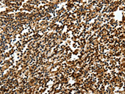
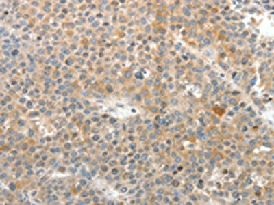
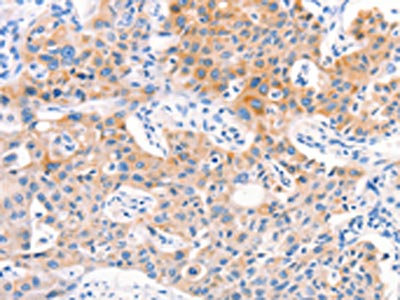
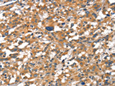
 IHC (Immunohiostchemistry)
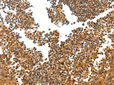
(The image on the left is immunohistochemistry of paraffin-embedded Human ovarian cancer tissue using AAA240581(BIRC6 Antibody) at dilution 1/30, on the right is treated with synthetic peptide. (Original magnification: ×200))
IHC (Immunohiostchemistry)
(The image on the left is immunohistochemistry of paraffin-embedded Human ovarian cancer tissue using AAA240581(BIRC6 Antibody) at dilution 1/30, on the right is treated with synthetic peptide. (Original magnification: ×200))
BIRC6, Polyclonal Antibody (Cat# AAA240581)
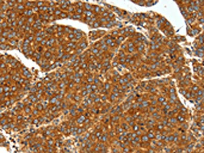
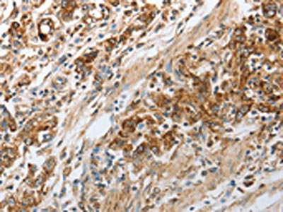
 IHC (Immunohiostchemistry)
(The image on the left is immunohistochemistry of paraffin-embedded Human lung cancer tissue using AAA240584(SV2A Antibody) at dilution 1/25, on the right is treated with synthetic peptide. (Original magnification: ×200))
IHC (Immunohiostchemistry)
(The image on the left is immunohistochemistry of paraffin-embedded Human lung cancer tissue using AAA240584(SV2A Antibody) at dilution 1/25, on the right is treated with synthetic peptide. (Original magnification: ×200))
SV2A, Polyclonal Antibody (Cat# AAA240584)
 IHC (Immunohiostchemistry)
(The image on the left is immunohistochemistry of paraffin-embedded Human ovarian cancer tissue using AAA240594(TNN Antibody) at dilution 1/50, on the right is treated with synthetic peptide. (Original magnification: ×200))
IHC (Immunohiostchemistry)
(The image on the left is immunohistochemistry of paraffin-embedded Human ovarian cancer tissue using AAA240594(TNN Antibody) at dilution 1/50, on the right is treated with synthetic peptide. (Original magnification: ×200))
TNN, Polyclonal Antibody (Cat# AAA240594)
 IHC (Immunohiostchemistry)
(The image on the left is immunohistochemistry of paraffin-embedded Human ovarian cancer tissue using AAA240596(TNR Antibody) at dilution 1/50, on the right is treated with synthetic peptide. (Original magnification: ×200))
IHC (Immunohiostchemistry)
(The image on the left is immunohistochemistry of paraffin-embedded Human ovarian cancer tissue using AAA240596(TNR Antibody) at dilution 1/50, on the right is treated with synthetic peptide. (Original magnification: ×200))
TNR, Polyclonal Antibody (Cat# AAA240596)
 IHC (Immunohiostchemistry)
(The image on the left is immunohistochemistry of paraffin-embedded Human esophagus cancer tissue using AAA240601(TERT Antibody) at dilution 1/40, on the right is treated with synthetic peptide. (Original magnification: ×200))
IHC (Immunohiostchemistry)
(The image on the left is immunohistochemistry of paraffin-embedded Human esophagus cancer tissue using AAA240601(TERT Antibody) at dilution 1/40, on the right is treated with synthetic peptide. (Original magnification: ×200))
TERT, Polyclonal Antibody (Cat# AAA240601)
 IHC (Immunohiostchemistry)
(The image on the left is immunohistochemistry of paraffin-embedded Human lung cancer tissue using AAA240605(F13A1 Antibody) at dilution 1/80, on the right is treated with synthetic peptide. (Original magnification: ×200))
IHC (Immunohiostchemistry)
(The image on the left is immunohistochemistry of paraffin-embedded Human lung cancer tissue using AAA240605(F13A1 Antibody) at dilution 1/80, on the right is treated with synthetic peptide. (Original magnification: ×200))
F13A1, Polyclonal Antibody (Cat# AAA240605)
 SDS-PAGE
(Gel: 12%SDS-PAGE, Lysate: 40 ug, Lane 1-3: Hela cells, MCF7 cells, human liver cancer tissue, Primary antibody: AAA240609(TXN Antibody) at dilution 1/600, Secondary antibody: Goat anti rabbit IgG at 1/8000 dilution, Exposure time: 20 seconds)
SDS-PAGE
(Gel: 12%SDS-PAGE, Lysate: 40 ug, Lane 1-3: Hela cells, MCF7 cells, human liver cancer tissue, Primary antibody: AAA240609(TXN Antibody) at dilution 1/600, Secondary antibody: Goat anti rabbit IgG at 1/8000 dilution, Exposure time: 20 seconds)
TXN, Polyclonal Antibody (Cat# AAA240609)
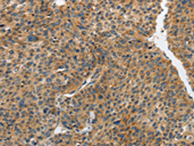
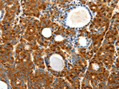
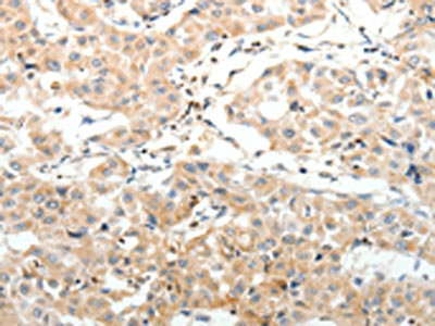
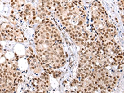
 IHC (Immunohiostchemistry)
(The image on the left is immunohistochemistry of paraffin-embedded Human tonsil tissue using AAA240494(PDCD2 Antibody) at dilution 1/50, on the right is treated with synthetic peptide. (Original magnification: ×200))
IHC (Immunohiostchemistry)
(The image on the left is immunohistochemistry of paraffin-embedded Human tonsil tissue using AAA240494(PDCD2 Antibody) at dilution 1/50, on the right is treated with synthetic peptide. (Original magnification: ×200))
PDCD2, Polyclonal Antibody (Cat# AAA240494)
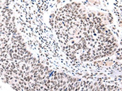
 IHC (Immunohiostchemistry)
(The image on the left is immunohistochemistry of paraffin-embedded Human ovarian cancer tissue using AAA240499(PDGFA Antibody) at dilution 1/70, on the right is treated with synthetic peptide. (Original magnification: ×200))
IHC (Immunohiostchemistry)
(The image on the left is immunohistochemistry of paraffin-embedded Human ovarian cancer tissue using AAA240499(PDGFA Antibody) at dilution 1/70, on the right is treated with synthetic peptide. (Original magnification: ×200))
PDGFA, Polyclonal Antibody (Cat# AAA240499)
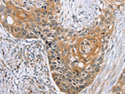
 IHC (Immunohiostchemistry)
(The image on the left is immunohistochemistry of paraffin-embedded Human ovarian cancer tissue using AAA240507(PMS2 Antibody) at dilution 1/50, on the right is treated with synthetic peptide. (Original magnification: ×200))
IHC (Immunohiostchemistry)
(The image on the left is immunohistochemistry of paraffin-embedded Human ovarian cancer tissue using AAA240507(PMS2 Antibody) at dilution 1/50, on the right is treated with synthetic peptide. (Original magnification: ×200))
PMS2, Polyclonal Antibody (Cat# AAA240507)
 SDS-PAGE
(Gel: 12%SDS-PAGE, Lysate: 40 ug, Lane: Jurkat cells, Primary antibody: AAA240510(PPP2CB Antibody) at dilution 1/1200, Secondary antibody: Goat anti rabbit IgG at 1/8000 dilution, Exposure time: 1 minute)
SDS-PAGE
(Gel: 12%SDS-PAGE, Lysate: 40 ug, Lane: Jurkat cells, Primary antibody: AAA240510(PPP2CB Antibody) at dilution 1/1200, Secondary antibody: Goat anti rabbit IgG at 1/8000 dilution, Exposure time: 1 minute)
PPP2CB, Polyclonal Antibody (Cat# AAA240510)
 IHC (Immunohiostchemistry)
(The image on the left is immunohistochemistry of paraffin-embedded Human ovarian cancer tissue using AAA240523(PTPRM Antibody) at dilution 1/80, on the right is treated with synthetic peptide. (Original magnification: ×200))
IHC (Immunohiostchemistry)
(The image on the left is immunohistochemistry of paraffin-embedded Human ovarian cancer tissue using AAA240523(PTPRM Antibody) at dilution 1/80, on the right is treated with synthetic peptide. (Original magnification: ×200))
PTPRM, Polyclonal Antibody (Cat# AAA240523)
 SDS-PAGE
(Gel: 12%SDS-PAGE, Lysate: 40 ug, Lane: Hela cells, Primary antibody: AAA240529(S100A4 Antibody) at dilution 1/500, Secondary antibody: Goat anti rabbit IgG at 1/8000 dilution, Exposure time: 40 seconds)
SDS-PAGE
(Gel: 12%SDS-PAGE, Lysate: 40 ug, Lane: Hela cells, Primary antibody: AAA240529(S100A4 Antibody) at dilution 1/500, Secondary antibody: Goat anti rabbit IgG at 1/8000 dilution, Exposure time: 40 seconds)
S100A4, Polyclonal Antibody (Cat# AAA240529)
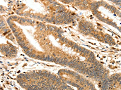
 IHC (Immunohiostchemistry)
(The image on the left is immunohistochemistry of paraffin-embedded Human lung cancer tissue using AAA240426(MUC3A Antibody) at dilution 1/60, on the right is treated with synthetic peptide. (Original magnification: ×200))
IHC (Immunohiostchemistry)
(The image on the left is immunohistochemistry of paraffin-embedded Human lung cancer tissue using AAA240426(MUC3A Antibody) at dilution 1/60, on the right is treated with synthetic peptide. (Original magnification: ×200))
MUC3A, Polyclonal Antibody (Cat# AAA240426)
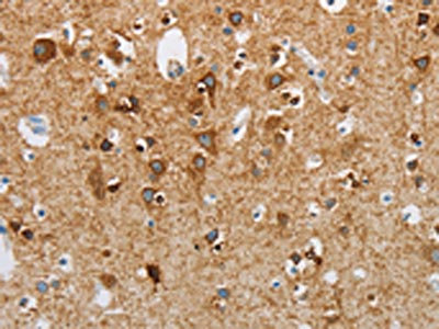
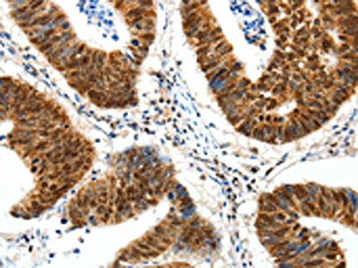
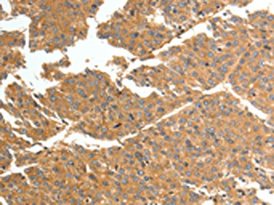
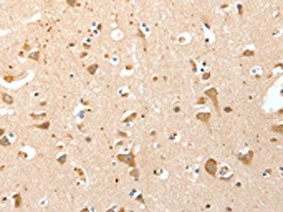
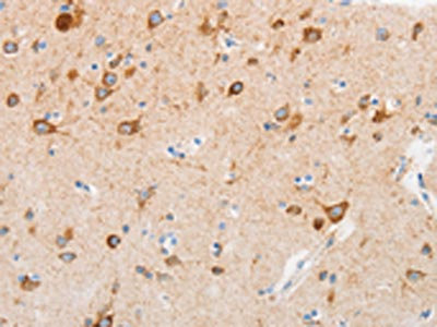 IHC (Immunohiostchemistry)
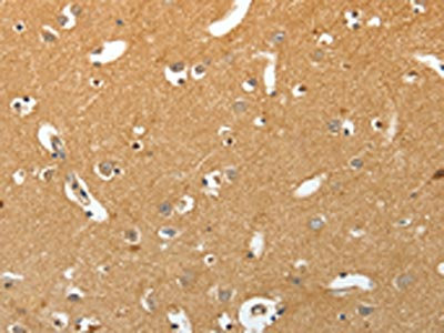
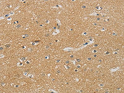
(The image on the left is immunohistochemistry of paraffin-embedded Human brain tissue using AAA240427(MUC13 Antibody) at dilution 1/30, on the right is treated with synthetic peptide. (Original magnification: ×200))
IHC (Immunohiostchemistry)
(The image on the left is immunohistochemistry of paraffin-embedded Human brain tissue using AAA240427(MUC13 Antibody) at dilution 1/30, on the right is treated with synthetic peptide. (Original magnification: ×200))
MUC13, Polyclonal Antibody (Cat# AAA240427)
 SDS-PAGE
(Gel: 10%SDS-PAGE, Lysate: 40 ug, Lane: Human fetal muscle tissue, Primary antibody: AAA240431(TRIM63 Antibody) at dilution 1/1050, Secondary antibody: Goat anti rabbit IgG at 1/8000 dilution, Exposure time: 50 seconds)
SDS-PAGE
(Gel: 10%SDS-PAGE, Lysate: 40 ug, Lane: Human fetal muscle tissue, Primary antibody: AAA240431(TRIM63 Antibody) at dilution 1/1050, Secondary antibody: Goat anti rabbit IgG at 1/8000 dilution, Exposure time: 50 seconds)
TRIM63, Polyclonal Antibody (Cat# AAA240431)
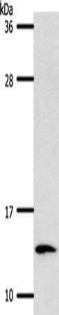
 SDS-PAGE
(Gel: 8%SDS-PAGE, Lysate: 40 ug, Lane: 293T cells, Primary antibody: AAA240433(MME Antibody) at dilution 1/700, Secondary antibody: Goat anti rabbit IgG at 1/8000 dilution, Exposure time: 1 minute)
SDS-PAGE
(Gel: 8%SDS-PAGE, Lysate: 40 ug, Lane: 293T cells, Primary antibody: AAA240433(MME Antibody) at dilution 1/700, Secondary antibody: Goat anti rabbit IgG at 1/8000 dilution, Exposure time: 1 minute)
MME, Polyclonal Antibody (Cat# AAA240433)
 SDS-PAGE
(Gel: 10%SDS-PAGE, Lysate: 40 ug, Lane: Human lymphoma tissue, Primary antibody: AAA240434(NDRG1 Antibody) at dilution 1/1000, Secondary antibody: Goat anti rabbit IgG at 1/8000 dilution, Exposure time: 10 minutes)
SDS-PAGE
(Gel: 10%SDS-PAGE, Lysate: 40 ug, Lane: Human lymphoma tissue, Primary antibody: AAA240434(NDRG1 Antibody) at dilution 1/1000, Secondary antibody: Goat anti rabbit IgG at 1/8000 dilution, Exposure time: 10 minutes)
NDRG1, Polyclonal Antibody (Cat# AAA240434)
 SDS-PAGE
(Gel: 10%SDS-PAGE, Lysate: 30 ug, Lane: Mouse heart tissue, Primary antibody: AAA240439(NDRG4 Antibody) at dilution 1/1200, Secondary antibody: Goat anti rabbit IgG at 1/8000 dilution, Exposure time: 10 seconds)
SDS-PAGE
(Gel: 10%SDS-PAGE, Lysate: 30 ug, Lane: Mouse heart tissue, Primary antibody: AAA240439(NDRG4 Antibody) at dilution 1/1200, Secondary antibody: Goat anti rabbit IgG at 1/8000 dilution, Exposure time: 10 seconds)
NDRG4, Polyclonal Antibody (Cat# AAA240439)
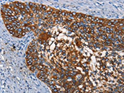
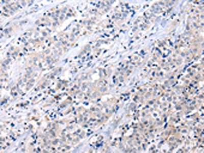
 IHC (Immunohiostchemistry)
(The image on the left is immunohistochemistry of paraffin-embedded Human gastric cancer tissue using AAA240440(SLC8A2 Antibody) at dilution 1/15, on the right is treated with synthetic peptide. (Original magnification: ×200))
IHC (Immunohiostchemistry)
(The image on the left is immunohistochemistry of paraffin-embedded Human gastric cancer tissue using AAA240440(SLC8A2 Antibody) at dilution 1/15, on the right is treated with synthetic peptide. (Original magnification: ×200))
SLC8A2, Polyclonal Antibody (Cat# AAA240440)
 IHC (Immunohiostchemistry)
(The image on the left is immunohistochemistry of paraffin-embedded Human gastric cancer tissue using AAA240450(TMEM8B Antibody) at dilution 1/15, on the right is treated with synthetic peptide. (Original magnification: ×200))
IHC (Immunohiostchemistry)
(The image on the left is immunohistochemistry of paraffin-embedded Human gastric cancer tissue using AAA240450(TMEM8B Antibody) at dilution 1/15, on the right is treated with synthetic peptide. (Original magnification: ×200))
TMEM8B, Polyclonal Antibody (Cat# AAA240450)
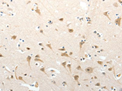
 SDS-PAGE
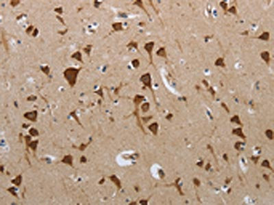
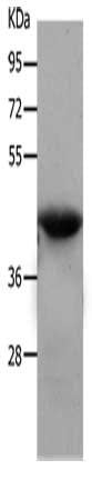
(Gel: 10%SDS-PAGE, Lysate: 60 ug, Lane: Human fetal brain tissue, Primary antibody: AAA240464(NPY1R Antibody) at dilution 1/500, Secondary antibody: Goat anti rabbit IgG at 1/8000 dilution, Exposure time: 2 minutes)
SDS-PAGE
(Gel: 10%SDS-PAGE, Lysate: 60 ug, Lane: Human fetal brain tissue, Primary antibody: AAA240464(NPY1R Antibody) at dilution 1/500, Secondary antibody: Goat anti rabbit IgG at 1/8000 dilution, Exposure time: 2 minutes)
NPY1R, Polyclonal Antibody (Cat# AAA240464)
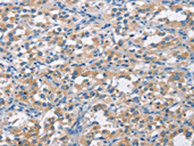
 IHC (Immunohiostchemistry)
(The image on the left is immunohistochemistry of paraffin-embedded Human gastric cancer tissue using AAA240389(CDH5 Antibody) at dilution 1/50, on the right is treated with synthetic peptide. (Original magnification: ×200))
IHC (Immunohiostchemistry)
(The image on the left is immunohistochemistry of paraffin-embedded Human gastric cancer tissue using AAA240389(CDH5 Antibody) at dilution 1/50, on the right is treated with synthetic peptide. (Original magnification: ×200))
CDH5, Polyclonal Antibody (Cat# AAA240389)
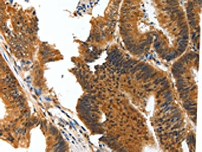
 IHC (Immunohiostchemistry)
(The image on the left is immunohistochemistry of paraffin-embedded Human gastric cancer tissue using AAA240396(CD226 Antibody) at dilution 1/60, on the right is treated with synthetic peptide. (Original magnification: ×200))
IHC (Immunohiostchemistry)
(The image on the left is immunohistochemistry of paraffin-embedded Human gastric cancer tissue using AAA240396(CD226 Antibody) at dilution 1/60, on the right is treated with synthetic peptide. (Original magnification: ×200))
CD226, Polyclonal Antibody (Cat# AAA240396)
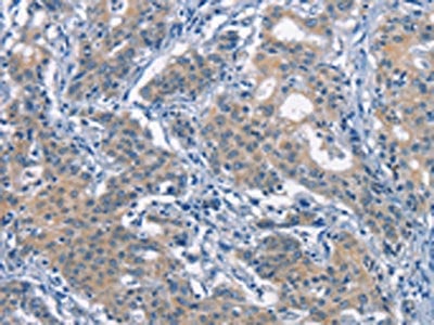
 IHC (Immunohiostchemistry)
(The image on the left is immunohistochemistry of paraffin-embedded Human thyroid cancer tissue using AAA240400(OLR1 Antibody) at dilution 1/25, on the right is treated with synthetic peptide. (Original magnification: ×200))
IHC (Immunohiostchemistry)
(The image on the left is immunohistochemistry of paraffin-embedded Human thyroid cancer tissue using AAA240400(OLR1 Antibody) at dilution 1/25, on the right is treated with synthetic peptide. (Original magnification: ×200))
OLR1, Polyclonal Antibody (Cat# AAA240400)
 SDS-PAGE
(Gel: 6%SDS-PAGE, Lysate: 40 ug, Lane: Human metastatic malignant melanoma tissue, Primary antibody: AAA240403(CEACAM1 Antibody) at dilution 1/300, Secondary antibody: Goat anti rabbit IgG at 1/8000 dilution, Exposure time: 20 seconds)
SDS-PAGE
(Gel: 6%SDS-PAGE, Lysate: 40 ug, Lane: Human metastatic malignant melanoma tissue, Primary antibody: AAA240403(CEACAM1 Antibody) at dilution 1/300, Secondary antibody: Goat anti rabbit IgG at 1/8000 dilution, Exposure time: 20 seconds)
CEACAM1, Polyclonal Antibody (Cat# AAA240403)
 IHC (Immunohiostchemistry)
(The image on the left is immunohistochemistry of paraffin-embedded Human lung cancer tissue using AAA240409(CD68 Antibody) at dilution 1/30, on the right is treated with synthetic peptide. (Original magnification: ×200))
IHC (Immunohiostchemistry)
(The image on the left is immunohistochemistry of paraffin-embedded Human lung cancer tissue using AAA240409(CD68 Antibody) at dilution 1/30, on the right is treated with synthetic peptide. (Original magnification: ×200))
CD68, Polyclonal Antibody (Cat# AAA240409)
 SDS-PAGE
(Gel: 6%SDS-PAGE, Lysate: 40 ug, Lane 1-2: Mouse brain tissue, RAW264.7 cells, Primary antibody: AAA240415(CDC25B Antibody) at dilution 1/250, Secondary antibody: Goat anti rabbit IgG at 1/8000 dilution, Exposure time: 20 seconds)
SDS-PAGE
(Gel: 6%SDS-PAGE, Lysate: 40 ug, Lane 1-2: Mouse brain tissue, RAW264.7 cells, Primary antibody: AAA240415(CDC25B Antibody) at dilution 1/250, Secondary antibody: Goat anti rabbit IgG at 1/8000 dilution, Exposure time: 20 seconds)
CDC25B, Polyclonal Antibody (Cat# AAA240415)
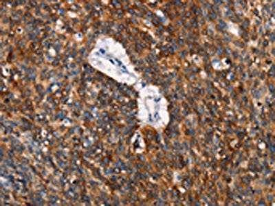
 SDS-PAGE
(Gel: 6%SDS-PAGE,Lysate: 40 ug,,Primary antibody: AAA240416(MET Antibody) at dilution 1/200 dilution,Secondary antibody: Goat anti rabbit IgG at 1/8000 dilution,Exposure time: 10 minutes)
SDS-PAGE
(Gel: 6%SDS-PAGE,Lysate: 40 ug,,Primary antibody: AAA240416(MET Antibody) at dilution 1/200 dilution,Secondary antibody: Goat anti rabbit IgG at 1/8000 dilution,Exposure time: 10 minutes)
MET, Polyclonal Antibody (Cat# AAA240416)
 SDS-PAGE
(Gel: 12%SDS-PAGE, Lysate: 40 ug, Lane: Human plasma solution, Primary antibody: AAA240420(Lambda Light chain Antibody) at dilution 1/200 dilution, Secondary antibody: Goat anti rabbit IgG at 1/8000 dilution, Exposure time: 10 seconds)
SDS-PAGE
(Gel: 12%SDS-PAGE, Lysate: 40 ug, Lane: Human plasma solution, Primary antibody: AAA240420(Lambda Light chain Antibody) at dilution 1/200 dilution, Secondary antibody: Goat anti rabbit IgG at 1/8000 dilution, Exposure time: 10 seconds)
IGLV1-51, Polyclonal Antibody (Cat# AAA240420)
 SDS-PAGE
(Gel: 12%SDS-PAGE, Lysate: 40 ug, Lane: Human plasma solution, Primary antibody: AAA240421(Lambda Light chain Antibody) at dilution 1/200 dilution, Secondary antibody: Goat anti rabbit IgG at 1/8000 dilution, Exposure time: 10 seconds)
SDS-PAGE
(Gel: 12%SDS-PAGE, Lysate: 40 ug, Lane: Human plasma solution, Primary antibody: AAA240421(Lambda Light chain Antibody) at dilution 1/200 dilution, Secondary antibody: Goat anti rabbit IgG at 1/8000 dilution, Exposure time: 10 seconds)
IGLV1-51, Polyclonal Antibody (Cat# AAA240421)
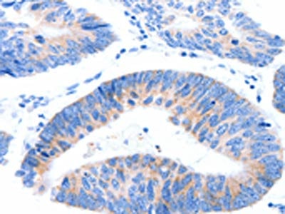
 IHC (Immunohiostchemistry)
(The image on the left is immunohistochemistry of paraffin-embedded Human tonsil tissue using AAA240423(MUC6 Antibody) at dilution 1/25, on the right is treated with synthetic peptide. (Original magnification: ×200))
IHC (Immunohiostchemistry)
(The image on the left is immunohistochemistry of paraffin-embedded Human tonsil tissue using AAA240423(MUC6 Antibody) at dilution 1/25, on the right is treated with synthetic peptide. (Original magnification: ×200))
MUC6, Polyclonal Antibody (Cat# AAA240423)
 IHC (Immunohiostchemistry)
(The image on the left is immunohistochemistry of paraffin-embedded Human thyroid cancer tissue using AAA241979(WNT2 Antibody) at dilution 1/35, on the right is treated with synthetic peptide. (Original magnification: ×200))
IHC (Immunohiostchemistry)
(The image on the left is immunohistochemistry of paraffin-embedded Human thyroid cancer tissue using AAA241979(WNT2 Antibody) at dilution 1/35, on the right is treated with synthetic peptide. (Original magnification: ×200))
WNT2, Polyclonal Antibody (Cat# AAA241979)
 IHC (Immunohiostchemistry)
(The image on the left is immunohistochemistry of paraffin-embedded Human esophagus cancer tissue using AAA241993(ZBTB2 Antibody) at dilution 1/40, on the right is treated with synthetic peptide. (Original magnification: ×200))
IHC (Immunohiostchemistry)
(The image on the left is immunohistochemistry of paraffin-embedded Human esophagus cancer tissue using AAA241993(ZBTB2 Antibody) at dilution 1/40, on the right is treated with synthetic peptide. (Original magnification: ×200))
ZBTB2, Polyclonal Antibody (Cat# AAA241993)
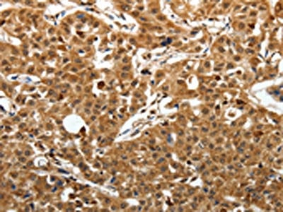
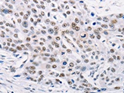
 IHC (Immunohiostchemistry)
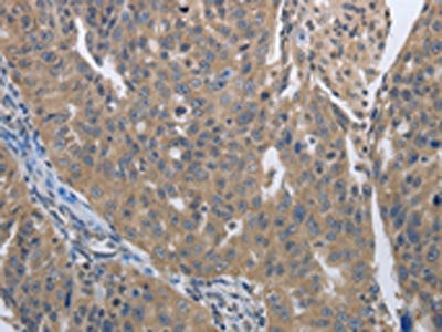
(The image on the left is immunohistochemistry of paraffin-embedded Human breast cancer tissue using AAA242027(ATF6 Antibody) at dilution 1/25, on the right is treated with synthetic peptide. (Original magnification: ×200))
IHC (Immunohiostchemistry)
(The image on the left is immunohistochemistry of paraffin-embedded Human breast cancer tissue using AAA242027(ATF6 Antibody) at dilution 1/25, on the right is treated with synthetic peptide. (Original magnification: ×200))
ATF6, Polyclonal Antibody (Cat# AAA242027)
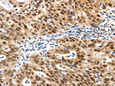
 IHC (Immunohiostchemistry)
(The image on the left is immunohistochemistry of paraffin-embedded Human lung cancer tissue using AAA242031(BRMS1 Antibody) at dilution 1/25, on the right is treated with synthetic peptide. (Original magnification: ×200))
IHC (Immunohiostchemistry)
(The image on the left is immunohistochemistry of paraffin-embedded Human lung cancer tissue using AAA242031(BRMS1 Antibody) at dilution 1/25, on the right is treated with synthetic peptide. (Original magnification: ×200))
BRMS1, Polyclonal Antibody (Cat# AAA242031)
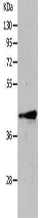
 SDS-PAGE
(Gel: 10%SDS-PAGE, Lysate: 40 ug, Lane: Human heart tissue, Primary antibody: AAA241636(RBM38 Antibody) at dilution 1/200, Secondary antibody: Goat anti rabbit IgG at 1/8000 dilution, Exposure time: 1 minute)
SDS-PAGE
(Gel: 10%SDS-PAGE, Lysate: 40 ug, Lane: Human heart tissue, Primary antibody: AAA241636(RBM38 Antibody) at dilution 1/200, Secondary antibody: Goat anti rabbit IgG at 1/8000 dilution, Exposure time: 1 minute)
RBM38, Polyclonal Antibody (Cat# AAA241636)
What are Polyclonal Antibodies?
Polyclonal antibodies are antibodies that come from multiple B cell clones of a host animal. The typical hosts used for the majority of polyclonal antibody production are rabbits, goats, sheep, and donkeys. These polyclonal antibodies, once having identified their target, will bind to different epitopes located at different regions or sequences on the same protein/antigen. As a result, they are ideal at locating and binding to the target, even if the target is in very low concentrations (due to many different antibodies being able to bind to the same target molecule, which allows for significant amplification of a downstream signal).
Polyclonal antibodies are typically produced by injecting an antigen into a host animal, which causes the animal’s immune system to attack the foreign antigen by mass generating antibodies against it. After a period of time, serum is collected from the animal and purified using physicochemical fractionation, class-specific affinity purification, and/or antigen-affinity purification.
Key Uses of Polyclonal Antibodies
- Western Blotting: This method is used to find specific proteins in biological samples after separating them by size.
- Immunohistochemistry: IHC helps visualize the location of proteins in tissue sections using various staining techniques.
- ELISA: (Enzyme-Linked Immunosorbent Assay) is typically used to identify specific protein quantities in a sample. ELISAs can be either “Quantitative” or “Qualitative”.
- Flow Cytometry: technique that identifies and measures the specific protein on the surface or inside the cells in a fluid suspension.
- Immunoprecipitation: IP isolates and studies a specific protein from a complex mixture using antibodies.
Why Buy Polyclonal Antibodies from AAA Biotech?
1. Ideal for Various Applications
Our antibodies are generally going to be validated for use in multiple types of assays, including ELISA, Western Blotting, Immunohistochemistry, Immunoprecipitation, amongst others. They are ideal for a wide range of research applications.
2. Rigorous Quality Control
All of the antibodies in our catalog undergo strict quality testing to ensure specificity, sensitivity, and consistent performance. We are confident in the ability of our antibodies to provide you with accurate results.
3. Wide Assortment of Antibodies
Antibodies in are catalog can be found for both common and exotic species, and these antibodies are also available in both conjugated and recombinant forms to suit many diverse experimental needs.
4. Highly Purified
Our antibodies are available in purified forms with over 85% purity, as confirmed by SDS-PAGE. They are also available with tags such as His, Flag, GST, or MBP. We cater to customers worldwide.
FAQ
1. How are polyclonal antibodies produced?
Traditionally, polyclonal antibodies are produced by injecting an antigen into a host animal (such as a rabbit or goat), which then triggers an immune response from the host animal. The animal’s B cells produce antibodies that will recognize different parts of the injected antigen. These antibodies are then collected from the animal’s blood and purified for use.
2. How do polyclonal antibodies differ from monoclonal antibodies?
Polyclonal antibodies are a mix of antibodies that bind to different locations (epitopes) of the same antigen, while monoclonal antibodies are identical and bind to just one specific epitope. This makes polyclonal antibodies more versatile and better at detecting proteins that may be present in low quantities or in altered/modified forms.
3. How should I store polyclonal antibodies?
Polyclonal antibodies should be stored at 4°C for short-term use (up to a few weeks) and at -20°C or -80°C for long-term storage. Avoid repeated freeze-thaw cycles by dividing them into small aliquots. Always check the datasheet for specific storage instructions.