Filters
▼Clonality
▼Type
▼Reactivity
▼Gene Name
▼Isotype
▼Host
▼Application
▼Clone
▼Polyclonal Antibodies
At AAA Biotech also known as AAA Bio or AAABio, we provide a broad range of purified polyclonal antibodies (pAbs) that are able to all be browsed online through our website. Due to their high specificity and strong binding affinity, these antibodies are ideal for wide swathes of research and experimental applications.
Our polyclonal antibodies can easily support your work, whether you use them for Western Blotting, Immunocytochemistry (with or without Immunofluorescence used in conjunction), Immunohistochemistry, Immunoprecipitation, and ELISA tests. We highly encourage you to browse our range of pAbs and choose the one that best suits your experimental model.
Viewing 8400-8450 of 96805 product results
 WB (Western Blot)
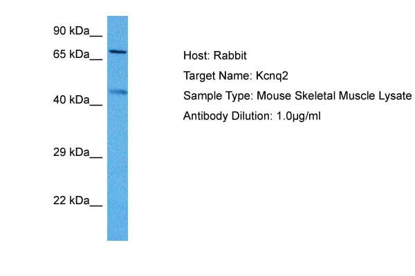
(WB Suggested Anti-KCNQ2 Antibody Titration: 1.25ug/mlELISA Titer: 1:62500Positive Control: HepG2 cell lysate)
WB (Western Blot)
(WB Suggested Anti-KCNQ2 Antibody Titration: 1.25ug/mlELISA Titer: 1:62500Positive Control: HepG2 cell lysate)
KCNQ2, Polyclonal Antibody (Cat# AAA197977)
 WB (Western Blot)
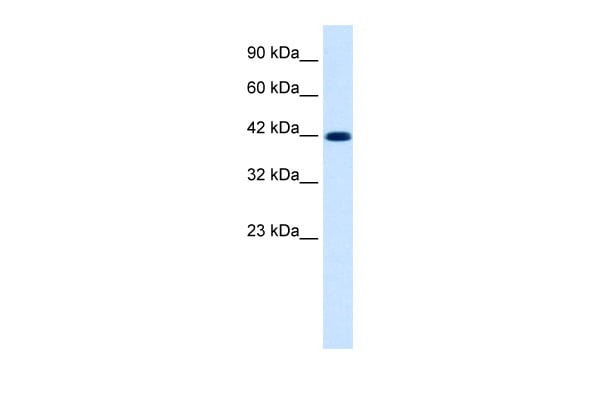
(Host: RabbitTarget Name: KCNG1Sample Tissue: Human PANC1 Whole CellAntibody Dilution: 1ug/ml)
WB (Western Blot)
(Host: RabbitTarget Name: KCNG1Sample Tissue: Human PANC1 Whole CellAntibody Dilution: 1ug/ml)
KCNG1, Polyclonal Antibody (Cat# AAA197981)
Predicted: Cow, Dog, Guinea Pig, Horse, Mouse, Rabbit, Rat
 WB (Western Blot)
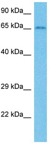
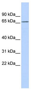
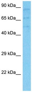
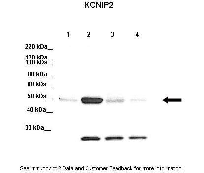
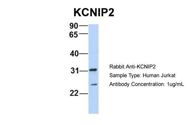
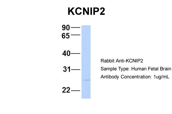
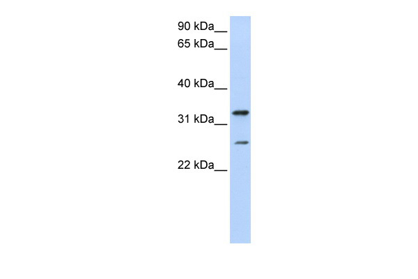
(WB Suggested Anti-KCNIP2 Antibody Titration: 0.2-1 ug/mlELISA Titer: 1:312500Positive Control: Jurkat cell lysateKCNIP2 is supported by BioGPS gene expression data to be expressed in Jurkat)
WB (Western Blot)
(WB Suggested Anti-KCNIP2 Antibody Titration: 0.2-1 ug/mlELISA Titer: 1:312500Positive Control: Jurkat cell lysateKCNIP2 is supported by BioGPS gene expression data to be expressed in Jurkat)
KCNIP2, Polyclonal Antibody (Cat# AAA197984)

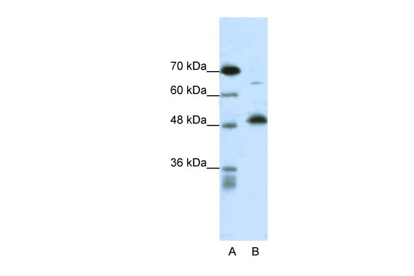
 WB (Western Blot)
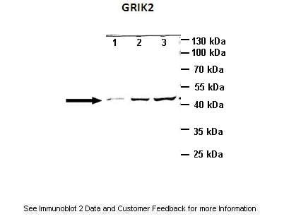
(Lanes:Lane1: 10ug mouse cortex brain lysateLane2: 25ug mouse cortex brain lysateLane3: 40ug mouse cortex brain lysatePrimary Antibody Dilution:1:1000Secondary Antibody:Anti-rabbit HRPSecondary Antibody Dilution:1:2000Gene Name:GRIK2Submitted by:Anonymous)
WB (Western Blot)
(Lanes:Lane1: 10ug mouse cortex brain lysateLane2: 25ug mouse cortex brain lysateLane3: 40ug mouse cortex brain lysatePrimary Antibody Dilution:1:1000Secondary Antibody:Anti-rabbit HRPSecondary Antibody Dilution:1:2000Gene Name:GRIK2Submitted by:Anonymous)
GRIK2, Polyclonal Antibody (Cat# AAA197987)
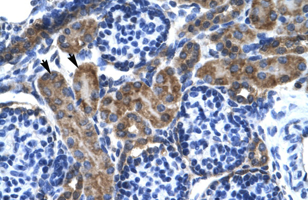
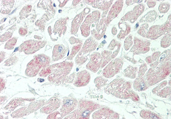
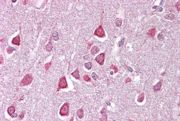
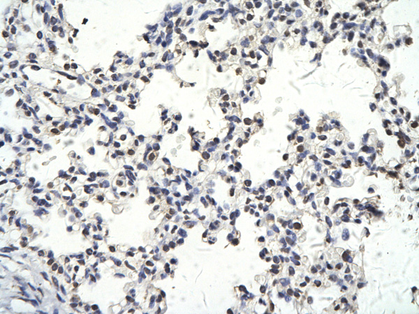
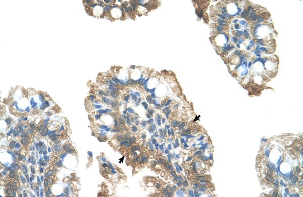
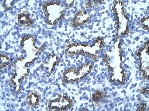
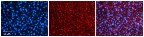
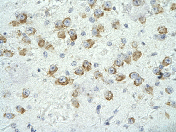
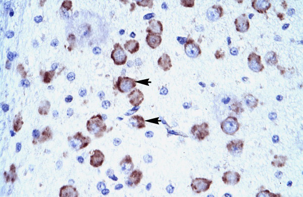
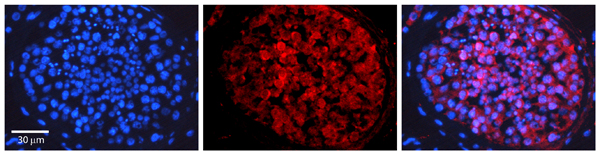
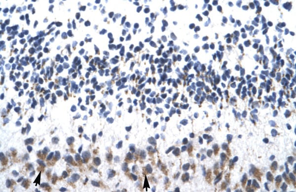
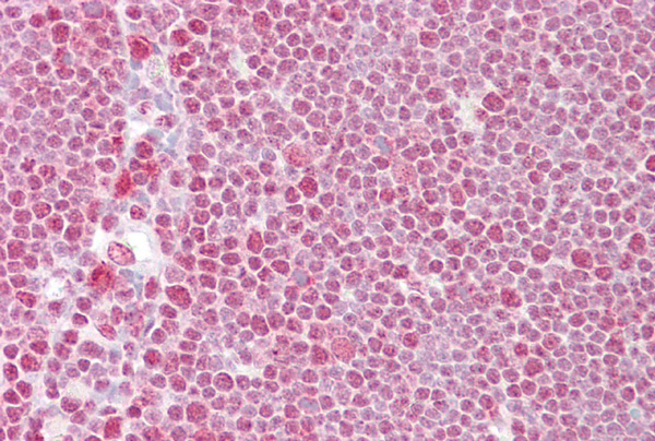
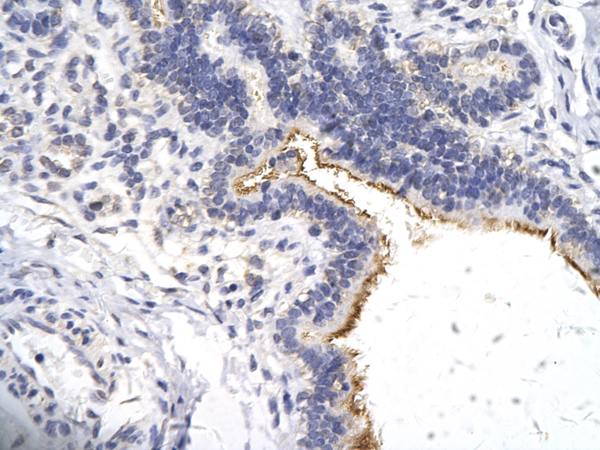
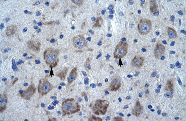 IHC (Immunohistochemistry)
(Human Brain)
IHC (Immunohistochemistry)
(Human Brain)
KCTD13, Polyclonal Antibody (Cat# AAA197990)
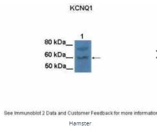
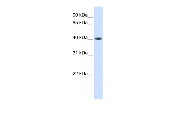
 WB (Western Blot)
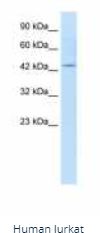
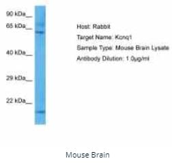
(WB Suggested Anti-KCNQ1 Antibody Titration: 1.25ug/mlELISA Titer: 1:62500Positive Control: Jurkat cell lysate)
WB (Western Blot)
(WB Suggested Anti-KCNQ1 Antibody Titration: 1.25ug/mlELISA Titer: 1:62500Positive Control: Jurkat cell lysate)
KCNQ1, Polyclonal Antibody (Cat# AAA197991)
 WB (Western Blot)
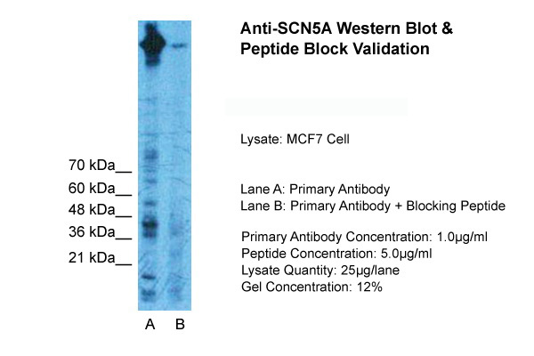
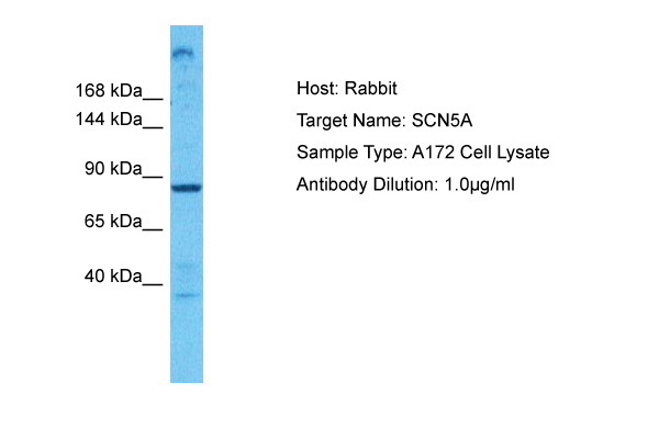
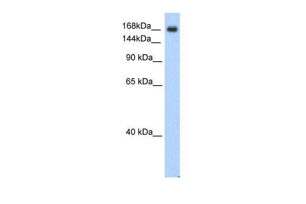
(WB Suggested Anti-SCN5A Antibody Titration: 0.2-1 ug/mlELISA Titer: 1:312500Positive Control: SW620 cell lysateSCN5A is supported by BioGPS gene expression data to be expressed in SW620)
WB (Western Blot)
(WB Suggested Anti-SCN5A Antibody Titration: 0.2-1 ug/mlELISA Titer: 1:312500Positive Control: SW620 cell lysateSCN5A is supported by BioGPS gene expression data to be expressed in SW620)
SCN5A, Polyclonal Antibody (Cat# AAA197994)
 WB (Western Blot)
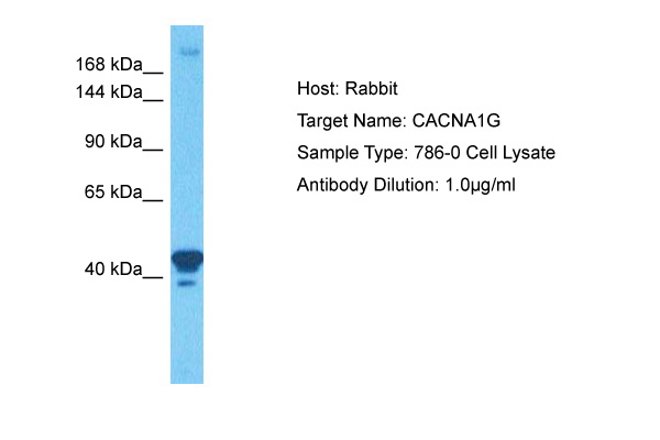
(WB Suggested Anti-CACNA1G Antibody Titration: 0.2-1 ug/mlELISA Titer: 1:312500Positive Control: Human Spleen)
WB (Western Blot)
(WB Suggested Anti-CACNA1G Antibody Titration: 0.2-1 ug/mlELISA Titer: 1:312500Positive Control: Human Spleen)
CACNA1G, Polyclonal Antibody (Cat# AAA197995)
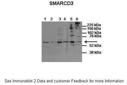
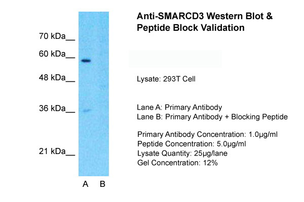
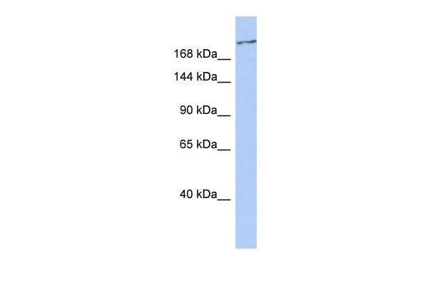
 WB (Western Blot)
(WB Suggested Anti-SMARCD3 Antibody Titration: 0.2-1 ug/mlELISA Titer: 1:312500Positive Control: 293T cell lysateSMARCD3 is supported by BioGPS gene expression data to be expressed in HEK293T)
WB (Western Blot)
(WB Suggested Anti-SMARCD3 Antibody Titration: 0.2-1 ug/mlELISA Titer: 1:312500Positive Control: 293T cell lysateSMARCD3 is supported by BioGPS gene expression data to be expressed in HEK293T)
SMARCD3, Polyclonal Antibody (Cat# AAA198001)
 WB (Western Blot)
(WB Suggested Anti-ZNF165 Antibody Titration: 0.2-1 ug/mlELISA Titer: 1:12500Positive Control: Hela cell lysate)
WB (Western Blot)
(WB Suggested Anti-ZNF165 Antibody Titration: 0.2-1 ug/mlELISA Titer: 1:12500Positive Control: Hela cell lysate)
ZNF165, Polyclonal Antibody (Cat# AAA198007)
 WB (Western Blot)
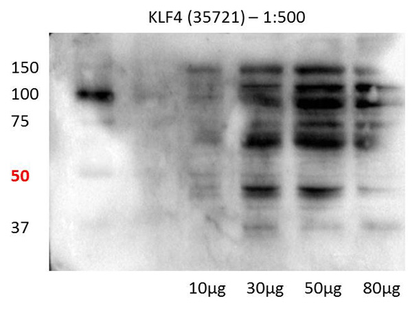
(WB Suggested Anti-KLF4 Antibody Titration: 2.5ug/mlELISA Titer: 1:312500Positive Control: Transfected 293T)
WB (Western Blot)
(WB Suggested Anti-KLF4 Antibody Titration: 2.5ug/mlELISA Titer: 1:312500Positive Control: Transfected 293T)
KLF4, Polyclonal Antibody (Cat# AAA198012)

 WB (Western Blot)
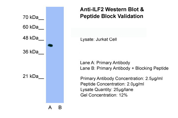
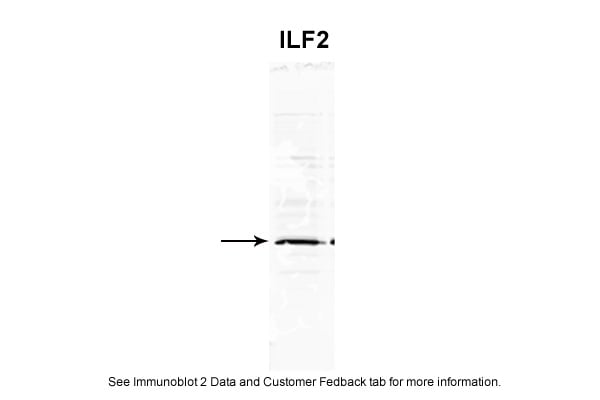
(WB Suggested Anti-ILF2 Antibody Titration: 1.25ug/mlELISA Titer: 1:62500Positive Control: Jurkat cell lysate)
WB (Western Blot)
(WB Suggested Anti-ILF2 Antibody Titration: 1.25ug/mlELISA Titer: 1:62500Positive Control: Jurkat cell lysate)
ILF2, Polyclonal Antibody (Cat# AAA198015)
 WB (Western Blot)
(WB Suggested Anti-KIF21A Antibody Titration: 0.2-1 ug/mlPositive Control: MCF7)
WB (Western Blot)
(WB Suggested Anti-KIF21A Antibody Titration: 0.2-1 ug/mlPositive Control: MCF7)
KIF21A, Polyclonal Antibody (Cat# AAA197695)
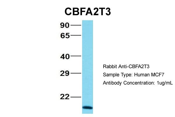
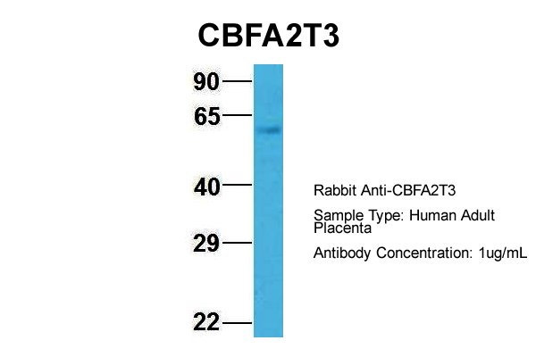
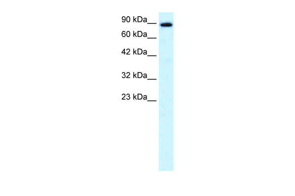
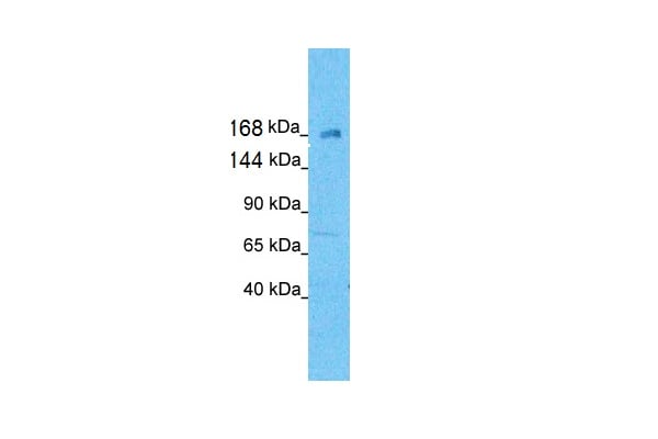
 WB (Western Blot)
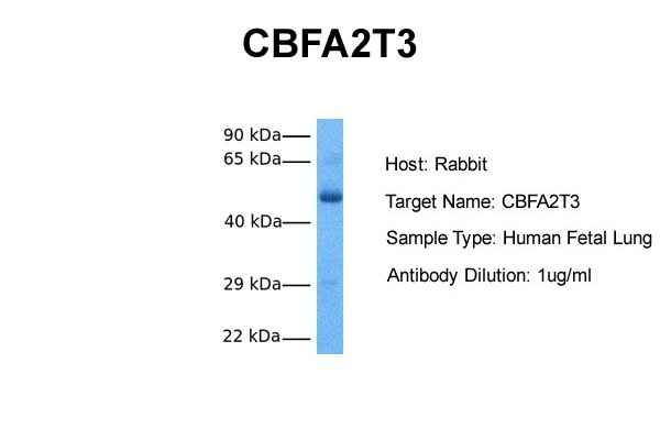
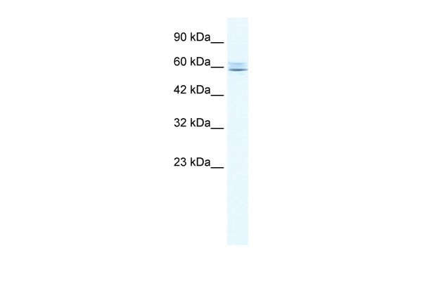
(WB Suggested Anti-CBFA2T3 Antibody Titration: 0.2-1 ug/mlPositive Control: Jurkat cell lysateCBFA2T3 is supported by BioGPS gene expression data to be expressed in Jurkat)
WB (Western Blot)
(WB Suggested Anti-CBFA2T3 Antibody Titration: 0.2-1 ug/mlPositive Control: Jurkat cell lysateCBFA2T3 is supported by BioGPS gene expression data to be expressed in Jurkat)
CBFA2T3, Polyclonal Antibody (Cat# AAA197703)
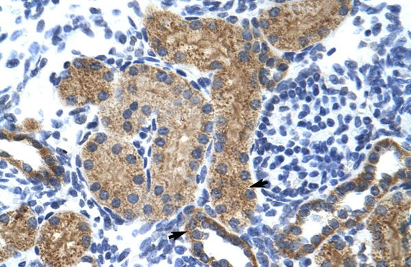
 WB (Western Blot)
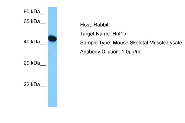
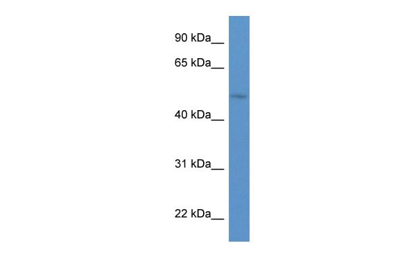
(WB Suggested Anti-HNF1B Antibody Titration: 0.2-1 ug/mlELISA Titer: 1:62500Positive Control: ACHN cell lysateHNF1B is supported by BioGPS gene expression data to be expressed in ACHN)
WB (Western Blot)
(WB Suggested Anti-HNF1B Antibody Titration: 0.2-1 ug/mlELISA Titer: 1:62500Positive Control: ACHN cell lysateHNF1B is supported by BioGPS gene expression data to be expressed in ACHN)
HNF1B, Polyclonal Antibody (Cat# AAA197707)
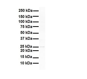
 WB (Western Blot)
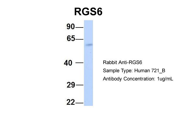
(WB Suggested Anti-RGS6 Antibody Titration: 0.2-1 ug/mlELISA Titer: 1:12500Positive Control: Jurkat cell lysate)
WB (Western Blot)
(WB Suggested Anti-RGS6 Antibody Titration: 0.2-1 ug/mlELISA Titer: 1:12500Positive Control: Jurkat cell lysate)
RGS6, Polyclonal Antibody (Cat# AAA197709)
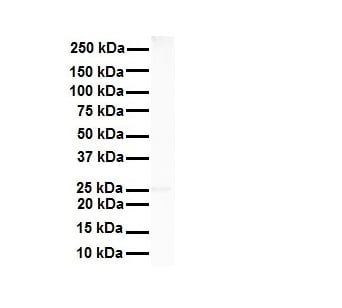
 WB (Western Blot)
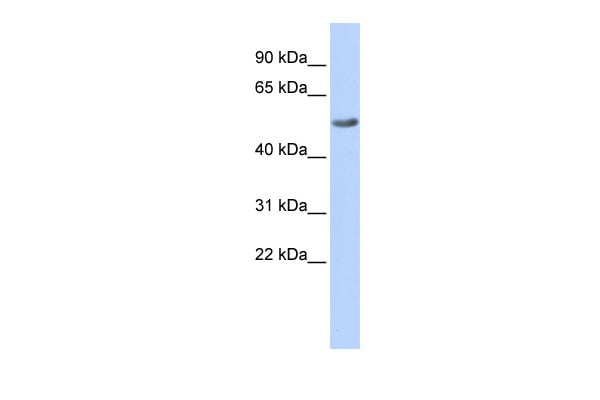
(WB Suggested Anti-RGS6 Antibody Titration: 0.2-1 ug/mlPositive Control: Jurkat cell lysate)
WB (Western Blot)
(WB Suggested Anti-RGS6 Antibody Titration: 0.2-1 ug/mlPositive Control: Jurkat cell lysate)
RGS6, Polyclonal Antibody (Cat# AAA197710)
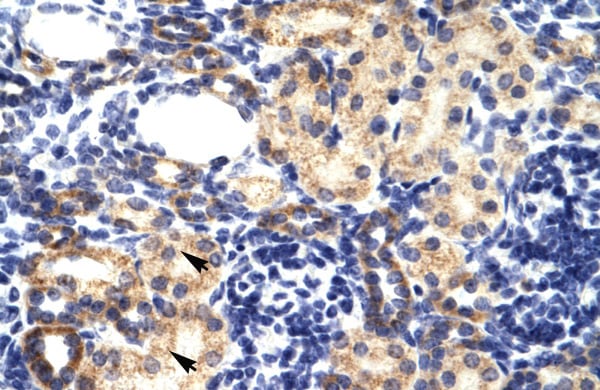
 WB (Western Blot)
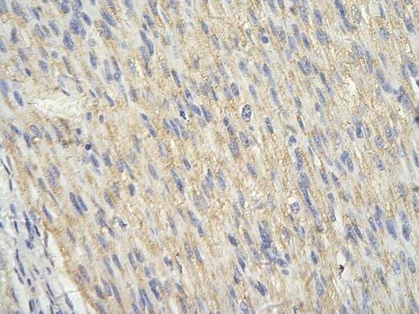
(WB Suggested Anti-TEAD3 antibody Titration: 1 ug/mLSample Type: Human heart)
WB (Western Blot)
(WB Suggested Anti-TEAD3 antibody Titration: 1 ug/mLSample Type: Human heart)
TEAD3, Polyclonal Antibody (Cat# AAA197714)
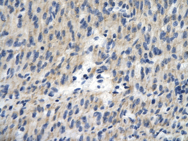
 WB (Western Blot)
(WB Suggested Anti-PARP6 Antibody Titration: 0.2-1 ug/mlPositive Control: HepG2 cell lysate)
WB (Western Blot)
(WB Suggested Anti-PARP6 Antibody Titration: 0.2-1 ug/mlPositive Control: HepG2 cell lysate)
PARP6, Polyclonal Antibody (Cat# AAA197717)
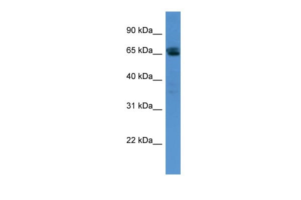
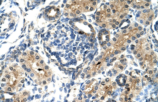
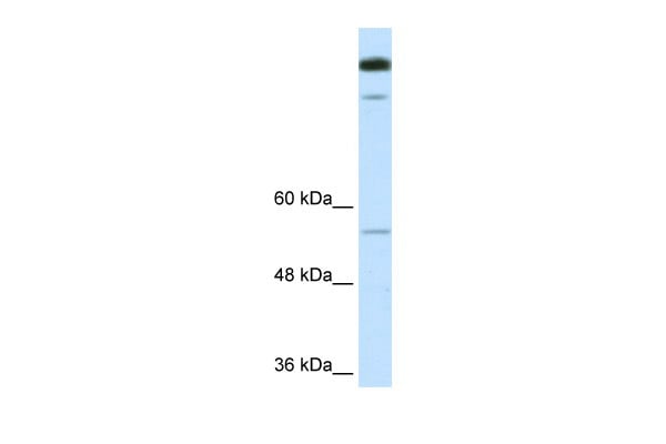
 WB (Western Blot)
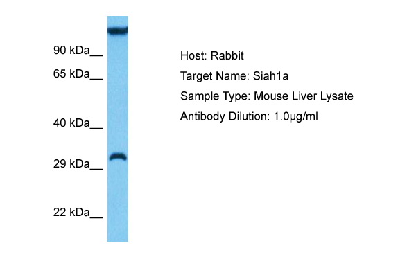
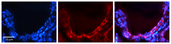
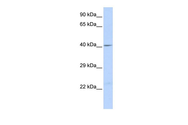
(Host: RabbitTarget Name: PGRC2Sample Type: Fetal Lung lysatesAntibody Dilution: 1ug/ml)
WB (Western Blot)
(Host: RabbitTarget Name: PGRC2Sample Type: Fetal Lung lysatesAntibody Dilution: 1ug/ml)
PGRMC2, Polyclonal Antibody (Cat# AAA197719)
Predicted Reactivity: Cow, Dog, Guinea Pig, Horse, Human, Mouse, Rabbit, Rat, Zebrafish
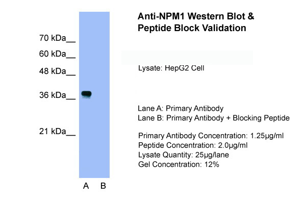
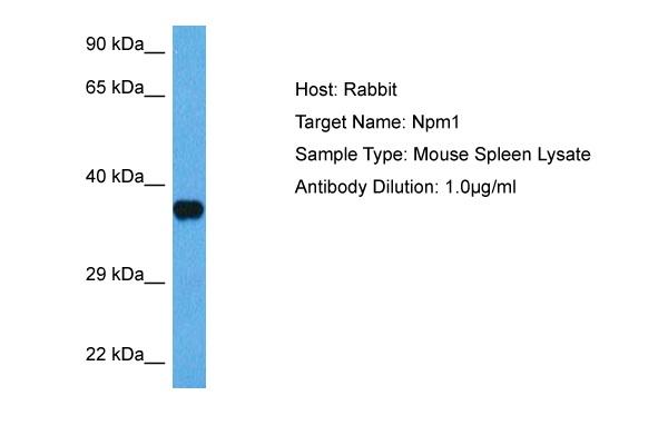
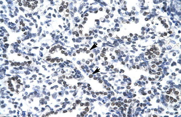
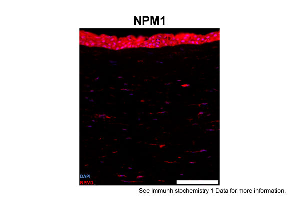
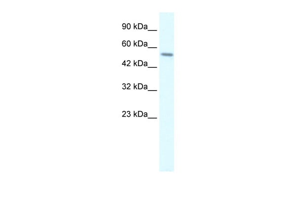
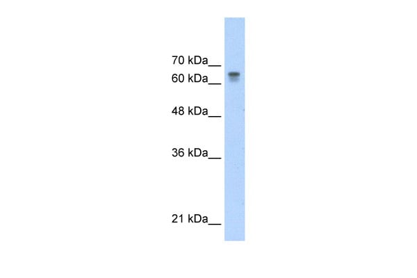
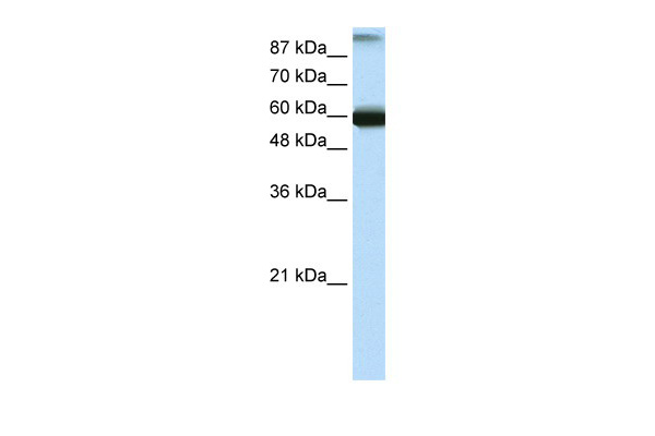
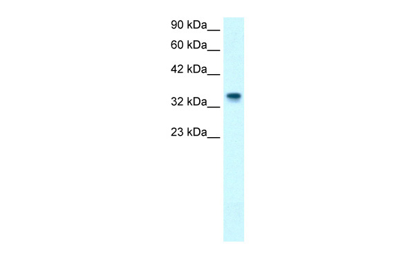 WB (Western Blot)
(WB Suggested Anti-NPM1 Antibody Titration: 1.0ug/mlPositive Control: HepG2 cell lysateThere is BioGPS gene expression data showing that NPM1 is expressed in HepG2)
WB (Western Blot)
(WB Suggested Anti-NPM1 Antibody Titration: 1.0ug/mlPositive Control: HepG2 cell lysateThere is BioGPS gene expression data showing that NPM1 is expressed in HepG2)
NPM1, Polyclonal Antibody (Cat# AAA197720)
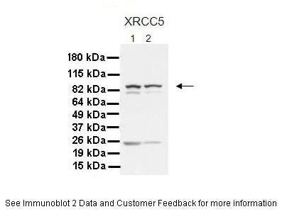
 WB (Western Blot)
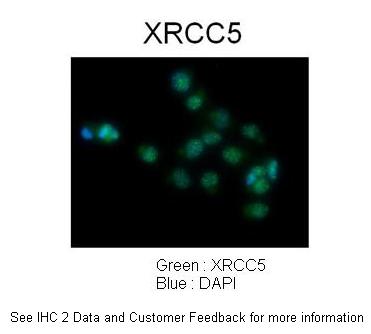
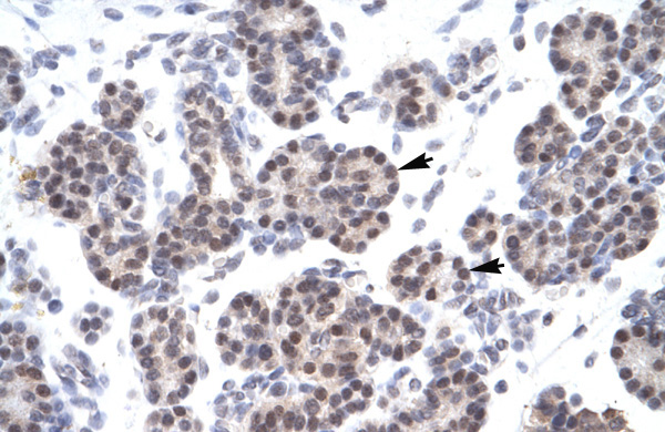
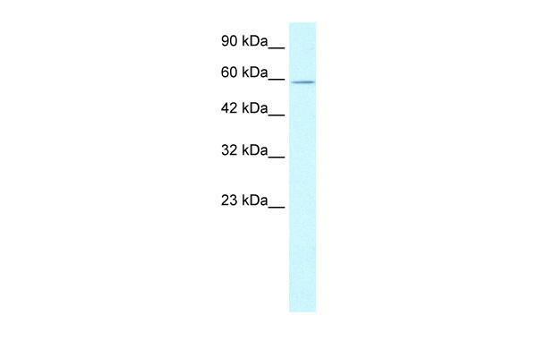
(WB Suggested Anti-XRCC5 Antibody Titration: 1.25ug/mlELISA Titer: 1:12500Positive Control: Jurkat cell lysateXRCC5 is strongly supported by BioGPS gene expression data to be expressed in Human Jurkat cells)
WB (Western Blot)
(WB Suggested Anti-XRCC5 Antibody Titration: 1.25ug/mlELISA Titer: 1:12500Positive Control: Jurkat cell lysateXRCC5 is strongly supported by BioGPS gene expression data to be expressed in Human Jurkat cells)
XRCC5, Polyclonal Antibody (Cat# AAA197726)
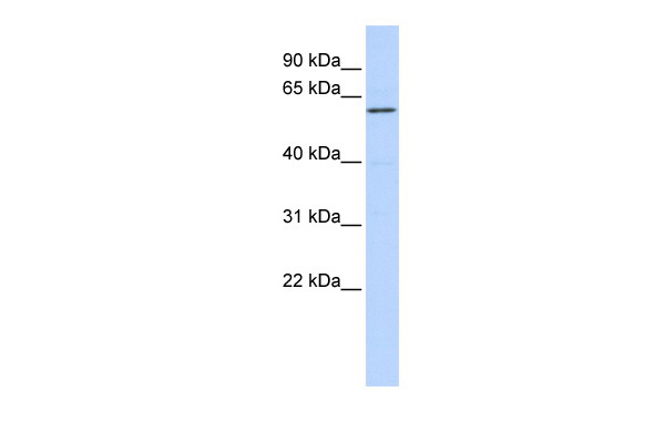
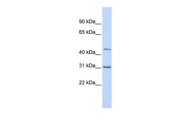
 WB (Western Blot)
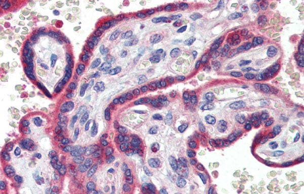
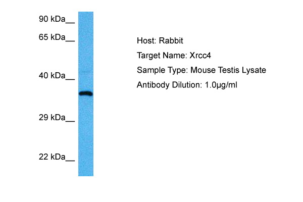
(WB Suggested Anti-XRCC4 Antibody Titration: 0.2-1 ug/mlELISA Titer: 1:2500Positive Control: Human Placenta)
WB (Western Blot)
(WB Suggested Anti-XRCC4 Antibody Titration: 0.2-1 ug/mlELISA Titer: 1:2500Positive Control: Human Placenta)
XRCC4, Polyclonal Antibody (Cat# AAA197728)

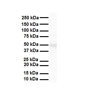
 WB (Western Blot)
(WB Suggested Anti-TPH2 Antibody Titration: 0.2-1 ug/mlELISA Titer: 1:500Positive Control: Human brain)
WB (Western Blot)
(WB Suggested Anti-TPH2 Antibody Titration: 0.2-1 ug/mlELISA Titer: 1:500Positive Control: Human brain)
TPH2, Polyclonal Antibody (Cat# AAA197735)
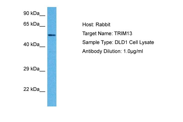
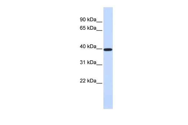
 WB (Western Blot)
(WB Suggested Anti-RFP2 Antibody Titration: 0.125ug/mlELISA Titer: 1:62500Positive Control: Human Lung)
WB (Western Blot)
(WB Suggested Anti-RFP2 Antibody Titration: 0.125ug/mlELISA Titer: 1:62500Positive Control: Human Lung)
RFP2, Polyclonal Antibody (Cat# AAA197736)
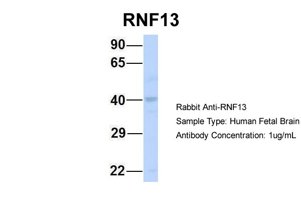
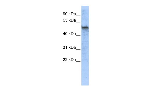
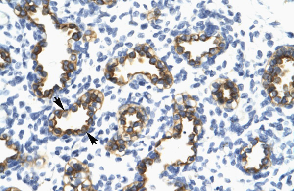
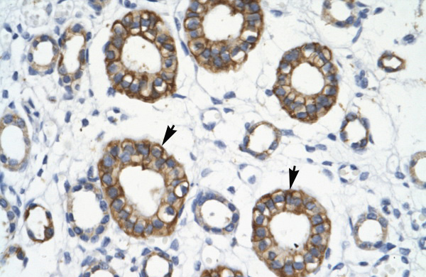
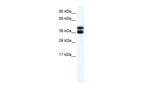
 WB (Western Blot)
(WB Suggested Anti-RNF13 Antibody Titration: 0.2-1 ug/mlELISA Titer: 1:312500Positive Control: Hela cell lysateRNF13 is supported by BioGPS gene expression data to be expressed in HeLa)
WB (Western Blot)
(WB Suggested Anti-RNF13 Antibody Titration: 0.2-1 ug/mlELISA Titer: 1:312500Positive Control: Hela cell lysateRNF13 is supported by BioGPS gene expression data to be expressed in HeLa)
RNF13, Polyclonal Antibody (Cat# AAA197737)
 WB (Western Blot)
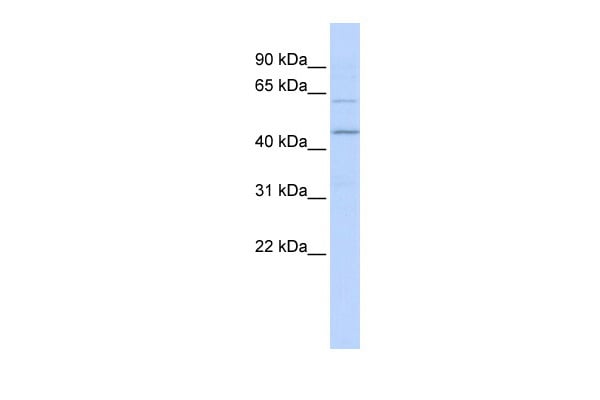
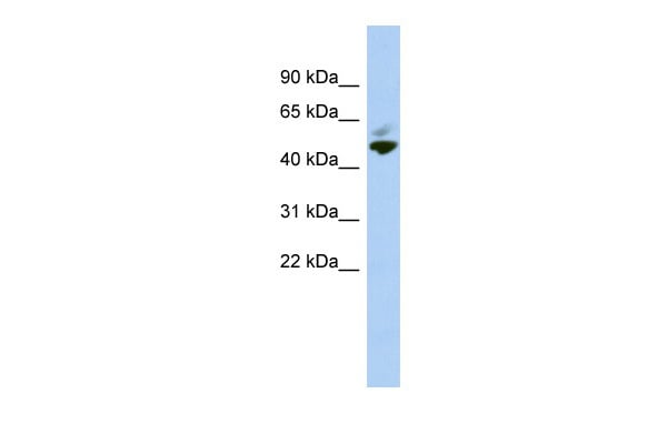
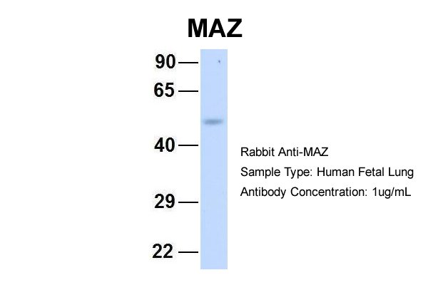
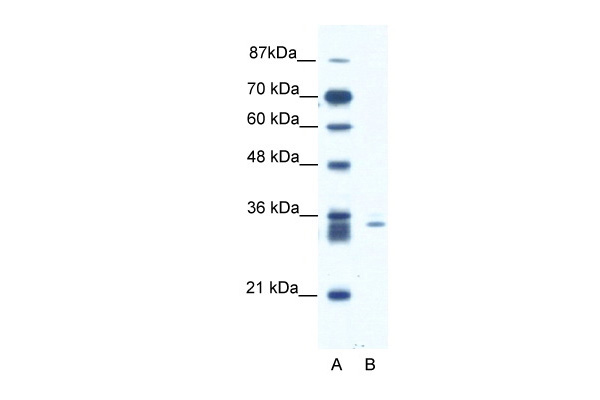
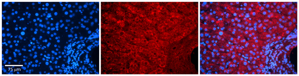
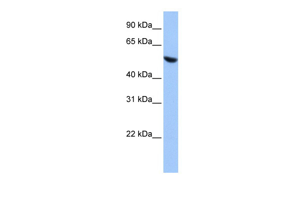
(WB Suggested Anti-MAZ antibody Titration: 1 ug/mLSample Type: Human liver)
WB (Western Blot)
(WB Suggested Anti-MAZ antibody Titration: 1 ug/mLSample Type: Human liver)
MAZ, Polyclonal Antibody (Cat# AAA197740)
 WB (Western Blot)
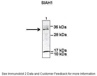
(WB Suggested Anti-SIAH1 Antibody Titration: 1.25ug/mlELISA Titer: 1:312500Positive Control: Jurkat cell lysate)
WB (Western Blot)
(WB Suggested Anti-SIAH1 Antibody Titration: 1.25ug/mlELISA Titer: 1:312500Positive Control: Jurkat cell lysate)
SIAH1, Polyclonal Antibody (Cat# AAA197741)
 WB (Western Blot)
(WB Suggested Anti-TIMELESS Antibody Titration: 0.2-1 ug/mlELISA Titer: 1:312500Positive Control: Transfected 293T)
WB (Western Blot)
(WB Suggested Anti-TIMELESS Antibody Titration: 0.2-1 ug/mlELISA Titer: 1:312500Positive Control: Transfected 293T)
TIMELESS, Polyclonal Antibody (Cat# AAA197747)
 WB (Western Blot)
(WB Suggested Anti-COPS2 Antibody Titration: 0.2-1 ug/mlELISA Titer: 1:62500Positive Control: HepG2 cell lysate)
WB (Western Blot)
(WB Suggested Anti-COPS2 Antibody Titration: 0.2-1 ug/mlELISA Titer: 1:62500Positive Control: HepG2 cell lysate)
COPS2, Polyclonal Antibody (Cat# AAA197748)
 WB (Western Blot)
(WB Suggested Anti-COPS2 AntibodyTitration: 0.5 ug/mlPositive Control: Fetal Liver)
WB (Western Blot)
(WB Suggested Anti-COPS2 AntibodyTitration: 0.5 ug/mlPositive Control: Fetal Liver)
COPS2, Polyclonal Antibody (Cat# AAA197749)
 WB (Western Blot)
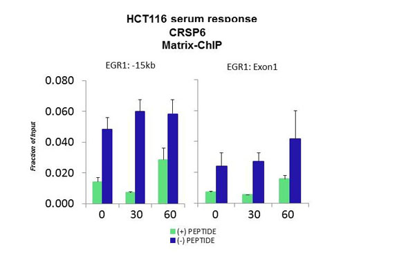
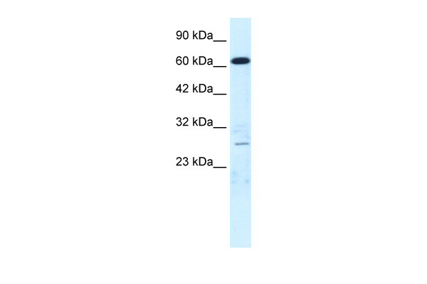
(WB Suggested Anti-CRSP6 Antibody Titration: 0.2-1 ug/mlELISA Titer: 1:12500Positive Control: HepG2 cell lysateMED17 is supported by BioGPS gene expression data to be expressed in HepG2)
WB (Western Blot)
(WB Suggested Anti-CRSP6 Antibody Titration: 0.2-1 ug/mlELISA Titer: 1:12500Positive Control: HepG2 cell lysateMED17 is supported by BioGPS gene expression data to be expressed in HepG2)
CRSP6, Polyclonal Antibody (Cat# AAA197751)
 WB (Western Blot)
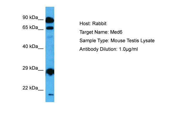
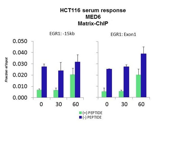
(WB Suggested Anti-MED6 Antibody Titration: 0.2-1 ug/mlELISA Titer: 1:2500Positive Control: Transfected 293T)
WB (Western Blot)
(WB Suggested Anti-MED6 Antibody Titration: 0.2-1 ug/mlELISA Titer: 1:2500Positive Control: Transfected 293T)
MED6, Polyclonal Antibody (Cat# AAA197759)

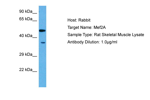
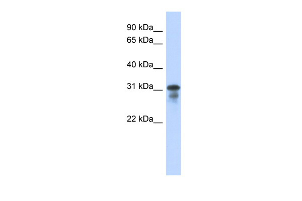
 WB (Western Blot)
(WB Suggested Anti-MEF2A Antibody Titration: 0.2-1 ug/mlELISA Titer: 1:12500Positive Control: Transfected 293T)
WB (Western Blot)
(WB Suggested Anti-MEF2A Antibody Titration: 0.2-1 ug/mlELISA Titer: 1:12500Positive Control: Transfected 293T)
MEF2A, Polyclonal Antibody (Cat# AAA197761)
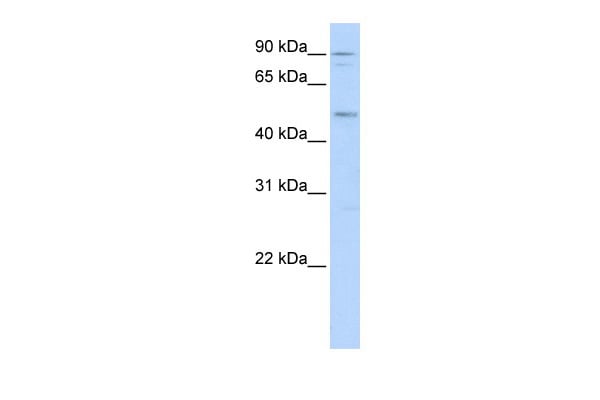
 WB (Western Blot)
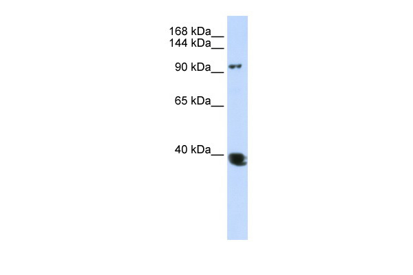
(WB Suggested Anti-HDAC4 Antibody Titration: 0.2-1 ug/mlELISA Titer: 1:312500Positive Control: Hela cell lysate)
WB (Western Blot)
(WB Suggested Anti-HDAC4 Antibody Titration: 0.2-1 ug/mlELISA Titer: 1:312500Positive Control: Hela cell lysate)
HDAC4, Polyclonal Antibody (Cat# AAA197762)
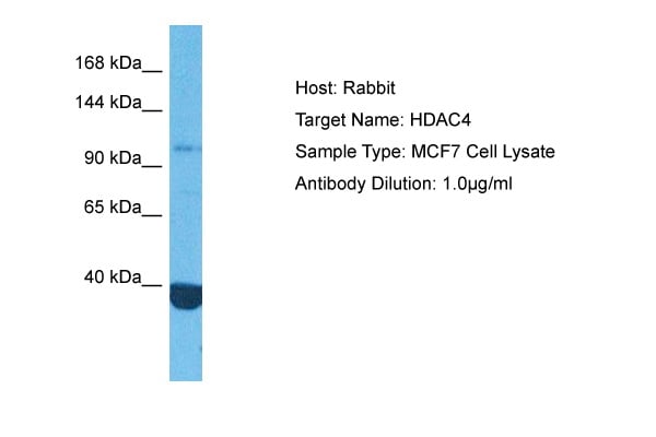
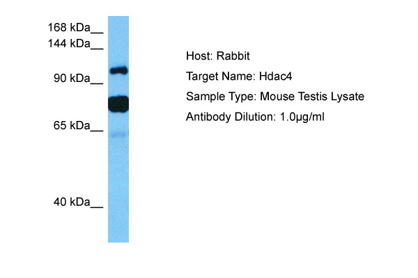
 WB (Western Blot)
(WB Suggested Anti-HDAC4 Antibody Titration: 0.2-1 ug/mlELISA Titer: 1:12500Positive Control: Hela cell lysate)
WB (Western Blot)
(WB Suggested Anti-HDAC4 Antibody Titration: 0.2-1 ug/mlELISA Titer: 1:12500Positive Control: Hela cell lysate)
HDAC4, Polyclonal Antibody (Cat# AAA197763)
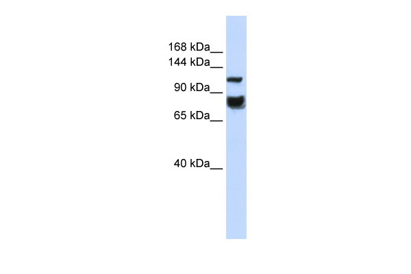
 WB (Western Blot)
(WB Suggested Anti-RNF2 Antibody Titration: 1.25ug/mlELISA Titer: 1:312500Positive Control: HepG2 cell lysate)
WB (Western Blot)
(WB Suggested Anti-RNF2 Antibody Titration: 1.25ug/mlELISA Titer: 1:312500Positive Control: HepG2 cell lysate)
RNF2, Polyclonal Antibody (Cat# AAA197769)
 WB (Western Blot)
(WB Suggested Anti-TCEAL1 Antibody Titration: 0.2-1 ug/mlELISA Titer: 1:1562500Positive Control: Human Placenta)
WB (Western Blot)
(WB Suggested Anti-TCEAL1 Antibody Titration: 0.2-1 ug/mlELISA Titer: 1:1562500Positive Control: Human Placenta)
TCEAL1, Polyclonal Antibody (Cat# AAA198018)
 WB (Western Blot)
(WB Suggested Anti-ZNF234 Antibody Titration: 0.2-1 ug/mlELISA Titer: 1:62500Positive Control: Human Pancreas)
WB (Western Blot)
(WB Suggested Anti-ZNF234 Antibody Titration: 0.2-1 ug/mlELISA Titer: 1:62500Positive Control: Human Pancreas)
ZNF234, Polyclonal Antibody (Cat# AAA198026)
 WB (Western Blot)
(WB Suggested Anti-ZNF266 Antibody Titration: 0.2-1 ug/mlELISA Titer: 1:1562500Positive Control: HepG2 cell lysate)
WB (Western Blot)
(WB Suggested Anti-ZNF266 Antibody Titration: 0.2-1 ug/mlELISA Titer: 1:1562500Positive Control: HepG2 cell lysate)
ZNF266, Polyclonal Antibody (Cat# AAA198027)
 WB (Western Blot)
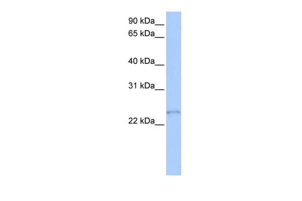
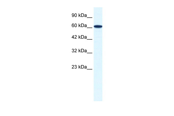
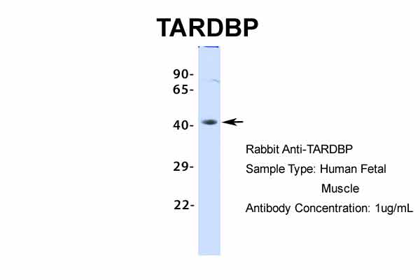
(WB Suggested Anti-TARDBP Antibody Titration: 0.2-1 ug/mlELISA Titer: 1:12500Positive Control: Human Muscle)
WB (Western Blot)
(WB Suggested Anti-TARDBP Antibody Titration: 0.2-1 ug/mlELISA Titer: 1:12500Positive Control: Human Muscle)
TARDBP, Polyclonal Antibody (Cat# AAA198033)
 WB (Western Blot)
(WB Suggested Anti-GTF3C5 Antibody Titration: 0.2-1 ug/mlELISA Titer: 1:312500Positive Control: Transfected 293T)
WB (Western Blot)
(WB Suggested Anti-GTF3C5 Antibody Titration: 0.2-1 ug/mlELISA Titer: 1:312500Positive Control: Transfected 293T)
GTF3C5, Polyclonal Antibody (Cat# AAA198035)
 WB (Western Blot)
(WB Suggested Anti-ZNF180 Antibody Titration: 0.2-1 ug/mlELISA Titer: 1:1562500Positive Control: 293T cell lysateZNF180 is supported by BioGPS gene expression data to be expressed in HEK293T)
WB (Western Blot)
(WB Suggested Anti-ZNF180 Antibody Titration: 0.2-1 ug/mlELISA Titer: 1:1562500Positive Control: 293T cell lysateZNF180 is supported by BioGPS gene expression data to be expressed in HEK293T)
ZNF180, Polyclonal Antibody (Cat# AAA198037)
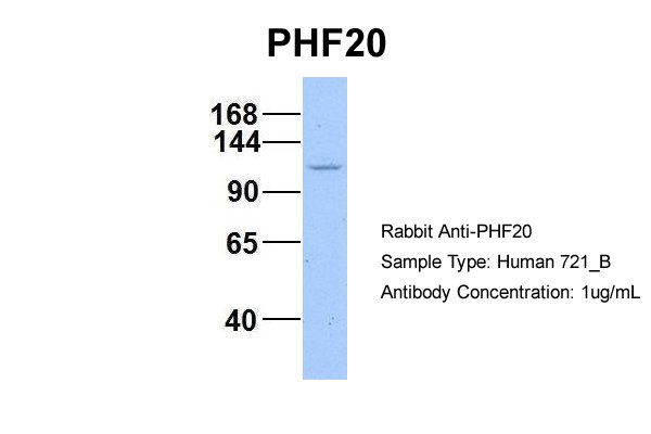
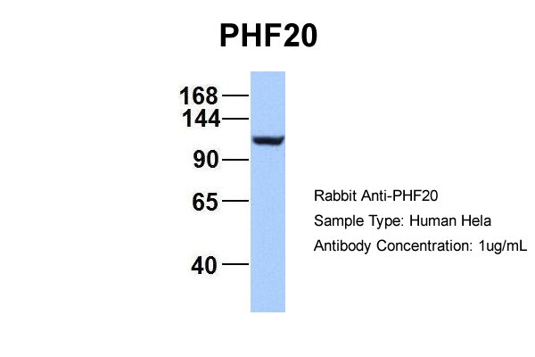
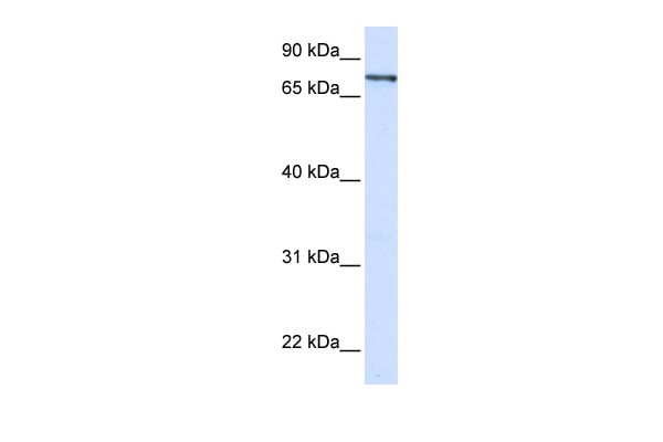
 WB (Western Blot)
(WB Suggested Anti-PHF20 Antibody Titration: 0.2-1 ug/mlELISA Titer: 1:2500Positive Control: Jurkat cell lysate)
WB (Western Blot)
(WB Suggested Anti-PHF20 Antibody Titration: 0.2-1 ug/mlELISA Titer: 1:2500Positive Control: Jurkat cell lysate)
PHF20, Polyclonal Antibody (Cat# AAA198043)
 WB (Western Blot)
(WB Suggested Anti-ZNF167 Antibody Titration: 1.25ug/mlELISA Titer: 1:312500Positive Control: Jurkat cell lysate)
WB (Western Blot)
(WB Suggested Anti-ZNF167 Antibody Titration: 1.25ug/mlELISA Titer: 1:312500Positive Control: Jurkat cell lysate)
ZNF167, Polyclonal Antibody (Cat# AAA198045)
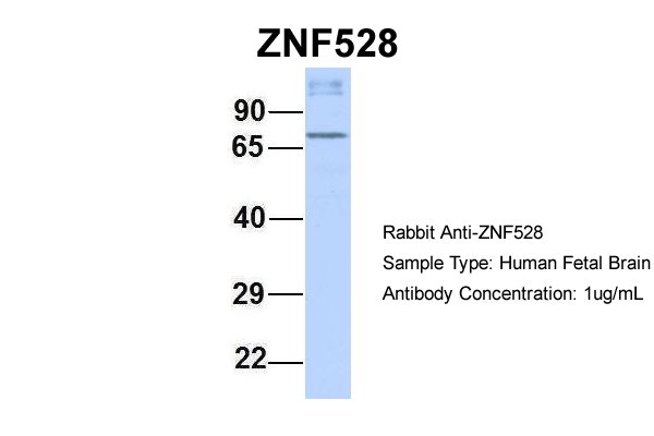
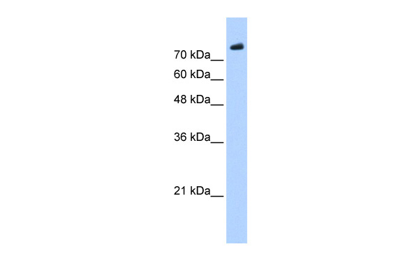
 WB (Western Blot)
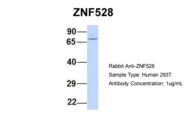
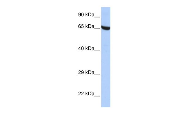
(WB Suggested Anti-ZNF528 Antibody Titration: 0.2-1 ug/mlELISA Titer: 1:1562500Positive Control: HepG2 cell lysate)
WB (Western Blot)
(WB Suggested Anti-ZNF528 Antibody Titration: 0.2-1 ug/mlELISA Titer: 1:1562500Positive Control: HepG2 cell lysate)
ZNF528, Polyclonal Antibody (Cat# AAA198052)
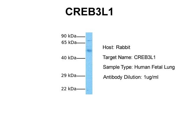
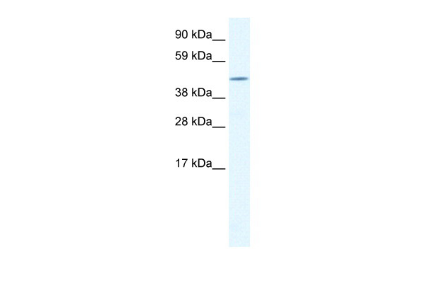 WB (Western Blot)
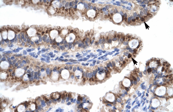
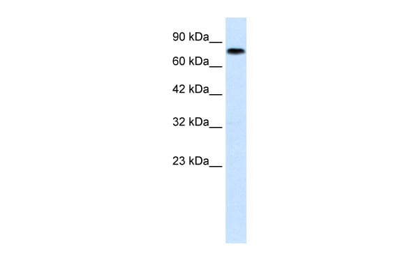
(WB Suggested Anti-CREB3L1 Antibody Titration: 1.25ug/mlELISA Titer: 1:1562500Positive Control: Human Small Intestine)
WB (Western Blot)
(WB Suggested Anti-CREB3L1 Antibody Titration: 1.25ug/mlELISA Titer: 1:1562500Positive Control: Human Small Intestine)
CREB3L1, Polyclonal Antibody (Cat# AAA198053)
 WB (Western Blot)
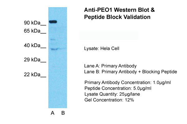
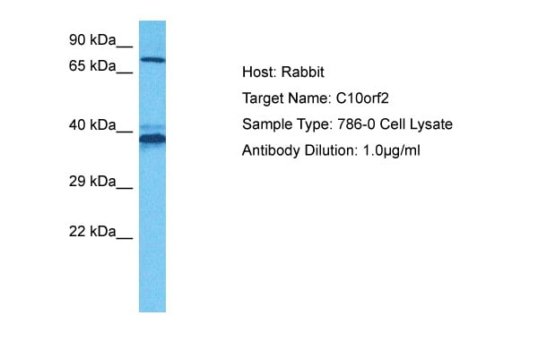
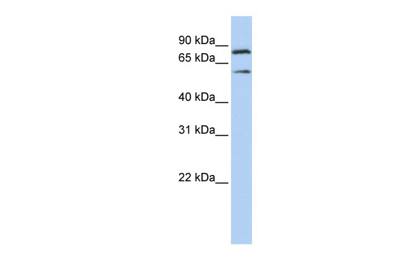
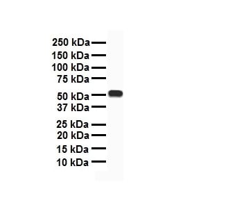
(WB Suggested Anti-PEO1 Antibody Titration: 0.2-1 ug/mlELISA Titer: 1:312500Positive Control: Jurkat cell lysate)
WB (Western Blot)
(WB Suggested Anti-PEO1 Antibody Titration: 0.2-1 ug/mlELISA Titer: 1:312500Positive Control: Jurkat cell lysate)
TWNK, Polyclonal Antibody (Cat# AAA198079)
 WB (Western Blot)
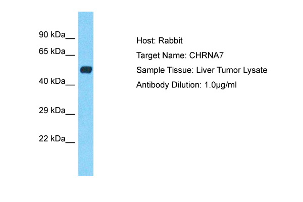
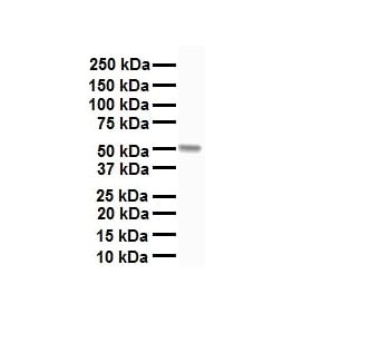
(WB Suggested Anti-CHRNA7 antibody Titration: 1 ug/mLSample Type: Human liver)
WB (Western Blot)
(WB Suggested Anti-CHRNA7 antibody Titration: 1 ug/mLSample Type: Human liver)
CHRNA7, Polyclonal Antibody (Cat# AAA198093)
 WB (Western Blot)
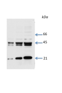
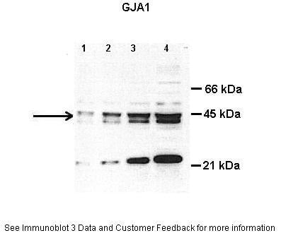
(WB Suggested Anti-GJA1 Antibody Titration: 0.2-1 ug/mlELISA Titer: 1:62500Positive Control: Human Liver)
WB (Western Blot)
(WB Suggested Anti-GJA1 Antibody Titration: 0.2-1 ug/mlELISA Titer: 1:62500Positive Control: Human Liver)
GJA1, Polyclonal Antibody (Cat# AAA198094)
What are Polyclonal Antibodies?
Polyclonal antibodies are antibodies that come from multiple B cell clones of a host animal. The typical hosts used for the majority of polyclonal antibody production are rabbits, goats, sheep, and donkeys. These polyclonal antibodies, once having identified their target, will bind to different epitopes located at different regions or sequences on the same protein/antigen. As a result, they are ideal at locating and binding to the target, even if the target is in very low concentrations (due to many different antibodies being able to bind to the same target molecule, which allows for significant amplification of a downstream signal).
Polyclonal antibodies are typically produced by injecting an antigen into a host animal, which causes the animal’s immune system to attack the foreign antigen by mass generating antibodies against it. After a period of time, serum is collected from the animal and purified using physicochemical fractionation, class-specific affinity purification, and/or antigen-affinity purification.
Key Uses of Polyclonal Antibodies
- Western Blotting: This method is used to find specific proteins in biological samples after separating them by size.
- Immunohistochemistry: IHC helps visualize the location of proteins in tissue sections using various staining techniques.
- ELISA: (Enzyme-Linked Immunosorbent Assay) is typically used to identify specific protein quantities in a sample. ELISAs can be either “Quantitative” or “Qualitative”.
- Flow Cytometry: technique that identifies and measures the specific protein on the surface or inside the cells in a fluid suspension.
- Immunoprecipitation: IP isolates and studies a specific protein from a complex mixture using antibodies.
Why Buy Polyclonal Antibodies from AAA Biotech?
1. Ideal for Various Applications
Our antibodies are generally going to be validated for use in multiple types of assays, including ELISA, Western Blotting, Immunohistochemistry, Immunoprecipitation, amongst others. They are ideal for a wide range of research applications.
2. Rigorous Quality Control
All of the antibodies in our catalog undergo strict quality testing to ensure specificity, sensitivity, and consistent performance. We are confident in the ability of our antibodies to provide you with accurate results.
3. Wide Assortment of Antibodies
Antibodies in are catalog can be found for both common and exotic species, and these antibodies are also available in both conjugated and recombinant forms to suit many diverse experimental needs.
4. Highly Purified
Our antibodies are available in purified forms with over 85% purity, as confirmed by SDS-PAGE. They are also available with tags such as His, Flag, GST, or MBP. We cater to customers worldwide.
FAQ
1. How are polyclonal antibodies produced?
Traditionally, polyclonal antibodies are produced by injecting an antigen into a host animal (such as a rabbit or goat), which then triggers an immune response from the host animal. The animal’s B cells produce antibodies that will recognize different parts of the injected antigen. These antibodies are then collected from the animal’s blood and purified for use.
2. How do polyclonal antibodies differ from monoclonal antibodies?
Polyclonal antibodies are a mix of antibodies that bind to different locations (epitopes) of the same antigen, while monoclonal antibodies are identical and bind to just one specific epitope. This makes polyclonal antibodies more versatile and better at detecting proteins that may be present in low quantities or in altered/modified forms.
3. How should I store polyclonal antibodies?
Polyclonal antibodies should be stored at 4°C for short-term use (up to a few weeks) and at -20°C or -80°C for long-term storage. Avoid repeated freeze-thaw cycles by dividing them into small aliquots. Always check the datasheet for specific storage instructions.