Filters
▼Clonality
▼Type
▼Reactivity
▼Gene Name
▼Isotype
▼Host
▼Application
▼Clone
▼Polyclonal Antibodies
At AAA Biotech also known as AAA Bio or AAABio, we provide a broad range of purified polyclonal antibodies (pAbs) that are able to all be browsed online through our website. Due to their high specificity and strong binding affinity, these antibodies are ideal for wide swathes of research and experimental applications.
Our polyclonal antibodies can easily support your work, whether you use them for Western Blotting, Immunocytochemistry (with or without Immunofluorescence used in conjunction), Immunohistochemistry, Immunoprecipitation, and ELISA tests. We highly encourage you to browse our range of pAbs and choose the one that best suits your experimental model.
Viewing 8550-8600 of 96805 product results
 IHC (Immunohistochemisry)
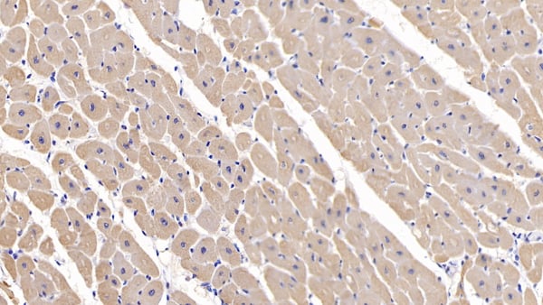
(DAB staining on IHC-P; Samples: Porcine Cardiac Muscle Tissue; Primary Ab: 20ug/ml Rabbit Anti-Porcine IL13 AntibodySecond Ab: 2ug/mL HRP-Linked Caprine Anti-Rabbit IgG Polyclonal Antibody)
IHC (Immunohistochemisry)
(DAB staining on IHC-P; Samples: Porcine Cardiac Muscle Tissue; Primary Ab: 20ug/ml Rabbit Anti-Porcine IL13 AntibodySecond Ab: 2ug/mL HRP-Linked Caprine Anti-Rabbit IgG Polyclonal Antibody)
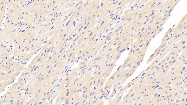
Interleukin 13 (IL13), Polyclonal Antibody (Cat# AAA149595)
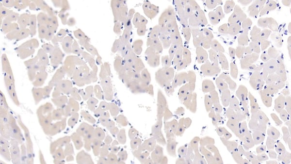
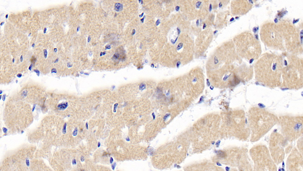 IHC (Immunohistochemisry)
(DAB staining on IHC-P; Samples: Human Cardiac Muscle Tissue; Primary Ab: 20ug/ml Rabbit Anti-Human OPG AntibodySecond Ab: 2ug/mL HRP-Linked Caprine Anti-Rabbit IgG Polyclonal Antibody)
IHC (Immunohistochemisry)
(DAB staining on IHC-P; Samples: Human Cardiac Muscle Tissue; Primary Ab: 20ug/ml Rabbit Anti-Human OPG AntibodySecond Ab: 2ug/mL HRP-Linked Caprine Anti-Rabbit IgG Polyclonal Antibody)
Osteoprotegerin (OPG), Polyclonal Antibody (Cat# AAA149605)
Stromal Cell Derived Factor 1 (SDF1), Polyclonal Antibody (Cat# AAA149606)
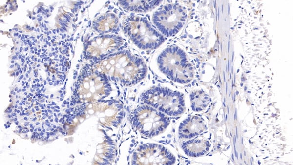 IHC (Immunohiostchemistry)
(DAB staining on IHC-P; Samples: Rat Colon Tissue; Primary Ab: 10ug/ml Rabbit Anti-Rat CEA Antibody Second Ab: 2ug/mL HRP-Linked Caprine Anti-Rabbit IgG Polyclonal Antibody)
IHC (Immunohiostchemistry)
(DAB staining on IHC-P; Samples: Rat Colon Tissue; Primary Ab: 10ug/ml Rabbit Anti-Rat CEA Antibody Second Ab: 2ug/mL HRP-Linked Caprine Anti-Rabbit IgG Polyclonal Antibody)
Carcinoembryonic Antigen (CEA), Polyclonal Antibody (Cat# AAA149610)
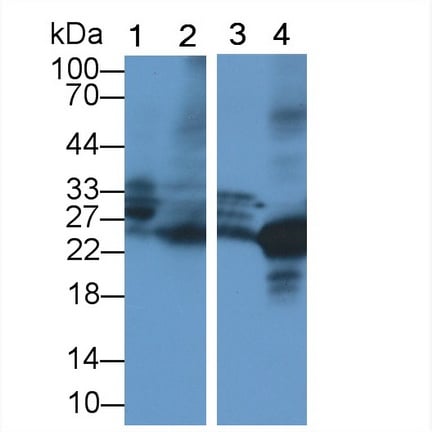 WB (Western Blot)
(Western Blot; Sample: Lane1: Mouse Lung lysate; Lane2: Mouse Spleen lysate; Lane3: Human Lung lysate; Lane4: Rat Lung lysatePrimary Ab: 1ug/ml Rabbit Anti-Mouse ELA2 AntibodySecond Ab: 0.2ug/mL HRP-Linked Caprine Anti-Rabbit IgG Polyclonal Antibody)
WB (Western Blot)
(Western Blot; Sample: Lane1: Mouse Lung lysate; Lane2: Mouse Spleen lysate; Lane3: Human Lung lysate; Lane4: Rat Lung lysatePrimary Ab: 1ug/ml Rabbit Anti-Mouse ELA2 AntibodySecond Ab: 0.2ug/mL HRP-Linked Caprine Anti-Rabbit IgG Polyclonal Antibody)
Neutrophil Elastase (NE), Polyclonal Antibody (Cat# AAA149612)
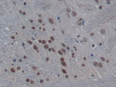 IHC (Immunohistochemistry)
(DAB staining on IHC-P; Samples: Rat Cerebrum Tissue; Primary Ab: 20ug/ml Rabbit Anti-Rat H3 AntibodySecond Ab: 2ug/mL HRP-Linked Caprine Anti-Rabbit IgG Polyclonal Antibody)
IHC (Immunohistochemistry)
(DAB staining on IHC-P; Samples: Rat Cerebrum Tissue; Primary Ab: 20ug/ml Rabbit Anti-Rat H3 AntibodySecond Ab: 2ug/mL HRP-Linked Caprine Anti-Rabbit IgG Polyclonal Antibody)
Histone H3 (H3), Polyclonal Antibody (Cat# AAA149617)
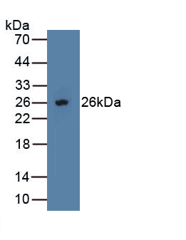
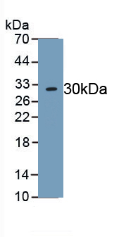
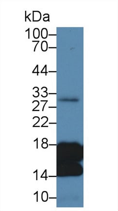
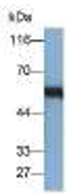
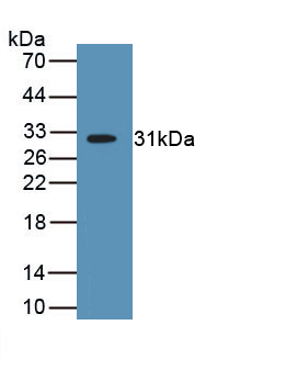
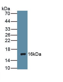
 WB (Western Blot)
(Western Blot; Sample: Recombinant GAL9, Canine.)
WB (Western Blot)
(Western Blot; Sample: Recombinant GAL9, Canine.)
Galectin 9 (GAL9), Polyclonal Antibody (Cat# AAA149619)
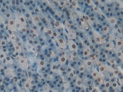
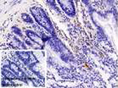
 IHC (Immunohiostchemistry)
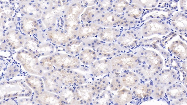
(DAB staining on fromalin fixed paraffin- embedded Kidney tissue)
IHC (Immunohiostchemistry)
(DAB staining on fromalin fixed paraffin- embedded Kidney tissue)
Myelin Basic Protein (MBP), Polyclonal Antibody (Cat# AAA149624)
Immunoglobulin E (IgE), Polyclonal Antibody (Cat# AAA149625)
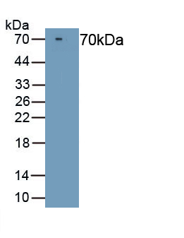
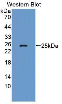
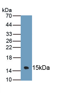
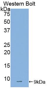
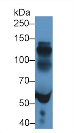 WB (Western Blot)
(Western Blot; Sample: Human Serum; Primary Ab: 5ug/ml Rabbit Anti-Porcine IgE Antibody Second Ab: 0)
WB (Western Blot)
(Western Blot; Sample: Human Serum; Primary Ab: 5ug/ml Rabbit Anti-Porcine IgE Antibody Second Ab: 0)
Immunoglobulin E (IgE), Polyclonal Antibody (Cat# AAA149626)
Fibroblast Growth Factor 2 (FGF2), Polyclonal Antibody (Cat# AAA149627)
Tumor Necrosis Factor Alpha (TNFa), Polyclonal Antibody (Cat# AAA149466)
Immunoglobulin E (IgE), Polyclonal Antibody (Cat# AAA149468)
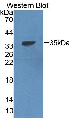
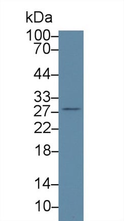
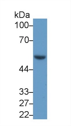
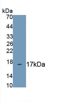
 WB (Western Blot)
(Western Blot;Sample: Rat Salivary gland lysatePrimary Ab: 0.1ug/ml Rabbit Anti-Mouse sIgA AntibodySecond Ab: 0.2ug/ml HRP-Linked Caprine Anti-Rabbit IgG Polyclonal Antibody)
WB (Western Blot)
(Western Blot;Sample: Rat Salivary gland lysatePrimary Ab: 0.1ug/ml Rabbit Anti-Mouse sIgA AntibodySecond Ab: 0.2ug/ml HRP-Linked Caprine Anti-Rabbit IgG Polyclonal Antibody)
Secretory Immunoglobulin A (sIgA), Polyclonal Antibody (Cat# AAA149469)
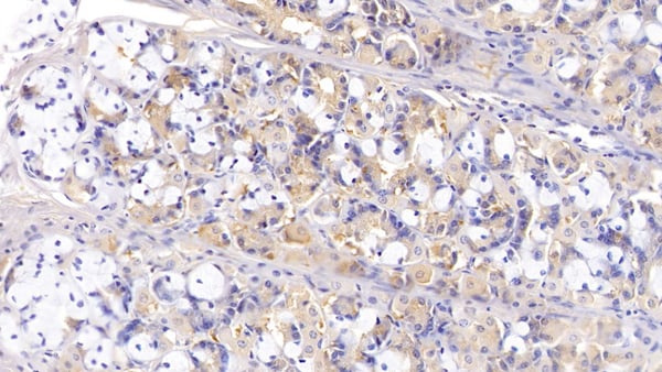
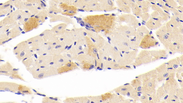
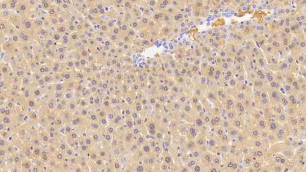
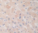
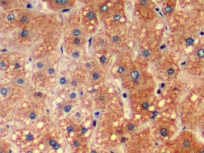 IHC (Immunohiostchemistry)
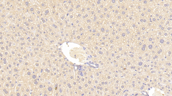
(DAB staining on IHC-P; Samples: Human Liver Tissue)
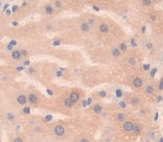
IHC (Immunohiostchemistry)
(DAB staining on IHC-P; Samples: Human Liver Tissue)
Hyaluronidase (HAase), Polyclonal Antibody (Cat# AAA149472)
Phosphatidylserine (PS), Polyclonal Antibody (Cat# AAA149474)
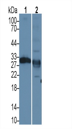
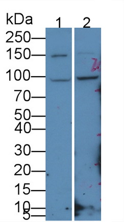
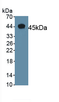
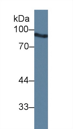
 WB (Western Blot)
(Western Blot: Sample: Lane1: Rat Pancreas lysate; Lane2: Mouse)
WB (Western Blot)
(Western Blot: Sample: Lane1: Rat Pancreas lysate; Lane2: Mouse)
Elastase 2A (ELA2A), Polyclonal Antibody (Cat# AAA149479)
Mucin 2 (MUC2), Polyclonal Antibody (Cat# AAA149807)
Integrin Alpha 7 (ITGa7), Polyclonal Antibody (Cat# AAA149811)
Advillin (AVIL), Polyclonal Antibody (Cat# AAA149814)
Heparin (Hep), Polyclonal Antibody (Cat# AAA149816)
Vanillylmandelic Acid (VMA), Polyclonal Antibody (Cat# AAA149832)
Coagulation Factor VII (F7), Polyclonal Antibody (Cat# AAA149834)
Concanavalin A (ConA), Polyclonal Antibody (Cat# AAA149835)
7-Dehydrocholesterol (7-DHC), Polyclonal Antibody (Cat# AAA149837)
Jasmonic Acid (JA), Polyclonal Antibody (Cat# AAA149842)
Procollagen I C-Terminal Propeptide (PICP), Polyclonal Antibody (Cat# AAA149845)
Dopamine (DA), Polyclonal Antibody (Cat# AAA149846)
Sjogren Syndrome Antigen A1 (SSA1), Polyclonal Antibody (Cat# AAA149847)
Lipase I (LIPI), Polyclonal Antibody (Cat# AAA149848)
Tissue Factor Pathway Inhibitor (TFPI), Polyclonal Antibody (Cat# AAA149852)
Pregnenolone (PGN), Polyclonal Antibody (Cat# AAA149858)
Angiotensin I Converting Enzyme (ACE), Polyclonal Antibody (Cat# AAA149859)
Matrix Metalloproteinase 2 (MMP2), Polyclonal Antibody (Cat# AAA149863)
Myeloperoxidase (MPO), Polyclonal Antibody (Cat# AAA149865)
Procalcitonin (PCT), Polyclonal Antibody (Cat# AAA149866)
Collagen Type II (COL2), Polyclonal Antibody (Cat# AAA149631)
 IHC (Immunohistochemisry)
(DAB staining on IHC-P; Samples: Human Cardiac Muscle Tissue; Primary Ab: 20?g/ml Rabbit Anti-Human GZMA Antibody Second Ab: 2ug/mL HRP-Linked Caprine Anti-Rabbit IgG Polyclonal Antibody)
IHC (Immunohistochemisry)
(DAB staining on IHC-P; Samples: Human Cardiac Muscle Tissue; Primary Ab: 20?g/ml Rabbit Anti-Human GZMA Antibody Second Ab: 2ug/mL HRP-Linked Caprine Anti-Rabbit IgG Polyclonal Antibody)
Granzyme A (GZMA), Polyclonal Antibody (Cat# AAA149632)
 IHC (Immunohiostchemistry)
(DAB staining on IHC-P; Samples: Human Cardiac Muscle Tissue; Primary Ab: 20?g/ml Rabbit Anti-Human CASP12 AntibodySecond Ab: 2ug/mL HRP-Linked Caprine Anti-Rabbit IgG Polyclonal Antibody)
IHC (Immunohiostchemistry)
(DAB staining on IHC-P; Samples: Human Cardiac Muscle Tissue; Primary Ab: 20?g/ml Rabbit Anti-Human CASP12 AntibodySecond Ab: 2ug/mL HRP-Linked Caprine Anti-Rabbit IgG Polyclonal Antibody)
Caspase 12 (CASP12), Polyclonal Antibody (Cat# AAA149638)


 IHC (Immunohistochemisry)
(DAB staining on IHC-P; Samples: Human Lymph node Tissue; Primary Ab: 20?g/ml Rabbit Anti-Human TLR4 AntibodySecond Ab: 2ug/mL HRP-Linked Caprine Anti-Rabbit IgG Polyclonal Antibody)
IHC (Immunohistochemisry)
(DAB staining on IHC-P; Samples: Human Lymph node Tissue; Primary Ab: 20?g/ml Rabbit Anti-Human TLR4 AntibodySecond Ab: 2ug/mL HRP-Linked Caprine Anti-Rabbit IgG Polyclonal Antibody)
Toll Like Receptor 4 (TLR4), Polyclonal Antibody (Cat# AAA149640)
 IHC (Immunohistochemisry)
(DAB staining on IHC-P; Samples: Human Skeletal muscle Tissue; Primary Ab: 10ug/ml Rabbit Anti-Human MYH2 AntibodySecond Ab: 2ug/mL HRP-Linked Caprine Anti-Rabbit IgG Polyclonal Antibody)
IHC (Immunohistochemisry)
(DAB staining on IHC-P; Samples: Human Skeletal muscle Tissue; Primary Ab: 10ug/ml Rabbit Anti-Human MYH2 AntibodySecond Ab: 2ug/mL HRP-Linked Caprine Anti-Rabbit IgG Polyclonal Antibody)
Myosin Heavy Chain 2 (MYH2), Polyclonal Antibody (Cat# AAA149641)
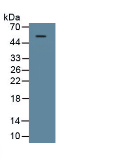
 WB (Western Blot)
(Western Blot; Sample: Lane1: Rat SerumPrimary Ab: 1ug/mL Rabbit Anti-Rat F10 AntibodySecond Ab: 0.2ug/mL HRP-Linked Caprine Anti-Rabbit IgG Polyclonal Antibody)
WB (Western Blot)
(Western Blot; Sample: Lane1: Rat SerumPrimary Ab: 1ug/mL Rabbit Anti-Rat F10 AntibodySecond Ab: 0.2ug/mL HRP-Linked Caprine Anti-Rabbit IgG Polyclonal Antibody)
Coagulation Factor X (F10), Polyclonal Antibody (Cat# AAA149644)
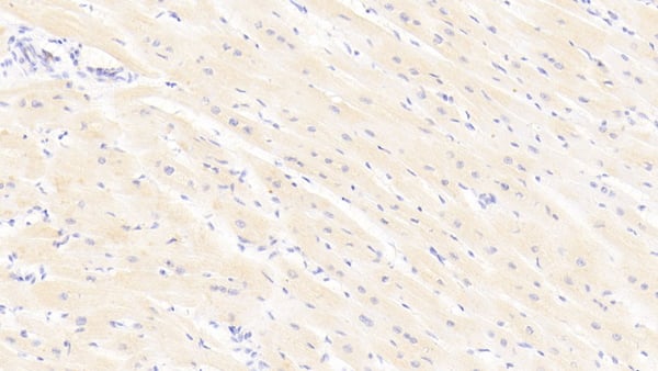
 IHC (Immunohistochemisry)
(DAB staining on IHC-P; Samples: Mouse Liver Tissue; Primary Ab: 20ug/ml Rabbit Anti-Mouse CGRP AntibodySecond Ab: 2ug/mL HRP-Linked Caprine Anti-Rabbit IgG Polyclonal Antibody)
IHC (Immunohistochemisry)
(DAB staining on IHC-P; Samples: Mouse Liver Tissue; Primary Ab: 20ug/ml Rabbit Anti-Mouse CGRP AntibodySecond Ab: 2ug/mL HRP-Linked Caprine Anti-Rabbit IgG Polyclonal Antibody)
Calcitonin Gene Related Peptide (CGRP), Polyclonal Antibody (Cat# AAA149646)
 IHC (Immunohistochemistry)
(DAB staining on IHC-P; Samples: Bovine Kidney Tissue; Primary Ab: 10ug/ml Rabbit Anti-Bovine OPN Antibody Second Ab: 2ug/mL HRP-Linked Caprine Anti-Rabbit IgG Polyclonal Antibody)
IHC (Immunohistochemistry)
(DAB staining on IHC-P; Samples: Bovine Kidney Tissue; Primary Ab: 10ug/ml Rabbit Anti-Bovine OPN Antibody Second Ab: 2ug/mL HRP-Linked Caprine Anti-Rabbit IgG Polyclonal Antibody)
Osteopontin (OPN), Polyclonal Antibody (Cat# AAA149648)
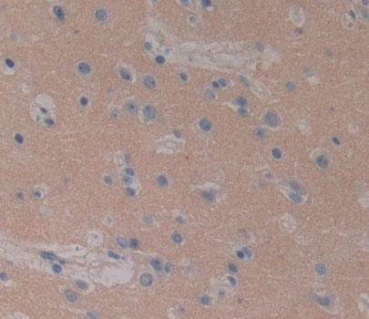
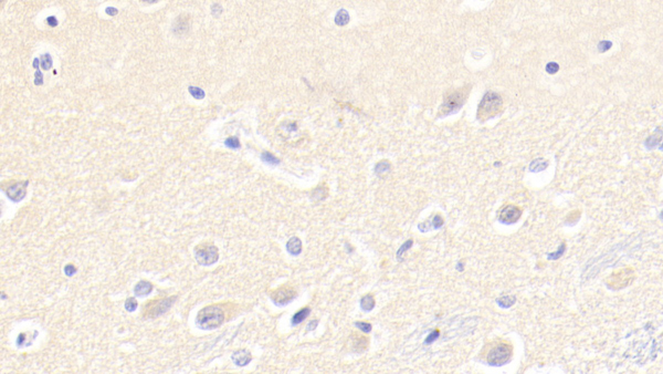
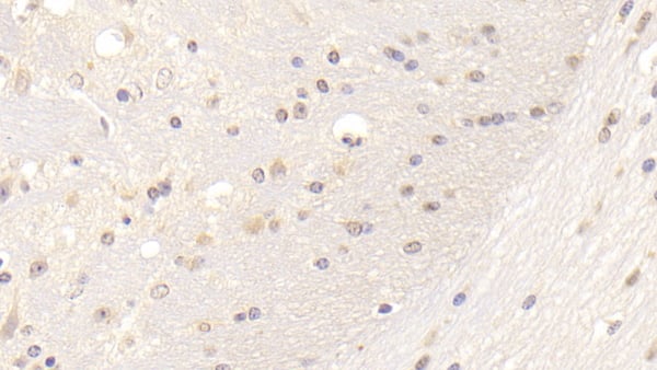 IHC (Immunohistochemisry)
(DAB staining on IHC-P; Samples: Mouse Cerebrum Tissue; Primary Ab: 20?g/ml Rabbit Anti-Mouse LDLR AntibodySecond Ab: 2ug/mL HRP-Linked Caprine Anti-Rabbit IgG Polyclonal Antibody)
IHC (Immunohistochemisry)
(DAB staining on IHC-P; Samples: Mouse Cerebrum Tissue; Primary Ab: 20?g/ml Rabbit Anti-Mouse LDLR AntibodySecond Ab: 2ug/mL HRP-Linked Caprine Anti-Rabbit IgG Polyclonal Antibody)
Low Density Lipoprotein Receptor (LDLR), Polyclonal Antibody (Cat# AAA149650)
 WB (Western Blot)
(Western Blot; Sample: Recombinant MUC3, Mouse.)
WB (Western Blot)
(Western Blot; Sample: Recombinant MUC3, Mouse.)
Mucin 3 (MUC3), Polyclonal Antibody (Cat# AAA149653)
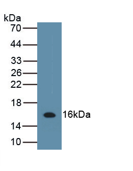
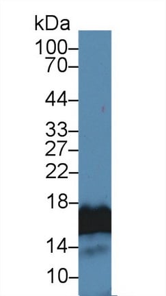
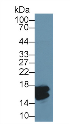
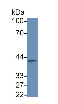
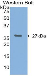
 WB (Western Blot)
(Western Blot;Sample: Recombinant MT1, Mouse.)
WB (Western Blot)
(Western Blot;Sample: Recombinant MT1, Mouse.)
Metallothionein 1 (MT1), Polyclonal Antibody (Cat# AAA149655)
 WB (Western Blot)
(Western Blot; Sample: Human Serum; Primary Ab: 3ug/ml Rabbit Anti-Human ANG AntibodySecond Ab: 0.2ug/mL HRP-Linked Caprine Anti-Rabbit IgG Polyclonal Antibody)
WB (Western Blot)
(Western Blot; Sample: Human Serum; Primary Ab: 3ug/ml Rabbit Anti-Human ANG AntibodySecond Ab: 0.2ug/mL HRP-Linked Caprine Anti-Rabbit IgG Polyclonal Antibody)
Angiostatin (ANG), Polyclonal Antibody (Cat# AAA149658)
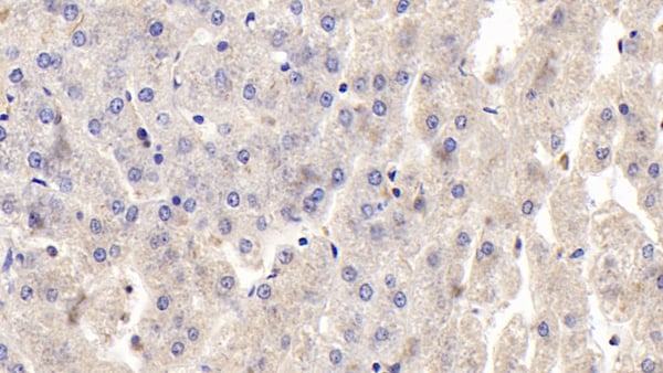
 IHC (Immunohiostchemistry)
(DAB staining on IHC-P; Samples: Human Liver Tissue)
IHC (Immunohiostchemistry)
(DAB staining on IHC-P; Samples: Human Liver Tissue)
Dopamine Receptor D1 (DRD1), Polyclonal Antibody (Cat# AAA149660)
 IHC (Immunohiostchemistry)
(DAB staining on fromalin fixed paraffin- embedded Kidney tissue)
IHC (Immunohiostchemistry)
(DAB staining on fromalin fixed paraffin- embedded Kidney tissue)
Chemokine (C-X-C Motif) Ligand 2 (CXCL2), Polyclonal Antibody (Cat# AAA149664)
What are Polyclonal Antibodies?
Polyclonal antibodies are antibodies that come from multiple B cell clones of a host animal. The typical hosts used for the majority of polyclonal antibody production are rabbits, goats, sheep, and donkeys. These polyclonal antibodies, once having identified their target, will bind to different epitopes located at different regions or sequences on the same protein/antigen. As a result, they are ideal at locating and binding to the target, even if the target is in very low concentrations (due to many different antibodies being able to bind to the same target molecule, which allows for significant amplification of a downstream signal).
Polyclonal antibodies are typically produced by injecting an antigen into a host animal, which causes the animal’s immune system to attack the foreign antigen by mass generating antibodies against it. After a period of time, serum is collected from the animal and purified using physicochemical fractionation, class-specific affinity purification, and/or antigen-affinity purification.
Key Uses of Polyclonal Antibodies
- Western Blotting: This method is used to find specific proteins in biological samples after separating them by size.
- Immunohistochemistry: IHC helps visualize the location of proteins in tissue sections using various staining techniques.
- ELISA: (Enzyme-Linked Immunosorbent Assay) is typically used to identify specific protein quantities in a sample. ELISAs can be either “Quantitative” or “Qualitative”.
- Flow Cytometry: technique that identifies and measures the specific protein on the surface or inside the cells in a fluid suspension.
- Immunoprecipitation: IP isolates and studies a specific protein from a complex mixture using antibodies.
Why Buy Polyclonal Antibodies from AAA Biotech?
1. Ideal for Various Applications
Our antibodies are generally going to be validated for use in multiple types of assays, including ELISA, Western Blotting, Immunohistochemistry, Immunoprecipitation, amongst others. They are ideal for a wide range of research applications.
2. Rigorous Quality Control
All of the antibodies in our catalog undergo strict quality testing to ensure specificity, sensitivity, and consistent performance. We are confident in the ability of our antibodies to provide you with accurate results.
3. Wide Assortment of Antibodies
Antibodies in are catalog can be found for both common and exotic species, and these antibodies are also available in both conjugated and recombinant forms to suit many diverse experimental needs.
4. Highly Purified
Our antibodies are available in purified forms with over 85% purity, as confirmed by SDS-PAGE. They are also available with tags such as His, Flag, GST, or MBP. We cater to customers worldwide.
FAQ
1. How are polyclonal antibodies produced?
Traditionally, polyclonal antibodies are produced by injecting an antigen into a host animal (such as a rabbit or goat), which then triggers an immune response from the host animal. The animal’s B cells produce antibodies that will recognize different parts of the injected antigen. These antibodies are then collected from the animal’s blood and purified for use.
2. How do polyclonal antibodies differ from monoclonal antibodies?
Polyclonal antibodies are a mix of antibodies that bind to different locations (epitopes) of the same antigen, while monoclonal antibodies are identical and bind to just one specific epitope. This makes polyclonal antibodies more versatile and better at detecting proteins that may be present in low quantities or in altered/modified forms.
3. How should I store polyclonal antibodies?
Polyclonal antibodies should be stored at 4°C for short-term use (up to a few weeks) and at -20°C or -80°C for long-term storage. Avoid repeated freeze-thaw cycles by dividing them into small aliquots. Always check the datasheet for specific storage instructions.