Filters
▼Clonality
▼Type
▼Reactivity
▼Gene Name
▼Isotype
▼Host
▼Application
▼Clone
▼Polyclonal Antibodies
At AAA Biotech also known as AAA Bio or AAABio, we provide a broad range of purified polyclonal antibodies (pAbs) that are able to all be browsed online through our website. Due to their high specificity and strong binding affinity, these antibodies are ideal for wide swathes of research and experimental applications.
Our polyclonal antibodies can easily support your work, whether you use them for Western Blotting, Immunocytochemistry (with or without Immunofluorescence used in conjunction), Immunohistochemistry, Immunoprecipitation, and ELISA tests. We highly encourage you to browse our range of pAbs and choose the one that best suits your experimental model.
Viewing 8650-8700 of 96805 product results
 IHC (Immunohiostchemistry)
(DAB staining on IHCP;Sample: Human Cerebrum Tissue; Primary Ab: 10ug/ml Rabbit AntiHuman GPR44 AntibodySecond Ab: 2ug/mL HRPLinked Caprine AntiRabbit IgG Polyclonal Antibody(Catalog: SAA544Rb19))
IHC (Immunohiostchemistry)
(DAB staining on IHCP;Sample: Human Cerebrum Tissue; Primary Ab: 10ug/ml Rabbit AntiHuman GPR44 AntibodySecond Ab: 2ug/mL HRPLinked Caprine AntiRabbit IgG Polyclonal Antibody(Catalog: SAA544Rb19))
G Protein Coupled Receptor 44 (GPR44), Polyclonal Antibody (Cat# AAA151935)
 IHC (Immunohistochemisry)
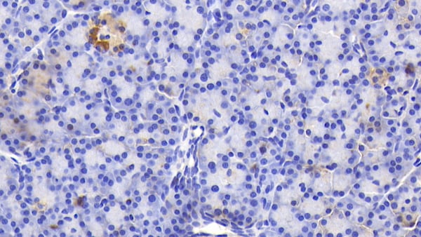
(DAB staining on IHCP;Sample: Porcine Stomach Tissue; Primary Ab: 20ug/ml Rabbit AntiMultispecies GIP AntibodySecond Ab: 2ug/mL HRPLinked Caprine AntiRabbit IgG Polyclonal Antibody(Catalog: SAA544Rb19))
IHC (Immunohistochemisry)
(DAB staining on IHCP;Sample: Porcine Stomach Tissue; Primary Ab: 20ug/ml Rabbit AntiMultispecies GIP AntibodySecond Ab: 2ug/mL HRPLinked Caprine AntiRabbit IgG Polyclonal Antibody(Catalog: SAA544Rb19))
Gastric Inhibitory Polypeptide (GIP), Polyclonal Antibody (Cat# AAA151942)
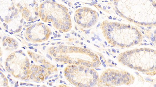 IHC (Immunohistochemisry)
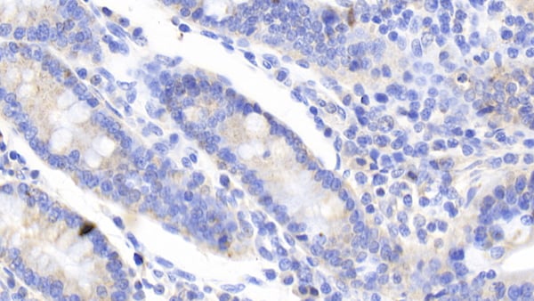
(DAB staining on IHCP;Samples: Human Stomach Tissue;Primary Ab: 20ug/ml Rabbit AntiHuman KRT19 AntibodySecond Ab: 2ug/mL HRPLinked Caprine AntiRabbit IgG Polyclonal Antibody(Catalog: SAA544Rb19))
IHC (Immunohistochemisry)
(DAB staining on IHCP;Samples: Human Stomach Tissue;Primary Ab: 20ug/ml Rabbit AntiHuman KRT19 AntibodySecond Ab: 2ug/mL HRPLinked Caprine AntiRabbit IgG Polyclonal Antibody(Catalog: SAA544Rb19))
Cytokeratin 19 (CK19), Polyclonal Antibody (Cat# AAA151948)
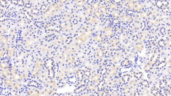
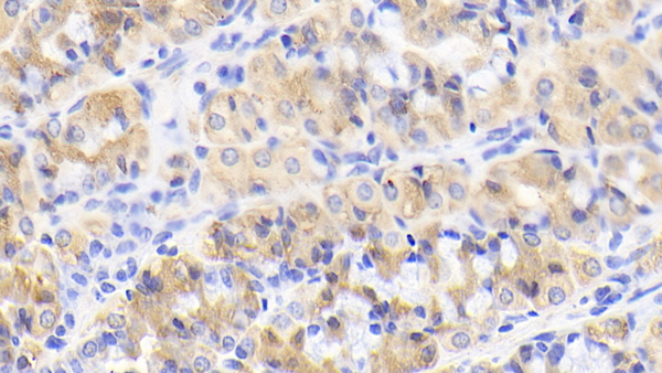
 IHC (Immunohiostchemistry)
(DAB staining on IHCP;Samples: Rat Liver Tissue;Primary Ab: 20ug/ml Rabbit AntiRat APOC2 AntibodySecond Ab: 2ug/mL HRPLinked Caprine AntiRabbit IgG Polyclonal Antibody(Catalog: SAA544Rb19))
IHC (Immunohiostchemistry)
(DAB staining on IHCP;Samples: Rat Liver Tissue;Primary Ab: 20ug/ml Rabbit AntiRat APOC2 AntibodySecond Ab: 2ug/mL HRPLinked Caprine AntiRabbit IgG Polyclonal Antibody(Catalog: SAA544Rb19))
Apolipoprotein C2 (APOC2), Polyclonal Antibody (Cat# AAA151955)
Meropenem (Mer), Polyclonal Antibody (Cat# AAA151973)
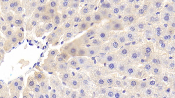
 IHC (Immunohiostchemistry)
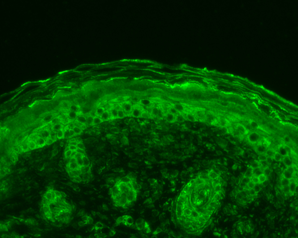
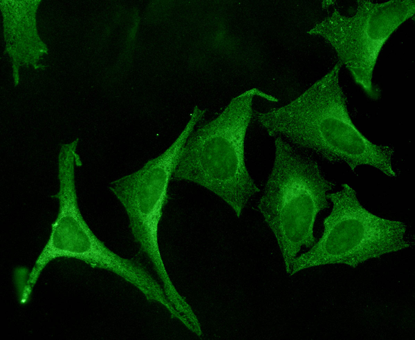
(Immunohistochemistry analysis using Rabbit Anti-SOD2 Polyclonal Antibody (SPC-117). Tissue: backskin. Species: Mouse. Fixation: Bouin's Fixative Solution. Primary Antibody: Rabbit Anti-SOD2 Polyclonal Antibody (SPC-117) at 1:100 for 1 hour at RT. Secondary Antibody: FITC Goat Anti-Rabbit (green) at 1:50 for 1 hour at RT. Localization: Mitochondrion matrix.)
IHC (Immunohiostchemistry)
(Immunohistochemistry analysis using Rabbit Anti-SOD2 Polyclonal Antibody (SPC-117). Tissue: backskin. Species: Mouse. Fixation: Bouin's Fixative Solution. Primary Antibody: Rabbit Anti-SOD2 Polyclonal Antibody (SPC-117) at 1:100 for 1 hour at RT. Secondary Antibody: FITC Goat Anti-Rabbit (green) at 1:50 for 1 hour at RT. Localization: Mitochondrion matrix.)
SOD (Mn), Polyclonal Antibody (Cat# AAA103559)
 WB (Western Blot)
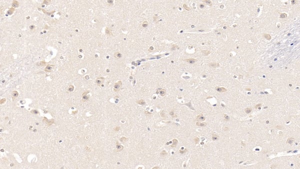
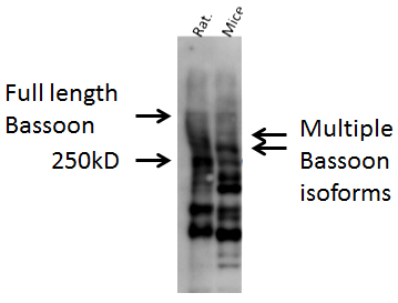
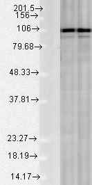
(Western blot analysis of Mouse, Rat brain cell lysates showing detection of Bassoon protein using Rabbit Anti-Bassoon Polyclonal Antibody (SPC-198). Primary Antibody: Rabbit Anti-Bassoon Polyclonal Antibody (SPC-198) at 1:1000.)
WB (Western Blot)
(Western blot analysis of Mouse, Rat brain cell lysates showing detection of Bassoon protein using Rabbit Anti-Bassoon Polyclonal Antibody (SPC-198). Primary Antibody: Rabbit Anti-Bassoon Polyclonal Antibody (SPC-198) at 1:1000.)
Bassoon, Polyclonal Antibody (Cat# AAA103563)
 WB (Western Blot)
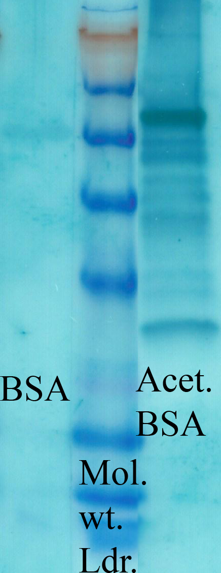
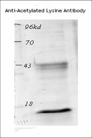
(Western blot analysis of Mouse Spleen lysates showing detection of Acetylated Lysine protein using Rabbit Anti-Acetylated Lysine Polyclonal Antibody (SPC-155). Primary Antibody: Rabbit Anti-Acetylated Lysine Polyclonal Antibody (SPC-155) at 1:1000.)
WB (Western Blot)
(Western blot analysis of Mouse Spleen lysates showing detection of Acetylated Lysine protein using Rabbit Anti-Acetylated Lysine Polyclonal Antibody (SPC-155). Primary Antibody: Rabbit Anti-Acetylated Lysine Polyclonal Antibody (SPC-155) at 1:1000.)
Acetylated Lysine, Polyclonal Antibody (Cat# AAA103581)
 WB (Western Blot)
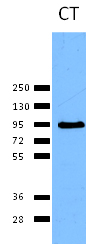
(Western blot analysis of Rat Tissue lysates showing detection of Calnexin protein using Rabbit Anti-Calnexin Polyclonal Antibody (SPC-108). Load: 15 ugprotein. Block: 1.5% BSA. Primary Antibody: Rabbit Anti-Calnexin Polyclonal Antibody (SPC-108) at 1:1000 for 2 hours at RT. Secondary Antibody: Donkey Anti-Rabbit IgG: HRP for 1 hour at RT.)
WB (Western Blot)
(Western blot analysis of Rat Tissue lysates showing detection of Calnexin protein using Rabbit Anti-Calnexin Polyclonal Antibody (SPC-108). Load: 15 ugprotein. Block: 1.5% BSA. Primary Antibody: Rabbit Anti-Calnexin Polyclonal Antibody (SPC-108) at 1:1000 for 2 hours at RT. Secondary Antibody: Donkey Anti-Rabbit IgG: HRP for 1 hour at RT.)
Calnexin-CT, Polyclonal Antibody (Cat# AAA103592)
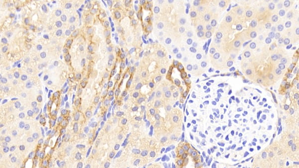
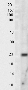
 WB (Western Blot)
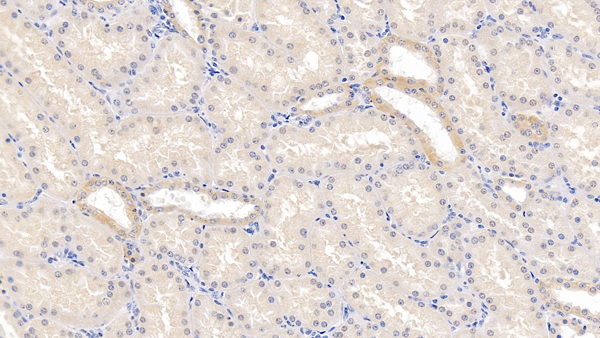
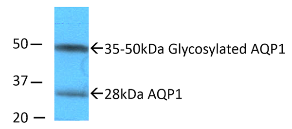
(Western blot analysis of Rat kidney inner medullary homogenates showing detection of Aquaporin 1 protein using Rabbit Anti-Aquaporin 1 Polyclonal Antibody (SPC-502). Primary Antibody: Rabbit Anti-Aquaporin 1 Polyclonal Antibody (SPC-502) at 1:2000. Showing glycosylated and non-glycosylated bands.)
WB (Western Blot)
(Western blot analysis of Rat kidney inner medullary homogenates showing detection of Aquaporin 1 protein using Rabbit Anti-Aquaporin 1 Polyclonal Antibody (SPC-502). Primary Antibody: Rabbit Anti-Aquaporin 1 Polyclonal Antibody (SPC-502) at 1:2000. Showing glycosylated and non-glycosylated bands.)
Aquaporin 1, Polyclonal Antibody (Cat# AAA103704)

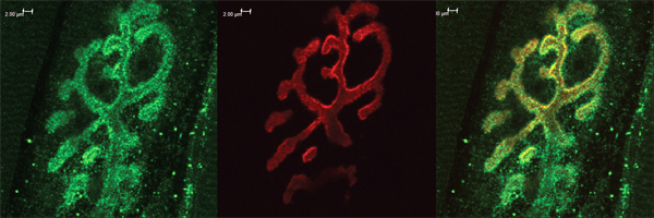
 IHC (Immunohiostchemistry)
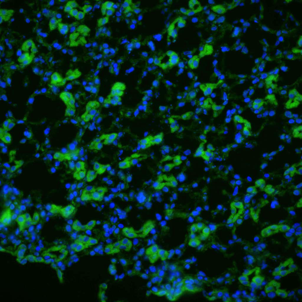
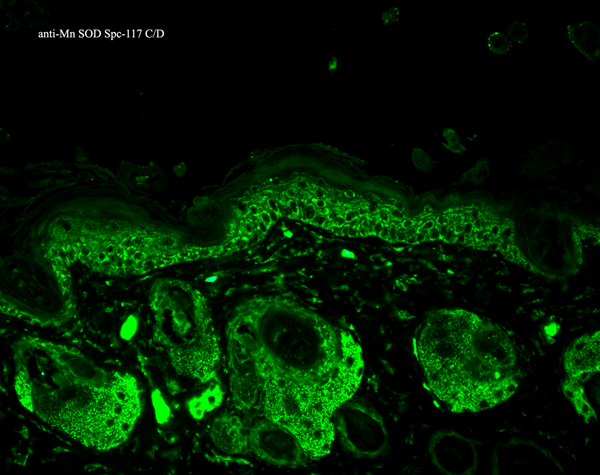
(Immunohistochemistry analysis using Rabbit Anti-SOD2 Polyclonal Antibody (SPC-117). Tissue: backskin. Species: Mouse. Fixation: Bouin's Fixative Solution. Primary Antibody: Rabbit Anti-SOD2 Polyclonal Antibody (SPC-117) at 1:100 for 1 hour at RT. Secondary Antibody: FITC Goat Anti-Rabbit (green) at 1:50 for 1 hour at RT. Localization: Mitochondrion matrix.)
IHC (Immunohiostchemistry)
(Immunohistochemistry analysis using Rabbit Anti-SOD2 Polyclonal Antibody (SPC-117). Tissue: backskin. Species: Mouse. Fixation: Bouin's Fixative Solution. Primary Antibody: Rabbit Anti-SOD2 Polyclonal Antibody (SPC-117) at 1:100 for 1 hour at RT. Secondary Antibody: FITC Goat Anti-Rabbit (green) at 1:50 for 1 hour at RT. Localization: Mitochondrion matrix.)
SOD (Mn), Polyclonal Antibody (Cat# AAA103717)
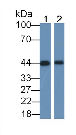
 WB (Western Blot)
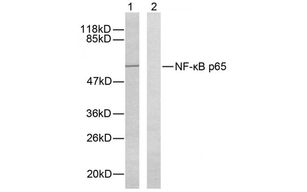
(Western blot analysis of extracts from HeLa cells. Lane 1: Using NF-kB p65 antibody. Lane 2: The same antibody preincubated with synthesized peptide.)
WB (Western Blot)
(Western blot analysis of extracts from HeLa cells. Lane 1: Using NF-kB p65 antibody. Lane 2: The same antibody preincubated with synthesized peptide.)
NFKP p65, Polyclonal Antibody (Cat# AAA108299)
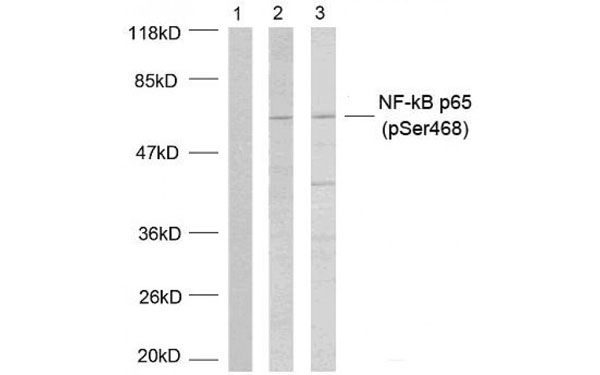 WB (Western Blot)
(Western blot analysis of extracts using NF-kB p65 (phospho-Ser468) antibody.)
WB (Western Blot)
(Western blot analysis of extracts using NF-kB p65 (phospho-Ser468) antibody.)
NFKP p65, Polyclonal Antibody (Cat# AAA108325)
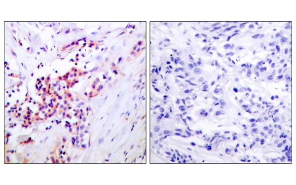
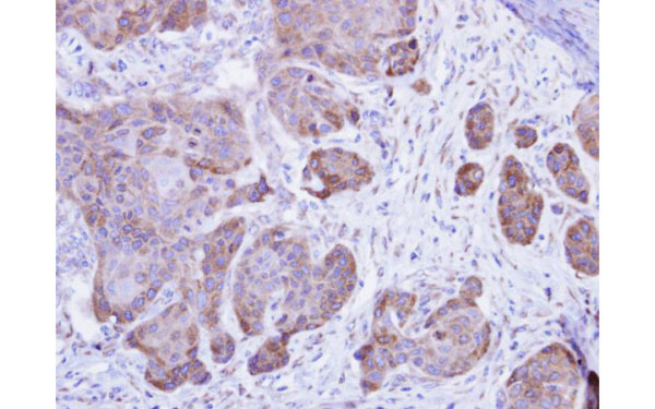
 IHC (Immunohistochemistry)
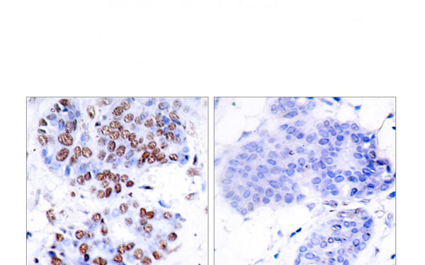
(Immunohistochemical analysis of paraffin-embedded human breast carcinoma tissue using E cadherin pan antibody)
IHC (Immunohistochemistry)
(Immunohistochemical analysis of paraffin-embedded human breast carcinoma tissue using E cadherin pan antibody)
E cadherin pan, Polyclonal Antibody (Cat# AAA108337)
 WB (Western Blot)
Fam55d, Polyclonal Antibody (Cat# AAA108357)
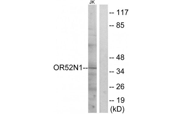
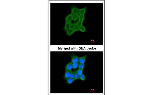
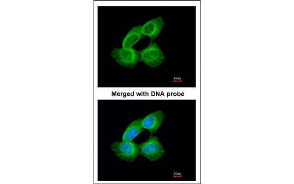
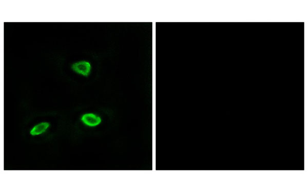 IF (Immunofluorescence)
(Immunofluorescence analysis of LOVO cells, using OR52N1 antibody.)
IF (Immunofluorescence)
(Immunofluorescence analysis of LOVO cells, using OR52N1 antibody.)
OR52N1, Polyclonal Antibody (Cat# AAA108393)
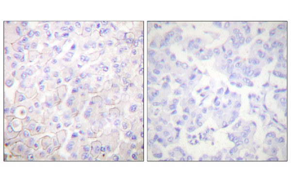
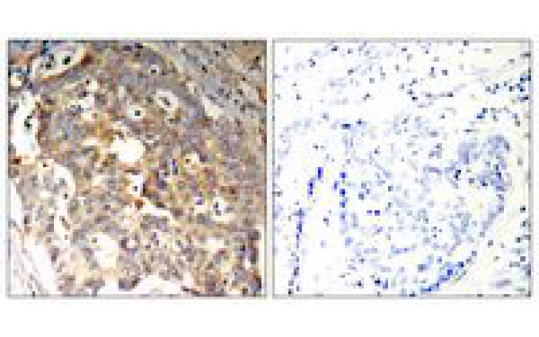
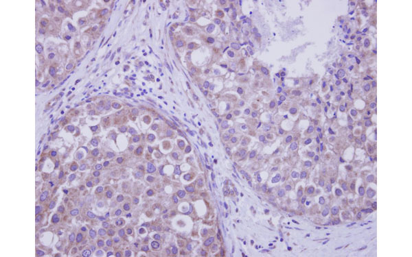 IHC (Immunohistochemisry)
(Immunohistochemical staining of paraffin-embedded Breast ca using LIM kinase 2 antibody at a dilution of 1:250)
IHC (Immunohistochemisry)
(Immunohistochemical staining of paraffin-embedded Breast ca using LIM kinase 2 antibody at a dilution of 1:250)
LIM kinase 2, Polyclonal Antibody (Cat# AAA107842)
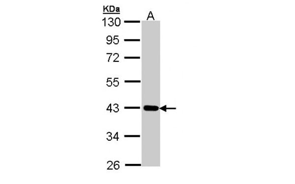
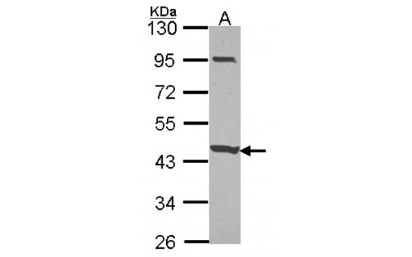
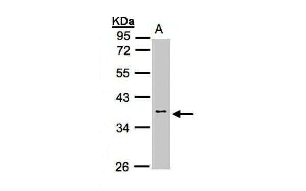 WB (Western Blot)
(Western blot analysis of 30 ug whole cell lysate (A:H1299) using a 10 % SDS PAGE gel and Calsequestrin 2 antibody at a dilution of 1:1000)
WB (Western Blot)
(Western blot analysis of 30 ug whole cell lysate (A:H1299) using a 10 % SDS PAGE gel and Calsequestrin 2 antibody at a dilution of 1:1000)
Calsequestrin 2, Polyclonal Antibody (Cat# AAA107875)
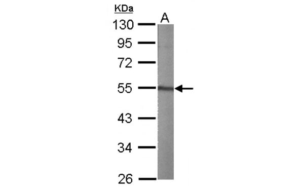
 WB (Western Blot)
(Western blot analysis of 30 ug of whole cell lysate (A: A431) using a 10 % SDS PAGE gel and Cartilage associated protein antibody at a dilution of 1:1000)
WB (Western Blot)
(Western blot analysis of 30 ug of whole cell lysate (A: A431) using a 10 % SDS PAGE gel and Cartilage associated protein antibody at a dilution of 1:1000)
Cartilage associated, Polyclonal Antibody (Cat# AAA107877)
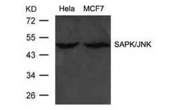 WB (Western Blot)
(Western Blot analysis of extracts from Hela and MCF cells using SAPK/JNK antibody)
WB (Western Blot)
(Western Blot analysis of extracts from Hela and MCF cells using SAPK/JNK antibody)
SAPK/JNK, Polyclonal Antibody (Cat# AAA107932)
 WB (Western Blot)
(Western blot analysis of 30 ug of whole cell lysate (A: JurKat) using a 10 % SDS PAGE gel and CCDC83 antibody at a dilution of 1:1000)
WB (Western Blot)
(Western blot analysis of 30 ug of whole cell lysate (A: JurKat) using a 10 % SDS PAGE gel and CCDC83 antibody at a dilution of 1:1000)
CCDC83, Polyclonal Antibody (Cat# AAA107938)

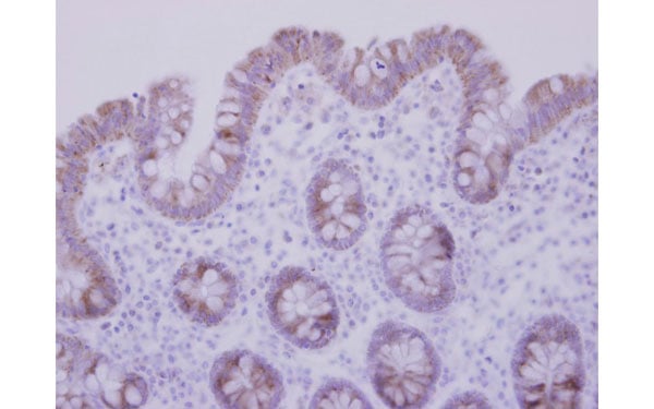
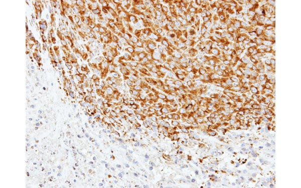 IHC (Immunohistochemistry)
(Immunohistochemical staining of paraffin-embedded SAS xenograft using alpha Tubulin 1A antibody at a dilution of 1:500)
IHC (Immunohistochemistry)
(Immunohistochemical staining of paraffin-embedded SAS xenograft using alpha Tubulin 1A antibody at a dilution of 1:500)
alpha Tubulin 1A, Polyclonal Antibody (Cat# AAA107950)
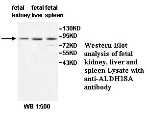
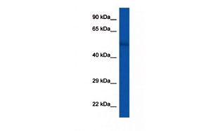
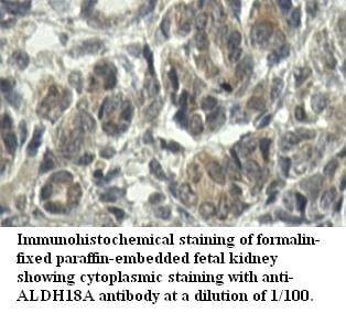 Application Data
ALDH18A, Polyclonal Antibody (Cat# AAA112216)
Predicted: Mouse, Rat
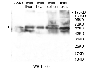
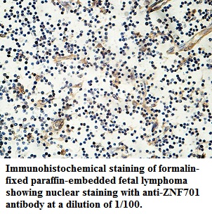 Application Data
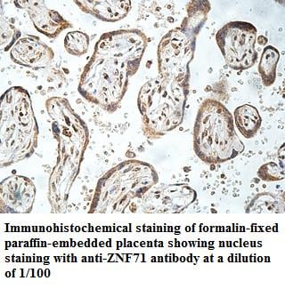
ZNF701, Polyclonal Antibody (Cat# AAA112221)
Predicted: Mouse, Rat
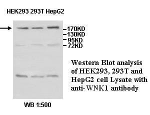
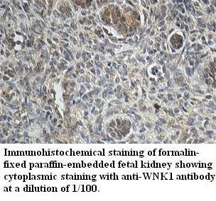 Application Data
WNK1, Polyclonal Antibody (Cat# AAA112226)
Predicted: Mouse, Rat
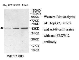
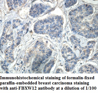 Application Data
FBXW12, Polyclonal Antibody (Cat# AAA112229)
Predicted: Mouse, Rat
 Application Data
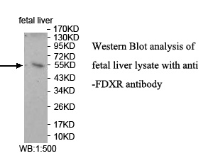
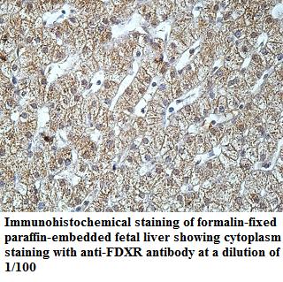
FDXR, Polyclonal Antibody (Cat# AAA112005)
Predicted: Mouse, Rat
 Application Data
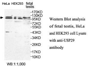
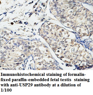
USP29, Polyclonal Antibody (Cat# AAA112008)
Predicted: Mouse, Rat
 Application Data
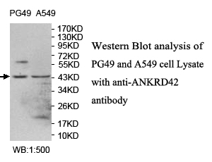
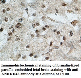
ANKRD42, Polyclonal Antibody (Cat# AAA112014)
 Application Data
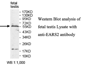
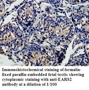
EARS2, Polyclonal Antibody (Cat# AAA112030)
Predicted: Mouse, Rat
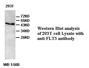
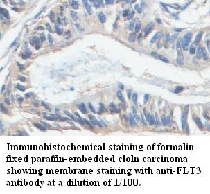 Application Data
FLT3, Polyclonal Antibody (Cat# AAA112032)

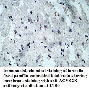 Application Data
ACVR2B, Polyclonal Antibody (Cat# AAA112034)
Predicted: Mouse, Rat
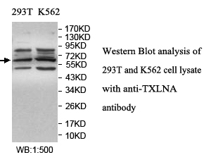
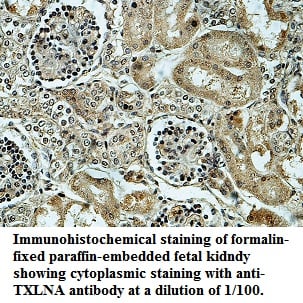 Application Data
TXLNA, Polyclonal Antibody (Cat# AAA112041)
Predicted: Mouse, Rat
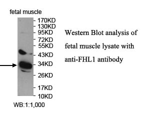
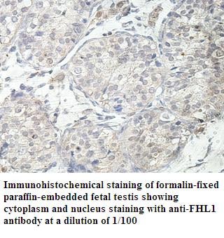 Application Data
FHL1, Polyclonal Antibody (Cat# AAA112050)
Predicted: Mouse, Rat
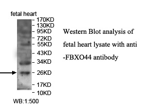
 Application Data
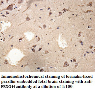
FBXO44, Polyclonal Antibody (Cat# AAA111770)
Predicted: Mouse, Rat
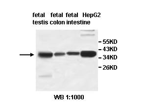
 Application Data
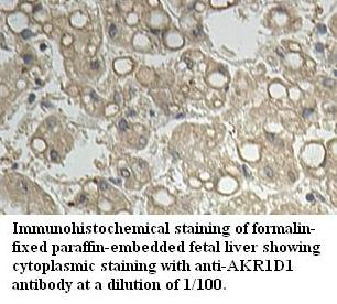
AKR1D1, Polyclonal Antibody (Cat# AAA111776)
Predicted: Mouse, Rat
 Application Data
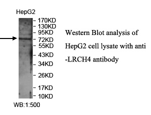
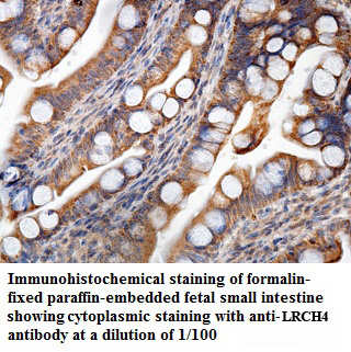
LRCH4, Polyclonal Antibody (Cat# AAA111777)
Predicted: Mouse, Rat
 Application Data
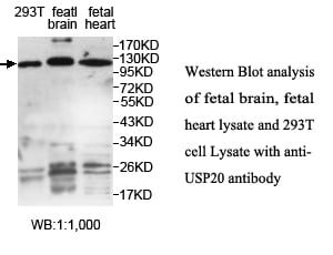
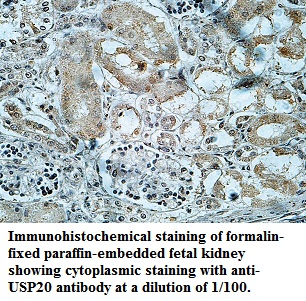
USP20, Polyclonal Antibody (Cat# AAA111786)
Predicted: Mouse, Rat
 Application Data
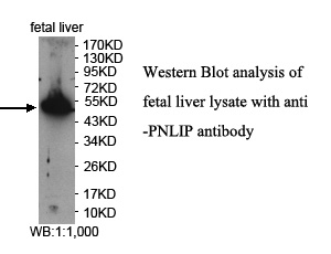
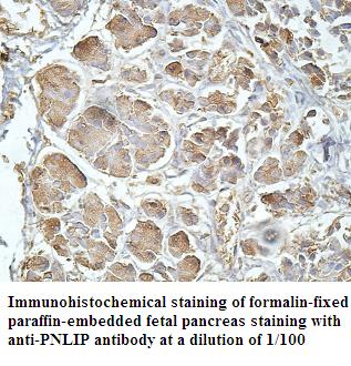
PNLIP, Polyclonal Antibody (Cat# AAA111788)
Predicted: Mouse, Rat
 Application Data
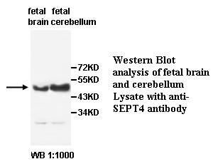
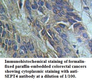
SEPT4, Polyclonal Antibody (Cat# AAA111795)
 Application Data
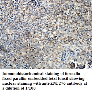
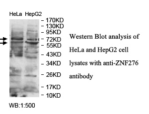
ZNF276, Polyclonal Antibody (Cat# AAA111799)
Predicted: Mouse, Rat
 Application Data
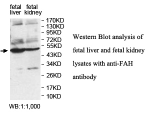
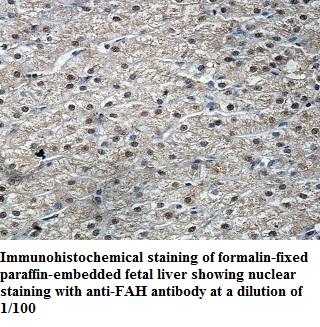
FAH, Polyclonal Antibody (Cat# AAA111804)
Predicted: Mouse, Rat
 Application Data
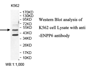
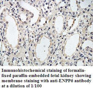
ENPP6, Polyclonal Antibody (Cat# AAA111807)
Predicted: Mouse, Rat
 Application Data
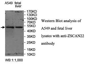
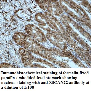
ZSCAN22, Polyclonal Antibody (Cat# AAA111808)
Predicted: Mouse, Rat
 Application Data
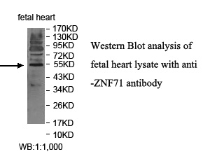
ZNF71, Polyclonal Antibody (Cat# AAA111818)
Predicted: Mouse, Rat
 Application Data
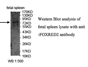
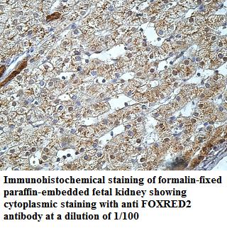
FOXRED2, Polyclonal Antibody (Cat# AAA111825)
Predicted: Mouse, Rat
 Application Data
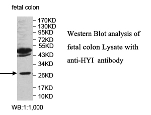
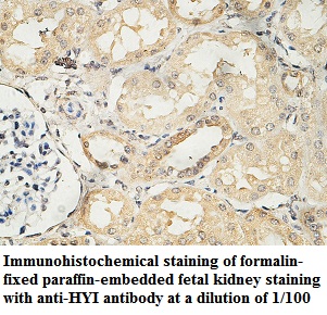
HYI, Polyclonal Antibody (Cat# AAA111827)
Predicted: Mouse, Rat
 Application Data
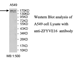
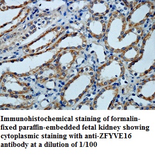
ZFYVE16, Polyclonal Antibody (Cat# AAA111830)
Predicted: Mouse, Rat
 Application Data
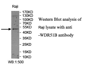
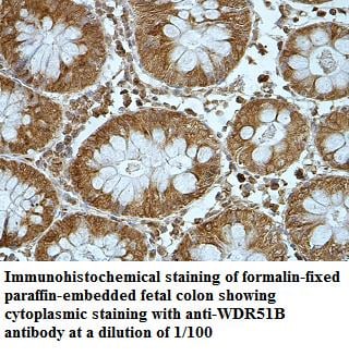
WDR51B, Polyclonal Antibody (Cat# AAA111836)
Predicted: Mouse, Rat
 Application Data
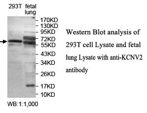
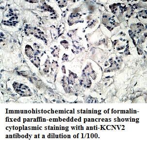
KCNV2, Polyclonal Antibody (Cat# AAA111840)
Predicted: Mouse, Rat
What are Polyclonal Antibodies?
Polyclonal antibodies are antibodies that come from multiple B cell clones of a host animal. The typical hosts used for the majority of polyclonal antibody production are rabbits, goats, sheep, and donkeys. These polyclonal antibodies, once having identified their target, will bind to different epitopes located at different regions or sequences on the same protein/antigen. As a result, they are ideal at locating and binding to the target, even if the target is in very low concentrations (due to many different antibodies being able to bind to the same target molecule, which allows for significant amplification of a downstream signal).
Polyclonal antibodies are typically produced by injecting an antigen into a host animal, which causes the animal’s immune system to attack the foreign antigen by mass generating antibodies against it. After a period of time, serum is collected from the animal and purified using physicochemical fractionation, class-specific affinity purification, and/or antigen-affinity purification.
Key Uses of Polyclonal Antibodies
- Western Blotting: This method is used to find specific proteins in biological samples after separating them by size.
- Immunohistochemistry: IHC helps visualize the location of proteins in tissue sections using various staining techniques.
- ELISA: (Enzyme-Linked Immunosorbent Assay) is typically used to identify specific protein quantities in a sample. ELISAs can be either “Quantitative” or “Qualitative”.
- Flow Cytometry: technique that identifies and measures the specific protein on the surface or inside the cells in a fluid suspension.
- Immunoprecipitation: IP isolates and studies a specific protein from a complex mixture using antibodies.
Why Buy Polyclonal Antibodies from AAA Biotech?
1. Ideal for Various Applications
Our antibodies are generally going to be validated for use in multiple types of assays, including ELISA, Western Blotting, Immunohistochemistry, Immunoprecipitation, amongst others. They are ideal for a wide range of research applications.
2. Rigorous Quality Control
All of the antibodies in our catalog undergo strict quality testing to ensure specificity, sensitivity, and consistent performance. We are confident in the ability of our antibodies to provide you with accurate results.
3. Wide Assortment of Antibodies
Antibodies in are catalog can be found for both common and exotic species, and these antibodies are also available in both conjugated and recombinant forms to suit many diverse experimental needs.
4. Highly Purified
Our antibodies are available in purified forms with over 85% purity, as confirmed by SDS-PAGE. They are also available with tags such as His, Flag, GST, or MBP. We cater to customers worldwide.
FAQ
1. How are polyclonal antibodies produced?
Traditionally, polyclonal antibodies are produced by injecting an antigen into a host animal (such as a rabbit or goat), which then triggers an immune response from the host animal. The animal’s B cells produce antibodies that will recognize different parts of the injected antigen. These antibodies are then collected from the animal’s blood and purified for use.
2. How do polyclonal antibodies differ from monoclonal antibodies?
Polyclonal antibodies are a mix of antibodies that bind to different locations (epitopes) of the same antigen, while monoclonal antibodies are identical and bind to just one specific epitope. This makes polyclonal antibodies more versatile and better at detecting proteins that may be present in low quantities or in altered/modified forms.
3. How should I store polyclonal antibodies?
Polyclonal antibodies should be stored at 4°C for short-term use (up to a few weeks) and at -20°C or -80°C for long-term storage. Avoid repeated freeze-thaw cycles by dividing them into small aliquots. Always check the datasheet for specific storage instructions.