Filters
▼Clonality
▼Type
▼Reactivity
▼Gene Name
▼Isotype
▼Host
▼Application
▼Clone
▼Polyclonal Antibodies
At AAA Biotech also known as AAA Bio or AAABio, we provide a broad range of purified polyclonal antibodies (pAbs) that are able to all be browsed online through our website. Due to their high specificity and strong binding affinity, these antibodies are ideal for wide swathes of research and experimental applications.
Our polyclonal antibodies can easily support your work, whether you use them for Western Blotting, Immunocytochemistry (with or without Immunofluorescence used in conjunction), Immunohistochemistry, Immunoprecipitation, and ELISA tests. We highly encourage you to browse our range of pAbs and choose the one that best suits your experimental model.
Viewing 8850-8900 of 96805 product results

 Application Data
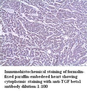
TGF beta1, Polyclonal Antibody (Cat# AAA109864)
 Application Data
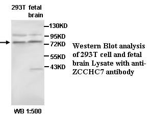
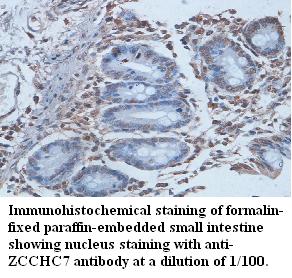
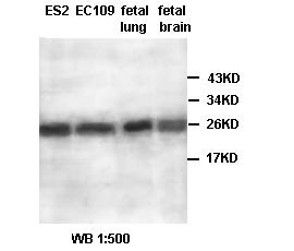
ZCCHC7, Polyclonal Antibody (Cat# AAA109872)
Predicted: Mouse, Rat
 Application Data
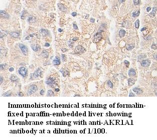
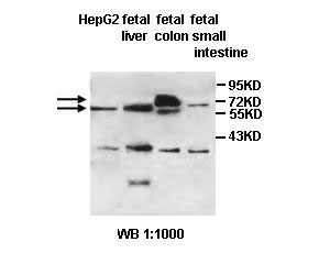
AKR1A1, Polyclonal Antibody (Cat# AAA109881)
 Application Data
ABCG5, Polyclonal Antibody (Cat# AAA109884)
 Application Data
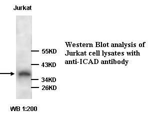
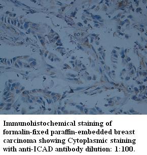
DFFA/ICAD, Polyclonal Antibody (Cat# AAA109886)
 WB (Western Blot)
(WB (1:1000) analysis of protein OsTip3-1 expression in rice (CV. 9311) root tissue at the seeding stage with Anti-OsTip3-1.)
WB (Western Blot)
(WB (1:1000) analysis of protein OsTip3-1 expression in rice (CV. 9311) root tissue at the seeding stage with Anti-OsTip3-1.)
OsTip3-1, Polyclonal Antibody (Cat# AAA109907)
 Application Data
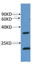
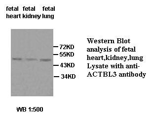
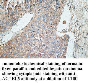
ACTBL3, Polyclonal Antibody (Cat# AAA110021)
 Application Data
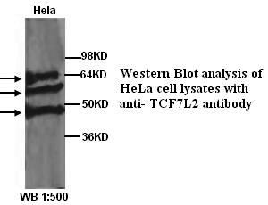
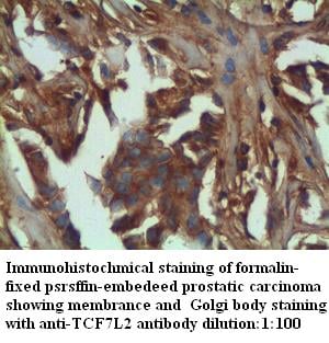
TCF7L2, Polyclonal Antibody (Cat# AAA110029)
 Application Data
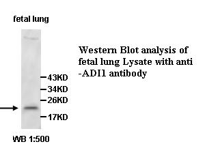
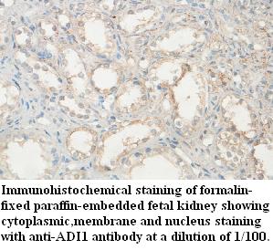
ADI1, Polyclonal Antibody (Cat# AAA110040)
Predicted: Mouse, Rat
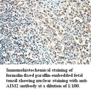 Application Data
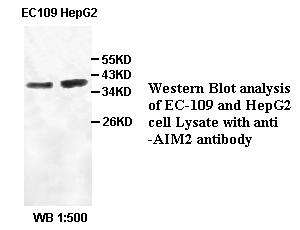
AIM2, Polyclonal Antibody (Cat# AAA110047)
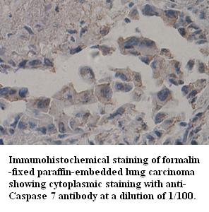
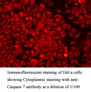 Application Data
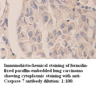
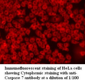
Caspase 7, Polyclonal Antibody (Cat# AAA110050)
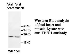
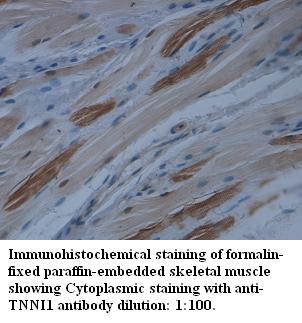 Application Data
TNNI1, Polyclonal Antibody (Cat# AAA110063)
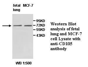
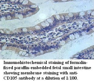 Application Data
CD105, Polyclonal Antibody (Cat# AAA110076)
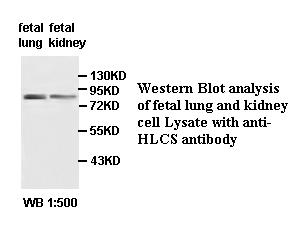
 Application Data
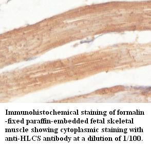
HLCS, Polyclonal Antibody (Cat# AAA110082)
Predicted: Mouse, Rat
 Application Data
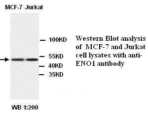
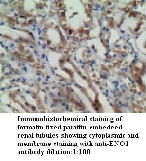
ENO1, Polyclonal Antibody (Cat# AAA110088)
 Application Data
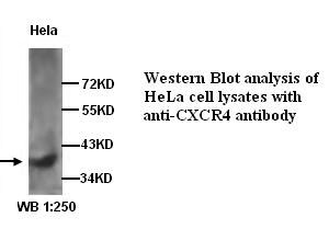
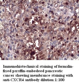
CXCR4, Polyclonal Antibody (Cat# AAA109925)
 Application Data
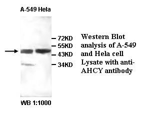
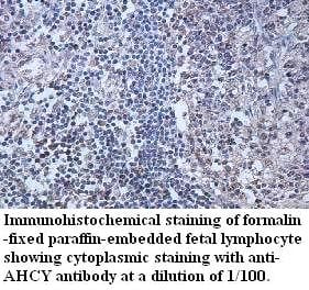
AHCY, Polyclonal Antibody (Cat# AAA109928)
Predicted: Mouse, Rat
 Application Data
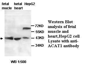
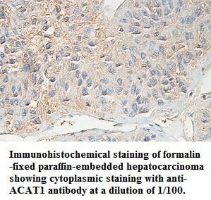
ACAT1, Polyclonal Antibody (Cat# AAA109932)
Predicted: Mouse, Rat
 Application Data
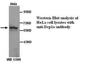
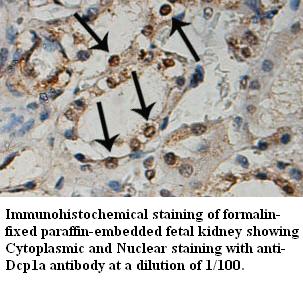
Dcp1a, Polyclonal Antibody (Cat# AAA109951)
 Application Data
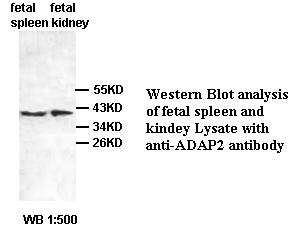
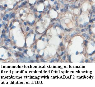
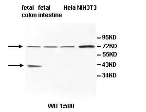
ADAP2, Polyclonal Antibody (Cat# AAA109953)
Predicted: Mouse, Rat
 Application Data
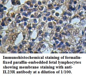
IL23R, Polyclonal Antibody (Cat# AAA109956)
Predicted: Mouse, Rat
 Application Data
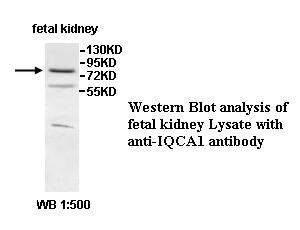
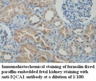
IQCA1, Polyclonal Antibody (Cat# AAA109992)
Predicted: Mouse, Rat
 Application Data
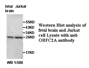
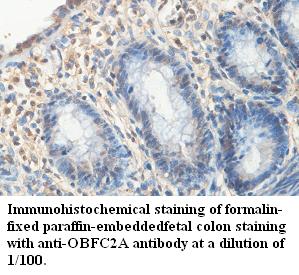
OBFC2A, Polyclonal Antibody (Cat# AAA109999)
Predicted: Mouse, Rat
 Application Data
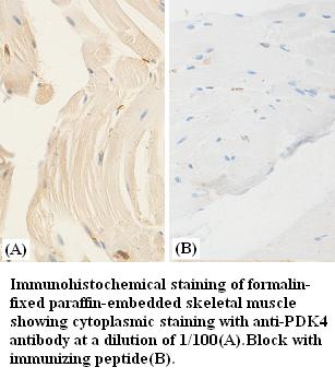
PDK4, Polyclonal Antibody (Cat# AAA110372)
 Application Data
Caspase7, Polyclonal Antibody (Cat# AAA110374)
 Application Data
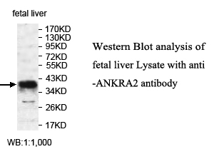
ANKRA2, Polyclonal Antibody (Cat# AAA110376)
Predicted: Mouse, Rat
 Application Data
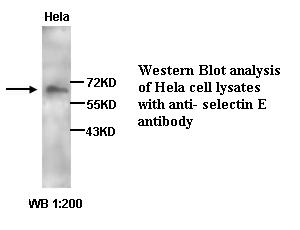
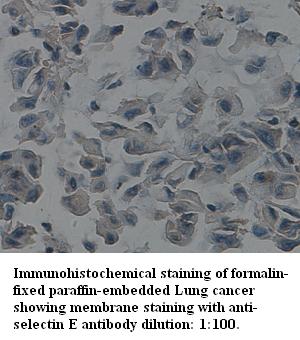
selectin E, Polyclonal Antibody (Cat# AAA110385)

 Application Data
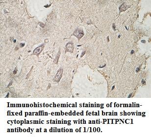
PITPNC1, Polyclonal Antibody (Cat# AAA110386)
Predicted: Mouse, Rat
 Application Data
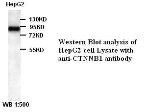
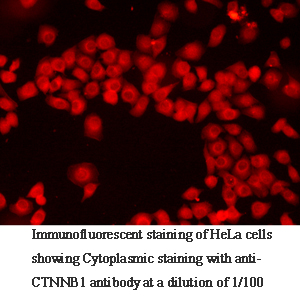
CTNNB1, Polyclonal Antibody (Cat# AAA110387)
 Application Data
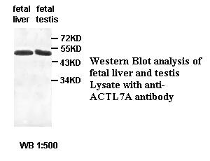
ACTL7A, Polyclonal Antibody (Cat# AAA110392)
Predicted: Mouse, Rat
 Application Data
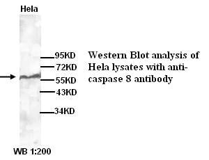
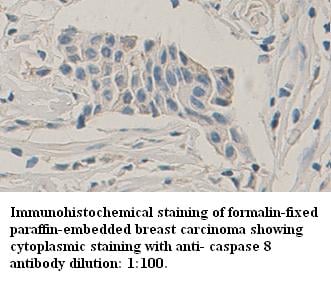
caspase 8, Polyclonal Antibody (Cat# AAA110397)
 Application Data
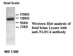
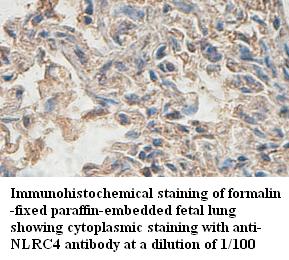
NLRC4, Polyclonal Antibody (Cat# AAA110402)
Predicted: Mouse, Rat
 Application Data
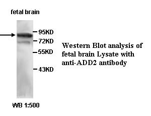
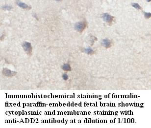
ADD2, Polyclonal Antibody (Cat# AAA110404)
Predicted: Mouse, Rat
 Application Data
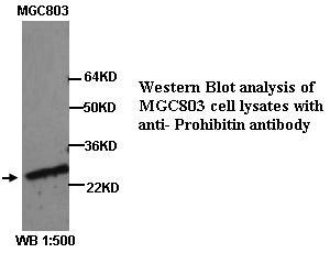
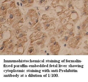
Prohibitin, Polyclonal Antibody (Cat# AAA110193)

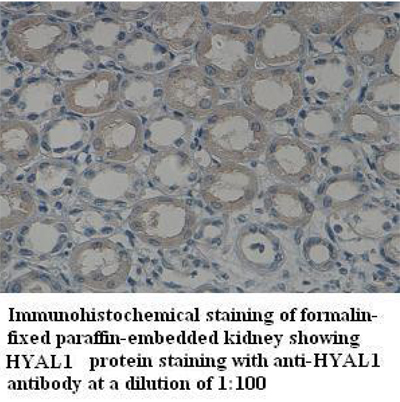 IHC (Immunohiostchemistry)
HYAL1, Polyclonal Antibody (Cat# AAA110209)
 Application Data
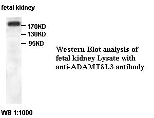
ADAMTSL3, Polyclonal Antibody (Cat# AAA110215)
Predicted: Mouse, Rat
Not yet tested in other species
 Application Data
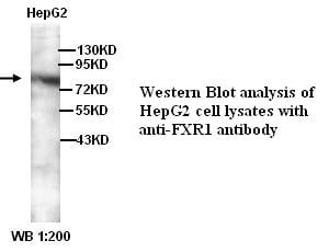
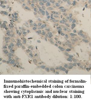
FXR1, Polyclonal Antibody (Cat# AAA110246)
 Application Data
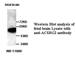
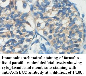
ACSBG2, Polyclonal Antibody (Cat# AAA110254)
Predicted: Mouse, Rat
 Application Data
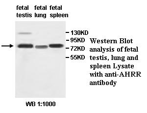
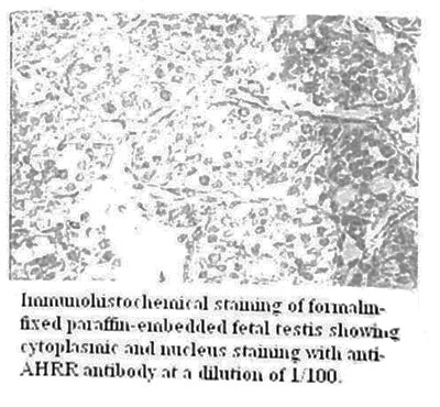
AHRR, Polyclonal Antibody (Cat# AAA110258)
 Application Data
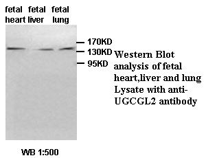
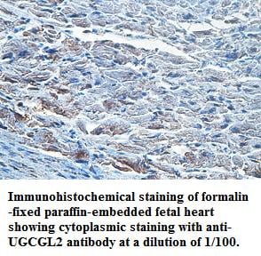
UGCGL2, Polyclonal Antibody (Cat# AAA110262)
Predicted: Mouse, Rat
 Application Data
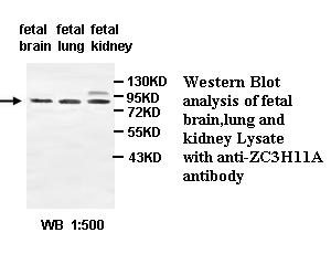
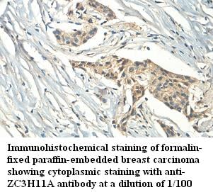
ZC3H11A, Polyclonal Antibody (Cat# AAA110272)
Predicted: Mouse, Rat
 Application Data
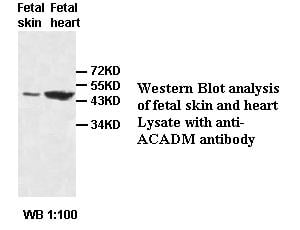
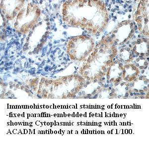
ACADM, Polyclonal Antibody (Cat# AAA110275)
Predicted: Mouse, Rat

 Application Data
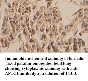
AP1G1, Polyclonal Antibody (Cat# AAA110286)
 Application Data
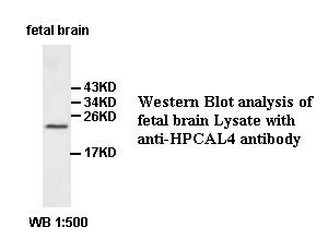
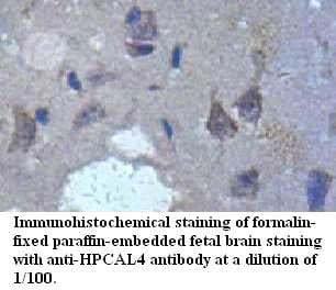
HPCAL4, Polyclonal Antibody (Cat# AAA110287)
Predicted: Mouse, Rat
 Application Data
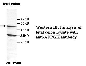
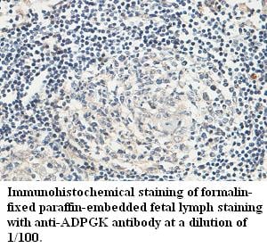
ADPGK, Polyclonal Antibody (Cat# AAA110319)
Predicted: Mouse, Rat
 Application Data
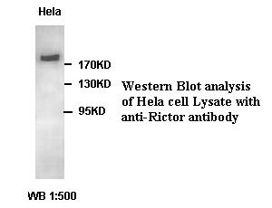
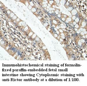
Rictor, Polyclonal Antibody (Cat# AAA110322)
 Application Data
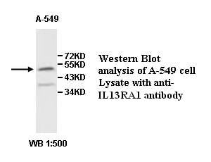
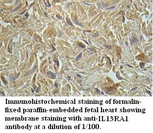
IL13RA1, Polyclonal Antibody (Cat# AAA110324)
Predicted: Mouse, Rat
 Application Data
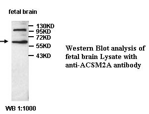
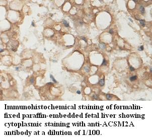
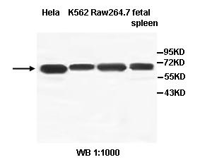
ACSM2A, Polyclonal Antibody (Cat# AAA110100)
Predicted: Mouse, Rat
 Application Data
IL18RAP, Polyclonal Antibody (Cat# AAA110102)
Predicted: Mouse, Rat
 Application Data
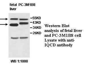
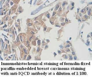
IQCD, Polyclonal Antibody (Cat# AAA110110)
Predicted: Mouse, Rat
What are Polyclonal Antibodies?
Polyclonal antibodies are antibodies that come from multiple B cell clones of a host animal. The typical hosts used for the majority of polyclonal antibody production are rabbits, goats, sheep, and donkeys. These polyclonal antibodies, once having identified their target, will bind to different epitopes located at different regions or sequences on the same protein/antigen. As a result, they are ideal at locating and binding to the target, even if the target is in very low concentrations (due to many different antibodies being able to bind to the same target molecule, which allows for significant amplification of a downstream signal).
Polyclonal antibodies are typically produced by injecting an antigen into a host animal, which causes the animal’s immune system to attack the foreign antigen by mass generating antibodies against it. After a period of time, serum is collected from the animal and purified using physicochemical fractionation, class-specific affinity purification, and/or antigen-affinity purification.
Key Uses of Polyclonal Antibodies
- Western Blotting: This method is used to find specific proteins in biological samples after separating them by size.
- Immunohistochemistry: IHC helps visualize the location of proteins in tissue sections using various staining techniques.
- ELISA: (Enzyme-Linked Immunosorbent Assay) is typically used to identify specific protein quantities in a sample. ELISAs can be either “Quantitative” or “Qualitative”.
- Flow Cytometry: technique that identifies and measures the specific protein on the surface or inside the cells in a fluid suspension.
- Immunoprecipitation: IP isolates and studies a specific protein from a complex mixture using antibodies.
Why Buy Polyclonal Antibodies from AAA Biotech?
1. Ideal for Various Applications
Our antibodies are generally going to be validated for use in multiple types of assays, including ELISA, Western Blotting, Immunohistochemistry, Immunoprecipitation, amongst others. They are ideal for a wide range of research applications.
2. Rigorous Quality Control
All of the antibodies in our catalog undergo strict quality testing to ensure specificity, sensitivity, and consistent performance. We are confident in the ability of our antibodies to provide you with accurate results.
3. Wide Assortment of Antibodies
Antibodies in are catalog can be found for both common and exotic species, and these antibodies are also available in both conjugated and recombinant forms to suit many diverse experimental needs.
4. Highly Purified
Our antibodies are available in purified forms with over 85% purity, as confirmed by SDS-PAGE. They are also available with tags such as His, Flag, GST, or MBP. We cater to customers worldwide.
FAQ
1. How are polyclonal antibodies produced?
Traditionally, polyclonal antibodies are produced by injecting an antigen into a host animal (such as a rabbit or goat), which then triggers an immune response from the host animal. The animal’s B cells produce antibodies that will recognize different parts of the injected antigen. These antibodies are then collected from the animal’s blood and purified for use.
2. How do polyclonal antibodies differ from monoclonal antibodies?
Polyclonal antibodies are a mix of antibodies that bind to different locations (epitopes) of the same antigen, while monoclonal antibodies are identical and bind to just one specific epitope. This makes polyclonal antibodies more versatile and better at detecting proteins that may be present in low quantities or in altered/modified forms.
3. How should I store polyclonal antibodies?
Polyclonal antibodies should be stored at 4°C for short-term use (up to a few weeks) and at -20°C or -80°C for long-term storage. Avoid repeated freeze-thaw cycles by dividing them into small aliquots. Always check the datasheet for specific storage instructions.