Filters
▼Clonality
▼Type
▼Reactivity
▼Gene Name
▼Isotype
▼Host
▼Application
▼Clone
▼Polyclonal Antibodies
At AAA Biotech also known as AAA Bio or AAABio, we provide a broad range of purified polyclonal antibodies (pAbs) that are able to all be browsed online through our website. Due to their high specificity and strong binding affinity, these antibodies are ideal for wide swathes of research and experimental applications.
Our polyclonal antibodies can easily support your work, whether you use them for Western Blotting, Immunocytochemistry (with or without Immunofluorescence used in conjunction), Immunohistochemistry, Immunoprecipitation, and ELISA tests. We highly encourage you to browse our range of pAbs and choose the one that best suits your experimental model.
Viewing 9000-9050 of 96805 product results

 Application Data
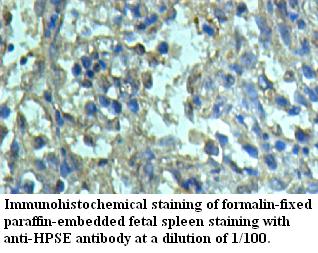
HPSE, Polyclonal Antibody (Cat# AAA109112)
Predicted: Mouse, Rat
 Application Data
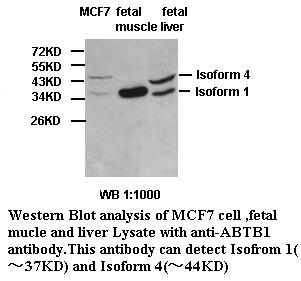
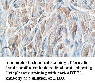
ABTB1, Polyclonal Antibody (Cat# AAA109143)
Predicted: Mouse, Rat
 Application Data
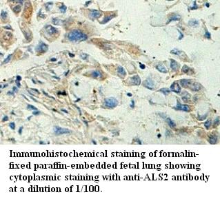
ALS2, Polyclonal Antibody (Cat# AAA109146)
Predicted: Mouse, Rat
 Application Data
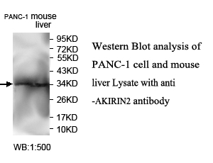
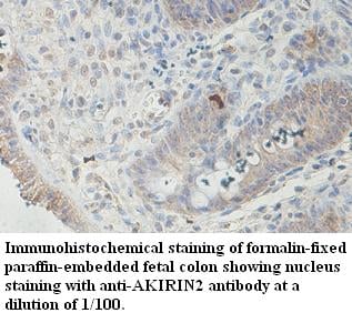
AKIRIN2, Polyclonal Antibody (Cat# AAA109147)
Predicted: Mouse, Rat
 Application Data
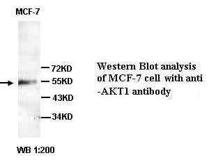
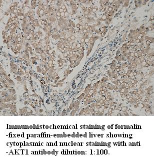
AKT1, Polyclonal Antibody (Cat# AAA109179)
 Application Data
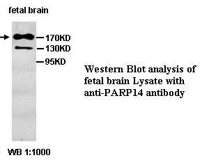
PARP14, Polyclonal Antibody (Cat# AAA109362)
 Application Data
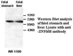
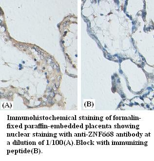
ZNF668, Polyclonal Antibody (Cat# AAA109370)
 Application Data
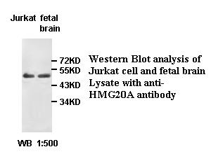
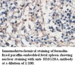
HMG20A, Polyclonal Antibody (Cat# AAA109421)
Predicted: Mouse, Rat
 Application Data
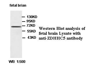
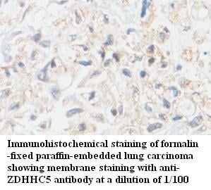
ZDHHC5, Polyclonal Antibody (Cat# AAA109685)
Predicted: Mouse, Rat
 Application Data
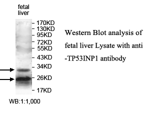
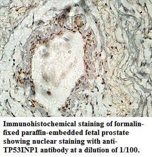
TP53INP1, Polyclonal Antibody (Cat# AAA109700)
 Application Data
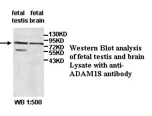
ADAM18, Polyclonal Antibody (Cat# AAA109720)
Predicted: Mouse, Rat
Not yet tested in other species.
 Application Data
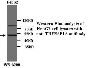
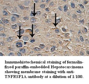
TNFRSF1A, Polyclonal Antibody (Cat# AAA109742)
 Application Data
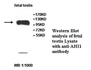
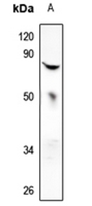
AHI1, Polyclonal Antibody (Cat# AAA109489)
 WB (Western Blot)
(Western blot analysis of IL-2 expression in CTLL2 (A) whole cell lysates.)
WB (Western Blot)
(Western blot analysis of IL-2 expression in CTLL2 (A) whole cell lysates.)
IL-2, Polyclonal Antibody (Cat# AAA104381)
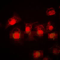
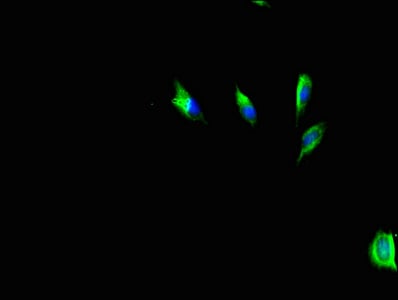
 IF (Immunofluorescence)
(Immunofluorescent analysis of PFKP staining in NIH3T3 cells. Formalin-fixed cells were permeabilized with 0.1% Triton X-100 in TBS for 5-10 minutes and blocked with 3% BSA-PBS for 30 minutes at room temperature. Cells were probed with the primary antibody in 3% BSA-PBS and incubated overnight at 4 °C in a humidified chamber. Cells were washed with PBST and incubated with a DyLight 594-conjugated secondary antibody (red) in PBS at room temperature in the dark. DAPI was used to stain the cell nuclei (blue).)
IF (Immunofluorescence)
(Immunofluorescent analysis of PFKP staining in NIH3T3 cells. Formalin-fixed cells were permeabilized with 0.1% Triton X-100 in TBS for 5-10 minutes and blocked with 3% BSA-PBS for 30 minutes at room temperature. Cells were probed with the primary antibody in 3% BSA-PBS and incubated overnight at 4 °C in a humidified chamber. Cells were washed with PBST and incubated with a DyLight 594-conjugated secondary antibody (red) in PBS at room temperature in the dark. DAPI was used to stain the cell nuclei (blue).)
PFKP, Polyclonal Antibody (Cat# AAA104391)
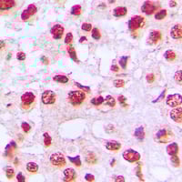
 IHC (Immunohiostchemistry)
(Immunohistochemical analysis of TIMP4 staining in human breast cancer formalin fixed paraffin embedded tissue section. The section was pre-treated using heat mediated antigen retrieval with sodium citrate buffer (pH 6.0). The section was then incubated with the antibody at room temperature and detected using an HRP conjugated compact polymer system. DAB was used as the chromogen. The section was then counterstained with haematoxylin and mounted with DPX.)
IHC (Immunohiostchemistry)
(Immunohistochemical analysis of TIMP4 staining in human breast cancer formalin fixed paraffin embedded tissue section. The section was pre-treated using heat mediated antigen retrieval with sodium citrate buffer (pH 6.0). The section was then incubated with the antibody at room temperature and detected using an HRP conjugated compact polymer system. DAB was used as the chromogen. The section was then counterstained with haematoxylin and mounted with DPX.)
TIMP4, Polyclonal Antibody (Cat# AAA104392)
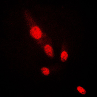
 IF (Immunofluorescence)
(Immunofluorescent analysis of DAXX staining in HeLa cells. Formalin-fixed cells were permeabilized with 0.1% Triton X-100 in TBS for 5-10 minutes and blocked with 3% BSA-PBS for 30 minutes at room temperature. Cells were probed with the primary antibody in 3% BSA-PBS and incubated overnight at 4 °C in a humidified chamber. Cells were washed with PBST and incubated with a DyLight 594-conjugated secondary antibody (red) in PBS at room temperature in the dark.)
IF (Immunofluorescence)
(Immunofluorescent analysis of DAXX staining in HeLa cells. Formalin-fixed cells were permeabilized with 0.1% Triton X-100 in TBS for 5-10 minutes and blocked with 3% BSA-PBS for 30 minutes at room temperature. Cells were probed with the primary antibody in 3% BSA-PBS and incubated overnight at 4 °C in a humidified chamber. Cells were washed with PBST and incubated with a DyLight 594-conjugated secondary antibody (red) in PBS at room temperature in the dark.)
DAXX, Polyclonal Antibody (Cat# AAA104422)
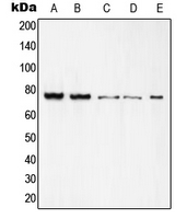
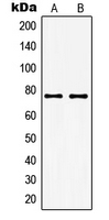
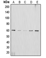
 WB (Western Blot)
(Western blot analysis of c-Rel expression in A549 (A), Hela (B), CT26 (C), PC12 (D) whole cell lysates.)
WB (Western Blot)
(Western blot analysis of c-Rel expression in A549 (A), Hela (B), CT26 (C), PC12 (D) whole cell lysates.)
c-Rel, Polyclonal Antibody (Cat# AAA104432)
 WB (Western Blot)
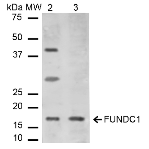
(Western blot analysis of Mouse, Rat Liver cell lysates showing detection of ~17.2 kDa FUNDC1 protein using Rabbit Anti-FUNDC1 Polyclonal Antibody . Lane 1: Molecular Weight Ladder (MW). Lane 2: Mouse Liver cell lysates. Lane 3: Rat Liver cell lysates. Load: 15 ug. Block: 5% Skim Milk in 1X TBST. Primary Antibody: Rabbit Anti-FUNDC1 Polyclonal Antibody at 1:1000 for 2 hours at RT. Secondary Antibody: Goat Anti-Rabbit IgG: HRP at 1:2000 for 60 min at RT. Color Development: ECL solution for 6 min in RT. Predicted/Observed Size: ~17.2 kDa.)
WB (Western Blot)
(Western blot analysis of Mouse, Rat Liver cell lysates showing detection of ~17.2 kDa FUNDC1 protein using Rabbit Anti-FUNDC1 Polyclonal Antibody . Lane 1: Molecular Weight Ladder (MW). Lane 2: Mouse Liver cell lysates. Lane 3: Rat Liver cell lysates. Load: 15 ug. Block: 5% Skim Milk in 1X TBST. Primary Antibody: Rabbit Anti-FUNDC1 Polyclonal Antibody at 1:1000 for 2 hours at RT. Secondary Antibody: Goat Anti-Rabbit IgG: HRP at 1:2000 for 60 min at RT. Color Development: ECL solution for 6 min in RT. Predicted/Observed Size: ~17.2 kDa.)
FUNDC1, Polyclonal Antibody (Cat# AAA104180)
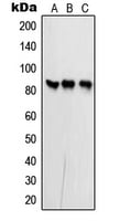
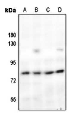
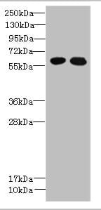
 WB (Western Blot)
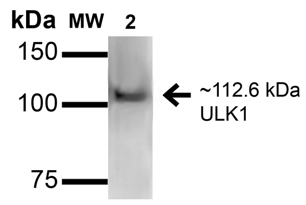
(Western blot analysis of Rat Brain cell lysates showing detection of ~112.6kDa ULK1 protein using Rabbit Anti-ULK1 Polyclonal Antibody (SPC-640). Lane 1: Molecular Weight Ladder (MW). Lane 2: Rat Brain cell lysates. Load: 15 ug. Block: 2% GE Healthcare Blocker (RT, 60 minutes). Primary Antibody: Rabbit Anti-ULK1 Polyclonal Antibody (SPC-640) at 1:1000 for 16 hours at 4 degree C. Secondary Antibody: Goat Anti-Rabbit IgG: HRP at 1/2000 for 60 min at RT. Color Development: ECL solution for 6 min at RT. Predicted/Observed Size: ~112.6kDa.)
WB (Western Blot)
(Western blot analysis of Rat Brain cell lysates showing detection of ~112.6kDa ULK1 protein using Rabbit Anti-ULK1 Polyclonal Antibody (SPC-640). Lane 1: Molecular Weight Ladder (MW). Lane 2: Rat Brain cell lysates. Load: 15 ug. Block: 2% GE Healthcare Blocker (RT, 60 minutes). Primary Antibody: Rabbit Anti-ULK1 Polyclonal Antibody (SPC-640) at 1:1000 for 16 hours at 4 degree C. Secondary Antibody: Goat Anti-Rabbit IgG: HRP at 1/2000 for 60 min at RT. Color Development: ECL solution for 6 min at RT. Predicted/Observed Size: ~112.6kDa.)
ULK1, Polyclonal Antibody (Cat# AAA104189)

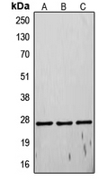
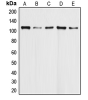
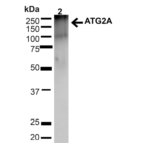
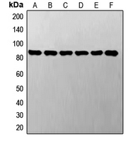
 WB (Western Blot)
(Western blot analysis of Human HeLa cell lysates showing detection of ~212.9kDa ATG2A protein using Rabbit Anti-ATG2A Polyclonal Antibody . Lane 1: MW Ladder. Lane 2: Human HeLa (20 ug). Load: 20 ug. Block: 5% milk + TBST for 1 hour at RT. Primary Antibody: Rabbit Anti-ATG2A Polyclonal Antibody at 1:1000 for 1 hour at RT. Secondary Antibody: Goat Anti-Rabbit: HRP at 1:2000 for 1 hour at RT. Color Development: TMB solution for 12 min at RT. Predicted/Observed Size: ~212.9kDa.)
WB (Western Blot)
(Western blot analysis of Human HeLa cell lysates showing detection of ~212.9kDa ATG2A protein using Rabbit Anti-ATG2A Polyclonal Antibody . Lane 1: MW Ladder. Lane 2: Human HeLa (20 ug). Load: 20 ug. Block: 5% milk + TBST for 1 hour at RT. Primary Antibody: Rabbit Anti-ATG2A Polyclonal Antibody at 1:1000 for 1 hour at RT. Secondary Antibody: Goat Anti-Rabbit: HRP at 1:2000 for 1 hour at RT. Color Development: TMB solution for 12 min at RT. Predicted/Observed Size: ~212.9kDa.)
ATG2A, Polyclonal Antibody (Cat# AAA103807)
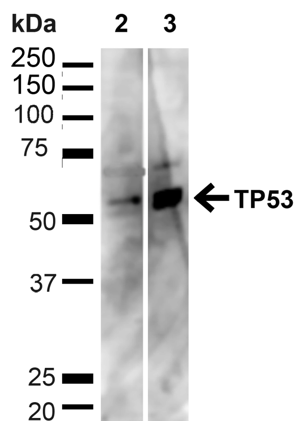
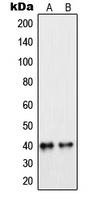
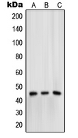
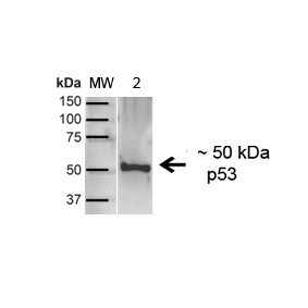 WB (Western Blot)
(Western blot analysis of Human A431 showing detection of ~43.7kDa p53 protein using Rabbit Anti-p53 Polyclonal Antibody (SPC-682). Lane 1: MW Ladder. Lane 2: Human A431 (20 ug). Load: 20 ug. Block: 5% milk + TBST for 1 hour at RT. Primary Antibody: Rabbit Anti-p53 Polyclonal Antibody (SPC-682) at 1:1000 for 1 hour at RT. Secondary Antibody: Goat Anti-Rabbit: HRP at 1:2000 for 1 hour at RT. Color Development: TMB solution for 12 min at RT. Predicted/Observed Size: ~43.7kDa.)
WB (Western Blot)
(Western blot analysis of Human A431 showing detection of ~43.7kDa p53 protein using Rabbit Anti-p53 Polyclonal Antibody (SPC-682). Lane 1: MW Ladder. Lane 2: Human A431 (20 ug). Load: 20 ug. Block: 5% milk + TBST for 1 hour at RT. Primary Antibody: Rabbit Anti-p53 Polyclonal Antibody (SPC-682) at 1:1000 for 1 hour at RT. Secondary Antibody: Goat Anti-Rabbit: HRP at 1:2000 for 1 hour at RT. Color Development: TMB solution for 12 min at RT. Predicted/Observed Size: ~43.7kDa.)
p53, Polyclonal Antibody (Cat# AAA103826)

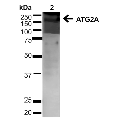 WB (Western Blot)
(Western blot analysis of Human HeLa cell lysates showing detection of ~212.9kDa ATG2A protein using Rabbit Anti-ATG2A Polyclonal Antibody . Lane 1: MW Ladder. Lane 2: Human HeLa (20 ug). Load: 20 ug. Block: 5% milk + TBST for 1 hour at RT. Primary Antibody: Rabbit Anti-ATG2A Polyclonal Antibody at 1:1000 for 1 hour at RT. Secondary Antibody: Goat Anti-Rabbit: HRP at 1:2000 for 1 hour at RT. Color Development: TMB solution for 12 min at RT. Predicted/Observed Size: ~212.9kDa.)
WB (Western Blot)
(Western blot analysis of Human HeLa cell lysates showing detection of ~212.9kDa ATG2A protein using Rabbit Anti-ATG2A Polyclonal Antibody . Lane 1: MW Ladder. Lane 2: Human HeLa (20 ug). Load: 20 ug. Block: 5% milk + TBST for 1 hour at RT. Primary Antibody: Rabbit Anti-ATG2A Polyclonal Antibody at 1:1000 for 1 hour at RT. Secondary Antibody: Goat Anti-Rabbit: HRP at 1:2000 for 1 hour at RT. Color Development: TMB solution for 12 min at RT. Predicted/Observed Size: ~212.9kDa.)
ATG2A, Polyclonal Antibody (Cat# AAA103854)
 WB (Western Blot)
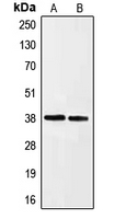
(Western blot analysis of MX2 expression in HEK293T (A) whole cell lysates.)
WB (Western Blot)
(Western blot analysis of MX2 expression in HEK293T (A) whole cell lysates.)
MX2, Polyclonal Antibody (Cat# AAA104301)
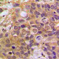
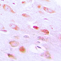
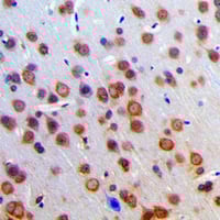
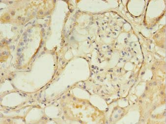
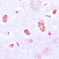 IHC (Immunohiostchemistry)
(Immunohistochemical analysis of GUCY1A3 staining in human brain formalin fixed paraffin embedded tissue section. The section was pre-treated using heat mediated antigen retrieval with sodium citrate buffer (pH 6.0). The section was then incubated with the antibody at room temperature and detected using an HRP conjugated compact polymer system. DAB was used as the chromogen. The section was then counterstained with haematoxylin and mounted with DPX.)
IHC (Immunohiostchemistry)
(Immunohistochemical analysis of GUCY1A3 staining in human brain formalin fixed paraffin embedded tissue section. The section was pre-treated using heat mediated antigen retrieval with sodium citrate buffer (pH 6.0). The section was then incubated with the antibody at room temperature and detected using an HRP conjugated compact polymer system. DAB was used as the chromogen. The section was then counterstained with haematoxylin and mounted with DPX.)
GUCY1A3, Polyclonal Antibody (Cat# AAA104317)
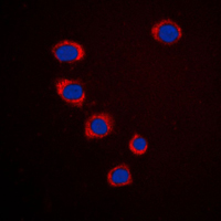 IF (Immunofluorescence)
(Immunofluorescent analysis of Ephrin A3 staining in HepG2 cells. Formalin-fixed cells were permeabilized with 0.1% Triton X-100 in TBS for 5-10 minutes and blocked with 3% BSA-PBS for 30 minutes at room temperature. Cells were probed with the primary antibody in 3% BSA-PBS and incubated overnight at 4 °C in a humidified chamber. Cells were washed with PBST and incubated with a DyLight 594-conjugated secondary antibody (red) in PBS at room temperature in the dark. DAPI was used to stain the cell nuclei (blue).)
IF (Immunofluorescence)
(Immunofluorescent analysis of Ephrin A3 staining in HepG2 cells. Formalin-fixed cells were permeabilized with 0.1% Triton X-100 in TBS for 5-10 minutes and blocked with 3% BSA-PBS for 30 minutes at room temperature. Cells were probed with the primary antibody in 3% BSA-PBS and incubated overnight at 4 °C in a humidified chamber. Cells were washed with PBST and incubated with a DyLight 594-conjugated secondary antibody (red) in PBS at room temperature in the dark. DAPI was used to stain the cell nuclei (blue).)
Ephrin A3, Polyclonal Antibody (Cat# AAA104323)
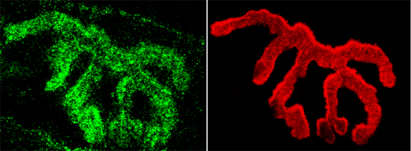
 IF (Immunofluorescence)
(Immunofluorescent analysis of AP2 alpha/beta staining in HepG2 cells. Formalin-fixed cells were permeabilized with 0.1% Triton X-100 in TBS for 5-10 minutes and blocked with 3% BSA-PBS for 30 minutes at room temperature. Cells were probed with the primary antibody in 3% BSA-PBS and incubated overnight at 4 °C in a humidified chamber. Cells were washed with PBST and incubated with a DyLight 594-conjugated secondary antibody (red) in PBS at room temperature in the dark.)
IF (Immunofluorescence)
(Immunofluorescent analysis of AP2 alpha/beta staining in HepG2 cells. Formalin-fixed cells were permeabilized with 0.1% Triton X-100 in TBS for 5-10 minutes and blocked with 3% BSA-PBS for 30 minutes at room temperature. Cells were probed with the primary antibody in 3% BSA-PBS and incubated overnight at 4 °C in a humidified chamber. Cells were washed with PBST and incubated with a DyLight 594-conjugated secondary antibody (red) in PBS at room temperature in the dark.)
STAT3, Polyclonal Antibody (Cat# AAA104222)
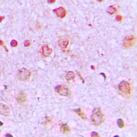
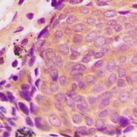
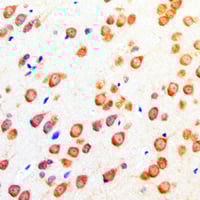
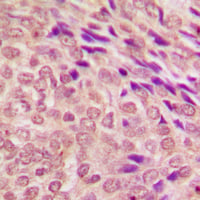
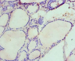
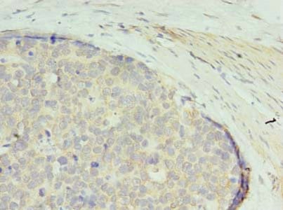
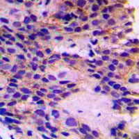 IHC (Immunohiostchemistry)
(Immunohistochemical analysis of PEX7 staining in human breast cancer formalin fixed paraffin embedded tissue section. The section was pre-treated using heat mediated antigen retrieval with sodium citrate buffer (pH 6.0). The section was then incubated with the antibody at room temperature and detected using an HRP conjugated compact polymer system. DAB was used as the chromogen. The section was then counterstained with haematoxylin and mounted with DPX.)
IHC (Immunohiostchemistry)
(Immunohistochemical analysis of PEX7 staining in human breast cancer formalin fixed paraffin embedded tissue section. The section was pre-treated using heat mediated antigen retrieval with sodium citrate buffer (pH 6.0). The section was then incubated with the antibody at room temperature and detected using an HRP conjugated compact polymer system. DAB was used as the chromogen. The section was then counterstained with haematoxylin and mounted with DPX.)
PEX7, Polyclonal Antibody (Cat# AAA104233)
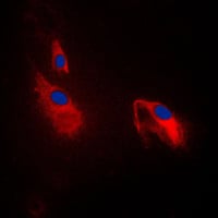
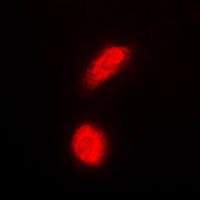
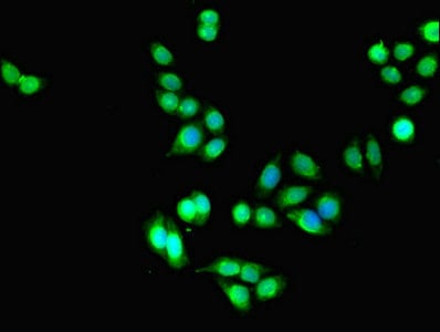
 IF (Immunofluorescence)
(Immunofluorescent analysis of AKT2 staining in HepG2 cells. Formalin-fixed cells were permeabilized with 0.1% Triton X-100 in TBS for 5-10 minutes and blocked with 3% BSA-PBS for 30 minutes at room temperature. Cells were probed with the primary antibody in 3% BSA-PBS and incubated overnight at 4 °C in a humidified chamber. Cells were washed with PBST and incubated with a DyLight 594-conjugated secondary antibody (red) in PBS at room temperature in the dark.)
IF (Immunofluorescence)
(Immunofluorescent analysis of AKT2 staining in HepG2 cells. Formalin-fixed cells were permeabilized with 0.1% Triton X-100 in TBS for 5-10 minutes and blocked with 3% BSA-PBS for 30 minutes at room temperature. Cells were probed with the primary antibody in 3% BSA-PBS and incubated overnight at 4 °C in a humidified chamber. Cells were washed with PBST and incubated with a DyLight 594-conjugated secondary antibody (red) in PBS at room temperature in the dark.)
AKT2, Polyclonal Antibody (Cat# AAA104258)
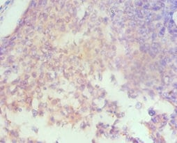
 IHC (Immunohiostchemistry)
Mu Opioid Receptor (pS375), Polyclonal Antibody (Cat# AAA104285)
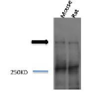 WB (Western Blot)
(Western blot analysis of Mouse, Rat brain cell lysates showing detection of Piccolo protein using Rabbit Anti-Piccolo Polyclonal Antibody (SPC-197). Primary Antibody: Rabbit Anti-Piccolo Polyclonal Antibody (SPC-197) at 1:1000.)
WB (Western Blot)
(Western blot analysis of Mouse, Rat brain cell lysates showing detection of Piccolo protein using Rabbit Anti-Piccolo Polyclonal Antibody (SPC-197). Primary Antibody: Rabbit Anti-Piccolo Polyclonal Antibody (SPC-197) at 1:1000.)
Piccolo, Polyclonal Antibody (Cat# AAA103761)
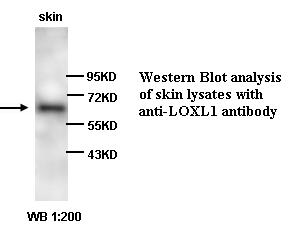
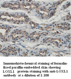 Application Data
LOXL1, Polyclonal Antibody (Cat# AAA108893)
 Application Data
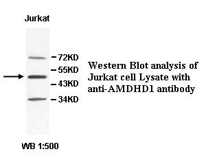
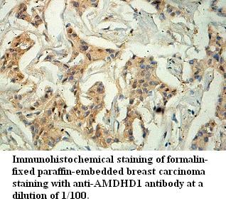
AMDHD1, Polyclonal Antibody (Cat# AAA108905)
Predicted: Mouse, Rat
 Application Data
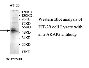
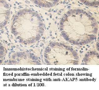
AKAP5, Polyclonal Antibody (Cat# AAA108912)
Predicted: Mouse, Rat
 Application Data
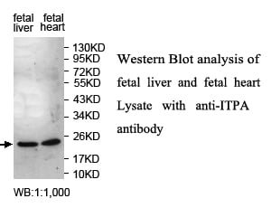
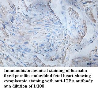
ITPA, Polyclonal Antibody (Cat# AAA108914)
Predicted: Mouse, Rat
 Application Data
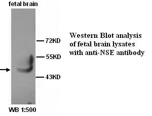
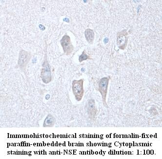
NSE, Polyclonal Antibody (Cat# AAA108915)
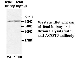
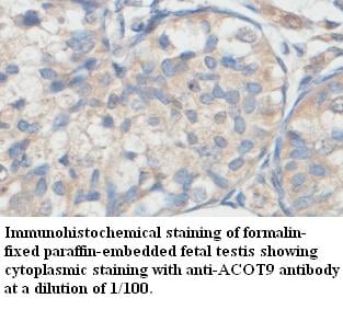 Application Data
ACOT9, Polyclonal Antibody (Cat# AAA108933)
Predicted: Mouse, Rat
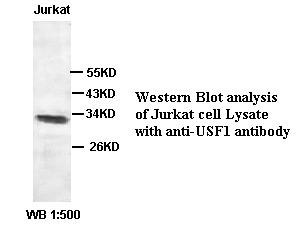
 Application Data
USF1, Polyclonal Antibody (Cat# AAA108943)
 Application Data
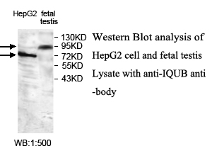
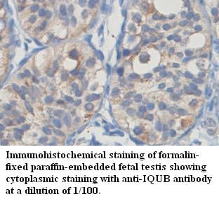
IQUB, Polyclonal Antibody (Cat# AAA109032)
Predicted: Mouse, Rat
 Application Data
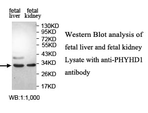
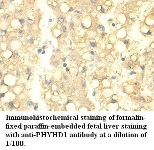
PHYHD1, Polyclonal Antibody (Cat# AAA109059)
Predicted: Mouse, Rat
 Application Data
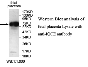
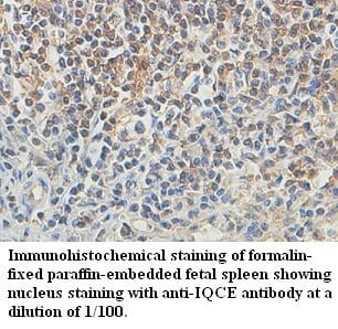
IQCE, Polyclonal Antibody (Cat# AAA109069)
Predicted: Mouse, Rat
 Application Data
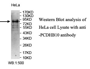
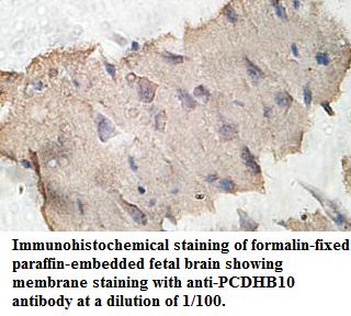
PCDHB10, Polyclonal Antibody (Cat# AAA109087)
Predicted: Mouse, Rat
 Application Data
PAAF1, Polyclonal Antibody (Cat# AAA108953)
Predicted: Mouse, Rat
 Application Data
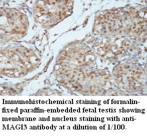
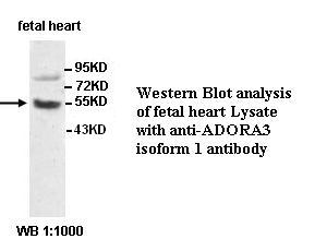
ADORA3, Polyclonal Antibody (Cat# AAA108954)
 Application Data
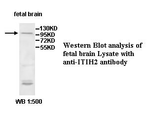
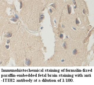
ITIH2, Polyclonal Antibody (Cat# AAA109005)
Predicted: Mouse, Rat
 Application Data
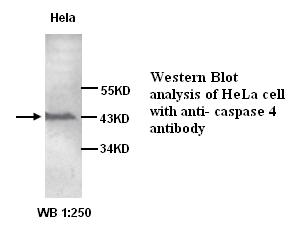
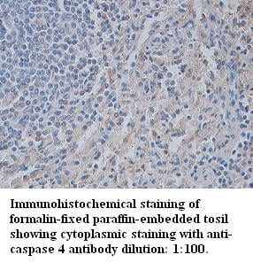
Caspase 4, Polyclonal Antibody (Cat# AAA109006)
 Application Data
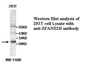
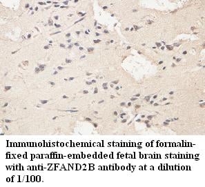
ZFAND2B, Polyclonal Antibody (Cat# AAA109007)
Predicted: Mouse, Rat
 IF (Immunofluorescence)
(Immunofluorescent analysis of PC3 cells using AAA114161 at a dilution of 1:100 and Alexa Fluor 488-congugated AffiniPure Goat Anti-Rabbit IgG(H+L))
IF (Immunofluorescence)
(Immunofluorescent analysis of PC3 cells using AAA114161 at a dilution of 1:100 and Alexa Fluor 488-congugated AffiniPure Goat Anti-Rabbit IgG(H+L))
DLEU1, Polyclonal Antibody (Cat# AAA114161)
 IHC (Immunohistochemisry)
(Immunofluorescent analysis of U251 cells using AAA114166 at a dilution of 1:100 and Alexa Fluor 488-congugated AffiniPure Goat Anti-Rabbit IgG(H+L))
IHC (Immunohistochemisry)
(Immunofluorescent analysis of U251 cells using AAA114166 at a dilution of 1:100 and Alexa Fluor 488-congugated AffiniPure Goat Anti-Rabbit IgG(H+L))
Hyaluronan synthase 3, Polyclonal Antibody (Cat# AAA114166)
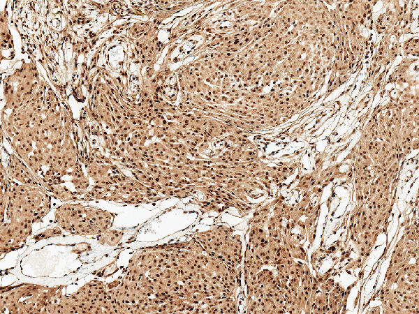
 IHC (Immunohiostchemistry)
(Immunohistochemistry of paraffin-embedded human gastric cancer using AAA114169 at dilution of 1:100)
IHC (Immunohiostchemistry)
(Immunohistochemistry of paraffin-embedded human gastric cancer using AAA114169 at dilution of 1:100)
GTPBP6, Polyclonal Antibody (Cat# AAA114169)
What are Polyclonal Antibodies?
Polyclonal antibodies are antibodies that come from multiple B cell clones of a host animal. The typical hosts used for the majority of polyclonal antibody production are rabbits, goats, sheep, and donkeys. These polyclonal antibodies, once having identified their target, will bind to different epitopes located at different regions or sequences on the same protein/antigen. As a result, they are ideal at locating and binding to the target, even if the target is in very low concentrations (due to many different antibodies being able to bind to the same target molecule, which allows for significant amplification of a downstream signal).
Polyclonal antibodies are typically produced by injecting an antigen into a host animal, which causes the animal’s immune system to attack the foreign antigen by mass generating antibodies against it. After a period of time, serum is collected from the animal and purified using physicochemical fractionation, class-specific affinity purification, and/or antigen-affinity purification.
Key Uses of Polyclonal Antibodies
- Western Blotting: This method is used to find specific proteins in biological samples after separating them by size.
- Immunohistochemistry: IHC helps visualize the location of proteins in tissue sections using various staining techniques.
- ELISA: (Enzyme-Linked Immunosorbent Assay) is typically used to identify specific protein quantities in a sample. ELISAs can be either “Quantitative” or “Qualitative”.
- Flow Cytometry: technique that identifies and measures the specific protein on the surface or inside the cells in a fluid suspension.
- Immunoprecipitation: IP isolates and studies a specific protein from a complex mixture using antibodies.
Why Buy Polyclonal Antibodies from AAA Biotech?
1. Ideal for Various Applications
Our antibodies are generally going to be validated for use in multiple types of assays, including ELISA, Western Blotting, Immunohistochemistry, Immunoprecipitation, amongst others. They are ideal for a wide range of research applications.
2. Rigorous Quality Control
All of the antibodies in our catalog undergo strict quality testing to ensure specificity, sensitivity, and consistent performance. We are confident in the ability of our antibodies to provide you with accurate results.
3. Wide Assortment of Antibodies
Antibodies in are catalog can be found for both common and exotic species, and these antibodies are also available in both conjugated and recombinant forms to suit many diverse experimental needs.
4. Highly Purified
Our antibodies are available in purified forms with over 85% purity, as confirmed by SDS-PAGE. They are also available with tags such as His, Flag, GST, or MBP. We cater to customers worldwide.
FAQ
1. How are polyclonal antibodies produced?
Traditionally, polyclonal antibodies are produced by injecting an antigen into a host animal (such as a rabbit or goat), which then triggers an immune response from the host animal. The animal’s B cells produce antibodies that will recognize different parts of the injected antigen. These antibodies are then collected from the animal’s blood and purified for use.
2. How do polyclonal antibodies differ from monoclonal antibodies?
Polyclonal antibodies are a mix of antibodies that bind to different locations (epitopes) of the same antigen, while monoclonal antibodies are identical and bind to just one specific epitope. This makes polyclonal antibodies more versatile and better at detecting proteins that may be present in low quantities or in altered/modified forms.
3. How should I store polyclonal antibodies?
Polyclonal antibodies should be stored at 4°C for short-term use (up to a few weeks) and at -20°C or -80°C for long-term storage. Avoid repeated freeze-thaw cycles by dividing them into small aliquots. Always check the datasheet for specific storage instructions.