Filters
▼Clonality
▼Type
▼Reactivity
▼Gene Name
▼Isotype
▼Host
▼Application
▼Clone
▼Polyclonal Antibodies
At AAA Biotech also known as AAA Bio or AAABio, we provide a broad range of purified polyclonal antibodies (pAbs) that are able to all be browsed online through our website. Due to their high specificity and strong binding affinity, these antibodies are ideal for wide swathes of research and experimental applications.
Our polyclonal antibodies can easily support your work, whether you use them for Western Blotting, Immunocytochemistry (with or without Immunofluorescence used in conjunction), Immunohistochemistry, Immunoprecipitation, and ELISA tests. We highly encourage you to browse our range of pAbs and choose the one that best suits your experimental model.
Viewing 9050-9100 of 96805 product results
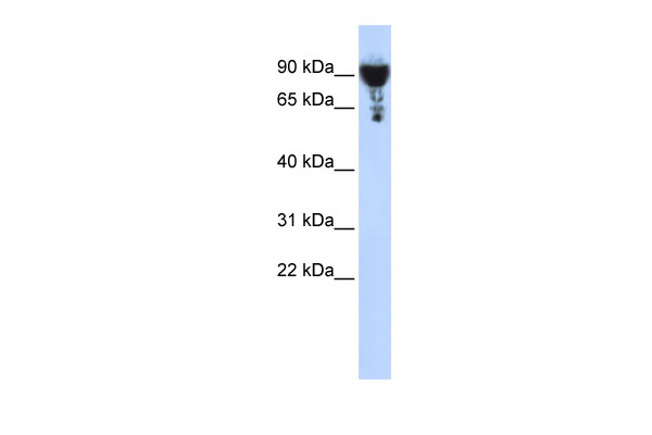
 WB (Western Blot)
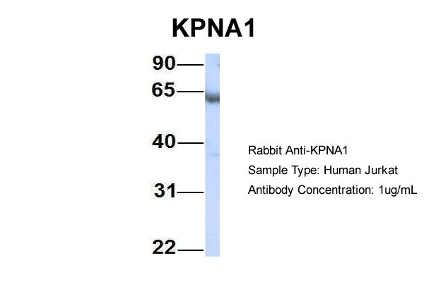
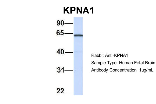
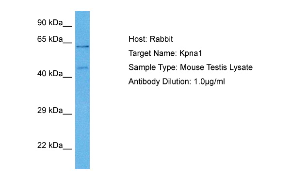
(WB Suggested Anti-KPNA1 Antibody Titration: 0.2-1 ug/mlELISA Titer: 1:312500Positive Control: HepG2 cell lysate)
WB (Western Blot)
(WB Suggested Anti-KPNA1 Antibody Titration: 0.2-1 ug/mlELISA Titer: 1:312500Positive Control: HepG2 cell lysate)
KPNA1, Polyclonal Antibody (Cat# AAA200395)
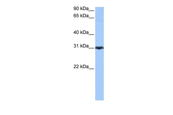
 WB (Western Blot)
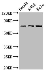
(WB Suggested Anti-CBLN4 Antibody Titration: 1 ug/mlPositive Control: Hela cell lysate)
WB (Western Blot)
(WB Suggested Anti-CBLN4 Antibody Titration: 1 ug/mlPositive Control: Hela cell lysate)
CBLN4, Polyclonal Antibody (Cat# AAA200252)
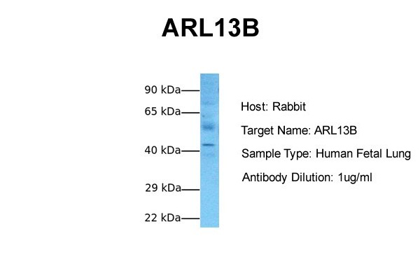
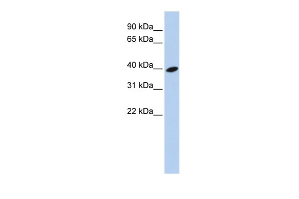 WB (Western Blot)
(WB Suggested Anti-ARL13B Antibody Titration: 0.2-1 ug/mlELISA Titer: 1:312500Positive Control: 721_B cell lysate)
WB (Western Blot)
(WB Suggested Anti-ARL13B Antibody Titration: 0.2-1 ug/mlELISA Titer: 1:312500Positive Control: 721_B cell lysate)
ARL13B, Polyclonal Antibody (Cat# AAA200260)
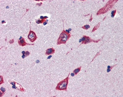
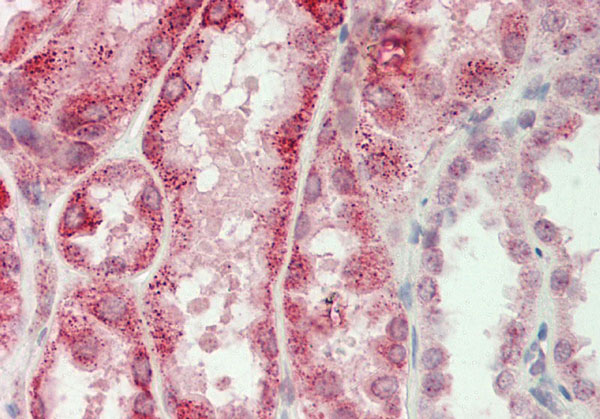
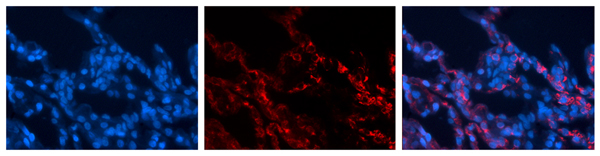
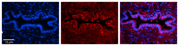
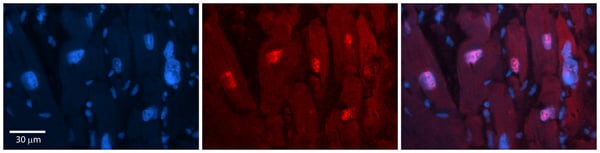
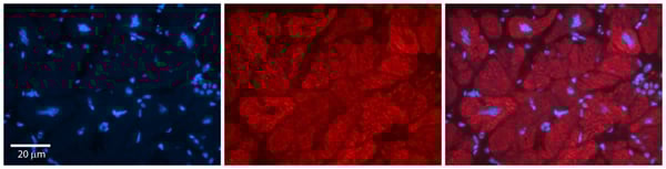
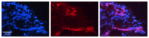
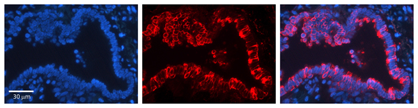
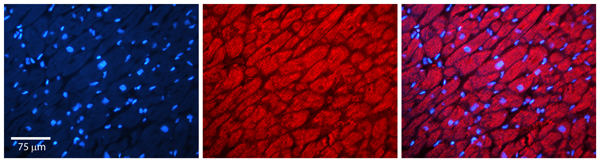
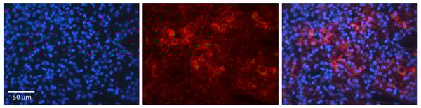
 IHC (Immunohistochemistry)
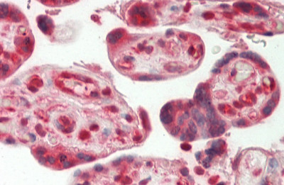
(Rabbit Anti-MLKL antibody Catalog Number: ARP53092 Formalin Fixed Paraffin Embedded Tissue: Human Placenta Primary antibody Concentration: 1:100 Secondary Antibody: Donkey anti-Rabbit-Cy3 Secondary Antibody Concentration: 1:200 Magnification: 20x Exposure Time: 0.5-2.0sec)
IHC (Immunohistochemistry)
(Rabbit Anti-MLKL antibody Catalog Number: ARP53092 Formalin Fixed Paraffin Embedded Tissue: Human Placenta Primary antibody Concentration: 1:100 Secondary Antibody: Donkey anti-Rabbit-Cy3 Secondary Antibody Concentration: 1:200 Magnification: 20x Exposure Time: 0.5-2.0sec)
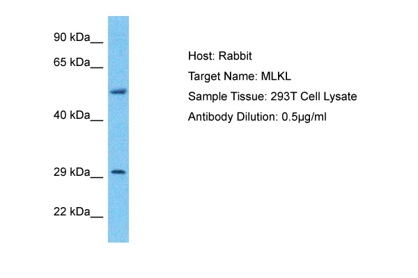
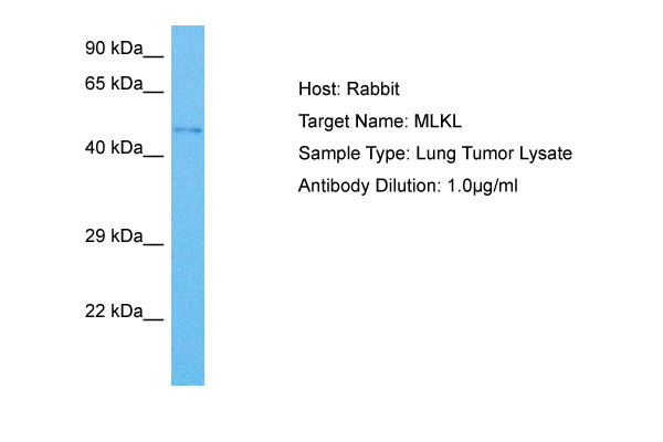
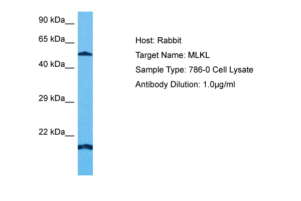
MLKL, Polyclonal Antibody (Cat# AAA200270)
Predicted Species Reactivity: Human, Rat, Cow, Horse
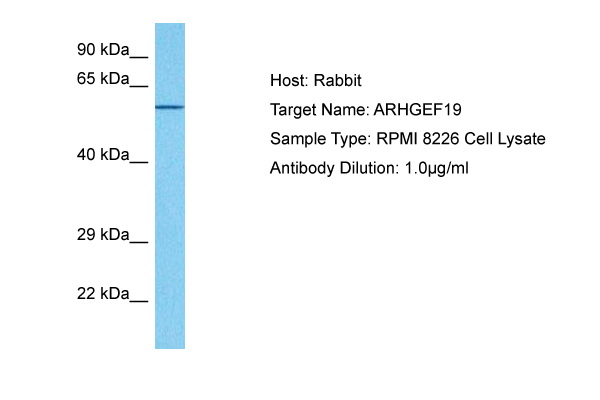
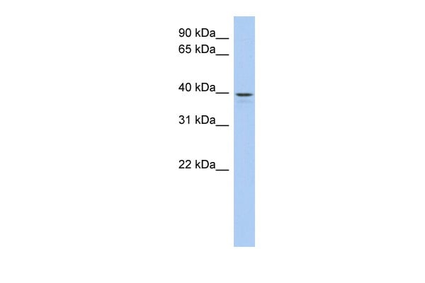
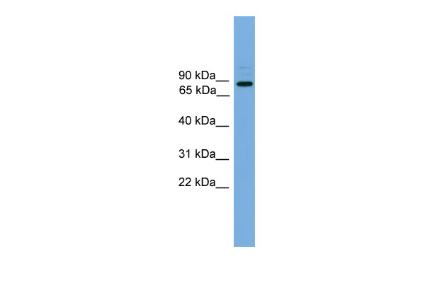 WB (Western Blot)
(WB Suggested Anti-ARHGEF19 Antibody Titration: 0.2-1 ug/mlPositive Control: Hela cell lysate)
WB (Western Blot)
(WB Suggested Anti-ARHGEF19 Antibody Titration: 0.2-1 ug/mlPositive Control: Hela cell lysate)
ARHGEF19, Polyclonal Antibody (Cat# AAA200274)
 WB (Western Blot)
(WB Suggested Anti-LIX1L Antibody Titration: 0.2-1 ug/mlELISA Titer: 1:1562500Positive Control: Human Lung)
WB (Western Blot)
(WB Suggested Anti-LIX1L Antibody Titration: 0.2-1 ug/mlELISA Titer: 1:1562500Positive Control: Human Lung)
LIX1L, Polyclonal Antibody (Cat# AAA200277)
Predicted: Cow, Dog, Guinea Pig, Human, Mouse, Rabbit, Rat, Zebrafish
 WB (Western Blot)
(WB Suggested Anti-FAM36A Antibody Titration: 0.2-1 ug/mlELISA Titer: 1:62500Positive Control: Human Muscle)
WB (Western Blot)
(WB Suggested Anti-FAM36A Antibody Titration: 0.2-1 ug/mlELISA Titer: 1:62500Positive Control: Human Muscle)
FAM36A, Polyclonal Antibody (Cat# AAA200296)
Predicted Reactivity: Horse, Human, Mouse, Rabbit, Rat
 WB (Western Blot)
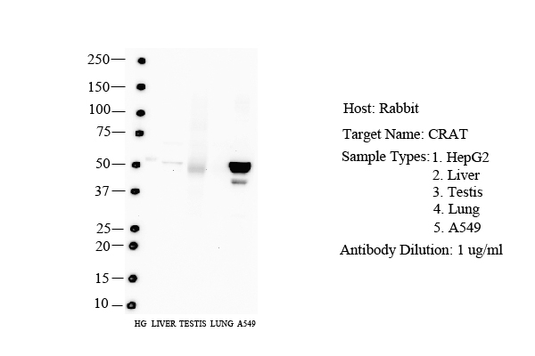
(WB Suggested Anti-CRAT Antibody Titration: 0.2-1 ug/mlPositive Control: HepG2 cell lysate)
WB (Western Blot)
(WB Suggested Anti-CRAT Antibody Titration: 0.2-1 ug/mlPositive Control: HepG2 cell lysate)
CRAT, Polyclonal Antibody (Cat# AAA200300)
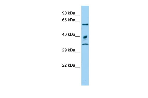
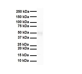
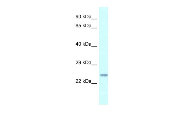 WB (Western Blot)
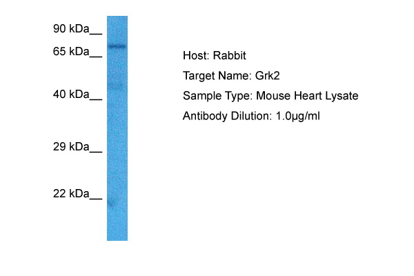
(WB Suggested Anti-Cdkn3 AntibodyTitration: 1.0 ug/mlPositive Control: Mouse Heart)
WB (Western Blot)
(WB Suggested Anti-Cdkn3 AntibodyTitration: 1.0 ug/mlPositive Control: Mouse Heart)
Cdkn3, Polyclonal Antibody (Cat# AAA200309)
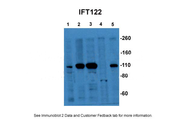
 WB (Western Blot)
(WB Suggested Anti-IFT122 Antibody Titration: 0.2-1 ug/mlPositive Control: 721_B cell lysate)
WB (Western Blot)
(WB Suggested Anti-IFT122 Antibody Titration: 0.2-1 ug/mlPositive Control: 721_B cell lysate)
IFT122, Polyclonal Antibody (Cat# AAA200331)
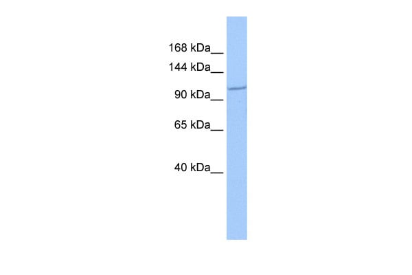
 WB (Western Blot)
(WB Suggested Anti-CPN1 Antibody Titration: 0.2-1 ug/mlELISA Titer: 1:1562500Positive Control: Transfected 293T)
WB (Western Blot)
(WB Suggested Anti-CPN1 Antibody Titration: 0.2-1 ug/mlELISA Titer: 1:1562500Positive Control: Transfected 293T)
CPN1, Polyclonal Antibody (Cat# AAA200192)
 WB (Western Blot)
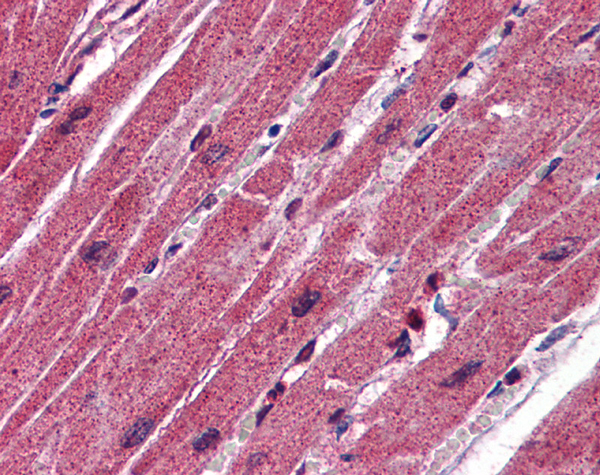
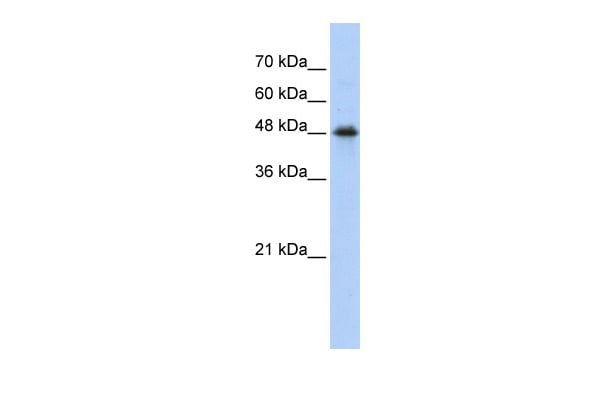
(WB Suggested Anti-ADRBK1 Antibody Titration: 0.2-1 ug/mlELISA Titer: 1:1562500Positive Control: PANC1 cell lysateADRBK1 is strongly supported by BioGPS gene expression data to be expressed in Human PANC1 cells)
WB (Western Blot)
(WB Suggested Anti-ADRBK1 Antibody Titration: 0.2-1 ug/mlELISA Titer: 1:1562500Positive Control: PANC1 cell lysateADRBK1 is strongly supported by BioGPS gene expression data to be expressed in Human PANC1 cells)
GRK2, Polyclonal Antibody (Cat# AAA200201)
 WB (Western Blot)
(WB Suggested Anti-EXOC3 Antibody Titration: 0.2-1 ug/mlELISA Titer: 1:312500Positive Control: Hela cell lysateEXOC3 is supported by BioGPS gene expression data to be expressed in HeLa)
WB (Western Blot)
(WB Suggested Anti-EXOC3 Antibody Titration: 0.2-1 ug/mlELISA Titer: 1:312500Positive Control: Hela cell lysateEXOC3 is supported by BioGPS gene expression data to be expressed in HeLa)
EXOC3, Polyclonal Antibody (Cat# AAA200241)
 WB (Western Blot)
(WB Suggested Anti-PRDX3 Antibody Titration: 0.2-1 ug/mlPositive Control: Jurkat cell lysatePRDX3 is supported by BioGPS gene expression data to be expressed in Jurkat)
WB (Western Blot)
(WB Suggested Anti-PRDX3 Antibody Titration: 0.2-1 ug/mlPositive Control: Jurkat cell lysatePRDX3 is supported by BioGPS gene expression data to be expressed in Jurkat)
PRDX3, Polyclonal Antibody (Cat# AAA200243)
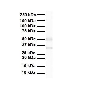 WB (Western Blot)
(WB Suggested Anti-Arih2 antibody Titration: 1 ug/mLSample Type: Human heart)
WB (Western Blot)
(WB Suggested Anti-Arih2 antibody Titration: 1 ug/mLSample Type: Human heart)
Arih2, Polyclonal Antibody (Cat# AAA200759)
 WB (Western Blot)
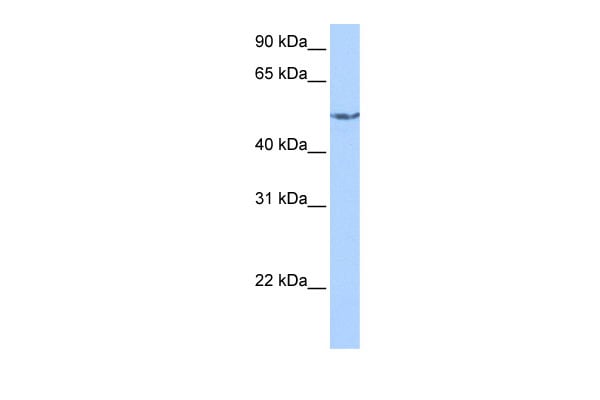
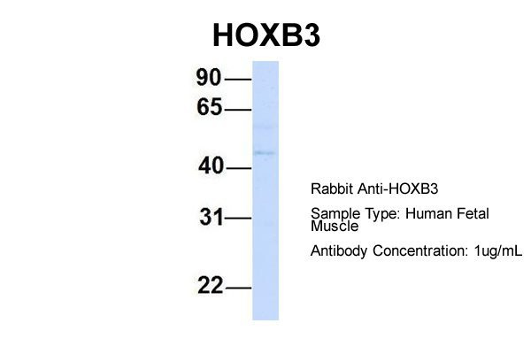
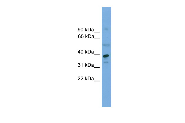
(WB Suggested Anti-HOXB3 antibody Titration: 1 ug/mLSample Type: Human liver)
WB (Western Blot)
(WB Suggested Anti-HOXB3 antibody Titration: 1 ug/mLSample Type: Human liver)
HOXB3, Polyclonal Antibody (Cat# AAA200766)
 WB (Western Blot)
(WB Suggested Anti-APEX1 Antibody Titration: 0.2-1 ug/mlELISA Titer: 1:1562500Positive Control: 293T cell lysateAPEX1 is supported by BioGPS gene expression data to be expressed in HEK293T)
WB (Western Blot)
(WB Suggested Anti-APEX1 Antibody Titration: 0.2-1 ug/mlELISA Titer: 1:1562500Positive Control: 293T cell lysateAPEX1 is supported by BioGPS gene expression data to be expressed in HEK293T)
APEX1, Polyclonal Antibody (Cat# AAA200767)
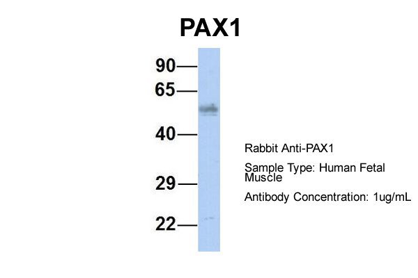
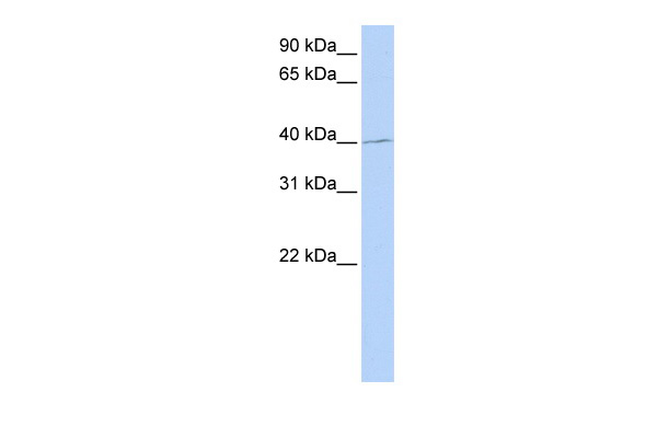 WB (Western Blot)
(WB Suggested Anti-PAX1 Antibody Titration: 0.2-1 ug/mlELISA Titer: 1:312500Positive Control: 721_B cell lysatePAX1 is supported by BioGPS gene expression data to be expressed in 721_B)
WB (Western Blot)
(WB Suggested Anti-PAX1 Antibody Titration: 0.2-1 ug/mlELISA Titer: 1:312500Positive Control: 721_B cell lysatePAX1 is supported by BioGPS gene expression data to be expressed in 721_B)
PAX1, Polyclonal Antibody (Cat# AAA200769)
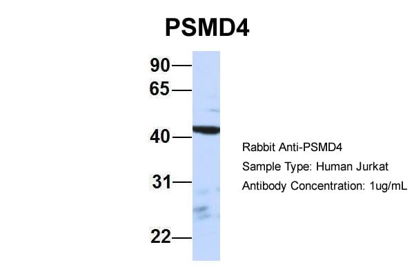
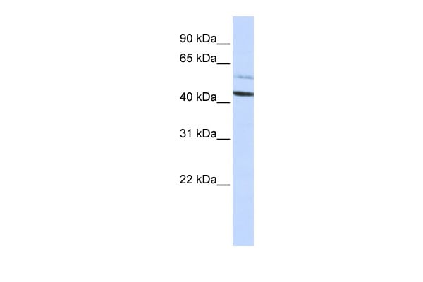 WB (Western Blot)
(WB Suggested Anti-PSMD4 Antibody Titration: 0.2-1 ug/mlELISA Titer: 1:312500Positive Control: HepG2 cell lysate)
WB (Western Blot)
(WB Suggested Anti-PSMD4 Antibody Titration: 0.2-1 ug/mlELISA Titer: 1:312500Positive Control: HepG2 cell lysate)
PSMD4, Polyclonal Antibody (Cat# AAA200770)


 WB (Western Blot)
(WB Suggested Anti-KLF9 Antibody Titration: 0.2-1 ug/mlELISA Titer: 1:62500Positive Control: 721_B cell lysateThere is BioGPS gene expression data showing that KLF9 is expressed in 721_B)
WB (Western Blot)
(WB Suggested Anti-KLF9 Antibody Titration: 0.2-1 ug/mlELISA Titer: 1:62500Positive Control: 721_B cell lysateThere is BioGPS gene expression data showing that KLF9 is expressed in 721_B)
KLF9, Polyclonal Antibody (Cat# AAA200772)
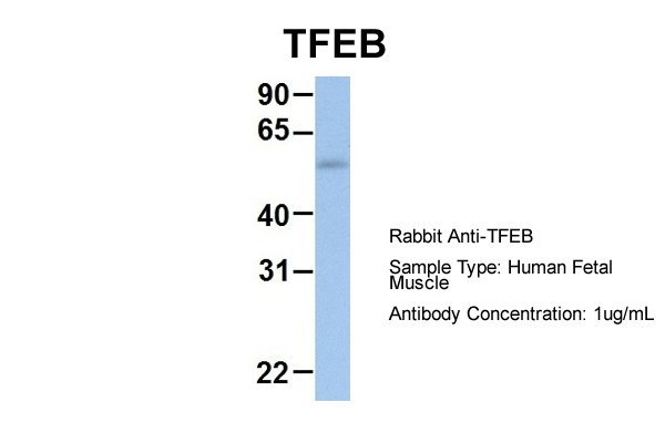
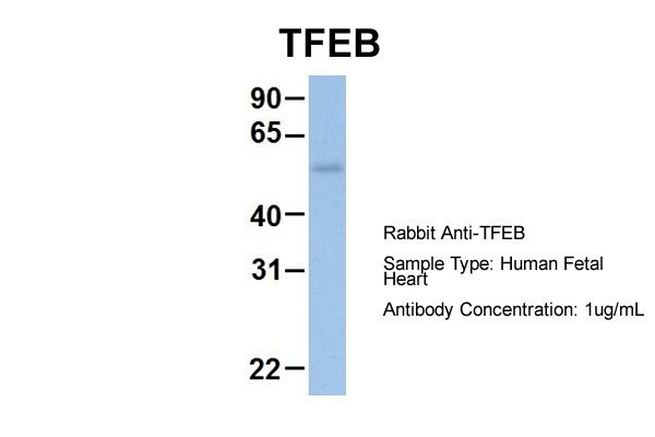
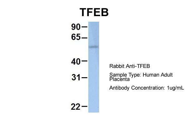
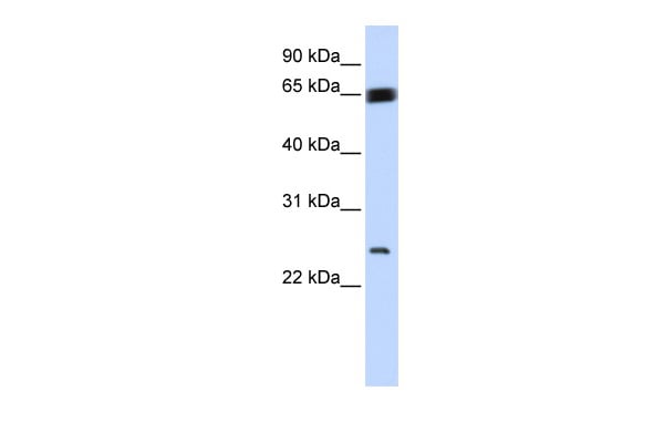
 WB (Western Blot)
(WB Suggested Anti-TFEB Antibody Titration: 0.2-1 ug/mlPositive Control: PANC1 cell lysate)
WB (Western Blot)
(WB Suggested Anti-TFEB Antibody Titration: 0.2-1 ug/mlPositive Control: PANC1 cell lysate)
TFEB, Polyclonal Antibody (Cat# AAA200777)
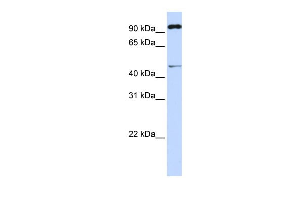
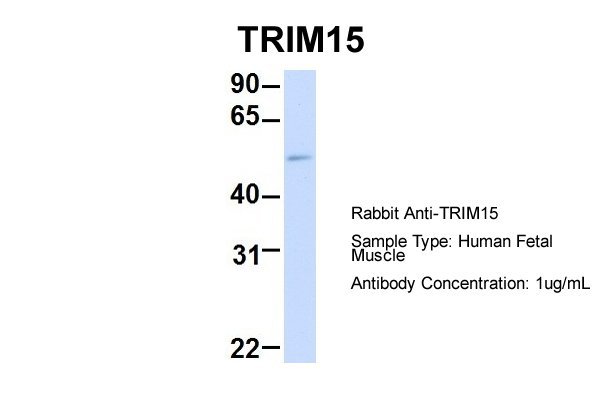
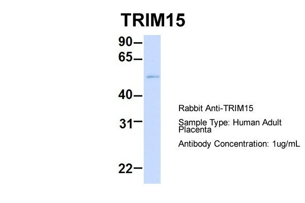
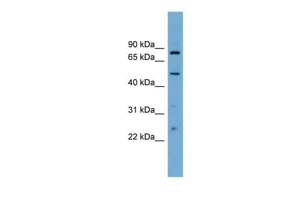
 WB (Western Blot)
(WB Suggested Anti-TRIM15 antibody Titration: 1 ug/mLSample Type: Human heart)
WB (Western Blot)
(WB Suggested Anti-TRIM15 antibody Titration: 1 ug/mLSample Type: Human heart)
TRIM15, Polyclonal Antibody (Cat# AAA200783)
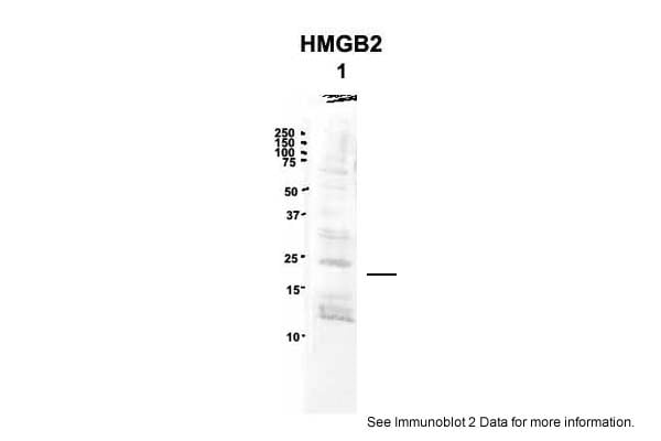
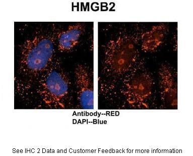
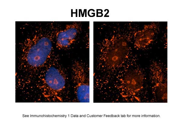
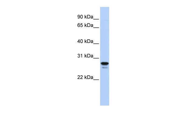 WB (Western Blot)
(WB Suggested Anti-HMGB2 Antibody Titration: 0.2-1 ug/mlELISA Titer: 1:12500Positive Control: Jurkat cell lysateHMGB2 is supported by BioGPS gene expression data to be expressed in Jurkat)
WB (Western Blot)
(WB Suggested Anti-HMGB2 Antibody Titration: 0.2-1 ug/mlELISA Titer: 1:12500Positive Control: Jurkat cell lysateHMGB2 is supported by BioGPS gene expression data to be expressed in Jurkat)
HMGB2, Polyclonal Antibody (Cat# AAA200786)
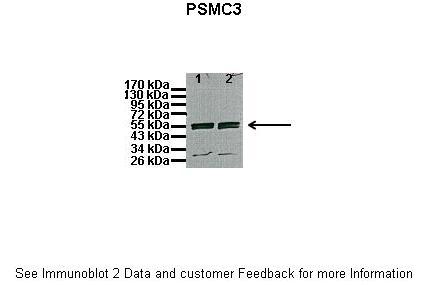
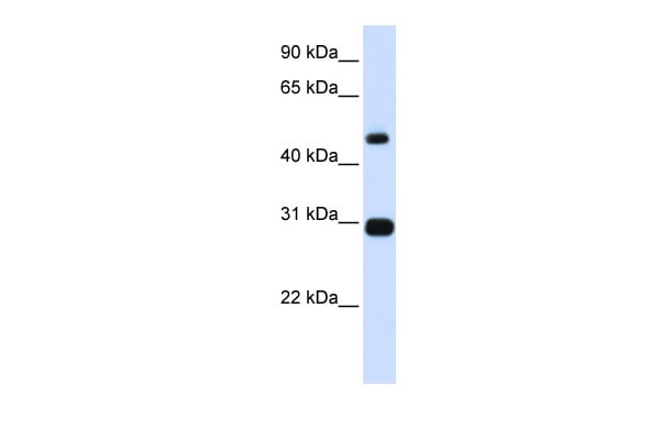 WB (Western Blot)
(WB Suggested Anti-PSMC3 Antibody Titration: 0.2-1 ug/mlELISA Titer: 1:62500Positive Control: Human Placenta)
WB (Western Blot)
(WB Suggested Anti-PSMC3 Antibody Titration: 0.2-1 ug/mlELISA Titer: 1:62500Positive Control: Human Placenta)
PSMC3, Polyclonal Antibody (Cat# AAA200787)
 WB (Western Blot)
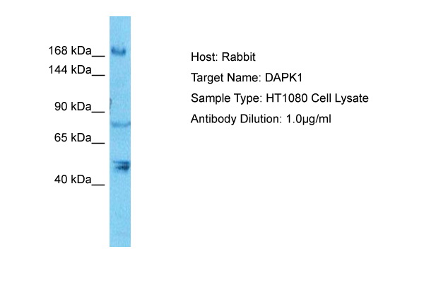
(WB Suggested Anti-DAPK1 Antibody Titration: 0.2-1 ug/mlELISA Titer: 1:1562500Positive Control: THP-1 cell lysate)
WB (Western Blot)
(WB Suggested Anti-DAPK1 Antibody Titration: 0.2-1 ug/mlELISA Titer: 1:1562500Positive Control: THP-1 cell lysate)
DAPK1, Polyclonal Antibody (Cat# AAA200790)
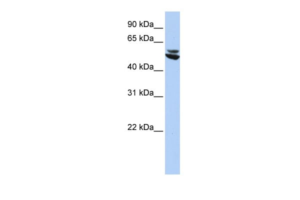 WB (Western Blot)
(WB Suggested Anti-KRT7 Antibody Titration: 0.2-1 ug/mlELISA Titer: 1:1562500Positive Control: HepG2 cell lysate)
WB (Western Blot)
(WB Suggested Anti-KRT7 Antibody Titration: 0.2-1 ug/mlELISA Titer: 1:1562500Positive Control: HepG2 cell lysate)
KRT7, Polyclonal Antibody (Cat# AAA200791)
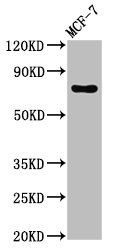
 WB (Western Blot)
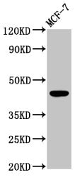
(WB Suggested Anti-SUMO3 Antibody Titration: 0.2-1 ug/mlELISA Titer: 1:62500Positive Control: MCF7 cell lysateSUMO3 is strongly supported by BioGPS gene expression data to be expressed in Human MCF7 cells)
WB (Western Blot)
(WB Suggested Anti-SUMO3 Antibody Titration: 0.2-1 ug/mlELISA Titer: 1:62500Positive Control: MCF7 cell lysateSUMO3 is strongly supported by BioGPS gene expression data to be expressed in Human MCF7 cells)
SUMO3, Polyclonal Antibody (Cat# AAA200796)
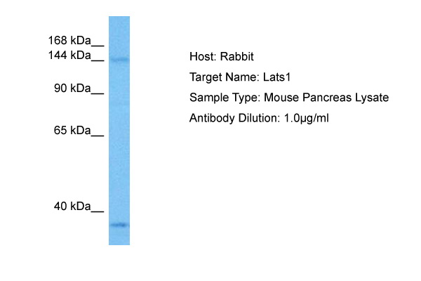
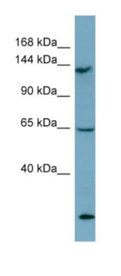 WB (Western Blot)
(WB Suggested Anti-LATS1 AntibodyTitration: 0.2-1 ug/mlELISA Titer: 1:12500Positive Control: THP-1 cell lysate)
WB (Western Blot)
(WB Suggested Anti-LATS1 AntibodyTitration: 0.2-1 ug/mlELISA Titer: 1:12500Positive Control: THP-1 cell lysate)
LATS1, Polyclonal Antibody (Cat# AAA200800)
Predicted: Cow, Dog, Horse, Rabbit, Rat, Sheep, Yeast, Zebrafish
 WB (Western Blot)
(WB Suggested Anti-SYCP3 Antibody Titration: 0.2-1 ug/mlELISA Titer: 1:312500Positive Control: Jurkat cell lysate)
WB (Western Blot)
(WB Suggested Anti-SYCP3 Antibody Titration: 0.2-1 ug/mlELISA Titer: 1:312500Positive Control: Jurkat cell lysate)
SYCP3, Polyclonal Antibody (Cat# AAA200802)
 WB (Western Blot)
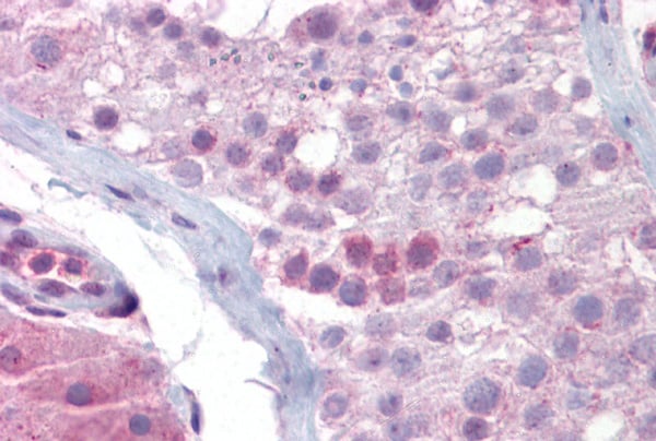
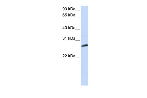
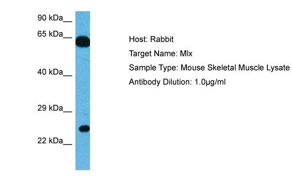
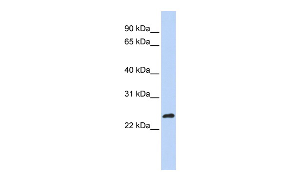
(WB Suggested Anti-MLX Antibody Titration: 0.2-1 ug/mlELISA Titer: 1:62500Positive Control: MCF7 cell lysateMLX is strongly supported by BioGPS gene expression data to be expressed in Human MCF7 cells)
WB (Western Blot)
(WB Suggested Anti-MLX Antibody Titration: 0.2-1 ug/mlELISA Titer: 1:62500Positive Control: MCF7 cell lysateMLX is strongly supported by BioGPS gene expression data to be expressed in Human MCF7 cells)
MLX, Polyclonal Antibody (Cat# AAA200806)
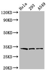
 WB (Western Blot)
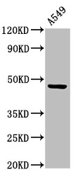
(WB Suggested Anti-EVI1 antibody Titration: 1 ug/mLSample Type: Human A549)
WB (Western Blot)
(WB Suggested Anti-EVI1 antibody Titration: 1 ug/mLSample Type: Human A549)
EVI1, Polyclonal Antibody (Cat# AAA200807)
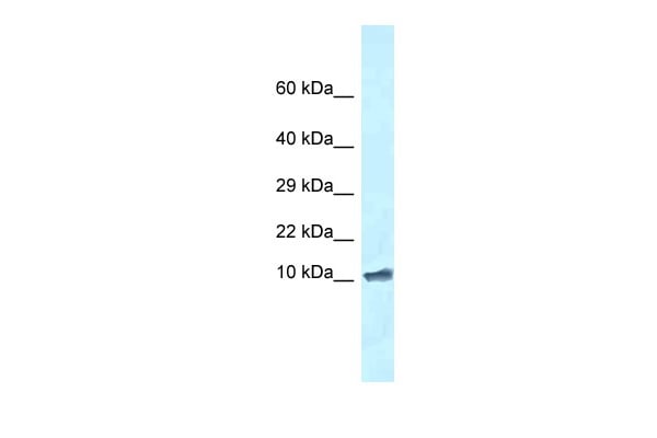 WB (Western Blot)
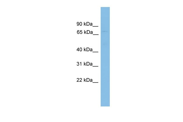
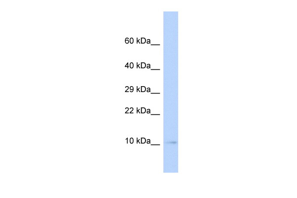
(WB Suggested Anti-Romo1 AntibodyTitration: 1.0 ug/mlPositive Control: Mouse Heart)
WB (Western Blot)
(WB Suggested Anti-Romo1 AntibodyTitration: 1.0 ug/mlPositive Control: Mouse Heart)
Romo1, Polyclonal Antibody (Cat# AAA200813)
Predicted Species Reactivity: Cow, Dog, Guinea Pig, Horse, Human, Mouse, Rabbit, Rat, Zebrafish
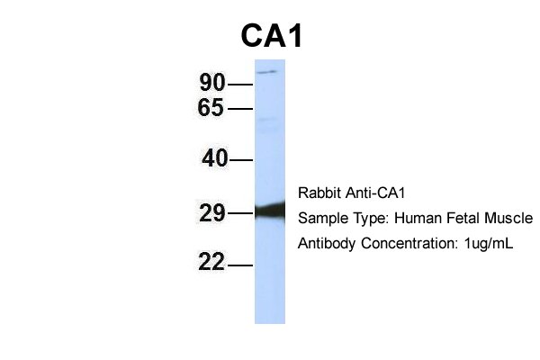
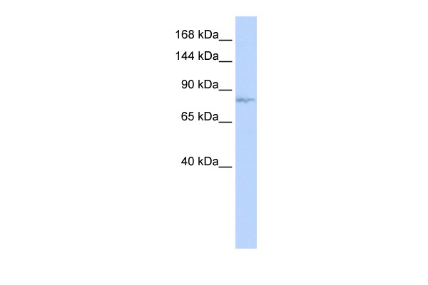
 WB (Western Blot)
(WB Suggested Anti-CA1 Antibody Titration: 0.2-1 ug/mlELISA Titer: 1:312500Positive Control: Human Placenta)
WB (Western Blot)
(WB Suggested Anti-CA1 Antibody Titration: 0.2-1 ug/mlELISA Titer: 1:312500Positive Control: Human Placenta)
CA1, Polyclonal Antibody (Cat# AAA200814)
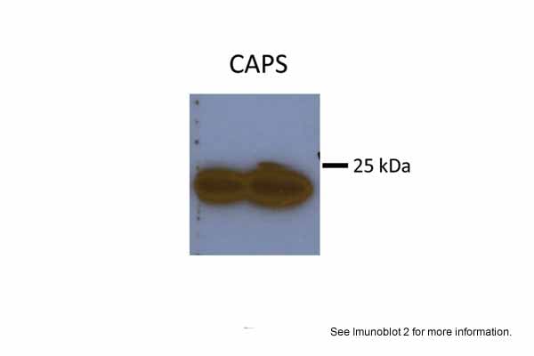
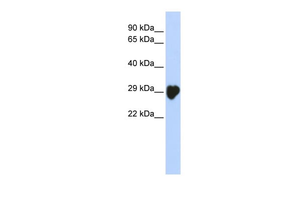
 WB (Western Blot)
(WB Suggested Anti-CAPS Antibody Titration: 0.2-1 ug/mlELISA Titer: 1:312500Positive Control: 293T cell lysate)
WB (Western Blot)
(WB Suggested Anti-CAPS Antibody Titration: 0.2-1 ug/mlELISA Titer: 1:312500Positive Control: 293T cell lysate)
CAPS, Polyclonal Antibody (Cat# AAA200815)
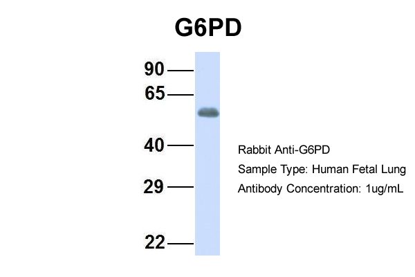
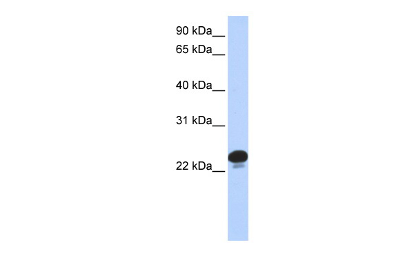
 WB (Western Blot)
(WB Suggested Anti-G6PD Antibody Titration: 0.2-1 ug/mlELISA Titer: 1:62500Positive Control: MCF7 cell lysateG6PD is supported by BioGPS gene expression data to be expressed in MCF7)
WB (Western Blot)
(WB Suggested Anti-G6PD Antibody Titration: 0.2-1 ug/mlELISA Titer: 1:62500Positive Control: MCF7 cell lysateG6PD is supported by BioGPS gene expression data to be expressed in MCF7)
G6PD, Polyclonal Antibody (Cat# AAA200821)
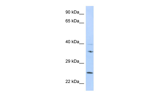
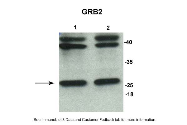
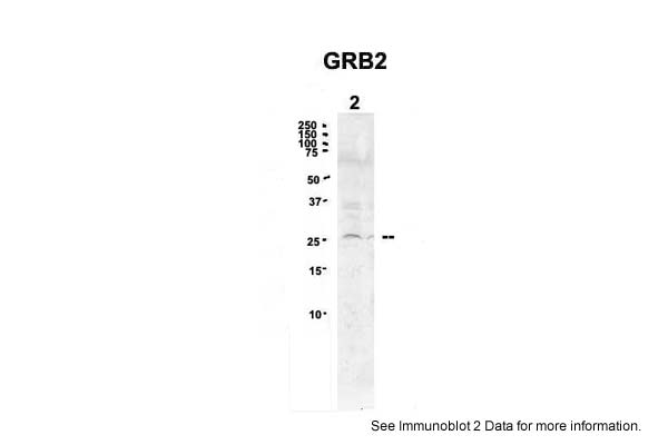
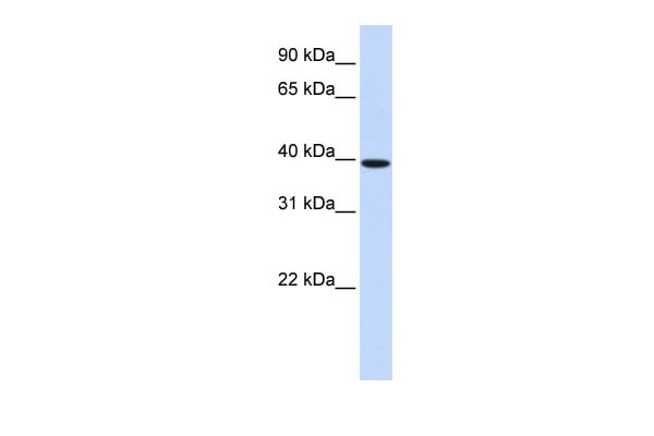
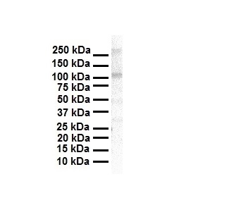
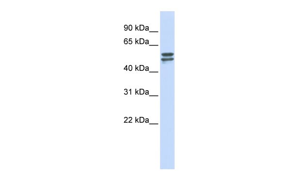
 WB (Western Blot)
(Host: MouseTarget Name: GRB2Sample Tissue: Mouse KidneyAntibody Dilution: 1ug/ml)
WB (Western Blot)
(Host: MouseTarget Name: GRB2Sample Tissue: Mouse KidneyAntibody Dilution: 1ug/ml)
GRB2, Polyclonal Antibody (Cat# AAA200823)
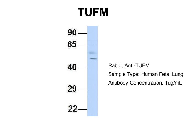
 WB (Western Blot)
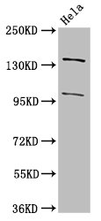
(WB Suggested Anti-TUFM Antibody Titration: 0.2-1 ug/mlELISA Titer: 1:1562500Positive Control: Hela cell lysateTUFM is supported by BioGPS gene expression data to be expressed in HeLa)
WB (Western Blot)
(WB Suggested Anti-TUFM Antibody Titration: 0.2-1 ug/mlELISA Titer: 1:1562500Positive Control: Hela cell lysateTUFM is supported by BioGPS gene expression data to be expressed in HeLa)
TUFM, Polyclonal Antibody (Cat# AAA200829)

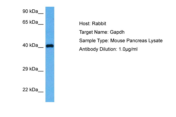
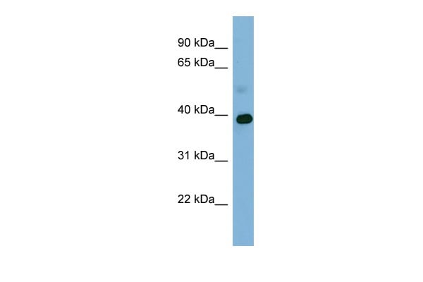 WB (Western Blot)
(WB Suggested Anti-GAPDH Antibody Titration: 0.2-1 ug/mlELISA Titer: 1:312500Positive Control: NCI-H226 cell lysate)
WB (Western Blot)
(WB Suggested Anti-GAPDH Antibody Titration: 0.2-1 ug/mlELISA Titer: 1:312500Positive Control: NCI-H226 cell lysate)
GAPDH, Polyclonal Antibody (Cat# AAA200831)
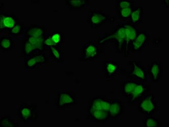
 IHC (Immunohistochemisry)
(Immunofluorescent analysis of HepG2 cells using AAA234024 at a dilution of 1:100 and Alexa Fluor 488-congugated AffiniPure Goat Anti-Rabbit IgG(H+L))
IHC (Immunohistochemisry)
(Immunofluorescent analysis of HepG2 cells using AAA234024 at a dilution of 1:100 and Alexa Fluor 488-congugated AffiniPure Goat Anti-Rabbit IgG(H+L))
ARHGEF12, Polyclonal Antibody (Cat# AAA234024)
 IHC (Immunohistochemisry)
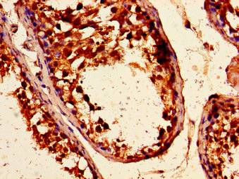
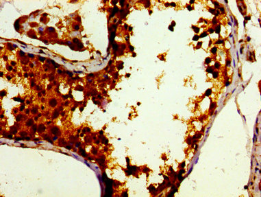
(Immunohistochemistry of paraffin-embedded human testis tissue using AAA234025 at dilution of 1:100)
IHC (Immunohistochemisry)
(Immunohistochemistry of paraffin-embedded human testis tissue using AAA234025 at dilution of 1:100)
TUBGCP4, Polyclonal Antibody (Cat# AAA234025)
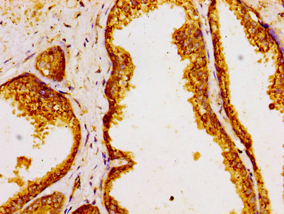
 IHC (Immunohistochemisry)
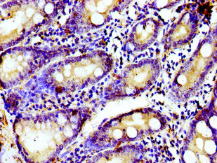
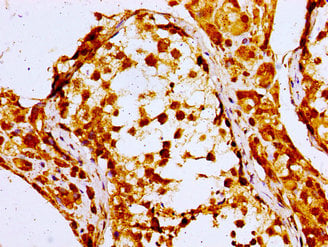
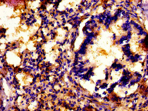
(Immunohistochemistry of paraffin-embedded human prostate tissue using AAA234027 at dilution of 1:100)
IHC (Immunohistochemisry)
(Immunohistochemistry of paraffin-embedded human prostate tissue using AAA234027 at dilution of 1:100)
GOLPH3, Polyclonal Antibody (Cat# AAA234027)
 IHC (Immunohiostchemistry)
(Immunohistochemistry of paraffin-embedded human testis tissue using AAA234033 at dilution of 1:100)
IHC (Immunohiostchemistry)
(Immunohistochemistry of paraffin-embedded human testis tissue using AAA234033 at dilution of 1:100)
DBF4, Polyclonal Antibody (Cat# AAA234033)
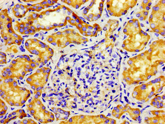
 IHC (Immunohistochemisry)
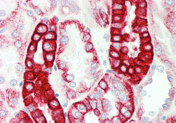
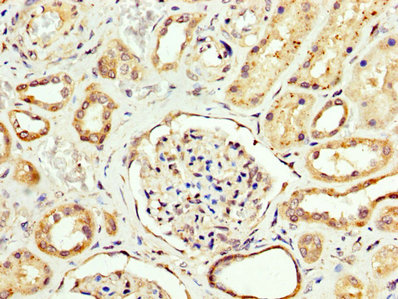
(Immunohistochemistry of paraffin-embedded human adrenal gland tissue using AAA234034 at dilution of 1:100)
IHC (Immunohistochemisry)
(Immunohistochemistry of paraffin-embedded human adrenal gland tissue using AAA234034 at dilution of 1:100)
NPC1, Polyclonal Antibody (Cat# AAA234034)
 IHC (Immunohistochemisry)
(Immunofluorescent analysis of MCF-7 cells using AAA234040 at a dilution of 1:100 and Alexa Fluor 488-congugated AffiniPure Goat Anti-Rabbit IgG(H+L))
IHC (Immunohistochemisry)
(Immunofluorescent analysis of MCF-7 cells using AAA234040 at a dilution of 1:100 and Alexa Fluor 488-congugated AffiniPure Goat Anti-Rabbit IgG(H+L))
TUBD1, Polyclonal Antibody (Cat# AAA234040)
 IHC (Immunohiostchemistry)
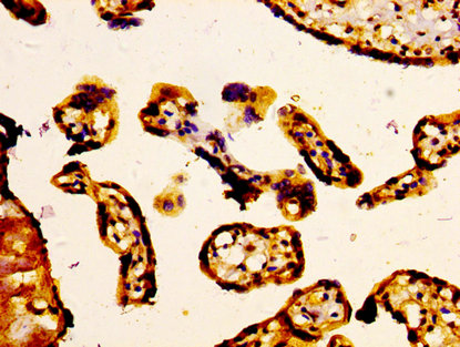
(IHC image of AAA234042 diluted at 1:400 and staining in paraffin-embedded human placenta tissue performed on a Leica BondTM system. After dewaxing and hydration, antigen retrieval was mediated by high pressure in a citrate buffer (pH 6.0). Section was blocked with 10% normal goat serum 30min at RT. Then primary antibody (1% BSA) was incubated at 4 degree C overnight. The primary is detected by a biotinylated secondary antibody and visualized using an HRP conjugated SP system.)
IHC (Immunohiostchemistry)
(IHC image of AAA234042 diluted at 1:400 and staining in paraffin-embedded human placenta tissue performed on a Leica BondTM system. After dewaxing and hydration, antigen retrieval was mediated by high pressure in a citrate buffer (pH 6.0). Section was blocked with 10% normal goat serum 30min at RT. Then primary antibody (1% BSA) was incubated at 4 degree C overnight. The primary is detected by a biotinylated secondary antibody and visualized using an HRP conjugated SP system.)
ABCC5, Polyclonal Antibody (Cat# AAA234042)
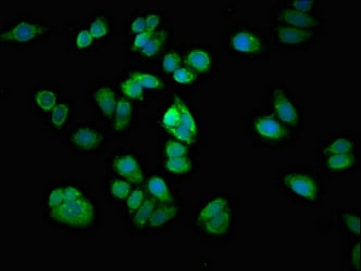
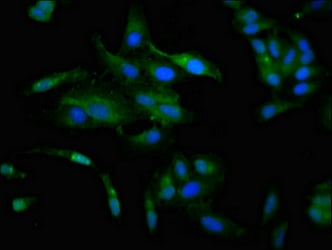
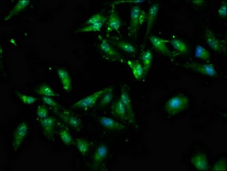 IF (Immunofluorescence)
(Immunofluorescence staining of Hela cells with AAA234044 at 1:100, counter-stained with DAPI. The cells were fixed in 4% formaldehyde, permeabilized using 0.2% Triton X-100 and blocked in 10% normal Goat Serum. The cells were then incubated with the antibody overnight at 4 degree C.The secondary antibody was Alexa Fluor 488-congugated AffiniPure Goat Anti-Rabbit IgG (H+L).)
IF (Immunofluorescence)
(Immunofluorescence staining of Hela cells with AAA234044 at 1:100, counter-stained with DAPI. The cells were fixed in 4% formaldehyde, permeabilized using 0.2% Triton X-100 and blocked in 10% normal Goat Serum. The cells were then incubated with the antibody overnight at 4 degree C.The secondary antibody was Alexa Fluor 488-congugated AffiniPure Goat Anti-Rabbit IgG (H+L).)
ATP7A, Polyclonal Antibody (Cat# AAA234044)
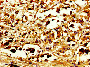
 IHC (Immunohiostchemistry)
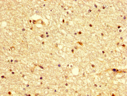
(IHC image of AAA234047 diluted at 1:300 and staining in paraffin-embedded human brain tissue performed on a Leica BondTM system. After dewaxing and hydration, antigen retrieval was mediated by high pressure in a citrate buffer (pH 6.0). Section was blocked with 10% normal goat serum 30min at RT. Then primary antibody (1% BSA) was incubated at 4 degree C overnight. The primary is detected by a biotinylated secondary antibody and visualized using an HRP conjugated SP system.)
IHC (Immunohiostchemistry)
(IHC image of AAA234047 diluted at 1:300 and staining in paraffin-embedded human brain tissue performed on a Leica BondTM system. After dewaxing and hydration, antigen retrieval was mediated by high pressure in a citrate buffer (pH 6.0). Section was blocked with 10% normal goat serum 30min at RT. Then primary antibody (1% BSA) was incubated at 4 degree C overnight. The primary is detected by a biotinylated secondary antibody and visualized using an HRP conjugated SP system.)
BDKRB2, Polyclonal Antibody (Cat# AAA234047)
 IHC (Immunohistochemisry)
(Immunofluorescence staining of Hela cells with AAA234048 at 1:133, counter-stained with DAPI. The cells were fixed in 4% formaldehyde, permeabilized using 0.2% Triton X-100 and blocked in 10% normal Goat Serum. The cells were then incubated with the antibody overnight at 4 degree C.The secondary antibody was Alexa Fluor 488-congugated AffiniPure Goat Anti-Rabbit IgG (H+L).)
IHC (Immunohistochemisry)
(Immunofluorescence staining of Hela cells with AAA234048 at 1:133, counter-stained with DAPI. The cells were fixed in 4% formaldehyde, permeabilized using 0.2% Triton X-100 and blocked in 10% normal Goat Serum. The cells were then incubated with the antibody overnight at 4 degree C.The secondary antibody was Alexa Fluor 488-congugated AffiniPure Goat Anti-Rabbit IgG (H+L).)
CCKAR, Polyclonal Antibody (Cat# AAA234048)
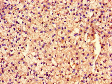
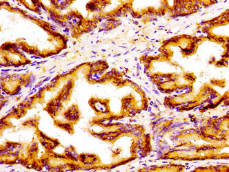
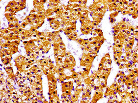
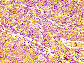
 IHC (Immunohiostchemistry)
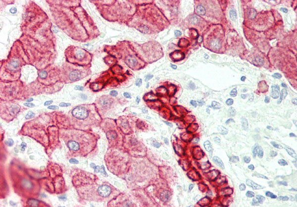
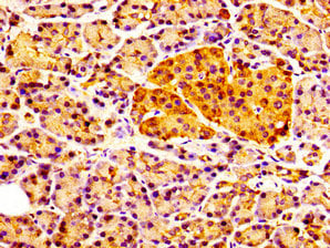
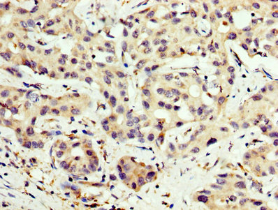
(IHC image of AAA234051 diluted at 1:200 and staining in paraffin-embedded human liver cancer performed on a Leica BondTM system. After dewaxing and hydration, antigen retrieval was mediated by high pressure in a citrate buffer (pH 6.0). Section was blocked with 10% normal goat serum 30min at RT. Then primary antibody (1% BSA) was incubated at 4 degree C overnight. The primary is detected by a biotinylated secondary antibody and visualized using an HRP conjugated SP system.)
IHC (Immunohiostchemistry)
(IHC image of AAA234051 diluted at 1:200 and staining in paraffin-embedded human liver cancer performed on a Leica BondTM system. After dewaxing and hydration, antigen retrieval was mediated by high pressure in a citrate buffer (pH 6.0). Section was blocked with 10% normal goat serum 30min at RT. Then primary antibody (1% BSA) was incubated at 4 degree C overnight. The primary is detected by a biotinylated secondary antibody and visualized using an HRP conjugated SP system.)
CPT2, Polyclonal Antibody (Cat# AAA234051)
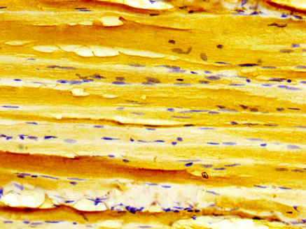
 IHC (Immunohiostchemistry)
(IHC image of AAA234054 diluted at 1:500 and staining in paraffin-embedded human heart tissue performed on a Leica BondTM system. After dewaxing and hydration, antigen retrieval was mediated by high pressure in a citrate buffer (pH 6.0). Section was blocked with 10% normal goat serum 30min at RT. Then primary antibody (1% BSA) was incubated at 4 degree C overnight. The primary is detected by a biotinylated secondary antibody and visualized using an HRP conjugated SP system.)
IHC (Immunohiostchemistry)
(IHC image of AAA234054 diluted at 1:500 and staining in paraffin-embedded human heart tissue performed on a Leica BondTM system. After dewaxing and hydration, antigen retrieval was mediated by high pressure in a citrate buffer (pH 6.0). Section was blocked with 10% normal goat serum 30min at RT. Then primary antibody (1% BSA) was incubated at 4 degree C overnight. The primary is detected by a biotinylated secondary antibody and visualized using an HRP conjugated SP system.)
DMPK, Polyclonal Antibody (Cat# AAA234054)
What are Polyclonal Antibodies?
Polyclonal antibodies are antibodies that come from multiple B cell clones of a host animal. The typical hosts used for the majority of polyclonal antibody production are rabbits, goats, sheep, and donkeys. These polyclonal antibodies, once having identified their target, will bind to different epitopes located at different regions or sequences on the same protein/antigen. As a result, they are ideal at locating and binding to the target, even if the target is in very low concentrations (due to many different antibodies being able to bind to the same target molecule, which allows for significant amplification of a downstream signal).
Polyclonal antibodies are typically produced by injecting an antigen into a host animal, which causes the animal’s immune system to attack the foreign antigen by mass generating antibodies against it. After a period of time, serum is collected from the animal and purified using physicochemical fractionation, class-specific affinity purification, and/or antigen-affinity purification.
Key Uses of Polyclonal Antibodies
- Western Blotting: This method is used to find specific proteins in biological samples after separating them by size.
- Immunohistochemistry: IHC helps visualize the location of proteins in tissue sections using various staining techniques.
- ELISA: (Enzyme-Linked Immunosorbent Assay) is typically used to identify specific protein quantities in a sample. ELISAs can be either “Quantitative” or “Qualitative”.
- Flow Cytometry: technique that identifies and measures the specific protein on the surface or inside the cells in a fluid suspension.
- Immunoprecipitation: IP isolates and studies a specific protein from a complex mixture using antibodies.
Why Buy Polyclonal Antibodies from AAA Biotech?
1. Ideal for Various Applications
Our antibodies are generally going to be validated for use in multiple types of assays, including ELISA, Western Blotting, Immunohistochemistry, Immunoprecipitation, amongst others. They are ideal for a wide range of research applications.
2. Rigorous Quality Control
All of the antibodies in our catalog undergo strict quality testing to ensure specificity, sensitivity, and consistent performance. We are confident in the ability of our antibodies to provide you with accurate results.
3. Wide Assortment of Antibodies
Antibodies in are catalog can be found for both common and exotic species, and these antibodies are also available in both conjugated and recombinant forms to suit many diverse experimental needs.
4. Highly Purified
Our antibodies are available in purified forms with over 85% purity, as confirmed by SDS-PAGE. They are also available with tags such as His, Flag, GST, or MBP. We cater to customers worldwide.
FAQ
1. How are polyclonal antibodies produced?
Traditionally, polyclonal antibodies are produced by injecting an antigen into a host animal (such as a rabbit or goat), which then triggers an immune response from the host animal. The animal’s B cells produce antibodies that will recognize different parts of the injected antigen. These antibodies are then collected from the animal’s blood and purified for use.
2. How do polyclonal antibodies differ from monoclonal antibodies?
Polyclonal antibodies are a mix of antibodies that bind to different locations (epitopes) of the same antigen, while monoclonal antibodies are identical and bind to just one specific epitope. This makes polyclonal antibodies more versatile and better at detecting proteins that may be present in low quantities or in altered/modified forms.
3. How should I store polyclonal antibodies?
Polyclonal antibodies should be stored at 4°C for short-term use (up to a few weeks) and at -20°C or -80°C for long-term storage. Avoid repeated freeze-thaw cycles by dividing them into small aliquots. Always check the datasheet for specific storage instructions.